Aminoglycoside- and Cisplatin-InducedOtotoxicity: Mechanisms and OtoprotectiveStrategies
Corné J. Kros1 and Peter S. Steyger2,3
1Sussex Neuroscience, School of Life Sciences, University of Sussex, Falmer, Brighton BN1 9QG,United Kingdom
2Oregon Hearing Research Center, Oregon Health & Science University, Portland, Oregon 972393National Center for Rehabilitative Auditory Research, VA Portland Health Care System,Portland, Oregon 97239
Ototoxicity refers to damage of inner ear structures (i.e., the cochlea and vestibule) and theirfunction (hearing and balance) following exposure to specific in-hospital medications (i.e.,aminoglycoside antibiotics, platinum-based drugs), as well as a variety of environmental oroccupational exposures (e.g., metals and solvents). This review provides a narrative derivedfrom relevant papers describing factors contributing to (or increasing the risk of ) aminoglyco-side and cisplatin-induced ototoxicity. We also review current strategies to protect againstototoxicity induced by these indispensable pharmacotherapeutic treatments for life-threat-ening infections and solid tumors. We end by highlighting several interventional strategiesthat are currently in development, as well as the diverse challenges that still need to beovercome to prevent drug-induced hearing loss.
Specific medications to treat life-threateningconditions as well as several other com-
pounds can cause a debilitating side effect—oto-toxicity (i.e., the permanent loss of hearing orbalance) (Table 1). The socioeconomic cost ofototoxicity has to beweighed against the benefitsof surviving severe infections or cancer. Inthis review, we focus on two clinically essentialdrugs, the aminoglycosides and cisplatin, bothof which remain widely used despite the risk ofototoxicity.
Aminoglycosides, including gentamicin,amikacin, kanamycin, and tobramycin, are
broad-spectrum antibiotics used for treating sus-pected or confirmed acute serious infections,and the long-term management of recurrentrespiratory infections in cystic fibrosis and mul-tidrug-resistant tuberculosis (Escobaret al. 2000;Garinis et al. 2017a; Jiang et al. 2017). The oto-toxicity of the first aminoglycoside discovered,streptomycin (Schatz et al. 1944), was recog-nized soon after its initial use (Hinshaw andFeldman 1945). Newer aminoglycosides havevarying degrees of cochleotoxicity and vestib-ulotoxicity (Miller 1985). Estimates of the in-cidence of hearing loss after aminoglycoside
Editors: Guy P. Richardson and Christine PetitAdditional Perspectives on Function and Dysfunction of the Cochlea available at www.perspectivesinmedicine.org
treatment vary widely, due to differing dosingregimens and sensitivities of audiological tests,yet may be as high as 20%–50% of recipients(Huth et al. 2015). Vestibulotoxicity can occurin up to 60% of treatment courses (van Heckeet al. 2017), and patients often self-report bal-ance issues (and tinnitus) before any perceivedcochleotoxicity. The lack of appropriate vesti-bulometric, compared to audiometric, testingfacilities means that vestibulotoxicity remainswoefully underreported. Furthermore, most au-diometric diagnosis is within the 500–8000 Hz(conventional) testing range, underestimatingthe incidence of cochleotoxicity at higher fre-quencies where the onset of hearing loss typi-cally occurs (Schacht et al. 2012). Aminoglyco-side-induced ototoxicity is dose dependent inpreclinical models and, likely, also in humans(Wu et al. 2001; Garinis et al. 2017a,b).
Aminoglycosides are polycationicmoleculeswith molar masses between 300 and 600 g/mol,with a maximal cross-sectional diameter of∼0.8 nm (van Netten and Kros 2007; Jianget al. 2017). In bacteria, aminoglycosides bindto ribosomal RNA, causing misreading of mes-senger RNA (mRNA) and consequent accumu-lation of misfolded proteins that leads to cellularstress and bacterial lysis (Schacht et al. 2012). In
mammals, aminoglycosides are selectively toxicto kidney proximal tubule cells, and to sensoryhair cells of the inner ear. Drug-induced kidneydamage is reversible if detected early and med-ication discontinued, as proximal tubule cellscan regenerate, but this is not typically true formammalian hair cells (Naughton 2008; Groves2010; Kusaba et al. 2014; Lombardi et al. 2016).Since aminoglycosides target bacteria, they alsoreadily disrupt mitochondria within cells, caus-ing the release of proapoptotic factors and oxi-dative enzymes into the cytoplasm and the gen-eration of free radicals (Warchol 2010; Huthet al. 2011; Esterberg et al. 2016).
Cisplatin degrades solid tumors, often withvery high efficacy (e.g., >90% cure rates for tes-ticular carcinoma) (Cheng et al. 2018), yet theincidence of cochleotoxicity is very high withhearing loss in almost all treated patients (Cal-lejo et al. 2015; Paken et al. 2016). As for amino-glycosides, onset of cisplatin-induced hearingloss typically occurs first in the extended high-frequency range before affecting sensitivity atconversational (lower) frequencies, as evaluatedby distortion product otoacoustic emissions orconventional audiometry. Increasing the cumu-lative dose consistently leads to increasing riskof permanent hearing loss in humans (Brock
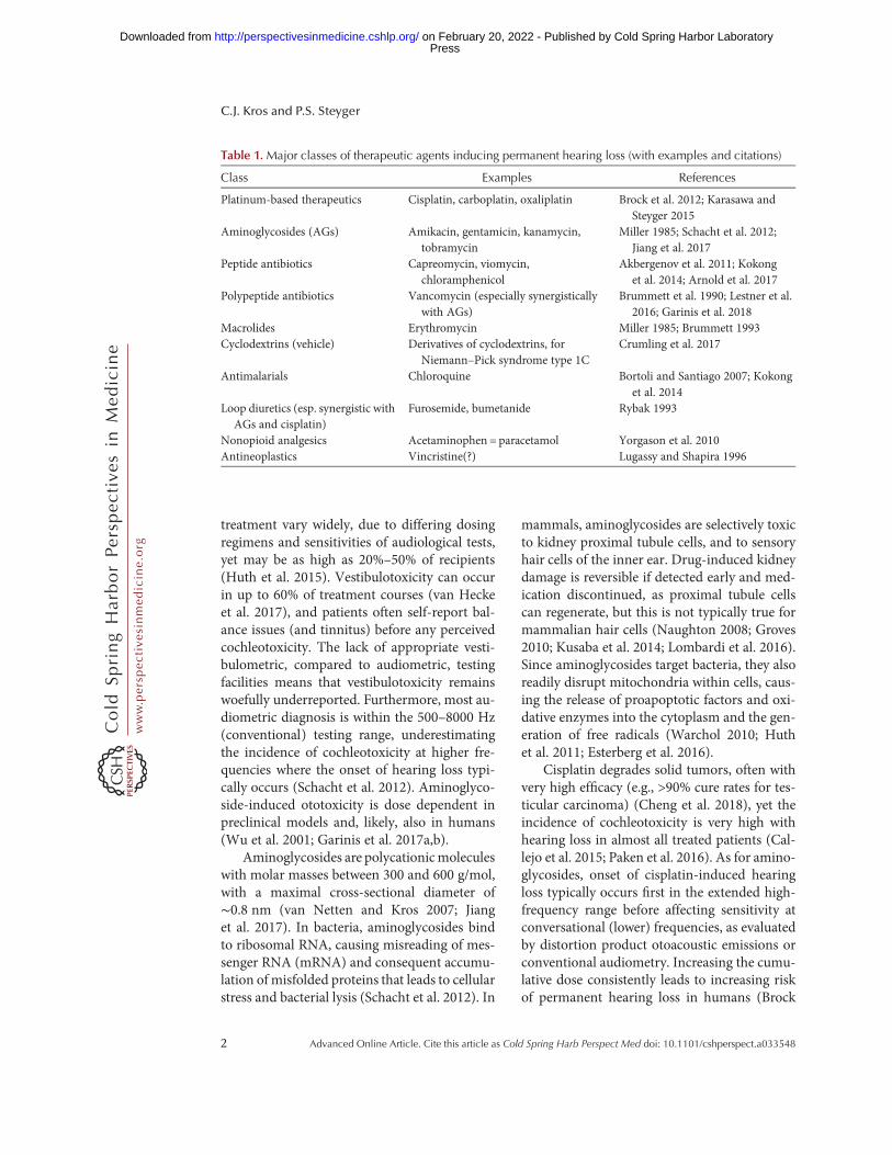
Table 1. Major classes of therapeutic agents inducing permanent hearing loss (with examples and citations)
et al. 2012; Garinis et al. 2017a). By contrast,there are few reports of vestibulotoxicity in pa-tients treated with cisplatin, despite dose-depen-dent loss of vestibular hair cells and vestibularfunction in animals (Callejo et al. 2017), perhapsdue to limitations in human vestibular testingprotocols (Prayuenyong et al. 2018). Cisplatinis smaller than the aminoglycosides, with a mo-lar mass of 300 g/mol and a molecular “diame-ter” of ∼0.5 nm. Cisplatin itself is not chargedbut the molecule is unstable and one or both ofthe Cl− ions can be replaced by H2O (aquation),resulting inmonovalent or divalent cations. Thisreaction is favored when Cl− concentrations arelow, as in the cytosol. Cisplatin can enter cells bypassive diffusion, and via a variety of transport-ers, the most prominent of which are coppertransporter CTR1 and organic cation trans-porter OCT2 (Waissbluth and Daniel 2013).Aquated cisplatin binds to nuclear DNA form-ing adducts that prevent transcription and rep-lication, ultimately resulting in apoptosis, thebasis of its cytotoxic antitumor activity, partic-ularly in proliferating cancer cells that are poor atrepairing DNA damage (Siddik 2003). Like ami-noglycosides, cisplatin can also induce nephro-toxicity and ototoxicity, with neurotoxicity anadditional outcome (Dzagnidze et al. 2007).These side effects are also likely due to the gen-eration of toxic levels of reactive oxygen speciesthat leads to apoptosis (Hazlitt et al. 2018a).
As aminoglycosides and cisplatin are typi-cally infused parenterally in a hospital setting(aminoglycosides are also administered viamid-dle ear [intratympanic] injections clinically),opportunities exist for coadministering thera-peutics to prevent or reduce ototoxic side effects.Yet, despite much experimentation, there are nodrugs that have been licensed for use in patientsto prevent ototoxicity. This review discussesrecent insights into how aminoglycosides andcisplatin damage sensory cells that respond tosound and motion. The intracellular mecha-nisms that lead to ototoxicity are also brieflydiscussed (with more detail in Schacht et al.2012; Karasawa and Steyger 2015; Jiang et al.2017). We end by considering how emerginginsights can promote development of novel oto-protective therapies.
TRAFFICKING OF CLINICALOTOTOXINSINTO THE INNER EAR
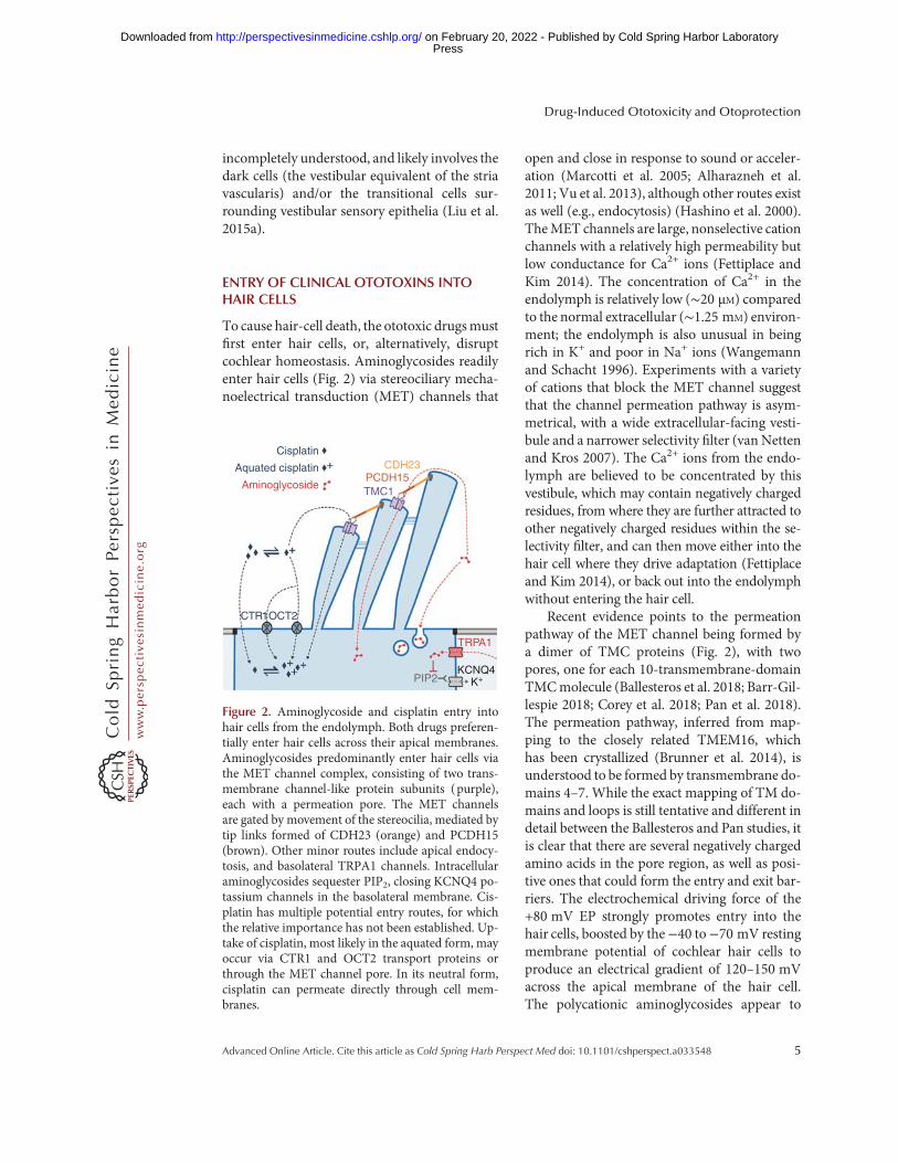
Ototoxicity results from drugs (ototoxins) en-tering inner ear tissues to exert their cytotoxiceffects. Ototoxins must first cross the blood–labyrinth barrier (BLB) that separates the blood-stream from inner ear tissues and fluids, pre-dominantly the endolymph and perilymph(Fig. 1). The BLB is a specialized structure com-posed of tight junction-coupled inner ear endo-thelial cells that prevents macromolecules andblood cells from entering cochlear tissues viapassive, paracellular extravasation from capillar-ies. Perilymph has an ionic composition similarto other extracellular fluids (high Na+, low K+,and millimolar Ca2+). In contrast, the endo-lymph has a high K+ concentration, low Na+
and ∼20 nM Ca2+, as well as a positive endolym-phatic potential (EP) of∼+80 mV in the cochlea(but not in the vestibule). The composition ofthe endolymph is actively maintained by cellswithin the adjacent stria vascularis in the co-chlea, and by dark cells in the vestibule (Wan-gemann 2006). Earlier cochlear studies reportedhigher aminoglycoside levels in perilymph thanin the endolymph, suggesting that aminoglyco-sides in perilymph were the primary source oftoxicity to hair cells (Tran Ba Huy et al. 1986).Although aminoglycosides enter perilymph fol-lowing systemic administration, they do notreadily enter hair cells via this route (Li andSteyger 2011). Experiments using fluorescentlylabeled gentamicin (gentamicin-Texas Red[GTTR]) revealed that systemically adminis-tered GTTR was more efficiently taken up byhair cells than GTTR perfused into the scalatympani. When applied systemically, GTTRreadily trafficked across the BLB into the striavascularis, and cleared into the endolymph priorto entering hair cells.
Surprisingly, following middle ear adminis-tration, auditory afferent fibers ipsilaterally, andvestibular efferent fibers bilaterally, transportaminoglycosides from cochlear perilymph tothe auditory brainstem, although it is not yetknown whether this leads to functional conse-quences (Zhang et al. 2012). Cochlear uptake ofaminoglycosides and ototoxicity are also strong-
Drug-Induced Ototoxicity and Otoprotection
Advanced Online Article. Cite this article as Cold Spring Harb Perspect Med doi: 10.1101/cshperspect.a033548 3
ww
w.p
ersp
ecti
vesi
nm
edic
ine.
org
Press on February 20, 2022 - Published by Cold Spring Harbor Laboratoryhttp://perspectivesinmedicine.cshlp.org/Downloaded from
ly increased during systemic inflammation thatvasodilates capillaries in the stria vascularis(Koo et al. 2015). How exactly aminoglycosidesenter the endolymph from the capillaries in thestria vascularis is currently not clear, but possi-bilities include ion channels, transporters, ortranscytosis (Koo et al. 2015).
Like aminoglycosides, cisplatin and relatedcompounds such as carboplatin and oxaliplatinhave also been detected in perilymph followingsystemic administration, suggesting that thesedrugs may enter cochlear hair cells via theirbasolateral membranes (Hellberg et al. 2009,2013). Yet, like aminoglycosides, various strandsof evidence suggest that the stria vascularis–en-dolymph route is more likely. Iontophoretic ap-plication of cisplatin to the endolymphatic com-partment of the guinea pig cochlea was morerapid and effective in reducing auditory nerveresponses to sound than perfusion of perilymphwith cisplatin-containing solution (McAlpine
and Johnstone 1990). When loop diureticswere coadministered with cisplatin, hearingloss was exacerbated in guinea pigs (McAlpineand Johnstone 1990). In young cancer patientstoo, cotreatment with furosemide increasedthe risk of hearing loss (Clemens et al. 2016).The CTR1 and OCT2 transporters have beenobserved in stria vascularis but not in the spiralligament, suggesting their possible involvementin cisplatin transport across the BLB into theendolymph (Ciarimboli et al. 2010; More et al.2010; Waissbluth and Daniel 2013). Measure-ment of platinum levels in cisplatin-treatedmice or patients revealed consistently higherlevels in the stria vascularis that were retainedformonths to years, especially in the basal, high-frequency region (Breglio et al. 2017). The EP ofcisplatin-treated mice dropped by up to 30 mVduring and after cisplatin treatment (Breglioet al. 2017). The entry of aminoglycosides andcisplatin into the vestibular endolymph remains
Cisplatin
CTR1?
OCT2?Reissner’smembrane
Scala vestibuliPerilymph 0 mV Stria
vascularis
Spiralligament
Haircells
Scala mediaEndolymph +80 mV
Organ of corti
Spiral ganglionneurons
Scala tympaniPerilymph 0 mV
Tectorialmembrane
Aminoglycoside
??
?
?
?
?
?
?
TRPV4?
TRPV1?
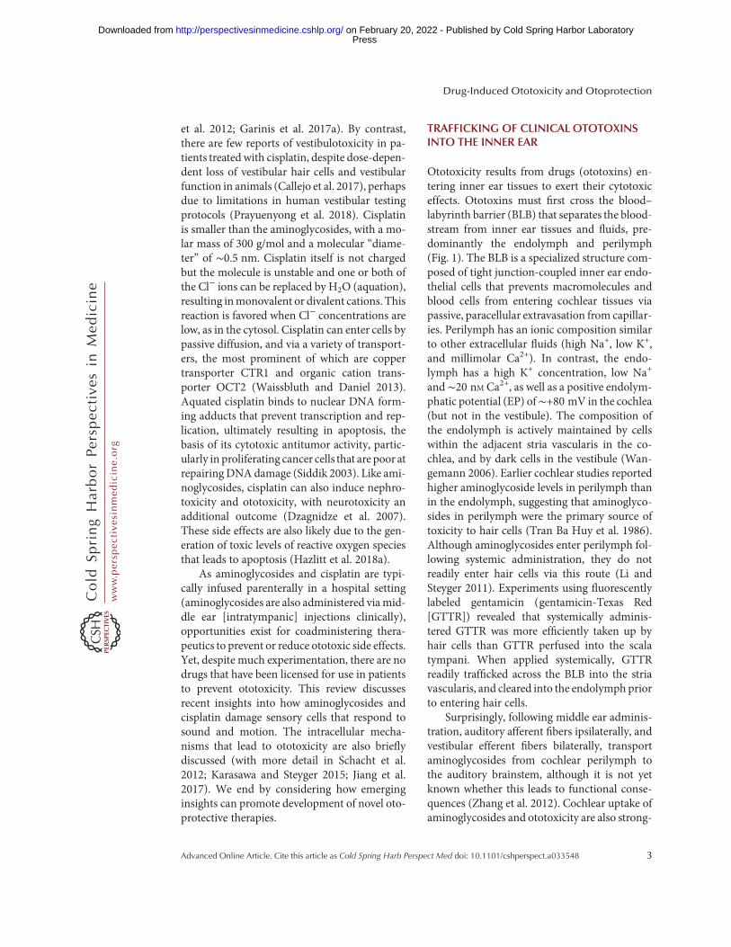
Figure 1.Main cochlear trafficking routes for systemic aminoglycosides and cisplatin. For both drugs, entry fromthe bloodstream into the cochlea occurs primarily from capillaries within the stria vascularis. Fromhere, cisplatinenters the endolymph in the scala media, potentially through organic cation transporter 2 (OCT2) and coppertransporter 1 (CTR1) transporters in the marginal cells. Aminoglycosides enter the endolymph through as-yet-unidentified ion channels or transporters, although several candidates exist (e.g., transient receptor potential V1[TRPV1] and TRPV4). From the endolymph, these ototoxins enter hair cells across their apical membranes.
C.J. Kros and P.S. Steyger
4 Advanced Online Article. Cite this article as Cold Spring Harb Perspect Med doi: 10.1101/cshperspect.a033548
ww
w.p
ersp
ecti
vesi
nm
edic
ine.
org
Press on February 20, 2022 - Published by Cold Spring Harbor Laboratoryhttp://perspectivesinmedicine.cshlp.org/Downloaded from
incompletely understood, and likely involves thedark cells (the vestibular equivalent of the striavascularis) and/or the transitional cells sur-rounding vestibular sensory epithelia (Liu et al.2015a).
ENTRY OF CLINICALOTOTOXINS INTOHAIR CELLS
To cause hair-cell death, the ototoxic drugsmustfirst enter hair cells, or, alternatively, disruptcochlear homeostasis. Aminoglycosides readilyenter hair cells (Fig. 2) via stereociliary mecha-noelectrical transduction (MET) channels that
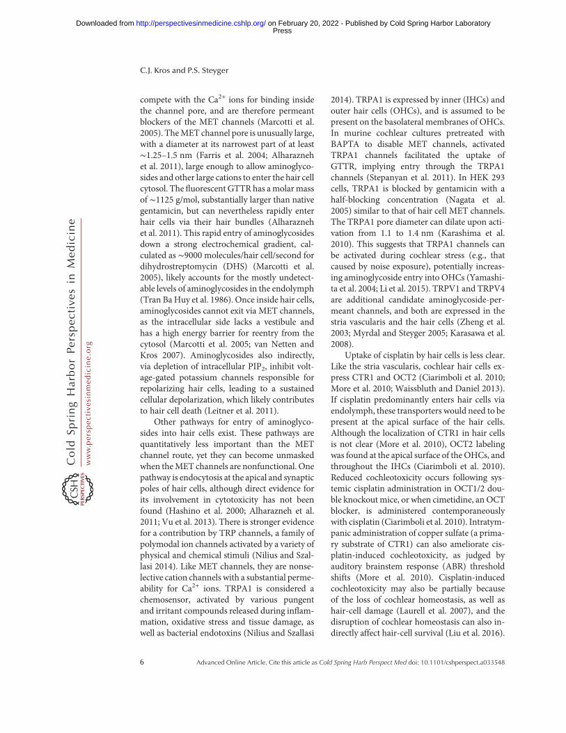
open and close in response to sound or acceler-ation (Marcotti et al. 2005; Alharazneh et al.2011; Vu et al. 2013), although other routes existas well (e.g., endocytosis) (Hashino et al. 2000).TheMET channels are large, nonselective cationchannels with a relatively high permeability butlow conductance for Ca2+ ions (Fettiplace andKim 2014). The concentration of Ca2+ in theendolymph is relatively low (∼20 µM) comparedto the normal extracellular (∼1.25 mM) environ-ment; the endolymph is also unusual in beingrich in K+ and poor in Na+ ions (Wangemannand Schacht 1996). Experiments with a varietyof cations that block the MET channel suggestthat the channel permeation pathway is asym-metrical, with a wide extracellular-facing vesti-bule and a narrower selectivity filter (van Nettenand Kros 2007). The Ca2+ ions from the endo-lymph are believed to be concentrated by thisvestibule, which may contain negatively chargedresidues, from where they are further attracted toother negatively charged residues within the se-lectivity filter, and can then move either into thehair cell where they drive adaptation (Fettiplaceand Kim 2014), or back out into the endolymphwithout entering the hair cell.
Recent evidence points to the permeationpathway of the MET channel being formed bya dimer of TMC proteins (Fig. 2), with twopores, one for each 10-transmembrane-domainTMCmolecule (Ballesteros et al. 2018; Barr-Gil-lespie 2018; Corey et al. 2018; Pan et al. 2018).The permeation pathway, inferred from map-ping to the closely related TMEM16, whichhas been crystallized (Brunner et al. 2014), isunderstood to be formed by transmembrane do-mains 4–7. While the exact mapping of TM do-mains and loops is still tentative and different indetail between the Ballesteros and Pan studies, itis clear that there are several negatively chargedamino acids in the pore region, as well as posi-tive ones that could form the entry and exit bar-riers. The electrochemical driving force of the+80 mV EP strongly promotes entry into thehair cells, boosted by the−40 to−70 mV restingmembrane potential of cochlear hair cells toproduce an electrical gradient of 120–150 mVacross the apical membrane of the hair cell.The polycationic aminoglycosides appear to
CDH23
CTR1OCT2
TRPA1
KCNQ4PIP2 K+
Cisplatin
Aquated cisplatin
AminoglycosidePCDH15TMC1
Figure 2. Aminoglycoside and cisplatin entry intohair cells from the endolymph. Both drugs preferen-tially enter hair cells across their apical membranes.Aminoglycosides predominantly enter hair cells viathe MET channel complex, consisting of two trans-membrane channel-like protein subunits (purple),each with a permeation pore. The MET channelsare gated by movement of the stereocilia, mediated bytip links formed of CDH23 (orange) and PCDH15(brown). Other minor routes include apical endocy-tosis, and basolateral TRPA1 channels. Intracellularaminoglycosides sequester PIP2, closing KCNQ4 po-tassium channels in the basolateral membrane. Cis-platin has multiple potential entry routes, for whichthe relative importance has not been established. Up-take of cisplatin, most likely in the aquated form, mayoccur via CTR1 and OCT2 transport proteins orthrough the MET channel pore. In its neutral form,cisplatin can permeate directly through cell mem-branes.
Drug-Induced Ototoxicity and Otoprotection
Advanced Online Article. Cite this article as Cold Spring Harb Perspect Med doi: 10.1101/cshperspect.a033548 5
ww
w.p
ersp
ecti
vesi
nm
edic
ine.
org
Press on February 20, 2022 - Published by Cold Spring Harbor Laboratoryhttp://perspectivesinmedicine.cshlp.org/Downloaded from
compete with the Ca2+ ions for binding insidethe channel pore, and are therefore permeantblockers of the MET channels (Marcotti et al.2005). TheMET channel pore is unusually large,with a diameter at its narrowest part of at least∼1.25–1.5 nm (Farris et al. 2004; Alharaznehet al. 2011), large enough to allow aminoglyco-sides and other large cations to enter the hair cellcytosol. The fluorescent GTTR has amolarmassof ∼1125 g/mol, substantially larger than nativegentamicin, but can nevertheless rapidly enterhair cells via their hair bundles (Alharaznehet al. 2011). This rapid entry of aminoglycosidesdown a strong electrochemical gradient, cal-culated as ∼9000 molecules/hair cell/second fordihydrostreptomycin (DHS) (Marcotti et al.2005), likely accounts for the mostly undetect-able levels of aminoglycosides in the endolymph(Tran Ba Huy et al. 1986). Once inside hair cells,aminoglycosides cannot exit via MET channels,as the intracellular side lacks a vestibule andhas a high energy barrier for reentry from thecytosol (Marcotti et al. 2005; van Netten andKros 2007). Aminoglycosides also indirectly,via depletion of intracellular PIP2, inhibit volt-age-gated potassium channels responsible forrepolarizing hair cells, leading to a sustainedcellular depolarization, which likely contributesto hair cell death (Leitner et al. 2011).
Other pathways for entry of aminoglyco-sides into hair cells exist. These pathways arequantitatively less important than the METchannel route, yet they can become unmaskedwhen theMET channels are nonfunctional. Onepathway is endocytosis at the apical and synapticpoles of hair cells, although direct evidence forits involvement in cytotoxicity has not beenfound (Hashino et al. 2000; Alharazneh et al.2011; Vu et al. 2013). There is stronger evidencefor a contribution by TRP channels, a family ofpolymodal ion channels activated by a variety ofphysical and chemical stimuli (Nilius and Szal-lasi 2014). Like MET channels, they are nonse-lective cation channels with a substantial perme-ability for Ca2+ ions. TRPA1 is considered achemosensor, activated by various pungentand irritant compounds released during inflam-mation, oxidative stress and tissue damage, aswell as bacterial endotoxins (Nilius and Szallasi
2014). TRPA1 is expressed by inner (IHCs) andouter hair cells (OHCs), and is assumed to bepresent on the basolateral membranes of OHCs.In murine cochlear cultures pretreated withBAPTA to disable MET channels, activatedTRPA1 channels facilitated the uptake ofGTTR, implying entry through the TRPA1channels (Stepanyan et al. 2011). In HEK 293cells, TRPA1 is blocked by gentamicin with ahalf-blocking concentration (Nagata et al.2005) similar to that of hair cell MET channels.The TRPA1 pore diameter can dilate upon acti-vation from 1.1 to 1.4 nm (Karashima et al.2010). This suggests that TRPA1 channels canbe activated during cochlear stress (e.g., thatcaused by noise exposure), potentially increas-ing aminoglycoside entry into OHCs (Yamashi-ta et al. 2004; Li et al. 2015). TRPV1 and TRPV4are additional candidate aminoglycoside-per-meant channels, and both are expressed in thestria vascularis and the hair cells (Zheng et al.2003; Myrdal and Steyger 2005; Karasawa et al.2008).
Uptake of cisplatin by hair cells is less clear.Like the stria vascularis, cochlear hair cells ex-press CTR1 and OCT2 (Ciarimboli et al. 2010;More et al. 2010; Waissbluth and Daniel 2013).If cisplatin predominantly enters hair cells viaendolymph, these transporters would need to bepresent at the apical surface of the hair cells.Although the localization of CTR1 in hair cellsis not clear (More et al. 2010), OCT2 labelingwas found at the apical surface of the OHCs, andthroughout the IHCs (Ciarimboli et al. 2010).Reduced cochleotoxicity occurs following sys-temic cisplatin administration in OCT1/2 dou-ble knockout mice, or when cimetidine, an OCTblocker, is administered contemporaneouslywith cisplatin (Ciarimboli et al. 2010). Intratym-panic administration of copper sulfate (a prima-ry substrate of CTR1) can also ameliorate cis-platin-induced cochleotoxicity, as judged byauditory brainstem response (ABR) thresholdshifts (More et al. 2010). Cisplatin-inducedcochleotoxicity may also be partially becauseof the loss of cochlear homeostasis, as well ashair-cell damage (Laurell et al. 2007), and thedisruption of cochlear homeostasis can also in-directly affect hair-cell survival (Liu et al. 2016).
C.J. Kros and P.S. Steyger
6 Advanced Online Article. Cite this article as Cold Spring Harb Perspect Med doi: 10.1101/cshperspect.a033548
ww
w.p
ersp
ecti
vesi
nm
edic
ine.
org
Press on February 20, 2022 - Published by Cold Spring Harbor Laboratoryhttp://perspectivesinmedicine.cshlp.org/Downloaded from
It is not known whether mammalian vestibularhair cells express either of these transporters.Since the size of cisplatin (∼0.5 nm) is muchsmaller than the MET channel pore, it is con-ceivable that the mono- and/or biaquated formsof cisplatin could be permeant blockers of theMET channel, like the aminoglycosides. Highdoses of cisplatin, near the solubility limit inaqueous solution, block MET currents of chickcochlear hair cells, with a half-blocking concen-tration of about 1.5 mM (Kimitsuki et al. 1993,1994) and a Hill coefficient of 2, indicating thattwo molecules block the channel cooperatively.Voltage dependencewas not investigated, so it isnot clear whether blockwas permeant. Zebrafishneuromast hair cells with functional MET chan-nels are killed by cisplatin, and zebrafish haircells do not express CTR1 or OCT2 (Thomaset al. 2013). It is not yet knownwhether cisplatinblocks mammalian MET channels.
MECHANISMS OF OTOTOXICITY
Ototoxicity can be multifactorial, inducingdamage to sensory hair cells or nonsensory cellswith homeostatic functions in the inner ear thatdirectly modulate hair cell function (e.g., in thestria vascularis). Ototoxicity can also occur inthe neural pathway between the peripheral innerear and the cortex of the brain, disruptingauditory and vestibular perception. Here we de-scribe mostly peripheral mechanisms of oto-toxicity that are better (but still incompletely)characterized.
Aminoglycosides in the perilymph canblockthe efferent synapses at the base of OHCs, dis-rupting the medial olivocochlear reflex that ap-pears to protect auditory hair cells from expo-sure to loud sounds (Avan et al. 1996; Blanchetet al. 2000). Once aminoglycosides enter haircells from the endolymph, they can also degradethe presynaptic ribbons prior to hair cell death(Liu et al. 2013; Oishi et al. 2015). This phenom-enon could contribute to auditory dysfunctionin cochlear regions with a high percentage ofsurviving hair cells (Nicol et al. 1992; Kooet al. 2015). Although the spontaneous repairof presynaptic ribbons in IHCs following low-dose gentamicin-induced cochleotoxicity has
been reported, further characterization of thiseffect is needed (Liu et al. 2015b).
Within cells, aminoglycosides bind to nu-merous proteins suggestive of multiple mech-anisms by which these drugs can induce haircell death (Karasawa et al. 2011; Jiang et al.2017). These include endoplasmic reticulumstress and disruption of mitochondrial integritycausing the generation of toxic reactive oxygenspecies that lead to cell death, particularly in haircells (Oishi et al. 2015; Esterberg et al. 2016).Selected mitochondrial mutations (predomi-nantly A1555G) in ribosomal RNA result in ahigher binding affinity for aminoglycosides andcan cause mistranslation of mRNA during pro-tein synthesis resulting in cell death (Hobbieet al. 2008; Qian and Guan 2009; Matt et al.2012).
Cisplatin-induced cytotoxicity is typicallydue to cisplatin binding to nuclear DNA, anddownstream signaling resulting in apoptosis,particularly in proliferating cells (Eastman1999). Hair cells are, however, postmitotic anddo not proliferate. Many studies also report dis-ruption of intracellular pathways and cisplatinbinding to various proteins that may also con-tribute to cellular dysregulation and cell death(Gibson 2009; Karasawa et al. 2013). In cochlearcells, NADPH oxidase 3 (NOX3), is a majorsource of reactive oxygen species, and is highlyexpressed in the organ of Corti and spiral gan-glion (Bánfi et al. 2004; Mukherjea et al. 2006).Cisplatin activates the transcription factor, sig-nal transducer and activator of transcription 1(STAT1) to trigger the TRPV1 and NOX3 sig-naling pathways that lead to cell death and hear-ing loss (Mukherjea et al. 2008, 2011).
In the avian papilla, hair cells lost due toototoxicity or noise trauma can be regeneratedby mitotic or nonmitotic (cellular transdifferen-tiation) mechanisms initiated by supportingcells (Girod et al. 1989, 1991; Tucci and Rubel1990; Cotanche et al. 1994; Janas et al. 1995;Adler et al. 1997). Intriguingly, however, whenthe avian papilla is exposed to cisplatin, hair-cellregeneration is exceedingly low, suggesting thatcisplatin-induced damage of supporting cellsprevents mitotic and nonmitotic mechanismsof restoring hair-cell populations (Slattery and
Drug-Induced Ototoxicity and Otoprotection
Advanced Online Article. Cite this article as Cold Spring Harb Perspect Med doi: 10.1101/cshperspect.a033548 7
ww
w.p
ersp
ecti
vesi
nm
edic
ine.
org
Press on February 20, 2022 - Published by Cold Spring Harbor Laboratoryhttp://perspectivesinmedicine.cshlp.org/Downloaded from
Warchol 2010). Cisplatin also damages support-ing cells in the mammalian vestibular system(utricle), and blocks the proliferation of residentstem cells in the utricle, reducing the potential toregenerate hair cells (Slattery et al. 2014). In-flammation potentiates cisplatin-induced oto-toxicity, as do loop diuretics (McAlpine andJohnstone 1990; Oh et al. 2011; Clemens et al.2016).
Cisplatin also damages the stria vascularis,diminishing the generation of the endolym-phatic potential essential for optimal auditoryperformance, and the spiral ganglion neurons(Campbell et al. 1999; Hamers et al. 2003;Sluyter et al. 2003; van Ruijven et al. 2005;Thomas et al. 2006; Laurell et al. 2007; Guthrieet al. 2008), suggesting that hearing loss due tocisplatin is multifactorial. Cisplatin also damagesmurine auditory cortical neurons in vitro, and rathippocampal neural networks involved in mem-ory invivo (Gopal et al. 2012;Hinduja et al. 2015).
OTOPROTECTION
Strategies for Otoprotection
Blocking the production and/or effects of freeradicals and proapoptotic factors have beenintensely investigated to prevent aminoglyco-side- and cisplatin-induced ototoxicity, andholdsome promise for therapeutic intervention (Ka-linec 2005; Sha et al. 2006; Chen et al. 2007;Huth et al. 2011; Brock et al. 2012). Alternative,potentially more specific, otoprotective strate-gies include blocking ototoxin entry into thecochlear fluids and hair cells, thus preventingthese ototoxic compounds from reaching theirtargets in the first place. Modification of thedrugs themselves to retain their desired efficacywhile ameliorating ototoxicity is another ap-proach that has been investigated. Below, webriefly discuss these and other strategies.
Modulating Drug Transport across the BLB
The surest way to avoid ototoxicity is to pre-vent entry of ototoxic agents into the innerear. Analogous to the blood–brain barrier(BBB), the inner ear is substantially protected
by the BLB. The main blood supply to theinner ear is via the spiral modiolar artery ra-diating arterioles to the spiral limbus, spiralligament (perilymph), and stria vascularis (en-dolymph). The permeability of the BLB can bemodulated by drugs like loop diuretics (Tayloret al. 2008), and in experimental models ofsystemic inflammation that mimic serious in-fection, to potentiate both aminoglycoside- andcisplatin-induced cochleotoxicity (Oh et al.2011; Koo et al. 2015). On the other hand, adetailed understanding of the relation betweenthe physicochemical properties of drugs andBLB permeability is lacking at present. In-creased understanding of the BLB will opti-mize trafficking of otoprotective compoundsacross the BLB into the inner ear for increasedefficacy.
Blocking the MET Channel
Designing competitive blockers to prevent oto-toxins entering hair cells via theirMET channelsis an attractive strategy, especially for aminogly-cosides, for which this is the major entry route(Gale et al. 2001; Marcotti et al. 2005; Alharaz-neh et al. 2011). Cisplatin, by contrast, likely hasmultiple entry mechanisms into cells, includinghair cells (Waissbluth and Daniel 2013). Ideally,a protective compound would be a nonper-meant, reversible blocker that could be coad-ministered with the ototoxin, either systemicallyor intratympanically. The arrow poison, D-tubo-curarine, is a nonpermeantMET channel block-er in turtle auditory hair cells (Farris et al. 2004),and permeates MET channels in murine OHCsin vitro at an order of magnitude slower than theaminoglycoside DHS (Kirkwood et al. 2017).Further optimization of this compound, whichprotects hair cells in zebrafish and cochlearcultures, for otoprotection in vivo via intra-tympanic injection would involve eliminatingpermeation through the MET channel and re-ducing its anticholinergic action to avoid block-ing the middle ear reflex. Whereas this blockmight cause a transient loss of hearing and re-modeling of the stereociliary bundle, the lattereffect is, at least in vitro, also reversible (Velez-Ortega et al. 2017).
C.J. Kros and P.S. Steyger
8 Advanced Online Article. Cite this article as Cold Spring Harb Perspect Med doi: 10.1101/cshperspect.a033548
ww
w.p
ersp
ecti
vesi
nm
edic
ine.
org
Press on February 20, 2022 - Published by Cold Spring Harbor Laboratoryhttp://perspectivesinmedicine.cshlp.org/Downloaded from
This strategy has been applied to both amino-glycosides and cisplatin. Although the clinicallyapproved aminoglycosides are reported to beeither more cochleotoxic or more vestibulotoxicin the clinic, all nevertheless still cause damageto both cochlear and vestibular hair cells to vary-ing degrees (Miller 1985). Apramycin, an ami-noglycoside currently licensed for veterinaryuse, combines potent and wide-spectrum anti-microbial activity with relatively little ototoxic-ity, probably because it has very little activityagainst mitochondrial ribosomes (Matt et al.2012). This feature could be explored as a basisfor the rational design of novel, less ototoxicaminoglycosides that still retain bactericidal ef-ficacy. Another promising approach optimizedan existing aminoglycoside, sisomicin (similarto gentamicin), by reducing the number of pos-itive charges to reduce uptake by hair cells. Onecompound that was designed (N1MS) had alower affinity for theMET channel than the par-ent compound and preserved hearing in mice,but at the expense of a narrowed spectrum ofantibacterial activity (Huth et al. 2015). Variouscisplatin congeners have been designed with theaim of reducing ototoxicity (e.g., carboplatinand oxaliplatin). Unfortunately, lower ototoxic-ity tends to come at the cost of reduced antitu-mor efficacy (Brock et al. 2012).
Drug Discovery and OptimizationUsing Zebrafish
A candidate otoprotective compound (PROTO-1) was originally discovered using a screenbased on larval zebrafish lateral-line neuromasts(Owens et al. 2008). Rational optimization ofPROTO-1 identified a derivative, ORC-13661(Chowdhury et al. 2018), that was recentlyawarded investigational new drug (IND) statusby the United States Food and Drug Adminis-tration (FDA), providing direction for others tofollow. The primary outcome measure was theconcentration of a compound required to pro-tect neuromast hair cells in the face of challengeby the aminoglycoside neomycin (Chowdhury
et al. 2018). The dose–response relationships fornumerous compounds in hair-cell survival as-says can be determined within days using zebra-fish, rather than over many months, with a rel-atively low degree of variability. However, leadcompounds from such assays do not always pro-vide otoprotection in mammals in vivo (Ma-jumder et al. 2017), though the aforementionedORC-13661 is very promising.
A similar approach by Kenyon et al. (2017)using zebrafish identified six hits from a screenof 160 known ion-channel modulators forpermeant blockers of murine hair cell METchannels. Whether MET channel block is theprimary otoprotective mechanism of these can-didates remains to be verified, as several of thesecompounds can also affect other ion channels(e.g., NMDA receptors). These compounds,plus two others that did not interact with theMET channel, protected OHCs from gentami-cin-induced ototoxicity in vitro, and were notthemselves cytotoxic to OHCs at high concen-trations.
Drug Discovery and OptimizationUsing Cell Lines
An alternative strategy for large-scale screeningfor candidate otoprotective compounds is to useimmortalized cell lines derived from the mousecochlea, such as the widely used HEI-OC1 (Ka-linec et al. 2003). Unlike zebrafish larval neuro-mast hair-cell screens, it is unlikely that suchscreens will yield compounds that interact withthe MET channels, as HEI-OC1 cells do notexpress functional MET channels. However,other otoprotectivemechanisms that are directlyrelevant to the mammalian inner ear may befound instead. CDK2 (cyclin-dependent kinase2) was identified as a target for protection fromcisplatin ototoxicity in a screen of over 4000small molecules in the HEI-OC1 cell line (Teitzet al. 2018). CDK2 inhibition reduces reactiveoxygen species production from mitochondriain response to cisplatin, and to date three suchinhibitors have been identified as candidate oto-protective compounds (Hazlitt et al. 2018b;Teitz et al. 2018).
Drug-Induced Ototoxicity and Otoprotection
Advanced Online Article. Cite this article as Cold Spring Harb Perspect Med doi: 10.1101/cshperspect.a033548 9
ww
w.p
ersp
ecti
vesi
nm
edic
ine.
org
Press on February 20, 2022 - Published by Cold Spring Harbor Laboratoryhttp://perspectivesinmedicine.cshlp.org/Downloaded from
Targeting intracellular signaling pathways thatlead to apoptotic hair cell death can be otopro-tective in preclinical studies. Inhibiting the JNKpathway protected mice from aminoglycoside-and noise-induced hearing loss (Wang et al.2003), but not from cisplatin-induced hearingloss (Wang et al. 2004). Inhibition of cell death–associated caspases (Wang et al. 2004) and p53(Benkafadar et al. 2017) did, however, protectmice from cisplatin ototoxicity. The p53 inhib-itor PFT-α protected hearing when adminis-tered intratympanically, or systemically, with-out interfering with the tumoritoxic efficacy ofcisplatin.
Antioxidants likeN-acetylcysteine and D-me-thionine also reduce aminoglycoside-inducedcochleotoxicity in preclinical models (Somdaset al. 2015; Campbell et al. 2016; Turan et al.2017), supporting the notion that drug-inducedgeneration of reactive oxygen species leads toototoxicity. D-methionine can also reduce cis-platin-induced disruption of central neuralpathways (Gopal et al. 2012; Hinduja et al.2015). Some antioxidants show otoprotectionagainst both aminoglycosides and cisplatin(Lorito et al. 2011; Tate et al. 2017).
Aminoglycoside treatment also leads to hy-poacetylation of nuclear histones, reducing tran-scription factor binding to DNA and decreasinggene expression (Chen et al. 2009). Histone de-acetylases (HDACs) remove histone acetylation,and specific inhibitors of HDACs are protectivein cochlear explants (Chen et al. 2009), yet oftenare not otoprotective in vivo (Yang et al. 2017).This dissociation between in vitro and in vivostudies demonstrates that the efficacyof candidateotoprotective agents must be verified in vivo.
Inducible expression of activation of heatshock proteins (HSPs) by supporting cells canpromote hair-cell survival against ototoxicity(Taleb et al. 2008; May et al. 2013). Inducedexpression of cochlear HSPs can be caused byexposure to sound levels sufficient to elicit tem-porary threshold shifts. This transiently stressesthe cochlea, yet significantly reduces the degreeof both aminoglycoside and cisplatin-inducedototoxicity (Roy et al. 2013).
Novel Otoprotective Strategies
Recently, several novel otoprotective strategieshave been reported. Steroids are otoprotective,yet may also decrease the tumoritoxicity ofplatin-based drugs when administered systemi-cally, or have safety issues when dosed chroni-cally (reviewed by Ramaswamy et al. 2017).Alternatively, locally delivered steroids can un-dergo rapid clearance from inner ear tissues(Salt and Plontke 2009). To circumvent theseissues, Ramaswamy et al. (2017) injected pred-nisolone-loaded magnetic nanoparticles intra-tympanically and used a contralateral magnetto pull the nananoparticles into the inner earfrom the middle ear space. This approach re-duced cisplatin-induced hearing loss in thehigh-frequency (basal) region of the cochlea.
A cell-penetrating peptide vaccine, GV1001,was recently shown to alleviate inflammatoryresponses, oxidative stress, and apoptosis, as re-viewed by Kim et al. (2018). Significantly re-duced hearing loss (and OHC loss) occurredwhen the peptide was parenterally administeredcontemporaneously with (or 1 or 3 days follow-ing) a single combined dose of aminoglycosideand furosemide that induces catastrophic hear-ing loss (Kim et al. 2018). Validating the mech-anism of action will be of great interest given thespatiotemporal separation between potent oto-toxin dosing and administration of the otopro-tective peptide vaccine.
Aqueous and gaseous hydrogen (H2) haveshown potential in preventing ischemia in sev-eral animal models of ototoxicity and nephro-toxicity, presumptively by reducing oxidativestress (reviewed by Fransson et al. 2017). Gas-eous H2 is safe at levels <4% of atmospheric air,and does not induce adverse reactions in hu-mans (Huang et al. 2011). The BLB, or othercochlear barriers, are not thought to pose a sig-nificant obstacle to the penetration of dissolvedgases like H2, CO2, or O2, among others. Frans-son et al. (2017) found that H2 inhalation (2% for60 min) abrogated cisplatin-induced loss ofCTR1 or synaptophysin, and loss of OHCs inguinea pigs. Crucially, inhaled H2 significantlyreduced cisplatin-induced hearing loss (Frans-son et al. 2017).
C.J. Kros and P.S. Steyger
10 Advanced Online Article. Cite this article as Cold Spring Harb Perspect Med doi: 10.1101/cshperspect.a033548
ww
w.p
ersp
ecti
vesi
nm
edic
ine.
org
Press on February 20, 2022 - Published by Cold Spring Harbor Laboratoryhttp://perspectivesinmedicine.cshlp.org/Downloaded from
Several challenges to preventing ototoxicity ex-ist. These are (1) insufficient understanding ofthe mechanisms of ototoxicity for individualototoxins, (2) knowing which otoprotectivestrategy is best for individual ototoxins, and (3)translating this knowledge into efficacious ther-apeutic interventions. Each are briefly discussed.
Mechanisms of Ototoxicity
Individual ototoxins have different (and oftenmultiple) mechanisms of cytotoxicity that canvary between cell type, cell status, and local en-vironment. This is typified by cisplatin, whichis preferentially cytotoxic in proliferating cells,yet remains toxic to terminally differentiated co-chlear hair cells no longer undergoing celldivision. In the cochlea, cisplatin’s cytotoxicityappears to primarily derive from its interferencewith cellular redox status and frombiasing cellu-lar inflammatory responses toward apoptosis(Ross et al. 2009; Ghosh et al. 2018). In addition,supporting cells in the avian basilar papilla ap-pear to (initially) survive exposure to cisplatin,yet cannot retain adequate proliferative capacityto initiate hair-cell regeneration typically seenafter noise or aminoglycoside exposure (Co-tanche et al. 1994; Woolley et al. 2001; Slatteryand Warchol 2010; Slattery et al. 2014). Howother ototoxins affect cochlear cell types remainto be determined. Aminoglycosides bind to largenumbers of intracellular proteins, and it is not yetclear which aminoglycoside-binding proteinsare themselves crucial for cell survival, andwhich can sequester the drug to promote cellularsurvival (Karasawa et al. 2010, 2011). More im-portantly, unlike hair cells and renal proximaltubule cells,most cells can clear aminoglycosidesfrom their cytosol, yet themechanisms by whichthis occurs remain unknown (Dai et al. 2006).
Mechanisms of Otoprotection
Mechanisms of otoprotection are often uncer-tain. For example, aspirin (salicyclate) is consid-ered to be an antioxidant that can sequester free
oxygen radicals to reduce the degree and extentof OHC loss (i.e., ototoxicity) (Sha and Schacht1999; Chen et al. 2007). However, loss of OHCsvia apoptosis is dependent on the translocationof cytoplasmic NF-κB to the nucleus, yet thistranslocation of NF-κB is inhibited by aspirinin other cell systems (Ramakrishnan and Jusko2001; Jiang et al. 2005), suggesting that aspirinhas additional mechanisms of otoprotectionbesides being an antioxidant. Thus, accurate in-terpretation of data is dependent on under-standing the full range of activities of candidateotoprotectants to avoid erroneous mechanisticassumptions and conclusions. The discovery ofnovel otoprotectants can reveal new mecha-nisms of ototoxicity. For example, the unexpect-ed efficacy of CDK2 inhibitors in protecting haircells suggest that cell cycle kinases can regulatedrug-induced apoptosis (Hazlitt et al. 2018b;Teitz et al. 2018). Thus, the role of this signalingpathway in the cytotoxicity of postmitotic haircells needs further characterization. As mecha-nisms of otoprotection become more fully un-derstood, individual candidate otoprotectantscould be used for multiple ototoxins with simi-lar mechanisms of action. The phenomenon ofincreased drug uptake by the cochlea duringimmunogenic inflammation (i.e., by bacterialor viral infections) may also promote an in-creased uptake of otoprotectants, akin to thatfor the inadvertent increased uptake of amino-glycosides that nefariously enhances cochleo-toxicity (Koo et al. 2015).
Another primary consideration for success-ful translation of a candidate otoprotectant intoclinical practice is that the protective efficacy ofthe candidate otoprotectant must not protectbacteria or tumors from the intended bacterici-dal effects of aminoglycosides or tumoritoxicityof platin-based drugs. This consideration hasdelayed the use of antioxidant-based otoprotec-tive candidates, like sodium thiosulfate, devel-oped to prevent cisplatin-induced ototoxicity(Freyer et al. 2017; Brock et al. 2018), and re-mains the subject of intense debate for antioxi-dants in general. It is for this reason that in vivopreclinical studies must closely mimic the med-ical setting in which these compounds will beused. This includes chronic dosing, inducing
Drug-Induced Ototoxicity and Otoprotection
Advanced Online Article. Cite this article as Cold Spring Harb Perspect Med doi: 10.1101/cshperspect.a033548 11
ww
w.p
ersp
ecti
vesi
nm
edic
ine.
org
Press on February 20, 2022 - Published by Cold Spring Harbor Laboratoryhttp://perspectivesinmedicine.cshlp.org/Downloaded from
live infection and/or subsequent inflammatoryresponse models for aminoglycosides (Koo etal. 2015), and xenografting appropriate tumormodels combined with subsequent inflammato-ry models (where appropriate) for platin-baseddrugs like cisplatin.
Translation into Clinical Practice
Translating preclinical mechanisms of otopro-tection into clinical practice remains challeng-ing on numerous levels. Unless the primary siteof otoprotection is at the BLB, candidate otopro-tectants must traverse the BLB at therapeuticlevels. If systemic administration is not practicalor safe, local delivery via intratympanic admin-istration may be a viable alternative, providingthe candidate otoprotectant remains efficaciouswhen primarily delivered to perilymph. Othermechanisms of preventing ototoxicity includeantibiotic stewardship to reduce aminoglycosidedosing and the risk of bacterial resistance toaminoglycosides (Pitiriga et al. 2017). Addition-al otoprotective strategies include the develop-ment of improved diagnosis for sepsis usingPCR detection of microbial DNA sequences(Trung et al. 2016) to reduce the incidence ofprophylactic aminoglycoside dosing, as well asgenetic screening for mutations known to en-hance susceptibility to aminoglycoside- or cis-platin-induced ototoxicity (Ross et al. 2009; Jinget al. 2015).
CONCLUSIONS
Over the last 70 years and since the discovery ofaminoglycosides and cisplatin, considerable in-formation has been acquired about the mecha-nisms of ototoxicity, underpinning novel strat-egies of cytoprotection specific for the inner ear.This foundation has poised the field for an ex-ponential growth of new knowledge regardingmechanisms of ototoxicity, and the discoveryof candidate otoprotective compounds. Thisgrowth phase provides unprecedented opportu-nities for academia and biotechnology com-panies to collaborate on multiple levels, fromgenetics and epidemiology to drug discovery,
toward preventing the devastating and debilitat-ing consequences of drug-induced hearing loss.
ACKNOWLEDGMENTS
This work was supported by MRC ProgrammeGrantNo.MR/K005561/1 (C.J.K.), and byNIH-NIDCD Grant No. R01 DC004555 (P.S.S.). Theillustrations were drafted by Karen Thiebes,Simplified Science Publishing. Funding agencieshad no role in the preparation of this manu-script, or decision to publish.
REFERENCES�Reference is also in this collection.
Adler HJ, KomedaM, Raphael Y. 1997. Further evidence forsupporting cell conversion in the damaged avian basilarpapilla. Int J Dev Neurosci 15: 375–385. doi:10.1016/S0736-5748(96)00098-6
Akbergenov R, Shcherbakov D, Matt T, Duscha S, Meyer M,Wilson DN, Böttger EC. 2011. Molecular basis for theselectivity of antituberculosis compounds capreomycinand viomycin. Antimicrob Agents Chemother 55: 4712–4717. doi:10.1128/AAC.00628-11
Alharazneh A, Luk L, Huth M, Monfared A, Steyger PS,Cheng AG, Ricci AJ. 2011. Functional hair cell mechano-transducer channels are required for aminoglycoside oto-toxicity. PLoS ONE 6: e22347. doi:10.1371/journal.pone.0022347
Arnold A, Cooke GS, Kon OM, Dedicoat M, Lipman M,Loyse A, Chis Ster I, Harrison TS. 2017. Adverse effectsand choice between the injectable agents amikacin andcapreomycin in multidrug-resistant tuberculosis. Anti-microb Agents Chemother 61: e02586. doi:10.1128/AAC.02586-16
Avan P, Erre JP, da Costa DL, Aran JM, Popelár J. 1996.The efferent-mediated suppression of otoacoustic emis-sions in awake guinea pigs and its reversible blockage bygentamicin. Exp Brain Res 109: 9–16. doi:10.1007/BF00228621
Ballesteros A, Fenollar-Ferrer C, Swartz KJ. 2018. Structuralrelationship between the putative hair cell mechanotrans-duction channel TMC1 and TMEM16 proteins. eLife 7:e38433. doi:10.7554/eLife.38433
Bánfi B,Malgrange B, Knisz J, Steger K, Dubois-DauphinM,Krause KH. 2004. NOX3, a superoxide-generatingNADPH oxidase of the inner ear. J Biol Chem 279:46065–46072. doi:10.1074/jbc.M403046200
Barr-Gillespie PG. 2018. Honing in on TMC as the hair cell’stransduction channel.Neuron 99: 628–629. doi:10.1016/j.neuron.2018.08.013
Benkafadar N, Menardo J, Bourien J, Nouvian R, François F,Decaudin D, Maiorano D, Puel JL, Wang J. 2017. Revers-ible p53 inhibition prevents cisplatin ototoxicity withoutblocking chemotherapeutic efficacy. EMBO Mol Med 9:7–26. doi:10.15252/emmm.201606230
C.J. Kros and P.S. Steyger
12 Advanced Online Article. Cite this article as Cold Spring Harb Perspect Med doi: 10.1101/cshperspect.a033548
ww
w.p
ersp
ecti
vesi
nm
edic
ine.
org
Press on February 20, 2022 - Published by Cold Spring Harbor Laboratoryhttp://perspectivesinmedicine.cshlp.org/Downloaded from
Blanchet C, Eróstegui C, Sugasawa M, Dulon D. 2000. Gen-tamicin blocks ACh-evoked K+ current in guinea-pig out-er hair cells by impairing Ca2+ entry at the cholinergicreceptor. J Physiol 525(Pt 3): 641–654. doi:10.1111/j.1469-7793.2000.t01-1-00641.x
Bortoli R, Santiago M. 2007. Chloroquine ototoxicity. ClinRheumatol 26: 1809–1810. doi:10.1007/s10067-007-0662-6
Breglio AM, Rusheen AE, Shide ED, Fernandez KA, Spiel-bauer KK,McLachlinKM,HallMD,Amable L, Cunning-ham LL. 2017. Cisplatin is retained in the cochlea indef-initely following chemotherapy. Nat Commun 8: 1654.doi:10.1038/s41467-017-01837-1
Brock PR, Knight KR, Freyer DR, Campbell KC, Steyger PS,Blakley BW, Rassekh SR, Chang KW, Fligor BJ, Rajput K,et al. 2012. Platinum-induced ototoxicity in children: Aconsensus review on mechanisms, predisposition, andprotection, including a new International Society of Pe-diatric Oncology Boston ototoxicity scale. J Clin Oncol 30:2408–2417. doi:10.1200/JCO.2011.39.1110
Brock PR, Maibach R, Childs M, Rajput K, Roebuck D,Sullivan MJ, Laithier V, Ronghe M, Dall’Igna P, HiyamaE, et al. 2018. Sodium thiosulfate for protection fromcisplatin-induced hearing loss. N Engl J Med 378: 2376–2385. doi:10.1056/NEJMoa1801109
Brummett RE. 1993. Ototoxic liability of erythromycin andanalogues. Otolaryngol Clin North Am 26: 811–819.
Brummett RE, Fox KE, Jacobs F, Kempton JB, Stokes Z,Richmond AB. 1990. Augmented gentamicin ototoxicityinduced by vancomycin in guinea pigs. Arch OtolaryngolHead Neck Surg 116: 61–64. doi:10.1001/archotol.1990.01870010065019
Brunner JD, Lim NK, Schenck S, Duerst A, Dutzler R. 2014.X-ray structure of a calcium-activated TMEM16 lipidscramblase. Nature 516: 207–212. doi:10.1038/nature13984
Callejo A, Sedó-Cabezón L, Juan ID, Llorens J. 2015. Cis-platin-induced ototoxicity: Effects, mechanisms and pro-tection strategies. Toxics 3: 268–293. doi:10.3390/toxics3030268
Callejo A, Durochat A, Bressieux S, Saleur A, Chabbert C,Domènech Juan I, Llorens J, Gaboyard-Niay S. 2017.Dose-dependent cochlear and vestibular toxicity oftrans-tympanic cisplatin in the rat. Neurotoxicology 60:1–9. doi:10.1016/j.neuro.2017.02.007
Campbell KC, Meech RP, Rybak LP, Hughes LF. 1999.D-Methionine protects against cisplatin damage to thestria vascularis. Hear Res 138: 13–28. doi:10.1016/S0378-5955(99)00142-2
Campbell KC,Martin SM,MeechRP,Hargrove TL, VerhulstSJ, Fox DJ. 2016. D-methionine (D-met) significantlyreduces kanamycin-induced ototoxicity in pigmentedguinea pigs. Int J Audiol 55: 273–278. doi:10.3109/14992027.2016.1143980
Chen Y, Huang WG, Zha DJ, Qiu JH, Wang JL, Sha SH,Schacht J. 2007. Aspirin attenuates gentamicin ototoxic-ity: From the laboratory to the clinic. Hear Res 226: 178–182. doi:10.1016/j.heares.2006.05.008
Chen FQ, Schacht J, Sha SH. 2009. Aminoglycoside-inducedhistone deacetylation and hair cell death in the mousecochlea. J Neurochem 108: 1226–1236. doi:10.1111/j.1471-4159.2009.05871.x
Chowdhury S, Owens KN, Herr RJ, Jiang Q, Chen X, John-son G, Groppi VE, Raible DW, Rubel EW, Simon JA.2018. Phenotypic optimization of urea-thiophene car-boxamides to yield potent, well tolerated, and orally activeprotective agents against aminoglycoside-induced hear-ing loss. J Med Chem 61: 84–97. doi:10.1021/acs.jmedchem.7b00932
Ciarimboli G, Deuster D, Knief A, SperlingM,HoltkampM,Edemir B, Pavenstadt H, Lanvers-Kaminsky C, amZehnhoff-Dinnesen A, Schinkel AH, et al. 2010. Organiccation transporter 2 mediates cisplatin-induced oto- andnephrotoxicity and is a target for protective interven-tions. Am J Pathol 176: 1169–1180. doi:10.2353/ajpath.2010.090610
Clemens E, de Vries AC, Pluijm SF, Am Zehnhoff-DinnesenA, Tissing WJ, Loonen JJ, van Dulmen-den Broeder E,Bresters D, Versluys B, Kremer LC, et al. 2016. Determi-nants of ototoxicity in 451 platinum-treated Dutchsurvivors of childhood cancer: A DCOG late-effectsstudy. Eur J Cancer 69: 77–85. doi:10.1016/j.ejca.2016.09.023
� Corey DP, Akyuz N, Holt JR. 2018. Function and dysfunc-tion of TMC channels in inner ear hair cells. Cold SpringHarb Perspect Med doi:10.1101/cshperspect.a033506
Cotanche DA, Lee KH, Stone JS, Picard DA. 1994. Hair cellregeneration in the bird cochlea following noise damageor ototoxic drug damage. Anat Embryol (Berl) 189: 1–18.doi:10.1007/BF00193125
Crumling MA, King KA, Duncan RK. 2017. Cyclodextrinsand iatrogenic hearing loss: New drugs with significantrisk. Front Cell Neurosci 11: 355. doi:10.3389/fncel.2017.00355
Dai CF, Mangiardi D, Cotanche DA, Steyger PS. 2006. Up-take of fluorescent gentamicin by vertebrate sensory cellsin vivo. Hear Res 213: 64–78. doi:10.1016/j.heares.2005.11.011
Eastman A. 1999. The mechanism of action of cisplatin:From adducts to apoptosis. In Cisplatin: Chemistry andbiochemistry of a leading anticancer drug (ed. Lippert B),pp. 111–134. Wiley, Zürich.
Escobar GJ, Li DK, Armstrong MA, Gardner MN, Folck BF,Verdi JE, Xiong B, Bergen R. 2000. Neonatal sepsis work-ups in infants ≥2000 grams at birth: A population-based study. Pediatrics 106: 256–263. doi:10.1542/peds.106.2.256
Fettiplace R, Kim KX. 2014. The physiology of mechano-electrical transduction channels in hearing. Physiol Rev94: 951–986. doi:10.1152/physrev.00038.2013
Fransson AE, Kisiel M, Pirttilä K, Pettersson C, VidehultPierre P, Laurell GFE. 2017. Hydrogen inhalation protectsagainst ototoxicity induced by intravenous cisplatin in theguinea pig. Front Cell Neurosci 11: 280. doi:10.3389/fncel.2017.00280
Freyer DR, Chen L, Krailo MD, Knight K, Villaluna D, BlissB, Pollock BH, Ramdas J, Lange B, VanHoff D, et al. 2017.Effects of sodium thiosulfate versus observation on devel-opment of cisplatin-induced hearing loss in children withcancer (ACCL0431): A multicentre, randomised, con-trolled, open-label, phase 3 trial. Lancet Oncol 18: 63–74. doi:10.1016/S1470-2045(16)30625-8
Gale JE, Marcotti W, Kennedy HJ, Kros CJ, Richardson GP.2001. FM1-43 dye behaves as a permeant blocker of thehair-cell mechanotransducer channel. J Neurosci 21:7013–7025. doi:10.1523/jneurosci.21-18-07013.2001
Garinis AC, Cross CP, Srikanth P, Carroll K, Feeney MP,Keefe DH, Hunter LL, Putterman DB, Cohen DM, GoldJA, et al. 2017a. The cumulative effects of intravenousantibiotic treatments on hearing in patients with cysticfibrosis. J Cyst Fibros 16: 401–409. doi:10.1016/j.jcf.2017.01.006
Garinis AC, Liao S, Cross CP, Galati J, Middaugh JL, MaceJC, Wood AM, McEvoy L, Moneta L, Lubianski T, et al.2017b. Effect of gentamicin and levels of ambient soundon hearing screening outcomes in the neonatal intensivecare unit: A pilot study. Int J Pediatr Otorhinolaryngol 97:42–50. doi:10.1016/j.ijporl.2017.03.025
Garinis AC, Kemph A, Tharpe AM, Weitkamp JH, McEvoyC, Steyger PS. 2018. Monitoring neonates for ototoxicity.Int J Audiol 57 (Suppl 4): S41–S48. doi:10.1080/14992027.2017.1339130
Ghosh S, Sheth S, Sheehan K, Mukherjea D, Dhukhwa A,Borse V, Rybak LP, Ramkumar V. 2018. The endocanna-binoid/cannabinoid receptor 2 system protects againstcisplatin-induced hearing loss. Front Cell Neurosci 12:271. doi:10.3389/fncel.2018.00271
Gibson D. 2009. The mechanism of action of platinum an-ticancer agents—What dowe really knowabout it?DaltonTrans 48: 10681–10689. doi:10.1039/b918871c
GirodDA,Duckert LG, Rubel EW. 1989. Possible precursorsof regenerated hair cells in the avian cochlea followingacoustic trauma. Hear Res 42: 175–194. doi:10.1016/0378-5955(89)90143-3
GrovesAK. 2010. The challenge of hair cell regeneration.ExpBiol Med (Maywood) 235: 434–446. doi:10.1258/ebm.2009.009281
Guthrie OW, Li-Korotky HS, Durrant JD, Balaban C. 2008.Cisplatin induces cytoplasmic to nuclear translocation ofnucleotide excision repair factors among spiral ganglion
neurons.Hear Res 239: 79–91. doi:10.1016/j.heares.2008.01.013
Hamers FP, Wijbenga J, Wolters FL, Klis SF, Sluyter S,Smoorenburg GF. 2003. Cisplatin ototoxicity involvesorgan of Corti, stria vascularis and spiral ganglion:Modulation by αMSH and ORG 2766. Audiol Neurotol8: 305–315. doi:10.1159/000073515
Hashino E, Shero M, Salvi RJ. 2000. Lysosomal augmen-tation during aminoglycoside uptake in cochlear haircells. Brain Res 887: 90–97. doi:10.1016/S0006-8993(00)02971-1
Hazlitt RA,Min J, Zuo J. 2018a. Progress in the developmentof preventative drugs for cisplatin-induced hearing loss.J Med Chem 61: 5512–5524. doi:10.1021/acs.jmedchem.7b01653
Hazlitt RA, Teitz T, Bonga JD, Fang J, Diao S, Iconaru LI,Yang L, Goktug AN, Currier DG, Chen T, et al. 2018b.Development of second-generation CDK2 inhibitorsfor the prevention of cisplatin-induced hearing loss.J Med Chem 61: 7700–7709. doi:10.1021/acs.jmedchem.8b00669
Hellberg V, Wallin I, Eriksson S, Hernlund E, Jerremalm E,BerndtssonM, Eksborg S, Arner ES, ShoshanM, EhrssonH, et al. 2009. Cisplatin and oxaliplatin toxicity: Impor-tance of cochlear kinetics as a determinant for ototoxicity.J Natl Cancer Inst 101: 37–47. doi:10.1093/jnci/djn418
Hellberg V, Wallin I, Ehrsson H, Laurell G. 2013. Cochlearpharmacokinetics of cisplatin: An in vivo study in theguinea pig. Laryngoscope 123: 3172–3177. doi:10.1002/lary.24235
Hinduja S, Kraus KS, Manohar S, Salvi RJ. 2015. D-Methio-nine protects against cisplatin-induced neurotoxicity inthe hippocampus of the adult rat. Neurotox Res 27: 199–204. doi:10.1007/s12640-014-9503-y
Hinshaw HC, Feldman WH. 1945. Streptomycin in treat-ment of clinical tuberculosis: A preliminary report. ProcMayo Clin 20: 313–318.
Hobbie SN, Bruell CM, Akshay S, Kalapala SK, ShcherbakovD, Bottger EC. 2008. Mitochondrial deafness alleles con-fermisreading of the genetic code. Proc Natl Acad Sci 105:3244–3249. doi:10.1073/pnas.0707265105
Huang Y, Xie K, Li J, Xu N, Gong G,WangG, Yu Y, DongH,Xiong L. 2011. Beneficial effects of hydrogen gas againstspinal cord ischemia-reperfusion injury in rabbits. BrainRes 1378: 125–136. doi:10.1016/j.brainres.2010.12.071
HuthME, Ricci AJ, ChengAG. 2011.Mechanisms of amino-glycoside ototoxicity and targets of hair cell protection.Int J Otolaryngol 2011: 937861.
Janas JD, Cotanche DA, Rubel EW. 1995. Avian cochlearhair cell regeneration: Stereological analyses of damageand recovery from a single high dose of gentamicin.Hear Res 92: 17–29. doi:10.1016/0378-5955(95)00190-5
Jiang H, Sha SH, Schacht J. 2005. NF-κB pathway protectscochlear hair cells from aminoglycoside-induced ototox-icity. J Neurosci Res 79: 644–651. doi:10.1002/jnr.20392
C.J. Kros and P.S. Steyger
14 Advanced Online Article. Cite this article as Cold Spring Harb Perspect Med doi: 10.1101/cshperspect.a033548
ww
w.p
ersp
ecti
vesi
nm
edic
ine.
org
Press on February 20, 2022 - Published by Cold Spring Harbor Laboratoryhttp://perspectivesinmedicine.cshlp.org/Downloaded from
Jiang M, Karasawa T, Steyger PS. 2017. Aminoglycoside-induced cochleotoxicity: A review. Front Cell Neurosci11: 308. doi:10.3389/fncel.2017.00308
Jing W, Zongjie H, Denggang F, Na H, Bin Z, Aifen Z,Xijiang H, Cong Y, Yunping D, Ring HZ, et al. 2015.Mitochondrial mutations associatedwith aminoglycosideototoxicity and hearing loss susceptibility identified bymeta-analysis. J Med Genet 52: 95–103. doi:10.1136/jmedgenet-2014-102753
Kalinec F. 2005. High-throughput screening of ototoxic andotoprotective pharmacological drugs. The Volta Review105: 385–408.
Kalinec GM,Webster P, Lim DJ, Kalinec F. 2003. A cochlearcell line as an in vitro system for drug ototoxicity screen-ing. Audiol Neurotol 8: 177–189. doi:10.1159/000071059
Karasawa T, Steyger PS. 2015. An integrated view of cisplat-in-induced nephrotoxicity and ototoxicity. Toxicol Lett237: 219–227. doi:10.1016/j.toxlet.2015.06.012
Karasawa T, Wang Q, Fu Y, Cohen DM, Steyger PS. 2008.TRPV4 enhances the cellular uptake of aminoglycosideantibiotics. J Cell Sci 121: 2871–2879. doi:10.1242/jcs.023705
KarasawaT,WangQ,David LL, Steyger PS. 2010. CLIMP-63is a gentamicin-binding protein that is involved in drug-induced cytotoxicity. Cell Death Dis 1: e102. doi:10.1038/cddis.2010.80
Karasawa T, Wang Q, David LL, Steyger PS. 2011. Calretic-ulin binds to gentamicin and reduces drug-induced oto-toxicity. Toxicol Sci 124: 378–387. doi:10.1093/toxsci/kfr196
Karasawa T, Sibrian-Vazquez M, Strongin RM, Steyger PS.2013. Identification of cisplatin-binding proteins usingagarose conjugates of platinum compounds. PLoS ONE8: e66220. doi:10.1371/journal.pone.0066220
Karashima Y, Prenen J, Talavera K, Janssens A, Voets T,Nilius B. 2010. Agonist-induced changes in Ca2+ perme-ation through the nociceptor cation channel TRPA1. Bio-phys J 98: 773–783. doi:10.1016/j.bpj.2009.11.007
Kenyon EJ, Kirkwood NK, Kitcher SR, O’Reilly M, DerudasM, Cantillon DM, Goodyear RJ, Secker A, Baxendale S,Bull JC, et al. 2017. Identification of ion-channel modu-lators that protect against aminoglycoside-induced haircell death. JCI Insight 2: 96773. doi:10.1172/jci.insight.96773
Kim SH, Jung G, Kim S, Koo JW. 2018. Novel peptide vac-cine GV1001 rescues hearing in kanamycin/furosemide-treated mice. Front Cell Neurosci 12: 3. doi:10.3389/fncel.2018.00003
Kimitsuki T, Nakagawa T, Hisashi K, Komune S, KomiyamaS. 1993. Cisplatin blocks mechano-electric transducercurrent in chick cochlear hair cells. Hear Res 71: 64–68.doi:10.1016/0378-5955(93)90021-R
Kimitsuki T, Nakagawa T, Hisashi K, Komune S, Uemura T.1994. The effects of ototoxic drugs on mechano-electrictransduction channels in chick cochlear hair cells. EurArch Otorhinolaryngol 251 (Suppl 1): S53–S56. doi:10.1007/BF02565220
Kirkwood NK, O’Reilly M, Derudas M, Kenyon EJ, Huck-vale R, vanNetten SM,Ward SE, RichardsonGP, Kros CJ.2017. D-Tubocurarine and berbamine: Alkaloids that arepermeant blockers of the hair cell’s mechano-electricaltransducer channel and protect from aminoglycoside tox-
icity. Front Cell Neurosci 11: 262. doi:10.3389/fncel.2017.00262
Kokong DD, Bakari A, Ahmad BM. 2014. Ototoxicity inNigeria: Why it persists. Ear Nose Throat J 93: 256–264.
Koo JW, Quintanilla-Dieck L, Jiang M, Liu J, Urdang ZD,Allensworth JJ, Cross CP, Li H, Steyger PS. 2015. Endo-toxemia-mediated inflammation potentiates aminogly-coside-induced ototoxicity. Sci Transl Med 7: 298ra118.doi:10.1126/scitranslmed.aac5546
Laurell G, Ekborn A, Viberg A, Canlon B. 2007. Effects of asingle high dose of cisplatin on the melanocytes of thestria vascularis in the guinea pig. Audiol Neurotol 12:170–178. doi:10.1159/000099020
Leitner MG, Halaszovich CR, Oliver D. 2011. Aminoglyco-sides inhibit KCNQ4 channels in cochlear outer hair cellsvia depletion of phosphatidylinositol(4,5)bisphosphate.Mol Pharmacol 79: 51–60. doi:10.1124/mol.110.068130
Lestner JM, Hill LF, Heath PT, SharlandM. 2016. Vancomy-cin toxicity in neonates: A review of the evidence. CurrOpin Infect Dis 29: 237–247. doi:10.1097/QCO.0000000000000263
Li H, Steyger PS. 2011. Systemic aminoglycosides are traf-ficked via endolymph into cochlear hair cells. Sci Rep 1:159. doi:10.1038/srep00159
Li H, Kachelmeier A, Furness DN, Steyger PS. 2015. Localmechanisms for loud sound-enhanced aminoglycosideentry into outer hair cells. Front Cell Neurosci 9: 130.
Liu K, Jiang X, Shi C, Shi L, Yang B, Shi L, Xu Y, Yang W,Yang S. 2013. Cochlear inner hair cell ribbon synapse isthe primary target of ototoxic aminoglycoside stimuli.Mol Neurobiol 48: 647–654. doi:10.1007/s12035-013-8454-2
Liu J, Kachelmeier A, Dai C, Li H, Steyger PS. 2015a. Uptakeof fluorescent gentamicin by peripheral vestibular cellsafter systemic administration. PLoS ONE 10: e0120612.
LiuK, ChenD,GuoW,YuN,WangX, Ji F, HouZ, YangWY,Yang S. 2015b. Spontaneous and partial repair of ribbonsynapse in cochlear inner hair cells after ototoxic with-drawal. Mol Neurobiol 52: 1680–1689. doi:10.1007/s12035-014-8951-y
Liu H, Li Y, Chen L, Zhang Q, Pan N, Nichols DH, ZhangWJ, Fritzsch B, He DZ. 2016. Organ of Corti and striavascularis: Is there an interdependence for survival? PLoSONE 11: e0168953.
Lombardi D, Becherucci F, Romagnani P. 2016. Howmuch can the tubule regenerate and who does it? Anopen question. Nephrol Dial Transplant 31: 1243–1250.doi:10.1093/ndt/gfv262
Lorito G, Hatzopoulos S, Laurell G, Campbell KC, Petruc-celli J, Giordano P, Kochanek K, Sliwa L, Martini A, Skar-zynski H. 2011. Dose-dependent protection on cisplatin-induced ototoxicity—an electrophysiological studyon theeffect of three antioxidants in the Sprague-Dawley ratanimal model. Med Sci Monit 17: BR179–BR186. doi:10.12659/MSM.881894
Drug-Induced Ototoxicity and Otoprotection
Advanced Online Article. Cite this article as Cold Spring Harb Perspect Med doi: 10.1101/cshperspect.a033548 15
ww
w.p
ersp
ecti
vesi
nm
edic
ine.
org
Press on February 20, 2022 - Published by Cold Spring Harbor Laboratoryhttp://perspectivesinmedicine.cshlp.org/Downloaded from
Lugassy G, Shapira A. 1996. A prospective cohort study ofthe effect of vincristine on audition. Anticancer Drugs 7:525–526. doi:10.1097/00001813-199607000-00005
Majumder P, Moore PA, Richardson GP, Gale JE. 2017.Protecting mammalian hair cells from aminoglycoside-toxicity: Assessing phenoxybenzamine’s potential. FrontCell Neurosci 11: 94. doi:10.3389/fncel.2017.00094
MarcottiW, vanNetten SM,Kros CJ. 2005. The aminoglyco-side antibiotic dihydrostreptomycin rapidly enters mouseouter hair cells through the mechano-electrical transduc-er channels. J Physiol 567: 505–521. doi:10.1113/jphysiol.2005.085951
Matt T, Ng CL, Lang K, Sha SH, Akbergenov R, ShcherbakovD, Meyer M, Duscha S, Xie J, Dubbaka SR, et al. 2012.Dissociation of antibacterial activity and aminoglycosideototoxicity in the 4-monosubstituted 2-deoxystreptamineapramycin. Proc Natl Acad Sci 109: 10984–10989.doi:10.1073/pnas.1204073109
May LA, Kramarenko II, Brandon CS, Voelkel-Johnson C,Roy S, Truong K, Francis SP, Monzack EL, Lee FS, Cun-ningham LL. 2013. Inner ear supporting cells protect haircells by secreting HSP70. J Clin Invest 123: 3577–3587.doi:10.1172/JCI68480
McAlpine D, Johnstone BM. 1990. The ototoxic mechanismof cisplatin. Hear Res 47: 191–203. doi:10.1016/0378-5955(90)90151-E
Miller JJ. 1985. Handbook of ototoxicity. CRC, Boca Raton.
More SS, Akil O, Ianculescu AG, Geier EG, Lustig LR, Gia-comini KM. 2010. Role of the copper transporter, CTR1,in platinum-induced ototoxicity. J Neurosci 30: 9500–9509. doi:10.1523/jneurosci.1544-10.2010
Mukherjea D, Whitworth CA, Nandish S, Dunaway GA,Rybak LP, Ramkumar V. 2006. Expression of the kidneyinjury molecule 1 in the rat cochlea and induction bycisplatin. Neuroscience 139: 733–740. doi:10.1016/j.neuroscience.2005.12.044
Mukherjea D, Jajoo S, Whitworth C, Bunch JR, Turner JG,Rybak LP, Ramkumar V. 2008. Short interfering RNAagainst transient receptor potential vanilloid 1 attenuatescisplatin-induced hearing loss in the rat. J Neurosci 28:13056–13065. doi:10.1523/jneurosci.1307-08.2008
Myrdal SE, Steyger PS. 2005. TRPV1 regulators mediategentamicin penetration of cultured kidney cells. HearRes 204: 170–182. doi:10.1016/j.heares.2005.02.005
Nagata K, Duggan A, Kumar G, Garcia-Anoveros J. 2005.Nociceptor and hair cell transducer properties of TRPA1,a channel for pain and hearing. J Neurosci 25: 4052–4061.doi:10.1523/jneurosci.0013-05.2005
Nicol KM, Hackney CM, Evans EF, Pratt SR. 1992. Behav-ioural evidence for recovery of auditory function in guin-ea pigs following kanamycin administration.Hear Res 61:117–131. doi:10.1016/0378-5955(92)90042-L
Nilius B, Szallasi A. 2014. Transient receptor potential chan-nels as drug targets: from the science of basic researchto the art of medicine. Pharmacol Rev 66: 676–814. doi:10.1124/pr.113.008268
Oh GS, KimHJ, Choi JH, Shen A, Kim CH, Kim SJ, Shin SR,Hong SH, Kim Y, Park C, et al. 2011. Activation of lipo-polysaccharide-TLR4 signaling accelerates the ototoxicpotential of cisplatin in mice. J Immunol 186: 1140–1150. doi:10.4049/jimmunol.1002183
Oishi N, Duscha S, Boukari H, Meyer M, Xie J, Wei G,Schrepfer T, Roschitzki B, Boettger EC, Schacht J. 2015.XBP1mitigates aminoglycoside-induced endoplasmic re-ticulum stress and neuronal cell death. Cell Death Dis 6:e1763. doi:10.1038/cddis.2015.108
Owens KN, Santos F, Roberts B, Linbo T, Coffin AB, KniselyAJ, Simon JA, Rubel EW, Raible DW. 2008. Identificationof genetic and chemical modulators of zebrafish mecha-nosensory hair cell death. PLoS Genet 4: e1000020.doi:10.1371/journal.pgen.1000020
Paken J, Govender CD, PillayM, SewramV. 2016. Cisplatin-associated ototoxicity: A review for the health profession-al. J Toxicol 2016: 1809394. doi:10.1155/2016/1809394
Pan B, Akyuz N, Liu XP, Asai Y, Nist-Lund C, Kurima K,Derfler BH, Gyorgy B, Limapichat W, Walujkar S, et al.2018. TMC1 forms the pore of mechanosensory trans-duction channels in vertebrate inner ear hair cells.Neuron99: 736–753.e6. doi:10.1016/j.neuron.2018.07.033
Pitiriga V, Dimitroulia E, Saroglou G, Tsakris A. 2017. Thechallenge of curbing aminoglycoside resistance: Can an-timicrobial stewardship programs play a critical role? Ex-pert Rev Anti Infect Ther 15: 947–954. doi:10.1080/14787210.2017.1382355
Prayuenyong P, Taylor JA, Pearson SE, Gomez R, Patel PM,Hall DA, Kasbekar AV, Baguley DM. 2018. Vestibulotox-icity associated with platinum-based chemotherapy insurvivors of cancer: A scoping review. Front Oncol 8:363. doi:10.3389/fonc.2018.00363
Qian Y, Guan MX. 2009. Interaction of aminoglycosideswith human mitochondrial 12S rRNA carrying the deaf-ness-associated mutation. Antimicrob Agents Chemother53: 4612–4618. doi:10.1128/AAC.00965-08
RamakrishnanR, JuskoWJ. 2001. Interactions of aspirin andsalicylic acid with prednisolone for inhibition of lympho-cyte proliferation. Int Immunopharmacol 1: 2035–2042.doi:10.1016/S1567-5769(01)00132-1
Ramaswamy B, Roy S, Apolo AB, Shapiro B, Depireux DA.2017. Magnetic nanoparticle mediated steroid deliverymitigates cisplatin induced hearing loss. Front Cell Neuro-sci 11: 268. doi:10.3389/fncel.2017.00268
Ross CJ, Katzov-Eckert H, Dube MP, Brooks B, Rassekh SR,Barhdadi A, Feroz-Zada Y, Visscher H, Brown AM,Rieder MJ, et al. 2009. Genetic variants in TPMT andCOMTare associated with hearing loss in children receiv-ing cisplatin chemotherapy. Nat Genet 41: 1345–1349.doi:10.1038/ng.478
Roy S, Ryals MM, van den Bruele AB, Fitzgerald TS,Cunningham LL. 2013. Sound preconditioning therapyinhibits ototoxic hearing loss in mice. J Clin Invest 123:4945–4949. doi:10.1172/JCI71353
Rybak LP. 1993. Ototoxicity of loop diuretics. OtolaryngolClin North Am 26: 829–844.
C.J. Kros and P.S. Steyger
16 Advanced Online Article. Cite this article as Cold Spring Harb Perspect Med doi: 10.1101/cshperspect.a033548
ww
w.p
ersp
ecti
vesi
nm
edic
ine.
org
Press on February 20, 2022 - Published by Cold Spring Harbor Laboratoryhttp://perspectivesinmedicine.cshlp.org/Downloaded from
Salt AN, Plontke SK. 2009. Principles of local drug deliveryto the inner ear. Audiol Neurotol 14: 350–360. doi:10.1159/000241892
Schacht J, Talaska AE, Rybak LP. 2012. Cisplatin and amino-glycoside antibiotics: Hearing loss and its prevention.Anat Rec (Hoboken) 295: 1837–1850. doi:10.1002/ar.22578
Schatz A, Bugie E, Waksman SA. 1944. Streptomycin, asubstance exhibiting antibiotic activity against gram-pos-itive and gram-negative bacteria. Clin Orthop Relat Res437: 3–6.
Sha SH, Qiu JH, Schacht J. 2006. Aspirin to prevent genta-micin-induced hearing loss. N Engl J Med 354: 1856–1857. doi:10.1056/NEJMc053428
Siddik ZH. 2003. Cisplatin: Mode of cytotoxic action andmolecular basis of resistance. Oncogene 22: 7265–7279.doi:10.1038/sj.onc.1206933
Slattery EL, Warchol ME. 2010. Cisplatin ototoxicity blockssensory regeneration in the avian inner ear. J Neurosci 30:3473–3481. doi:10.1523/jneurosci.4316-09.2010
Slattery EL, Oshima K, Heller S, WarcholME. 2014. Cisplat-in exposure damages resident stem cells of the mamma-lian inner ear. Dev Dyn 243: 1328–1337. doi:10.1002/dvdy.24150
Sluyter S, Klis SF, de Groot JC, Smoorenburg GF. 2003.Alterations in the stria vascularis in relation to cisplatinototoxicity and recovery. Hear Res 185: 49–56. doi:10.1016/S0378-5955(03)00260-0
Somdas MA, Korkmaz F, Gurgen SG, Sagit M, Akcadag A.2015.N-acetylcysteine prevents gentamicin ototoxicity ina rat model. J Int Adv Otol 11: 12–18. doi:10.5152/iao.2015.650
Taleb M, Brandon CS, Lee FS, Lomax MI, Dillmann WH,Cunningham LL. 2008. Hsp70 inhibits aminoglycoside-induced hair cell death and is necessary for the protectiveeffect of heat shock. J Assoc Res Otolaryngol 9: 277–289.doi:10.1007/s10162-008-0122-2
Taylor RR, Nevill G, Forge A. 2008. Rapid hair cell loss: Amousemodel for cochlear lesions. J Assoc Res Otolaryngol9: 44–64. doi:10.1007/s10162-007-0105-8
Teitz T, Fang J, Goktug AN, Bonga JD, Diao S, Hazlitt RA,Iconaru L, Morfouace M, Currier D, Zhou Y, et al. 2018.CDK2 inhibitors as candidate therapeutics for cisplatin-and noise-induced hearing loss. J Exp Med 215: 1187–1203. doi:10.1084/jem.20172246
Thomas JP, Lautermann J, Liedert B, Seiler F, Thomale J.2006. High accumulation of platinum-DNA adducts instrial marginal cells of the cochlea is an early event in
cisplatin but not carboplatin ototoxicity. Mol Pharmacol70: 23–29.
Thomas AJ, Hailey DW, Stawicki TM, Wu P, Coffin AB,Rubel EW, Raible DW, Simon JA, Ou HC. 2013. Func-tional mechanotransduction is required for cisplatin-in-duced hair cell death in the zebrafish lateral line. J Neuro-sci 33: 4405–4414. doi:10.1523/jneurosci.3940-12.2013
Tran Ba Huy P, Bernard P, Schacht J. 1986. Kinetics of gen-tamicin uptake and release in the rat. Comparison ofinner ear tissues and fluids with other organs. J Clin Invest77: 1492–1500. doi:10.1172/JCI112463
TrungNT,HienTT,HuyenTT,QuyenDT,Van SonT,HoanPQ, Phuong NT, Lien TT, Binh MT, Van Tong H, et al.2016. Enrichment of bacterial DNA for the diagnosis ofblood stream infections. BMC Infect Dis 16: 235. doi:10.1186/s12879-016-1568-1
Tucci DL, Rubel EW. 1990. Physiologic status of regeneratedhair cells in the avian inner ear following aminoglycosideototoxicity. Otolaryngol Head Neck Surg 103: 443–450.doi:10.1177/019459989010300317
Turan M, Ciger E, Arslanoglu S, Borekci H, Onal K. 2017.Could edaravone prevent gentamicin ototoxicity? Anexperimental study. Hum Exp Toxicol 36: 123–127.doi:10.1177/0960327116639360
van Hecke R, van Rompaey V, Wuyts FL, Leyssens L, MaesL. 2017. Systemic aminoglycosides-induced vestibulotox-icity in humans. Ear Hear 38: 653–662. doi:10.1097/AUD.0000000000000458
van Netten SM, Kros CJ. 2007. Insights into the pore ofthe hair cell transducer channel from experiments withpermeant blockers. Curr Top Membr 59: 375–398.doi:10.1016/S1063-5823(06)59013-1
van Ruijven MW, de Groot JC, Hendriksen F, SmoorenburgGF. 2005. Immunohistochemical detection of platinatedDNA in the cochlea of cisplatin-treated guinea pigs.HearRes 203: 112–121. doi:10.1016/j.heares.2004.12.007
Velez-Ortega AC, Freeman MJ, Indzhykulian AA, Gros-sheim JM, Frolenkov GI. 2017. Mechanotransductioncurrent is essential for stability of the transducing stereo-cilia in mammalian auditory hair cells. eLife 6: e24661.
Vu AA, Nadaraja GS, Huth ME, Luk L, Kim J, Chai R, RicciAJ, Cheng AG. 2013. Integrity and regeneration of me-chanotransduction machinery regulate aminoglycosideentry and sensory cell death. PLoS ONE 8: e54794.doi:10.1371/journal.pone.0054794
Waissbluth S, Daniel SJ. 2013. Cisplatin-induced ototoxicity:Transporters playing a role in cisplatin toxicity. Hear Res299: 37–45. doi:10.1016/j.heares.2013.02.002
Wang J, Van DeWater TR, Bonny C, de Ribaupierre F, PuelJL, Zine A. 2003. A peptide inhibitor of c-Jun N-terminalkinase protects against both aminoglycoside and acoustictrauma-induced auditory hair cell death and hearing loss.J Neurosci 23: 8596–8607. doi:10.1523/jneurosci.23-24-08596.2003
Wang J, Ladrech S, Pujol R, Brabet P, VanDeWater TR, PuelJL. 2004. Caspase inhibitors, but not c-Jun NH2-terminalkinase inhibitor treatment, prevent cisplatin-inducedhearing loss. Cancer Res 64: 9217–9224. doi:10.1158/0008-5472.CAN-04-1581
Wangemann P. 2006. Supporting sensory transduction: Co-chlear fluid homeostasis and the endocochlear potential. JPhysiol 576: 11–21. doi:10.1113/jphysiol.2006.112888
Drug-Induced Ototoxicity and Otoprotection
Advanced Online Article. Cite this article as Cold Spring Harb Perspect Med doi: 10.1101/cshperspect.a033548 17
ww
w.p
ersp
ecti
vesi
nm
edic
ine.
org
Press on February 20, 2022 - Published by Cold Spring Harbor Laboratoryhttp://perspectivesinmedicine.cshlp.org/Downloaded from
WangemannP, Schacht J. 1996.Homeostaticmechanisms inthe cochlea. In The cochlea (ed. Dallos P, Popper AN, FayRR), pp. 130–185. Springer, New York.
Warchol ME. 2010. Cellular mechanisms of aminoglycosideototoxicity. Curr Opin Otolaryngol Head Neck Surg 18:454–458. doi:10.1097/MOO.0b013e32833e05ec
Woolley SM, Wissman AM, Rubel EW. 2001. Hair cell re-generation and recovery of auditory thresholds followingaminoglycoside ototoxicity in Bengalese finches.Hear Res153: 181–195. doi:10.1016/S0378-5955(00)00217-3
Wu WJ, Sha SH, McLaren JD, Kawamoto K, Raphael Y,Schacht J. 2001. Aminoglycoside ototoxicity in adultCBA, C57BL and BALB mice and the Sprague-Dawleyrat. Hear Res 158: 165–178. doi:10.1016/S0378-5955(01)00303-3
Yamashita D, Jiang HY, Schacht J, Miller JM. 2004. Delayedproduction of free radicals following noise exposure.Brain Res 1019: 201–209. doi:10.1016/j.brainres.2004.05.104
Yang CH, Liu Z, Dong D, Schacht J, Arya D, Sha SH. 2017.Histone deacetylase inhibitors are protective in acute butnot in chronic models of ototoxicity. Front Cell Neurosci11: 315. doi:10.3389/fncel.2017.00315
Yorgason JG, Kalinec GM, Luxford WM, Warren FM, Kali-nec F. 2010. Acetaminophen ototoxicity after acetamino-phen/hydrocodone abuse: Evidence from two parallel invitro mouse models. Otolaryngol Head Neck Surg 142:814–819, 819.e1–2. doi:10.1016/j.otohns.2010.01.010
Zhang YB, Zhang R, Zhang WF, Steyger PS, Dai CF. 2012.Uptake of gentamicin by vestibular efferent neurons andsuperior olivary complex after transtympanic administra-tion in guinea pigs.Hear Res 283: 169–179. doi:10.1016/j.heares.2011.10.003
Zheng J, Dai C, Steyger PS, KimY, Vass Z, Ren T, Nuttall AL.2003. Vanilloid receptors in hearing: Altered cochlearsensitivity by vanilloids and expression of TRPV1 in theorgan of corti. J Neurophysiol 90: 444–455. doi:10.1152/jn.00919.2002
C.J. Kros and P.S. Steyger
18 Advanced Online Article. Cite this article as Cold Spring Harb Perspect Med doi: 10.1101/cshperspect.a033548
ww
w.p
ersp
ecti
vesi
nm
edic
ine.
org
Press on February 20, 2022 - Published by Cold Spring Harbor Laboratoryhttp://perspectivesinmedicine.cshlp.org/Downloaded from
published online December 17, 2018Cold Spring Harb Perspect Med Corné J. Kros and Peter S. Steyger Otoprotective StrategiesAminoglycoside- and Cisplatin-Induced Ototoxicity: Mechanisms and
Subject Collection Function and Dysfunction of the Cochlea
Etiologies and MechanismsHidden Hearing Loss: A Disorder with Multiple
et al.David C. Kohrman, Guoqiang Wan, Luis Cassinotti,
Convergent Extension to Planar PolarityDevelopment and Patterning of the Cochlea: From
Mireille Montcouquiol and Matthew W. Kelley
DysfunctionHair Cell Afferent Synapses: Function and
Mustapha, et al.Stuart L. Johnson, Saaid Safieddine, Mirna
FunctionHair-Bundle Links: Genetics as the Gateway to
Guy P. Richardson and Christine Petit
Active Biomechanics of Sensory Hair BundlesDolores Bozovic
StrategiesOtotoxicity: Mechanisms and Otoprotective Aminoglycoside- and Cisplatin-Induced
Corné J. Kros and Peter S. Steyger
and FunctionsThe Tectorial Membrane: Mechanical Properties
M. FreemanJonathan B. Sellon, Roozbeh Ghaffari and Dennis
Inner Ear Hair CellsFunction and Dysfunction of TMC Channels in
David P. Corey, Nurunisa Akyuz and Jeffrey R. Holt
The Epidemiology of DeafnessAbraham M. Sheffield and Richard J.H. Smith
Cochlear Gene TherapyLawrence Lustig and Omar Akil
Toward the Optical Cochlear Implant
MoserTobias Dombrowski, Vladan Rankovic and Tobias
Age-Related Hearing LossMichael R. Bowl and Sally J. Dawson
Outer Hair Cells and ElectromotilityJonathan Ashmore
FunctionDevelopment and Maintenance of Cochlear Inner Ear Connexin Channels: Roles in
Fabio Mammano
Sensory Cells of the Inner EarInteractions between Macrophages and the
Mark E. WarcholCochleaA Functional Perspective on the Evolution of the
Christine Köppl and Geoffrey A. Manley
http://perspectivesinmedicine.cshlp.org/cgi/collection/ For additional articles in this collection, see