This Capstone is brought to you for free and open access by the School of Nurse Anesthesia at DUNE: DigitalUNE. It has been accepted for inclusionin Nurse Anesthesia Capstones by an authorized administrator of DUNE: DigitalUNE. For more information, please contact [email protected].
Recommended CitationPasquariello, Vanessa, "Amniotic Fluid Embolism In The Parturient Patient" (2019). Nurse Anesthesia Capstones. 27.https://dune.une.edu/na_capstones/27
An amniotic fluid embolism (AFE) occurs when amniotic fluid enters maternal
circulation, causing a physical obstruction or an anaphylactoid reaction, both of which are often
detrimental to the parturient patient. This paper reviews a series of studies to examine the
incidence, risk factors, presentation, and management of an AFE in the healthcare setting.
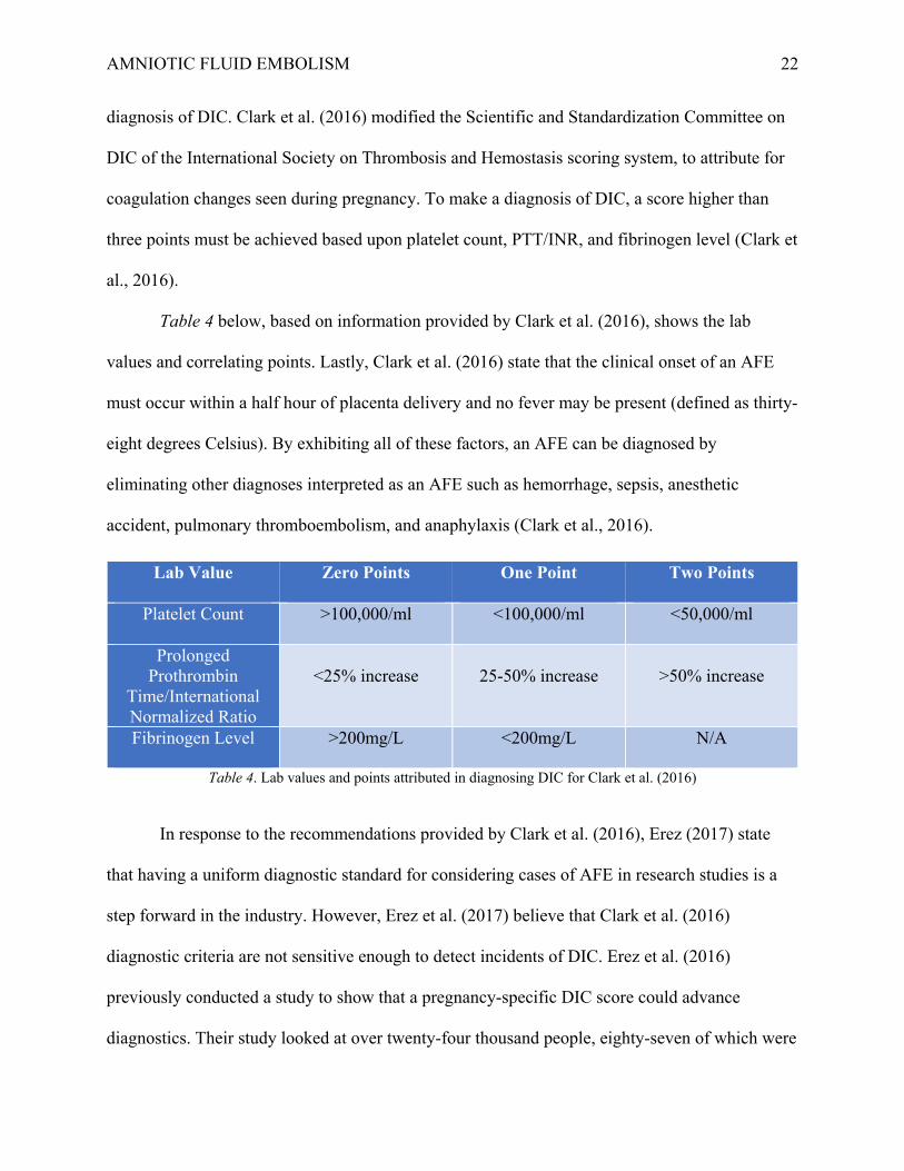
Diagnosing an AFE remains difficult as a universal diagnostic criterion does not exist (aside
from in reported research); thus, its identification is often made when another differential
diagnosis fails to manifest. The presentation of the following biomarkers: squamous cell
carcinoma antigen, CK13, and CK10/13 can aid in the investigation of an AFE event.
Management of an AFE takes a comprehensive approach with considerations to cardiac
resuscitation, post-cardiac arrest care, hemodynamic support, coagulopathy, and uterine atony.
New and unconventional methods in the treatment of an AFE have been suggested in three case
studies using C1INH, lipid emulsion therapy, and atropine/ondansetron/ketorolac.
AMNIOTIC FLUID EMBOLISM 3
Amniotic Fluid Embolism in the Parturient Patient
AFE Pathophysiology
According to Kanayama & Tamura (2014), lanugo, meconium, squamous cells, tissue
factors, and vernix are fetal materials that make up amniotic fluid. An amniotic fluid embolism
(AFE) develops as a direct result of amniotic fluid entering maternal circulation. Kanayama &
Tamura (2014), explain that this process occurs when the amniotic fluid leaks out of its sac and
joins maternal circulation via ruptured vessels in the uterine cavity or muscle. As this fluid
travels into the maternal circulation, its contents can cause physical obstructions or ensure an
anaphylactoid reaction that can develop into disseminated intravascular coagulation (DIC)
(Kanayama & Tamura, 2014). Two types of AFEs exist (1) those that are first attributed to
pulmonary symptoms and lead to cardiopulmonary collapse or (2) those that start with atonic
bleeding/DIC (Kanayama & Tamura, 2014).
Rath, Hofer, & Sinicina (2014) explain how an AFE recognized as pulmonary symptoms
can progress into cardiopulmonary collapse through two phases. In the first phase, pulmonary
vasoconstriction increases pulmonary resistance and pulmonary hypertension (Rath et al., 2014).
This increase causes the cardiac system to go into right heart failure attributed to pressure
overload, which can be viewed as right ventricle dilation and severe tricuspid insufficiency with
transesophageal echocardiography (Rath et al., 2014). Rath et al. (2014) go on to explain that as
the perfusion to the lungs is interrupted, inflammation develops that damages the alveoli,
contributing to respiratory failure and hypoxia. In phase two of this process, left heart failure and
resulting pulmonary edema are evident (Rath et al., 2014). Kanayama & Tamura (2014),
describe phase two in more detail, stating that left ventricle insufficiency causes an increase in
pulmonary wedge pressure and a decrease in left ventricle work index and systemic vascular
resistance.
AMNIOTIC FLUID EMBOLISM 4
The other type of AFE that exists presents with atonic bleeding/DIC. Rath et al. (2014)
state that the procoagulant substances within the amniotic fluid (tissue factor and
phosphatidylserine), can directly or indirectly cause the cascading effects of DIC. These
procoagulant substances occur directly via cytokines or indirectly by complement activation
from the extrinsic coagulation cascade leading to hyperfibrinolysis (Rath et al., 2014). Kanayama
& Tamura (2014) discuss this further by saying that when amniotic fluid associates with allergy-
associated cells, such as mast cells located within the cervix, it produces bradykinin and
inflammatory cytokines. The production of these mediators facilitates uterine muscle relaxation
and edema that causes incoagulable vaginal bleeding after delivery (Kanayama & Tamura,
2014).
Figure 1. Summarizing the two main types of AFEs and their related pathophysiology. Reprinted from “AFE: diagnosis and management” by Pacheco, L. D., Saade, G., Hankins, G. D., Clark, S. L., & Society for Maternal-Fetal Medicine SMFM, 2016, American journal of obstetrics and gynecology, 215(2), B16-B24.
AMNIOTIC FLUID EMBOLISM 5
Literature Review
Bonnet et al. (2018)
Bonnet et al. (2018) conducted a retrospective study over four years with the goals of
applying a new criterion for AFE and describing the epidemiology, characteristics, and
management of an AFE. The diagnostic criteria, proposed by the Society for Maternal-Fetal
Medicine and the AFE Foundation, was comprised of four conditions that must be present to
diagnose the patient as having an AFE. The four principles are as follows: (1) abrupt
cardiopulmonary arrest, or hypotension (systolic blood pressure less than ninety millimeters of
mercury) with respiratory compromise, (2) documentation of DIC after appearance of
preliminary symptoms with coagulopathy detected before significant loss of blood, (3) onset
during labor or within a half hour of placenta delivery, and (4) no fever (fever defined as greater
than thirty-eight degrees Celsius) (Bonnet et al., 2018).
Results.
Incidence. Bonnet et al. (2018) reported that there were close to four million live births
during their study period. Out of these live births, four hundred twenty-nine maternal deaths
occurred, thirty-nine of which being attributed to an AFE (Bonnet et al., 2018). Of the thirty-nine
deaths, thirty-six contained a complete medical record, and thus data was reported on these
thirty-six cases (Bonnet et al., 2018).
Risk Factors. In considering maternal age in death from an AFE, forty-two percent of
women were over the age of thirty-five and had a body mass index less than thirty before
pregnancy (Bonnet et al., 2018). Bonnet et al. (2018) pointed out that those in between one and
three parities represented fifty-eight percent of the patient population. Out of the thirty-two
patients who went into labor, sixteen had their labor induced, nine of which were induced with
prostaglandins (Bonnet et al., 2018). Out of the thirty-two patients who went into labor, eighteen
AMNIOTIC FLUID EMBOLISM 6
received oxytocin (Bonnet et al., 2018). Additionally, Bonnet et al. (2018) looked at the mode of
delivery; the majority of patients who suffered an AFE underwent a cesarean section (n=nineteen
during labor and three before labor, netting a total of twenty-two patients or about sixty-nine
percent). Interestingly, when looking at fetal characteristics, sixty-six percent of fetuses were of
male gender (Bonnet et al., 2018). Also, an AFE was less common with a gestational age shorter
than thirty-seven weeks (Bonnet et al., 2018).
Presentation. Out of the thirty-six patients that had an AFE, eighty-one percent exhibited
premonitory signs, such as neurological/respiratory signs, fainting, and fetal heart rate
abnormalities (Bonnet et al., 2018). Bonnet et al. (2018) looked into more detail about the
symptoms exhibited by these patients; ninety-seven percent of patients collapsed (n=eighteen as
a first sign), eighty-three percent showed signs of a clinical coagulopathy (n=eight as a first
sign), forty-four percent of patients had respiratory compromise (n=thirteen as a first sign), and
thirty-three percent suffered from a seizure (n=eleven as a first sign).
When looking at hemorrhage, Bonnet et al. (2018) stated that twenty patients exhibited
this symptom immediately, with eight patients displaying it at a later time. Out of the twenty-
four patients who had their platelet count drawn, fifty-percent showed a platelet count greater
than one hundred thousand (Bonnet et al., 2018). According to Bonnet et al. (2018), twenty-two
patients had a prolonged prothrombin time (PTT) and fibrinogen level drawn. Out of these
twenty-two patients described by Bonnet et al. (2018), nearly all showed more than a fifty
percent increase in their PTT (n=nineteen) and the majority had a fibrinogen level less than one
(n=sixteen).
The National Experts Committee concluded that fifty-six percent of patients diagnosed
with an AFE had suboptimal care (Bonnet et al., 2018). Bonnet et al. (2018) state that the most
popular reasons for suboptimal care included obstetrical care (n=sixteen/forty-four percent),
AMNIOTIC FLUID EMBOLISM 7
inadequate hemorrhage management (n=fifteen/forty-two percent), and delayed hysterectomy or
no hysterectomy when clinically indicated (n=thirteen/thirty-six percent). Of the six patients who
were in the intensive care unit, Bonnet et al. (2018) state suboptimal care was exhibited by a
products while lab work suggested coagulopathy (n=three), and cardiopulmonary resuscitation
being stopped early (n=two).
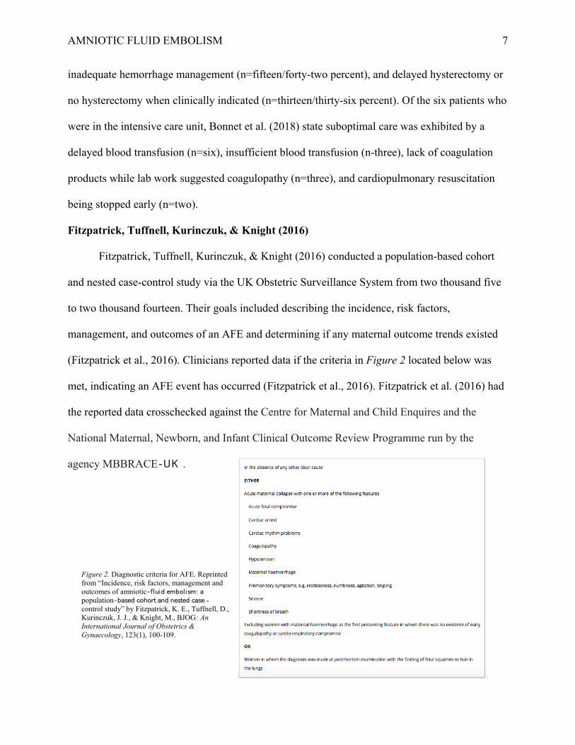
Fitzpatrick, Tuffnell, Kurinczuk, & Knight (2016)
Fitzpatrick, Tuffnell, Kurinczuk, & Knight (2016) conducted a population-based cohort
and nested case-control study via the UK Obstetric Surveillance System from two thousand five
to two thousand fourteen. Their goals included describing the incidence, risk factors,
management, and outcomes of an AFE and determining if any maternal outcome trends existed
(Fitzpatrick et al., 2016). Clinicians reported data if the criteria in Figure 2 located below was
met, indicating an AFE event has occurred (Fitzpatrick et al., 2016). Fitzpatrick et al. (2016) had
the reported data crosschecked against the Centre for Maternal and Child Enquires and the
National Maternal, Newborn, and Infant Clinical Outcome Review Programme run by the
agency MBBRACE‐UK .
Figure 2. Diagnostic criteria for AFE. Reprinted from “Incidence, risk factors, management and outcomes of amniotic‐fluid embolism: a population‐based cohort and nested case -control study” by Fitzpatrick, K. E., Tuffnell, D., Kurinczuk, J. J., & Knight, M., BJOG: An International Journal of Obstetrics & Gynaecology, 123(1), 100-109.
AMNIOTIC FLUID EMBOLISM 8
Results.
Incidence. Fitzpatrick et al. (2016) data showed that out of over seven million births, a
total of one hundred twenty confirmed cases of AFE transpired with twenty-three cases deemed
fatal. Fitzpatrick et al. (2016) data represent a total incidence of an AFE occurring one-point-
seven times per one hundred thousand people, and a fatality incidence occurring zero-point-three
times per one hundred thousand people.
Risk Factors. Similar to Bonnet et al. (2018) study, Fitzpatrick et al. (2016) study
showed that the majority of woman who suffered an AFE were less than thirty-five years old,
had a body mass index less than thirty, and had a parity of one or higher. Fitzpatrick et al. (2016)
looked further into the ethnicity of affected individuals, the majority being white (n=ninety-
two/seventy-seven percent) compared to black and other ethnic groups (n=twenty-five/twenty-
one percent). Also, Fitzpatrick et al. (2016) had a higher incidence of an AFE in those who never
smoked or quit smoking. Patients who were pregnant with one fetus were more likely than those
who were pregnant with multiple fetuses to develop an AFE (Fitzpatrick et al., 2016).
Additionally, Fitzpatrick et al. (2016) state that forty-nine patients (forty-one percent) had labor
induced. When looking at additional maternal conditions, all three patients who suffered from
placenta previa died from an AFE (Fitzpatrick et al., 2016).
As said by Fitzpatrick et al. (2016) in comparing the above risk factors, all results were
similar among those who survived and died from an AFE. Fitzpatrick et al. (2016) examined the
incidence of AFE with induction of labor. They found that there was an increased incidence of
AFE in women who received prostaglandin without oxytocin, oxytocin without prostaglandin,
and who received both prostaglandin and oxytocin (Fitzpatrick et al., 2016). Out of the women
diagnosed with an AFE and who received prostaglandin, Fitzpatrick et al. (2016) report that
ninety-five percent of patients received dinoprostone, and only one patient received misoprostol.
AMNIOTIC FLUID EMBOLISM 9
Presentation. Fitzpatrick et al. (2016) state that more than half of those diagnosed
(n=sixty-two) had an AFE present before/at delivery, compared to those (n=fifty-five) who
presented at a median of nineteen minutes post-delivery. In those diagnosed with an AFE before,
during, or post-delivery, the median age of gestation was thirty-nine weeks, and diagnosis ranged
from zero minutes to two days, with a median of thirty-three minutes (Fitzpatrick et al., 2016).
Women that presented with cardiac arrest as the first recognized symptom of an AFE were more
likely to die or have a permanent neurological injury (Fitzpatrick et al., 2016). Also, most
women (n=one hundred eight/ninety-one percent) had their membranes ruptured before AFE
presentation (Fitzpatrick et al., 2016). Similar to Bonnet et al. (2018) study, the highest incidence
of AFE was in those that delivered via cesarean section (Fitzpatrick et al., 2016). Those also at
an increased risk of having an AFE included those who delivered vaginally with instrumentation,
followed by spontaneous vaginal delivery (Fitzpatrick et al., 2016).
Management.
Coagulation. Fitzpatrick et al. (2016) mention that out of the women diagnosed with an
AFE, forty-four (thirty-seven percent) did not have any coagulopathy support. Of those women
that did have coagulopathy support, thirty-one patients (twenty-six percent) received only one
method of support, while twenty-one patients (eighteen percent) received more than one method
of coagulopathy management (Fitzpatrick et al., 2016). Cryoprecipitate was used in six cases
(twenty-percent) of those who died from an AFE, compared to forty-one cases (forty-six percent)
of those who survived an AFE (Fitzpatrick et al., 2016). According to Fitzpatrick et al. (2016),
platelets were used in eight cases (twenty-seven percent) of those who died from an AFE,
compared to thirty-six cases (forty percent) of those who survived an AFE. Fresh frozen plasma
was more likely to be used than cryoprecipitate and platelets as evidenced by its use in seventeen
cases (fifty-seven percent) of those who died from an AFE, compared to fifty-two cases (fifty-
AMNIOTIC FLUID EMBOLISM 10
eight percent) of those who survived an AFE (Fitzpatrick et al., 2016). Factor VIIa and
fibrinogen were less commonly used compared to the coagulopathy management methods used
above. Fitzpatrick et al. (2016) show the administration of Factor VIIa appeared in six cases
(twenty-percent) of those who died from an AFE, compared to twenty cases (twenty-two
percent) of those who survived an AFE. Similarly, fibrinogen was used in one case (three
percent) of those who died from an AFE, compared to one case (one percent) of those who
survived an AFE (Fitzpatrick et al., 2016).
Other. Aside from attempting to control an AFE with the coagulopathy methods
discussed above, additional management strategies exist. Fitzpatrick et al. (2016) indicate that
performing a hysterectomy was most common in those that died and survived an AFE (forty
percent and twenty-one percent respectively). Exchange transfusions, plasma exchanges, and
embolization were not a method used in those who died from an AFE but were used in those who
survived (Fitzpatrick et al., 2016). Misoprostol and Hemabate were two other management
techniques employed. Fitzpatrick et al. (2016) show the administration of Misoprostol occurred
in two cases (seven percent) of those who died from an AFE, compared to four cases (four
percent) of those who survived an AFE. Similarly, Hemabate was used in two instances (seven
percent) of those who died from an AFE, compared to six cases (seven percent) of those who
survived an AFE (Fitzpatrick et al., 2016). Fitzpatrick et al. (2016) express that intrauterine
balloons were common in those that survived an AFE compared to those who died (n=six and
n=one respectively). Tranexamic acid was used in one patient who died from an AFE, while it
was more frequently (n=five) used in patients who survived (Fitzpatrick et al., 2016).
AMNIOTIC FLUID EMBOLISM 11
Guillaume et al. (2013)
Guillaume et al. (2013) conducted a retrospective study over ten years with the goals of
providing up to date statistics on AFEs and determining ways to advance current practice
guidelines. Guillaume et al. (2013) defined an AFE as exhibiting the following symptoms: (1)
circulatory and/or cardiopulmonary arrest, (2) hypoxia with oxygen saturation less than ninety
percent, (3) a neurological component such as loss of consciousness/agitation/seizures/coma, and
(4) hemorrhage associated with DIC.
Results.
Incidence. According to Guillaume et al. (2013), data for this study was gathered based
upon the coding system of hospitals in conjunction with histology and pathology reports. During
the study period, there was close to thirty-nine thousand births, eleven of which met the
definition of an AFE (Guillaume et al., 2013). Using this data, Guillaume et al. (2013) concluded
that the incidence of an AFE was twenty-eight in one hundred thousand. Out of the eleven
patients who suffered an AFE, there were three fatalities, carrying a maternal mortality rate of
seven point eight in one hundred thousand people (Guillaume et al., 2013).
Risk Factors. Like discussed in the studies by Bonnet et al. (2018) and Fitzpatrick et al.
(2016), Guillaume et al. (2013) had similar risk factors that were noted in women who suffered
an AFE in regard to maternal age (median of thirty-two years), parity (median one point seven),
and gestational age (median forty-weeks). Similar to Bonnet et al. (2018) study, Guillaume et al.
(2013) reported that the majority of fetuses were male gender.
When looking at if inducing labor played a role in increasing the odds of an AFE,
Guillaume et al. (2013) state that six patients had labor induced. Three of these patients received
misoprostol; two received dinoprostone, and one received oxytocin (Guillaume et al., 2013).
Also, Guillaume et al. (2013) state that oxytocin was used to enhance labor in nine patients. This
AMNIOTIC FLUID EMBOLISM 12
data is similar to that of Fitzpatrick et al. (2016) study which showed that there was an increased
risk of developing an AFE in women who received prostaglandin without oxytocin, oxytocin
without prostaglandin, and both prostaglandin and oxytocin. Different from Fitzpatrick et al.
(2016) study was that more patients received misoprostol than dinoprostone (Guillaume et al.,
2013). Lastly, the patient’s membranes had been ruptured, either spontaneously or artificially, in
all cases of AFE (Guillaume et al., 2013).
Presentation. Guillaume et al. (2013) state that eight-cases of an AFEs occurred during
the peripartum period, two cases transpired in the postpartum period, with one incident taking
place during a scheduled cesarean section. This data is different from Bonnet et al. (2018) and
Fitzpatrick et al. (2016) studies which showed that AFEs were more likely to occur in those
undergoing a cesarean section. In the eight cases of AFE appearing in the peripartum period,
Guillaume et al. (2013) report that delivery of the fetus was performed in all cases within twenty
minutes, with five of those cases being within five minutes of when symptoms of an AFE first
appeared. Delivery methods included vacuum-assisted delivery (n=one) and emergency cesarean
sections (n=seven) (Guillaume et al., 2013). When Guillaume et al. (2013) looked at presenting
symptoms, six patients had a cardiopulmonary arrest, with this being the first symptom in two
patients. In every case of AFE, symptoms progressed to post-partum hemorrhage, with ten out of
the eleven patients having DIC (Guillaume et al., 2013). When looking at cytology and histology
reports, Guillaume et al. (2013) state that eight patients had fetal squamous cells present, with
one of these cases also manifesting meconium.
Management. Guillaume et al. (2013) report that seven of the eleven patients who
experienced post-partum hemorrhage required surgical intervention to establish hemostasis.
Methods to secure hemostasis included an immediate hysterectomy (five patients) and delayed
hysterectomy after ligation of uterine/hypogastric arteries failed (two patients who both later
AMNIOTIC FLUID EMBOLISM 13
died) (Guillaume et al., 2013). Out of the six patients who coded, cardiopulmonary resuscitation
was sufficient in one patient, and defibrillation was required in five patients (Guillaume et al.,
2013). Guillaume et al. (2013) explain that extracorporeal membrane oxygenation with veno-
venous cannulation was utilized in two patients, both of which later died. In every patient,
transfusions took place with a median of eleven units of packed red blood cells, eleven units of
fresh frozen plasma, and two units of platelets (Guillaume et al., 2013). Guillaume et al. (2013)
also say that the administration of fibrinogen occurred in six patients along with Factor VIIa in
two patients. In comparing the products transfused in this study to Fitzpatrick et al. (2016) study,
the use of fibrinogen was more commonly used in Guillaume et al. (2013) study.
McDonnell et al. (2015)
McDonnell et al. (2015) conducted a population-based descriptive study specific to the
countries of Australia and New Zealand as AFEs are one of the leading causes of maternal death,
yet there is no national population study currently in existence in these areas. To collect their
data, McDonnell et al. (2015) utilized the Australasian Maternity Outcomes Surveillance System
which they state represents about ninety-six percent of women delivering in Australia and one
hundred percent of women giving birth in hospitals in New Zealand. After identifying an AFE
event, a case-specific questionnaire was then filled out (McDonnell et al., 2015). McDonnell et
al. (2015) collected their data throughout two years, from the beginning of two thousand ten to
the end of two thousand eleven. They defined an AFE clinically by the presence of acute
hypotension/cardiac arrest, acute hypoxia, and coagulopathy without a source of explanation or
via lab tests in the postmortem period (McDonnell et al., 2015).
Results.
Incidence. McDonnell et al. (2015) results showed that out of over six hundred thousand
women giving birth, thirty-three confirmed cases of an AFE existed, with five cases resulting in
AMNIOTIC FLUID EMBOLISM 14
death. This data shows an incidence of an AFE occurring five point four times in one hundred
thousand people and a mortality rate of zero point eight deaths in one hundred thousand people
(McDonnell et al., 2015). When comparing these rates to studies previously mentioned, they are
higher than reported in the Fitzpatrick et al. (2016) study but less than reported in Guillaume et
al. (2013) study.
Risk Factors. McDonnell et al. (2015) state that gestational age (seventy-three percent
exceeded thirty-seven weeks) was a risk factor for developing an AFE. Two characteristics that
differed in comparison to the studies previously mentioned are that the majority of patients (fifty-
two percent) were over the age of thirty-five and (fifty-two percent) had a parity of zero
(McDonnell et al., 2015). Similar to Bonnet et al. (2018) and Fitzpatrick et al. (2016) studies,
body mass index was taken into consideration, but in this study body mass index was reported as
less than thirty-five (thirty-one patients) making it impossible to identify an association with the
other studies. Comparable to Fitzpatrick et al. (2016) study, nonsmokers were shown to have an
increased risk, and placenta previa (fifteen percent) was also established as a risk factor
(McDonnell et al., 2015). Moreover, consistent with Bonnet et al. (2018) and Guillaume et al.
(2013) study, the majority of fetus’s (sixty-four percent) were of male gender (McDonnell et al.,
2015). Furthermore, McDonnell et al. (2015) looked at the number of women who received
assistance with conceiving, which represented twenty-four percent. Lastly, induction of labor did
prove to be a risk factor for AFE as evidenced by fourteen women having labor induced or
augmented (McDonnell et al., 2015).
Presentation. McDonnell et al. (2015) looked at the location of where the AFE took
place, the majority of which took place in the operating room or birthing suite (both had fifteen
cases), with two events occurring at home, and one incident not being recorded. Five cases
occurred during the pre-labor period, four cases in the first stage of labor, four cases during the
AMNIOTIC FLUID EMBOLISM 15
second stage of labor, and the majority (seventeen cases) during the postpartum period
(McDonnell et al., 2015). The onset of an AFE was noted to occur within five minutes before or
after delivery in forty-eight percent of patients (McDonnell et al., 2015). McDonnell et al. (2015)
recorded the signs and symptoms that patients most commonly presented with as premonitory
(twenty-seven percent), hypotension (twenty-one percent), shortness of breath (fifteen percent),
and fetal compromise (fifteen percent). They went on to report that twenty-two patients (sixty-
seven percent) had a cesarean section, of which six were elective, eight were emergent, and eight
did not have a recorded reason (McDonnell et al., 2015). Like Bonnet et al. (2018) and
Fitzpatrick et al. (2016) studies, this showed a higher incidence of AFE with cesarean sections.
Associated features included seventy-nine percent experiencing hemorrhage, seventy-six percent
(two hundred eleven beats per minute), one-centimeter cervical dilation with no effacement and
intact membranes (Rezai et al., 2017). Rezai et al. (2017) stated that the patient was given
intravenous fluids and started on antibiotics. A decision was made for an emergency cesarean
section with general anesthesia when a nonreactive tracing with minimal variability and
persistent fetal tachycardia was observed (Rezai et al., 2017).
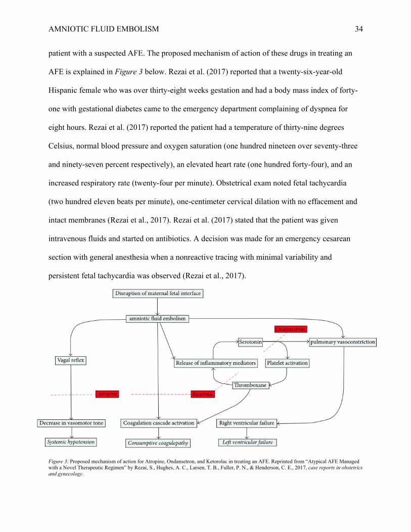
Figure 3: Proposed mechanism of action for Atropine, Ondansetron, and Ketorolac in treating an AFE. Reprinted from “Atypical AFE Managed with a Novel Therapeutic Regimen” by Rezai, S., Hughes, A. C., Larsen, T. B., Fuller, P. N., & Henderson, C. E., 2017, case reports in obstetrics and gynecology.
AMNIOTIC FLUID EMBOLISM 35
Rezai et al. (2017) reported that immediately after delivery of the fetus and before the
placenta was delivered the patient had the following vital signs: oxygen saturation of seventy-
two percent, blood pressure of seventy-two over forty-eight, and no end-tidal carbon dioxide
(equipment checked with no disconnections). Within one minute of these symptoms, two-tenths
of a milligram of Atropine, eight milligrams of Ondansetron, and fifteen milligrams of Ketorolac
were administered (Rezai et al., 2017). Rezai et al. (2017) state that within two to three minutes
of the patient receiving the A-OK medications the following vital signs were present: oxygen
saturation of ninety-seven percent, blood pressure of one hundred thirty-eight over sixty-eight,
and end-tidal carbon dioxide of thirty-two. Fifty units of Oxytocin, two doses of Carboprost,
three units of packed red blood cells, one unit of fresh frozen plasma, and three and a half liters
of fluid were used to treat uterine atony and hemorrhage as evidenced by an estimated blood loss
of two liters (Rezai et al., 2017). Rezai et al. (2017) report that the only vasopressor used was
phenylephrine with a total of eighteen hundred micrograms being administered in fifty-seven
minutes (before the start of the incision to the end of surgery). The patient was admitted to the
intensive care unit, extubated the following day, and sent home on a postoperative day three
(Rezai et al., 2017). Rezai et al. (2017) reported that a chest computed tomography scan, chest x-
ray, doppler ultrasound for thrombosis, lab work to suggest disseminated intravascular
coagulation, blood/urine/sputum cultures, and placenta pathology came back all negative.
Conclusion
The occurrence of an amniotic fluid embolism (AFE) causes a physical obstruction or an
anaphylactoid reaction within the parturient patient. Symptoms present in two forms: as either
cardiopulmonary collapse or DIC, both frequently leading to maternal death. An examination of
current research studies analyzed the incidence, risk factors, presentation, and management of an
AFE in the healthcare setting. From these studies, it shows that an AFE has an incidence rate
AMNIOTIC FLUID EMBOLISM 36
ranging between 1.7-28:100,000 women. Significant risk factors predisposing women to an AFE
include a maternal age less than thirty-five years old, a gestational age greater than thirty-seven
weeks, a parity greater than one, induction of labor, cesarean section, nonsmoking, and male
gender fetus. Additionally, it is shown that an AFE most often occurs prior to or during delivery
compared to the post-delivery period.
Making an in-hospital clinical diagnosis of an AFE remains challenging as a universal
diagnostic criterion has yet to exist. Thus, further research into identifying a diagnostic tool
would be beneficial. As there have been attempts made to establish a uniform guideline to
identify AFE events when reporting in research, this can be used as a stepping stone to build on.
It appears that the presentation of squamous cell carcinoma antigen, CK13, and CK10/13
biomarkers can aid in the investigation/confirmation of an AFE event. Management of an AFE
takes a comprehensive approach with attention to cardiac resuscitation, post-cardiac arrest care,
hemodynamic support, coagulopathy, and uterine atony. In three unique case reports, the
treatment of an AFE was proven successful with using C1INH, lipid emulsion therapy, and
atropine/ondansetron/ketorolac. By applying the information discussed into a nurse anesthetists’
clinical practice, it allows for a better understanding of what an AFE entails, and how to be
prepared in treating the multitude of symptoms that occur in an AFE event.
AMNIOTIC FLUID EMBOLISM 37
References
Akasaka, M., Osato, K., Sakamoto, M., Kihira, T., Ikeda, T., & Yamawaki, T. (2018). Practical
use of C1 esterase inhibitor concentrate for clinical AFE. Journal of Obstetrics and
Kanayama, N., & Tamura, N. (2014). AFE: pathophysiology and new strategies for
AMNIOTIC FLUID EMBOLISM 38
management. Journal of Obstetrics and Gynaecology Research, 40(6), 1507-1517.
doi:10.1111/jog.12428
Kaur, K., Bhardwaj, M., Kumar, P., Singhal, S., Singh, T., & Hooda, S. (2016). Amniotic fluid
embolism. Journal of Anaesthesiology, clinical pharmacology, 32(2), 153.
doi:10.4103/0970-9185.173356 Koike, N., Oi, H., Naruse, K., Kanayama, N., & Kobayashi, H. (2017). Squamous cell carcinoma
antigen as a novel candidate marker for AFE. Journal of Obstetrics
and Gynaecology Research, 43(12), 1815-1820. doi:10.1111/jog.13453 Lynch, W., McAllister, R. K., Lay, J. F., & Culp, W. C. (2017). Lipid emulsion rescue of
AFE-induced cardiac arrest: a case report. A&A Practice, 8(3), 64-66.
doi:10.1213/XAA.0000000000000427
McAllister, R. K., Lay Jr, J. F., & Culp Jr, W. C. (2018). Letter to 'Practical use of C1 esterase
inhibitor concentrate for clinical AFE'. The journal of obstetrics and
gynaecology research. Retrieved from https://obgyn.onlinelibrary.wiley.com/doi
/full/10.1111/jog.13821
McDonnell, N., Knight, M., Peek, M. J., Ellwood, D., Homer, C. S., McLintock, C., ... &
Sullivan, E. (2015). AFE: an Australian-New Zealand population-
based study. BMC pregnancy and childbirth, 15(1), 352. doi:10.1186/s12884-015-
0792-9.
Moore, L. E. (2018). AFE Differential Diagnoses. Medscape. Retrieved November 16, 2018
from https://emedicine.medscape.com/article/253068-differential