18
SH Workshop Chicago 2017 An Aggressive Burkitt-like Lymphoma SH2017-0137 Katrin Hüttl, MD German Ott, MD Robert-Bosch-Krankenhaus Stuttgart, Germany

SH Workshop Chicago 2017
An Aggressive Burkitt-likeLymphomaSH2017-0137
Katrin Hüttl, MDGerman Ott, MD
Robert-Bosch-Krankenhaus Stuttgart, Germany
Clinical Data• A 21-year old male patient
• 10/2015Presents with acute abdominal symptoms. Laboratory testsunremarkable with exception of slight anemia (Hb 9,3 g/dl ). Diagnosis of a tumor in the appendiceal region. Clinical stagingreveals Stage IVA disease with involvement of the omentum.
• 11/2015Opstipation, abdominal discomfort and pain. After diagnosisof an ileus, extended surgery with omentectomy and resectionof parts of terminal ileum and colon. Repeated staging revealsno enlarged lymph nodes cervical, mediastinal, abdominal. Nobone marrow involvement, no CNS involvement.
Histology reveals aggressive lymphoma.


Summary of Features
• Large tumor mass in the area of theterminal ileum, cecum and appendix
• Aggressive lymphoma with a „starry sky“ pattern
• Medium-sized to large cohesive blasts
Burkitt-Lymphoma?
DLBCL with „Burkitt-like“ features?

CD20
CD10
CD3
BCL2

Ki67 MYC

Summary of Immunohistochemistry
CD20+, CD10+, BCL2-, BCL6+, IRF4/MUM1-, Ki67 100%
But: No „significant“ over-expression of MYC
„Classical“ Burkitt phenotype

MYC-BAPMYC t(8;14)FISH:

Burkitt-Lymphoma without MYC Rearrangement?
These findings indicate the existenceof a molecular distinct subset of B-celllymphomas reminiscent of BL which ischaracterized by deregulation ofgenes in 11q (Blood. 2014;123(8):1187-1198).
A recurrent 11q aberration pattern characterizes a subsetof MYC-negative high-grade B-cell lymphomas resemblingBurkitt lymphoma
Pienkowska-Grela Med.Oncol. 2011Ferreiro Haematologica 2015Zajdel Tumour Biol 2015
Salaverria et al.
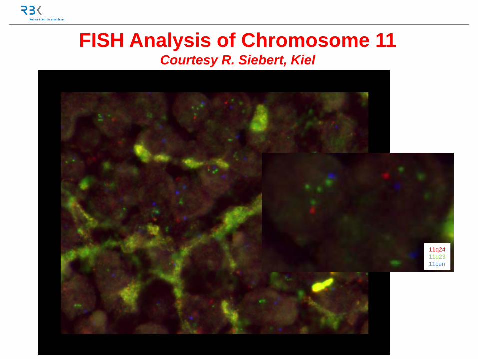
FISH Analysis of Chromosome 11Courtesy R. Siebert, Kiel
11q2411q2311cen

OncoScan Analysis of the LymphomaCourtesy R. Siebert, Kiel
Gain 3qGain 11q23
Del(11q24) ETS1
Gain 3q

Diagnosis:(WHO-Update 2016)
Burkitt-like Lymphoma with 11q Aberration (Provisional)
Swerdlow et al. Blood 2016
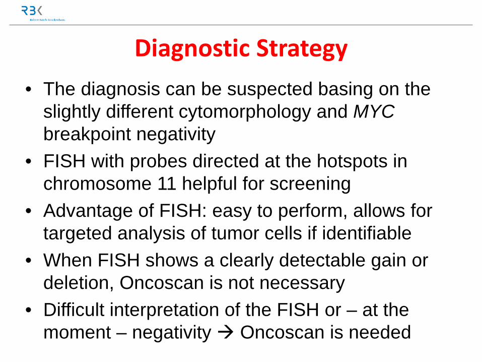
Diagnostic Strategy• The diagnosis can be suspected basing on the
slightly different cytomorphology and MYCbreakpoint negativity
• FISH with probes directed at the hotspots in chromosome 11 helpful for screening
• Advantage of FISH: easy to perform, allows fortargeted analysis of tumor cells if identifiable
• When FISH shows a clearly detectable gain ordeletion, Oncoscan is not necessary
• Difficult interpretation of the FISH or – at themoment – negativity Oncoscan is needed

• 11/2015 – 03/2016:Therapy with 8 cycles of chemotherapyaccording to the B-ALL protocol. Atypicalpneumonia after the fourth cycle.
• Last staging 08/2017: Complete Remission.
Follow-up
Prof. Reiner Siebert, MD and Rabea Wagener PhD, University of Ulm, GermanyMatthias Vöhringer, MD, Department of Hematooncology, Robert-Bosch-Krankenhaus, Stuttgart, GermanyAnnette Staiger PhD, and Heike Horn PhD, MolecularDiagnostics Unit, Robert-Bosch-Krankenhaus and Dr. Margarete-Fischer-Bosch Institute of Clinical Pathology, Stuttgart, Germany.
Acknowledgements

Burkitt-likeLymphoma with 11q
Aberration
Panel Diagnosis:


![Women and Bladder Cancer - Oncoscan yellow 8-20_.pdf · bladder cancer [18,170] than cervical cancer [11,270] > 500,000 people in the U.S. have/had bladder cancer – highest recurrent](https://static.documents.pub/doc/80x56/5f694c18ea002e289e4b0db0/women-and-bladder-cancer-yellow-8-20pdf-bladder-cancer-18170-than-cervical.jpg)

![When and how to test for C MYC in aggressive B cell lymphomasBCL-U according to the suggestion of Salaverria et al. [34]. Diffuse large B cell lymphoma, not otherwise specified In](https://static.documents.pub/doc/80x56/5f820394a05527210372eca9/when-and-how-to-test-for-c-myc-in-aggressive-b-cell-lymphomas-bcl-u-according-to.jpg)
