INTRODUCTION Partial edentulism can lead to multiple complications from the functional, biological and esthetic aspects. 1,2 After the loss of some natural teeth, the inter- and intra-arch relationships of the remaining dentition might be affected. The adjacent teeth tend to adapt physiologically to the changes by drifting or tip- ping toward the resultant space, while the antagonist teeth has the tendency to overerupt. 1,2 In addition to limit the space for any future prosthesis, occlusal interferences and disfigurement can be introduced. Subsequently, the rehabilitative treatment will be complicated by including invasive adjunctive therapies such as crown lengthening surgery, elective endodontic treat- ment, orthodontic movement, and increasing the vertical dimension of occlusion. 3 This clinical report demonstrates the application of well-planned rehabilitative treatment and coordinated team work of a patient with extreme features of partial edentulism. Due to the complexity of the initial presentation, the final treatment was accomplished after series of provisional prostheses in con- junction with crown lengthening surgery. The final treat- ment involved innovatively designed fixed and removable pros- theses that employ the concepts of milled surfaces, telescop- ic attachments, and strategic implant support. CASE REPORT A 49-year-old partially edentulous male was referred to the prosthodontic clinic for the management of his complex den- tal status. His chief complaint was dissatisfaction of his exist- ing dental condition that affected quality of his life. He requested a predictable treatment to improve his dental func- tion and restore the missing teeth. His medical status was unre- markable. Intraorally (Fig. 1A-D), the prominent features were as followed: all the remaining teeth were unopposed and almost contacting the residual ridges; absence of posterior or anterior teeth support, and no occlusal guidance. The exces- sive thickness of the attached gingival band indicated over-erup- tion of the remaining teeth which might be the cause of the sig- nificant disfigurement of the occlusal plane. The maxilla and the mandible were partially edentulous and classified as Kennedy Class II modification 1. None of the teeth were mobile. The remaining maxillary incisors and the left mandibu- lar premolar were extensively carious and deemed non-restor- able. The mandibular left first molar was carious on the mesial surface and required root canal treatment. Measuring the vertical dimensions revealed excessive loss of vertical dimen- sion of occlusion (VDO) which was manifested as excessive freeway space (FWS) (6 mm) (Fig. 1B). There was no sign of temporomandibular disorder. The phonetic assessment revealed DOI:10.4047/jap.2011.3.1.37 An innovative prostheses design for rehabilitation of severely mutilated dentition: a case report Jaafar Abduo*, BDS, DClinDent Prosthodontist, Private Practice, Faculty of Dentistry, University of Western Australia Partial edentulism has multiple implications in relation to function, esthetics and future rehabilitative treatment. This case report illustrates the management of a patient with extreme consequences of partial edentulism. The main clinical findings were unopposed remaining teeth, over- eruption of the remaining teeth, loss of vertical dimension of occlusion, and significant disfigurement of the occlusal plane. Following the diag- nostic procedure, a well-coordinated prosthodontic treatment involving liaison with other dental disciplines was indicated. The management involved an innovative combination of fixed and removable prostheses in conjunction with crown lengthening surgery and strategic implant place- ment. Series of provisional prostheses were applied to facilitate the transition to the final treatment. [J Adv Prosthodont 2011;3:37-42] 37 CASE REPORT J Adv Prosthodont 2011;3:37-42 KEY WORDS. Diagnostic wax-up, Strategic implant, Provisional prosthesis, Telescopic attachment, Precision prosthesis Corresponding author: Jaafar Abduo Prosthodontist, Private Practice, Faculty of Dentistry, University of Western Australia Private Practice, Suite 6, 20 Altona Street, West Perth, Western Australia, 6005 Tel. + 0061 8 9322: e-mail, [email protected]Received January 5, 2011 / Last Revison January 24, 2011 / Accepted January 27, 2011 ⓒ 2011 The Korean Academy of Prosthodontics This is an Open Access article distributed under the terms of the Creative Commons Attribution Non-Commercial License (http://creativecommons.org/licenses/by- nc/3.0) which permits unrestricted non-commercial use, distribution, and reproduction in any medium, provided the original work is properly cited.
Transcript
INTRODUCTION
Partial edentulism can lead to multiple complications fromthe functional, biological and esthetic aspects.1,2 After theloss of some natural teeth, the inter- and intra-arch relationshipsof the remaining dentition might be affected. The adjacent teethtend to adapt physiologically to the changes by drifting or tip-ping toward the resultant space, while the antagonist teeth hasthe tendency to overerupt.1,2 In addition to limit the space forany future prosthesis, occlusal interferences and disfigurementcan be introduced. Subsequently, the rehabilitative treatmentwill be complicated by including invasive adjunctive therapiessuch as crown lengthening surgery, elective endodontic treat-ment, orthodontic movement, and increasing the verticaldimension of occlusion.3
This clinical report demonstrates the application of well-plannedrehabilitative treatment and coordinated team work of apatient with extreme features of partial edentulism. Due to thecomplexity of the initial presentation, the final treatmentwas accomplished after series of provisional prostheses in con-junction with crown lengthening surgery. The final treat-ment involved innovatively designed fixed and removable pros-theses that employ the concepts of milled surfaces, telescop-ic attachments, and strategic implant support.
CASE REPORT
A 49-year-old partially edentulous male was referred tothe prosthodontic clinic for the management of his complex den-tal status. His chief complaint was dissatisfaction of his exist-ing dental condition that affected quality of his life. Herequested a predictable treatment to improve his dental func-tion and restore the missing teeth. His medical status was unre-markable. Intraorally (Fig. 1A-D), the prominent featureswere as followed: all the remaining teeth were unopposed andalmost contacting the residual ridges; absence of posterior oranterior teeth support, and no occlusal guidance. The exces-sive thickness of the attached gingival band indicated over-erup-tion of the remaining teeth which might be the cause of the sig-nificant disfigurement of the occlusal plane. The maxillaand the mandible were partially edentulous and classifiedas Kennedy Class II modification 1. None of the teeth weremobile. The remaining maxillary incisors and the left mandibu-lar premolar were extensively carious and deemed non-restor-able. The mandibular left first molar was carious on themesial surface and required root canal treatment. Measuringthe vertical dimensions revealed excessive loss of vertical dimen-sion of occlusion (VDO) which was manifested as excessivefreeway space (FWS) (6 mm) (Fig. 1B). There was no sign oftemporomandibular disorder. The phonetic assessment revealed
DOI:10.4047/jap.2011.3.1.37
An innovative prostheses design for rehabilitation ofseverely mutilated dentition: a case report
Jaafar Abduo*, BDS, DClinDent
Prosthodontist, Private Practice, Faculty of Dentistry, University of Western Australia
Partial edentulism has multiple implications in relation to function, esthetics and future rehabilitative treatment. This case report illustrates themanagement of a patient with extreme consequences of partial edentulism. The main clinical findings were unopposed remaining teeth, over-eruption of the remaining teeth, loss of vertical dimension of occlusion, and significant disfigurement of the occlusal plane. Following the diag-nostic procedure, a well-coordinated prosthodontic treatment involving liaison with other dental disciplines was indicated. The managementinvolved an innovative combination of fixed and removable prostheses in conjunction with crown lengthening surgery and strategic implant place-ment. Series of provisional prostheses were applied to facilitate the transition to the final treatment. [J Adv Prosthodont 2011;3:37-42]
Corresponding author: Jaafar AbduoProsthodontist, Private Practice, Faculty of Dentistry, University of Western AustraliaPrivate Practice, Suite 6, 20 Altona Street, West Perth, Western Australia, 6005Tel. + 0061 8 9322: e-mail, [email protected] January 5, 2011 / Last Revison January 24, 2011 / Accepted January 27, 2011
ⓒ 2011 The Korean Academy of ProsthodonticsThis is an Open Access article distributed under the terms of the Creative CommonsAttribution Non-Commercial License (http://creativecommons.org/licenses/by-nc/3.0) which permits unrestricted non-commercial use, distribution, and reproductionin any medium, provided the original work is properly cited.
difficulties in pronouncing the /s/ and /f/ sounds. However, thepatient adapted to the missing incisal edges and was able to pro-duce the sound with relative acuity. Radiographic assess-ment indicated limited vertical alveolar bone height in the sec-ond quadrant.
The patient was stabilized by extracting the non-restorableteeth and initiating root canal treatment for the mandibular leftmolar. Oral hygiene measures were demonstrated and empha-sized before considering any rehabilitative treatment.
After obtaining study models, occlusal rims were fabricat-ed to record the centric relation at the restored VDO (FWS =2 mm). With the aid of facebow transfer record, the study mod-els were mounted on a semi-adjustable articulator (ArtexArticulator, Jensen Dental, North Haven, CT, USA) (Fig.2A). Subsequently, it was possible to assess the potentialtreatment options closely and any adjunctive procedurerequired correction of the occlusal plane. The diagnosticwax-up was completed with simulated crown lengthening
38
An innovative prostheses design for rehabilitation of severely mutilated dentition: a case report
J Adv Prosthodont 2011;3:37-42
Abduo J
Fig. 1. Initial intraoral images. A: Frontal view of the dentition in occlusion, B: Frontal view of the dentition while the mandible is at rest, C: Rightlateral view, D: Left lateral view.
A B
C D
A B
Fig. 2. A: Articulated study models in centric relation, B: Diagnostic wax-up after restoring the VDO and simulated crown lengthening surgery.
surgery (Fig. 2B). The planned occlusal scheme was unilateralgroup function with long centric following the principles of bio-logical occlusion described by Becker and Kaiser.4 The bilat-eral group function was opted because of the tendency for ClassII incisal relationship hindering efficient canine guidance.Due to the excessive FWS, there was no need to consider fur-ther increase in the VDO. On the basis of the diagnosticwax-up, it was decided to extract the severely over-eruptedmandibular left second molar and crown lengthening of all theremaining teeth. Further, the diagnostic wax-up was utilizedto determine the location of the implants in the second and fourthquadrant. The next treatment options were presented:
1. Maxillary rehabilitation with crowns and precisionimplant-supported removable partial denture (RPD).Mandibular rehabilitation with crowns, fixed partial den-ture and implant fixed partial denture.
2. Extraction of the maxillary teeth and rehabilitation withimplant fixed prosthesis. Mandibular rehabilitation withcrowns, fixed partial denture and implant fixed partial den-ture.
3. Maxillary and mandibular RPDsFor the maxilla, despite the patient’s preference for the
fixed option, he was reluctant to undergo through extensive bonegrafting procedure. Therefore, the first option was selected. Asrecommended by several authors, strategic freestandingimplant placement was considered to modify the Kennedy clas-sification.5-7 Additional advantages included improving ofthe retention and stability, enhancing of patient comfort, andsimplifying of prosthesis design.5,7,8
Surgical templates were fabricated to guide the implantplacement and the crown lengthening surgery. For themandible, thee regular platform implants (Biomet 3i, Palm BeachGardens, FL, USA) were inserted in the region of lowerright canine, first premolar and first molar. For the maxilla, toovercome the limitation of compromised bone quantity, sin-gle wide platform implant (Southern Implants Ltd, Irene,
SA) was placed posterior to the maxillary sinus with angularorientation (Fig. 3). The angular implant placement allowedengaging of maximal amount of available bone.9 To minimizethe impact of poor bone quality, conventional healing periodwas followed as advised by Friberg et al..10
The first phase of the rehabilitative treatment involvedrestoring the VDO, stabilizing the occlusion and improving theesthetics by constructing provisional maxillary cobalt-chromi-um RPD and mandibular screw-retained metal reinforcedcomposite resin FPD (Fig. 4). The patient was monitoredclosely for 3 months. Through this period, mastication, com-fort, phonetics, and esthetics were closely assessed.
The second phase of the rehabilitation comprised of prepar-ing the maxillary and mandibular teeth for porcelain fused tometal prostheses. Due to the need to rectify the occlusalplane, it was necessary to prepare all the teeth simultaneous-ly. Chair-side provisional prostheses were fabricated (Luxatemp,DMG, Hamburg, Germany) and cemented with temporarycement (TempBond, Kerr Corporation, Orange, CA, USA). Themaxillary RPD was readapted to fit the provisional prosthesis.
The technician was instructed to follow the diagnostic wax-up closely to correct the occlusal plane. In this phase, the defin-itive mandibular prostheses were completed at the correct occlusalplane. The maxilla was provisionally restored with metal-rein-forced cross arch provisional prosthesis. As described byEmtiaz and Tarnow,11 a mesh of cobalt-chromium was constructedand veneered with acrylic resin (SR Ivocron, Ivoclar Vivadent,Schaan, Liechtenstein). This allowed the major portion of thefitting surface along with all margins to be covered withacrylic resin (Fig. 5A) facilitating future removal and possi-ble adjustments.
The mandibular definitive prostheses were cemented with glassionomer cement (Fuji I, GC Corporation, Tokyo, Japan).The maxillary prosthesis was tried in and cemented tem-porarily (TempBond, Kerr Corporation, Orange, CA, USA).Any necessary occlusal adjustments were performed on the max-
39
An innovative prostheses design for rehabilitation of severely mutilated dentition: a case report
J Adv Prosthodont 2011;3:37-42
Abduo J
Fig. 3. Panoramic radiograph demonstrating the location of the maxil-lary and mandibular implants.
Fig. 4. Frontal view of the first phase of rehabilitation.
illary provisional prosthesis. From the diagnostic perspective,the maxillary provisional prosthesis allowed more preciseassessment of the function, esthetics and phonetics (Fig. 5B).The patient was reviewed weekly for a period of one month anddemonstrated high level of oral hygiene.
The final phase of the rehabilitation involved the fabricationof definitive maxillary fixed prosthesis and precision RPD. Sincethe maxillary provisional prosthesis provided stable occlusion,it was utilized to obtain precise interocclusal record. Toenhance the predictability of the laboratory articulation,medium body silicone impression material (Exahiflex, GCCorporation, Tokyo, Japan) was applied on the occlusal sur-face and the patient was asked to occlude on the previouslyachieved occlusion. For the first quadrant, porcelain fused tometal crowns were fabricated with milled surfaces incorporatedon the palatal aspect (Fig. 6). As a future contingency planning,extra-coronal precision attachment (Bredent LTD, Chesterfield,UK) was incorporated mesial to the maxillary right canine. Forthe second quadrant, telescopic retention mechanism wasused.8 Common path of insertion and an occlusal conver-gence angle of 4 degrees were obtained for palatal milled sur-faces and the primary telescopic copings with the aid of a millingsystem attached to a surveyor table (AmannGirrbach AG,Bregenz, Austria).
The merit of applying telescopic attachment is omittingthe palatal major connector without compromising the rigid-ity of the final RPD framework. For the angulated implant, thetelescopic attachment was suitable to compensate for angulatedorientation without compromising the retention. Anotheradvantage of palatal milled surfaces and telescopic attachmentsis enhancing the stability and retention by restricting theRPD path of withdrawal and creating friction between the inti-mately fitting parallel surfaces.8,12 In addition, with well dis-tributed abutments, the occlusal forces are directed axially.12
The maxillary fixed prostheses were tried in and pick-up
impression was taken with polyether impression material(Impregum Penta Soft, 3M ESPE, St. Paul, MN, USA).Secondary copings were fabricated by electroforming processon the primary telescopic copings. Precision cobalt-chromi-um framework was constructed on the milled surfaces and sec-ondary copings (Fig. 7A). The framework try-in step revealedpassive fit of all the components. The definitive RPD wasdesigned to mimic the morphology and occlusion of the pro-visional prosthesis (Fig. 7B).
The fixed prostheses and the primary coping were cement-ed permanently with glass ionomer cement (Fuji I, GCCorporation, Tokyo, Japan). The implant primary copingwas fitted and the retaining screw was tightened to 35 Ncm.The telescope fitting surface of the framework and the exter-
40
An innovative prostheses design for rehabilitation of severely mutilated dentition: a case report
J Adv Prosthodont 2011;3:37-42
Abduo J
A B
Fig. 5. A: The fitting surface of the maxillary cross-arch provisional prosthesis revealing the acrylic resin margins and fitting surfaces, B: Frontalview after fitting the definitive mandibular prosthesis and the provisional maxillary prosthesis.
Fig. 6. Occlusal view of the maxillary definitive fixed prosthesis. The palatalmilled surfaces, the extracoronal precision attachment and the tele-scopic primary copings exhibit a common path of insertion.
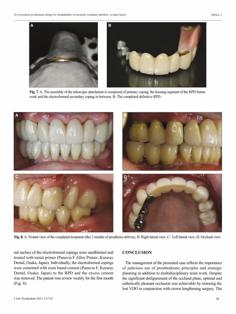
nal surface of the electroformed copings were sandblasted andtreated with metal primer (Panavia F Alloy Primer, KurarayDental, Osaka, Japan). Individually, the electroformed copingswere cemented with resin based cement (Panavia F, KurarayDental, Osaka, Japan) to the RPD and the excess cementwas removed. The patient was review weekly for the first month(Fig. 8).
CONCLUSION
The management of the presented case reflects the importanceof judicious use of prosthodontic principles and strategicplanning in addition to multidisciplinary team work. Despitethe significant disfigurement of the occlusal plane, optimal andesthetically pleasant occlusion was achievable by restoring thelost VDO in conjunction with crown lengthening surgery. The
41
An innovative prostheses design for rehabilitation of severely mutilated dentition: a case report
J Adv Prosthodont 2011;3:37-42
Abduo J
A B
Fig. 7. A: The assembly of the telescopic attachment is composed of primary coping, the housing segment of the RPD frame-work and the electroformed secondary coping in between, B: The completed definitive RPD.
Fig. 8. A: Frontal view of the completed treatment after 2 months of prostheses delivery, B: Right lateral view, C: Left lateral view, D: Occlusal view.
A B
C D
multiple provisional prostheses enhanced the predictability andpatient adaptation to the definitive prostheses.
2. Eckert SE. Sequelae of partial edentulism. Int J Prosthodont2007;20:356.
3. Turner KA, Missirlian DM. Restoration of the extremely worndentition. J Prosthet Dent 1984;52:467-74.
4. Becker CM, Kaiser DA. Evolution of occlusion and occlusal in-struments. J Prosthodont 1993;2:33-43.
5. Keltjens HM, Kayser AF, Hertel R, Battistuzzi PG. Distal ex-tension removable partial dentures supported by implants andresidual teeth: considerations and case reports. Int J OralMaxillofac Implants 1993;8:208-13.
6. Kuzmanovic DV, Payne AG, Purton DG. Distal implants to mod-ify the Kennedy classification of a removable partial denture: a
clinical report. J Prosthet Dent 2004;92:8-11.7. Kaufmann R, Friedli M, Hug S, Mericske-Stern R. Removable
dentures with implant support in strategic positions followed forup to 8 years. Int J Prosthodont 2009;22:233-41.
8. Zitzmann NU, Rohner U, Weiger R, Krastl G. When to choosewhich retention element to use for removable dental prostheses.Int J Prosthodont 2009;22:161-7.
9. Krekmanov L, Kahn M, Rangert B, Lindstrom H. Tilting of pos-terior mandibular and maxillary implants for improved prosthesissupport. Int J Oral Maxillofac Implants 2000;15:405-14.
10. Friberg B, Sennerby L, Meredith N, Lekholm U. A comparisonbetween cutting torque and resonance frequency measure-ments of maxillary implants. A 20-month clinical study. Int J OralMaxillofac Surg 1999;28:297-303.
11. Emtiaz S, Tarnow DP. Processed acrylic resin provisionalrestoration with lingual cast metal framework. J Prosthet Dent1998;79:484-8.
12. Langer A. Combinations of diverse retainers in removablepartial dentures. J Prosthet Dent 1978;40:378-84.
42
An innovative prostheses design for rehabilitation of severely mutilated dentition: a case report


![Full Mouth Rehabilitation with Modified Andrew’s Bridge ... · fixed prostheses [4,5]. In this clinical report, because of prevailing limitations, a full mouth rehabilitation after](https://static.documents.pub/doc/80x56/5f42560028e9b16b6a577f0b/full-mouth-rehabilitation-with-modified-andrewas-bridge-fixed-prostheses-45.jpg)