OPEN ACCESS Human & Veterinary Medicine International Journal of the Bioflux Society Research Article Volume 7 | Issue 3 Page 162 HVM Bioflux http://www.hvm.bioflux.com.ro/ Analysis of hematological and oxidative stress parameters in the evaluation of experimentally induced periapical lesions 1 Antonela M. Berar, 2 Dragomir C. David, 1 Liana Lascu, 3 Luminita Matros, 4 Radu S. Campian 1 Department of Prosthodontics, Faculty of Dentistry, ”Iuliu Haţieganu” University of Medicine and Pharmacy, Cluj-Napoca, Romania; 2 National Institute of Public Health-Regional Centre for Public Health, Cluj-Napoca, Romania; 3 Department of Microbiology, ”Iuliu Haţieganu” University of Medicine and Pharmacy, Cluj-Napoca, Romania; 4 Department of Oral Rehabilitation, Faculty of Dentistry, ”Iuliu Haţieganu” University of Medicine and Pharmacy, Cluj-Napoca, Romania. Introduction Periapical lesion (PL) is an inflammatory process caused by bac- terial infection of the root canal system, which develops around the apex of the necrotic or infected teeth. The endodontic environ- ment provides a selective habitat for the development of a mixed, predominantly anaerobic flora (Munir et al 2011). By colonizing the necrotic root canals, bacteria induce damage to periradicu- lar tissues and give rise to inflammatory changes (Siqueira & Rôças 2007). The nature and extension of PL is determined by the pathogenicity of bacteria and the host defense response. PL generally occurs following interaction between microbial fac- tors and the host defense system at the interface of the infected radicular pulp and periodontal ligament. This dynamic process leads to local inflammation, resorption of hard tissues and de- velopment of periapical lesions (Nair 2004). The host response against bacterial infection involves the inflammatory cells, the activation of chemical mediators of inflammation, which are important in the pathogenesis of periapical tissue destruction. The severity of the inflammatory reaction in the periapical area depends on the host’s defensive ability and biological status. Periapical reaction has two stages: acute and chronic, accord- ing to the evolution of the inflammatory process. During the acute stage, mediators of inflammation and pro-inflammatory cytokines are released from periapical tissues into bloodstream in response to the bacterial infection of root canal systems (Li et al 2000). In the later stage, the persistent microbial infection within the root canal system of the affected teeth can induce periodic low-grade bacteremia, which contributes to elevated pro-inflammatory cytokines leading to systemic manifestation and alteration of general status (Verdi 2013). Additionally, bac- terial products, including toxins, are absorbed into the systemic circulation and stimulate host defense. Abstract. Aim: To evaluate hematological and oxidative stress parameters in animal models associated with experimentally induced periapi- cal lesions (PL) in molar tooth and to investigate the influence of inflammatory periapical lesions on these parameters at different intervals. Material and method: 100 Adult Wistar rats was divided into four groups: group I, control rats with no lesions; group II, rats with induced peri- apical lesions and no pathology; group III, diabetic rats with periapical lesions; and group IV, ovariectomized rats with periapical lesions un- der bisphosphonates therapy. The periapical lesions in groups II, III and IV were induced in all animals by exposure of the pulp in right first mandibular molar to the oral microorganisms. The blood samples were collected from all groups; the group I was used as the control for the rest of other groups at time zero (T0), while in groups II,III, IV , the collection of blood samples was made by retro-orbital sinus puncture at 14 (T1), 30 (T2) and 60 (T3) days following pulp exposure. Hematological parameters (total leukocytes, neutrophils, lymphocytes, monocytes, eosinophils, erythrocytes, hemoglobin, hematocrit and platelets) and oxidative stress markers (malondialdehyde-MDA and glutathione-GSH) were determined. The presence of periapical lesions was confirmed radiographically in experimental animals. Results: Statistically significant differences between all groups were found following evaluation of hematological and oxidative stress parameters: total leukocytes p=0.043, neutrophils p=0.034, eosinophils p=0.022, platelets p=0.0001, erythrocytes p=0.001, hemoglobin p=0.016, as well as oxidative stress markers: MDA p<0.001, GSH p=0.022. There were no statistically significant differences regarding lymphocytes and monocytes between these groups, p>0.05. A statistically significant increase in neutrophils count was observed in group II compared to the control group I (p=0.036) at 60 days (T3), in group III compared to group I at 30 days (T2) (p=0.004), and in group IV at 14 days (T1) compared to group I (p=0.01). Conclusion: The animal model of experimentally induced PL in molar teeth influences the investigated hematological and oxidative stress parameters. The presence of PL was associated with variations in these parameters’ levels in the peripheral blood. Key Words: periapical lesions, hematological parameters, oxidative stress markers. Copyright: This is an open-access article distributed under the terms of the Creative Commons Attribution License, which permits unrestricted use, distribution, and reproduction in any medium, provided the original author and source are credited. Corresponding Authors: A. M. Berar, email: [email protected]
Transcript
OPEN ACCESSHuman & Veterinary MedicineInternational Journal of the Bioflux Society Research Article
Volume 7 | Issue 3 Page 162 HVM Bioflux
http://www.hvm.bioflux.com.ro/
Analysis of hematological and oxidative stress parameters in the evaluation of experimentally
induced periapical lesions
1Antonela M. Berar, 2Dragomir C. David, 1Liana Lascu, 3Luminita Matros, 4Radu S. Campian1 Department of Prosthodontics, Faculty of Dentistry, ”Iuliu Haţieganu” University of Medicine and Pharmacy, Cluj-Napoca, Romania; 2 National Institute of Public Health-Regional Centre for Public Health, Cluj-Napoca, Romania; 3 Department of Microbiology, ”Iuliu Haţieganu” University of Medicine and Pharmacy, Cluj-Napoca, Romania; 4 Department of Oral Rehabilitation, Faculty of Dentistry, ”Iuliu Haţieganu” University of Medicine and Pharmacy, Cluj-Napoca, Romania.
Introduction Periapical lesion (PL) is an inflammatory process caused by bac-terial infection of the root canal system, which develops around the apex of the necrotic or infected teeth. The endodontic environ-ment provides a selective habitat for the development of a mixed, predominantly anaerobic flora (Munir et al 2011). By colonizing the necrotic root canals, bacteria induce damage to periradicu-lar tissues and give rise to inflammatory changes (Siqueira & Rôças 2007). The nature and extension of PL is determined by the pathogenicity of bacteria and the host defense response. PL generally occurs following interaction between microbial fac-tors and the host defense system at the interface of the infected radicular pulp and periodontal ligament. This dynamic process leads to local inflammation, resorption of hard tissues and de-velopment of periapical lesions (Nair 2004). The host response against bacterial infection involves the inflammatory cells, the
activation of chemical mediators of inflammation, which are important in the pathogenesis of periapical tissue destruction. The severity of the inflammatory reaction in the periapical area depends on the host’s defensive ability and biological status. Periapical reaction has two stages: acute and chronic, accord-ing to the evolution of the inflammatory process. During the acute stage, mediators of inflammation and pro-inflammatory cytokines are released from periapical tissues into bloodstream in response to the bacterial infection of root canal systems (Li et al 2000). In the later stage, the persistent microbial infection within the root canal system of the affected teeth can induce periodic low-grade bacteremia, which contributes to elevated pro-inflammatory cytokines leading to systemic manifestation and alteration of general status (Verdi 2013). Additionally, bac-terial products, including toxins, are absorbed into the systemic circulation and stimulate host defense.
Abstract. Aim: To evaluate hematological and oxidative stress parameters in animal models associated with experimentally induced periapi-cal lesions (PL) in molar tooth and to investigate the influence of inflammatory periapical lesions on these parameters at different intervals. Material and method: 100 Adult Wistar rats was divided into four groups: group I, control rats with no lesions; group II, rats with induced peri-apical lesions and no pathology; group III, diabetic rats with periapical lesions; and group IV, ovariectomized rats with periapical lesions un-der bisphosphonates therapy. The periapical lesions in groups II, III and IV were induced in all animals by exposure of the pulp in right first mandibular molar to the oral microorganisms. The blood samples were collected from all groups; the group I was used as the control for the rest of other groups at time zero (T0), while in groups II,III, IV , the collection of blood samples was made by retro-orbital sinus puncture at 14 (T1), 30 (T2) and 60 (T3) days following pulp exposure. Hematological parameters (total leukocytes, neutrophils, lymphocytes, monocytes, eosinophils, erythrocytes, hemoglobin, hematocrit and platelets) and oxidative stress markers (malondialdehyde-MDA and glutathione-GSH) were determined. The presence of periapical lesions was confirmed radiographically in experimental animals. Results: Statistically significant differences between all groups were found following evaluation of hematological and oxidative stress parameters: total leukocytes p=0.043, neutrophils p=0.034, eosinophils p=0.022, platelets p=0.0001, erythrocytes p=0.001, hemoglobin p=0.016, as well as oxidative stress markers: MDA p<0.001, GSH p=0.022. There were no statistically significant differences regarding lymphocytes and monocytes between these groups, p>0.05. A statistically significant increase in neutrophils count was observed in group II compared to the control group I (p=0.036) at 60 days (T3), in group III compared to group I at 30 days (T2) (p=0.004), and in group IV at 14 days (T1) compared to group I (p=0.01). Conclusion: The animal model of experimentally induced PL in molar teeth influences the investigated hematological and oxidative stress parameters. The presence of PL was associated with variations in these parameters’ levels in the peripheral blood.
Copyright: This is an open-access article distributed under the terms of the Creative Commons Attribution License, which permits unrestricted use, distribution, and reproduction in any medium, provided the original author and source are credited.
Some studies have demonstrated a relationship between PL and several systemic conditions such as diabetes mellitus or cardiovascular disease (Chapple et al 2007). Diabetes mellitus may be associated with the development of more severe forms of periapical lesions. It has been demonstrated that metabolic conditions in type 2 diabetes may enhance the development of periradicular lesions (Iwama et al 2003). During periapical le-sion progression, the alveolar bone resorption is triggered by several inflammatory mediators (IL-1, IL-6, TNF-α) released by leucocytes, macrophages. In this study, we used an ovarec-tomized rat model induced osteoporosis and treated with bis-phosphonates. The ovariectomy causes the reduced alveolar bone density in female rats (Anbinder et al 2006; Omi & Ezawa 1995) and the estrogen deficiency induced osteoporosis in the female rats with similar response to that in a post-menopausal women. In addition, the oxidative stress markers contribute to the development of postmenopausal osteoporosis by increasing the bone resorption (Cervellati 2013). It has been demonstrated that antiresorptive therapy with bisphosphonates has beneficial effects in the management of alveolar bone destruction asso-ciated with periodontal destruction (Reddy 1995). Zoledronic acid is used in the systemic treatment of osteoporosis due to their ability to reduce bone resorption (Roux 2004). Oxidative stress is induced by an imbalance between exces-sive reactive oxygen species (ROS) production and antioxidant mechanisms. The presence of local inflammation and bone re-sorption in periapical area may lead to an increased production of free radicals and ROS (Basu et al 2001; D’Aiuto et al 2010). Increased ROS levels cause oxidative damage to proteins, lipids and DNA nucleic acid bases, contributing to the development of inflammatory process (Dalle et al 2006). The aim of the study was to analyze the systemic effects induced by periapical lesions in experimental rat models by measuring hematological and oxidative stress parameters. In this study, we present an animal model of inflammatory periapical lesion in which the systemic effects of local periapical inflammation on hematological parameters were evaluated. In parallel, oxi-dative stress markers (malondialdehyde MDA and glutathione GSH) were used for the quantification of the oxidative stress level and antioxidant activity.
Material and methodsFour groups of adult Wistar rats (body weight 220-250 grams, age 24 weeks) were used within this study. The animals were kept in plastic well-aerated cages (6 per cage), in a climate-controlled room (21 to 240C) with a 12 h light/12 h dark cycle. The animals had free access to water and were fed balanced food for laboratory rats (daily intake 20 g/animal). The study was carried out in the Experimental Research Laboratory of the Department of Physiology of UMF Cluj-Napoca. The experi-mental protocol was approved by the Medical Ethics Committee of the “Iuliu Hatieganu” University of Medicine and Pharmacy Cluj-Napoca (Protocol No. 421/17.12.2014). Initially, the experimental animals were divided into four groups, as detailed below: Group I (control group) included 10 healthy rats that received no intervention or treatment, but these animals were kept in the same environment as other groups.
Group II included 30 healthy animals with experimentally in-duced PL in the right first mandibular molar, which received no treatment for the entire experimental period of the study. Group III included 30 experimentally induced diabetic rats. Diabetes was induced by administration of an intraperitoneal injection of streptozotocin (Sigma Aldrich Chemie GmbH Munich, Germany), 4 mg/kg body weight. Blood glucose moni-toring was performed in all diabetic rats and the levels of glu-cose were measured using the Accutrend Plus® Glucometer (Roche Diagnostics GmbH, Germany), with blood obtained from the rat’s tail vein (Lenzen 2008)Group IV included 30 ovariectomized female rats treated with subcutaneous injections of zoledronic acid monohydrate (Sigma Aldrich Chemie GmbH Munich, Germany), at a dose of 0.25 mg/kg-1. The bilateral ovarectomy in the female rats were per-formed as previously described by Anbinder 2006. The first injection with zoledronic acid was administered 7 days after ovariectomy and at three-day intervals, during the entire ex-perimental period.For the induction of periapical lesions, at time 0, the experimen-tal animals were anesthetized with a mixture of 0.1 ml ketamine hydrochloride (Vetased, SC Pasteur-Filipesti SRL) and 0.05 ml xylazine hydrochloride (Alfasan International BV, Netherlands) per 100 mg body weight, administered intramuscularly. PL was induced by preparing a class I occlusal cavity in the right first mandibular molar, using a round bur to open the pulp cham-ber. Then, the cavity was left open to the oral environment for bacterial contamination (Tanni-Ishii 1994). The presence of PL was confirmed radiographically.A total of 1.5-2 ml blood samples were collected from the group I at time 0 (T0) of the experiment (used as control at T0 for the other groups), at 14 (T1), 30 (T2) and 60 (T3) days after the lesion induction under general anesthesia by retro-orbital si-nus puncture. The blood was stored into tubes with ethylene diaminotetraacetic acid (EDTA) as anticoagulant. Thirty min-utes after blood collection the animals were checked for post-operative complications and periorbital lesions (Parasuraman et al 2010). After blood collection, the animals were sacrificed with a ketamine overdose. Hematological analysis was carried out in the Laboratory of the Department of Pathophysiology of the Faculty of Veterinary Medicine. Complete blood count was performed with the Abacus Junior Vet automatic analyzer (Diatron Messtechnik, Hungary). Blood smears were stained using Diff Quick and Giemsa stain-ing. The following hematological parameters were assessed: total leukocytes, neutrophils, lymphocytes, monocytes, eosinophils, erythrocytes, hemoglobin, hematocrit and platelets. Oxidative stress parameters were determined in the Oxidative Stress Laboratory of the Department of Physiology, UMF Cluj-Napoca. Oxidative stress was quantified by measuring the con-centration of malondialdehyde (MDA), and antioxidant activity of reduced gluthatione (GSH) using a Perkin Elmer spectropho-tometer. The concentration of MDA and GSH was expressed as nmol/ml.Measurement of MDA concentration: Malondialdehyde (MDA) was determined using a fluorimetric method with 2-thiobarbi-turic acid 10 mM in K2HPO4 75 mM pH3. After cooling, the reaction product was extracted into n-butanol. Its concentra-tion was determined in the organic phase after separation by
Berar et al 2015
Volume 7 | Issue 3 Page 164 HVM Bioflux
http://www.hvm.bioflux.com.ro/
centrifugation. The measurement of emission intensity was cal-culated at 534 nm by the synchronous fluorescence technique at a wavelength difference between excitation and emission (Δλ) of 14 nm (Conti et al 1991) . Measurement of GSH level: The antioxidant capacity was quan-tified using reduced gluthatione GSH level. A volume of plas-ma was mixed with 10% tricarboxylic acid (TCA), and after 10 minutes it was centrifuged. The supernatant was separated and 1.7 ml phosphate buffer pH 8 and 1 ml o-phthalaldehyde were added. After 15 minutes, the emission intensity at 420 nm was measured at an excitation of 350 nm (Hu 1994). Statistically analysis was performed using Starts Direct v 2.7.2 software and the Excel application (Microsoft Office 2007). All values are presented as median, standard deviation (SD), mini-mum and maximum. The comparisons between the groups were performed in the case of normal distribution data using Anova test or non-parametric Kruskal-Wallis test in the case of non-uniform distribution values. In the case of two paired samples, the Student’s t test was applied for normal distribution data, and the non-parametric Wilcoxon test was used for non-uniform distribution values. The Student’s t test for unpaired samples was used for normal distribution data, and the non-parametric Mann-Whitney test was for non-uniform distribution values.
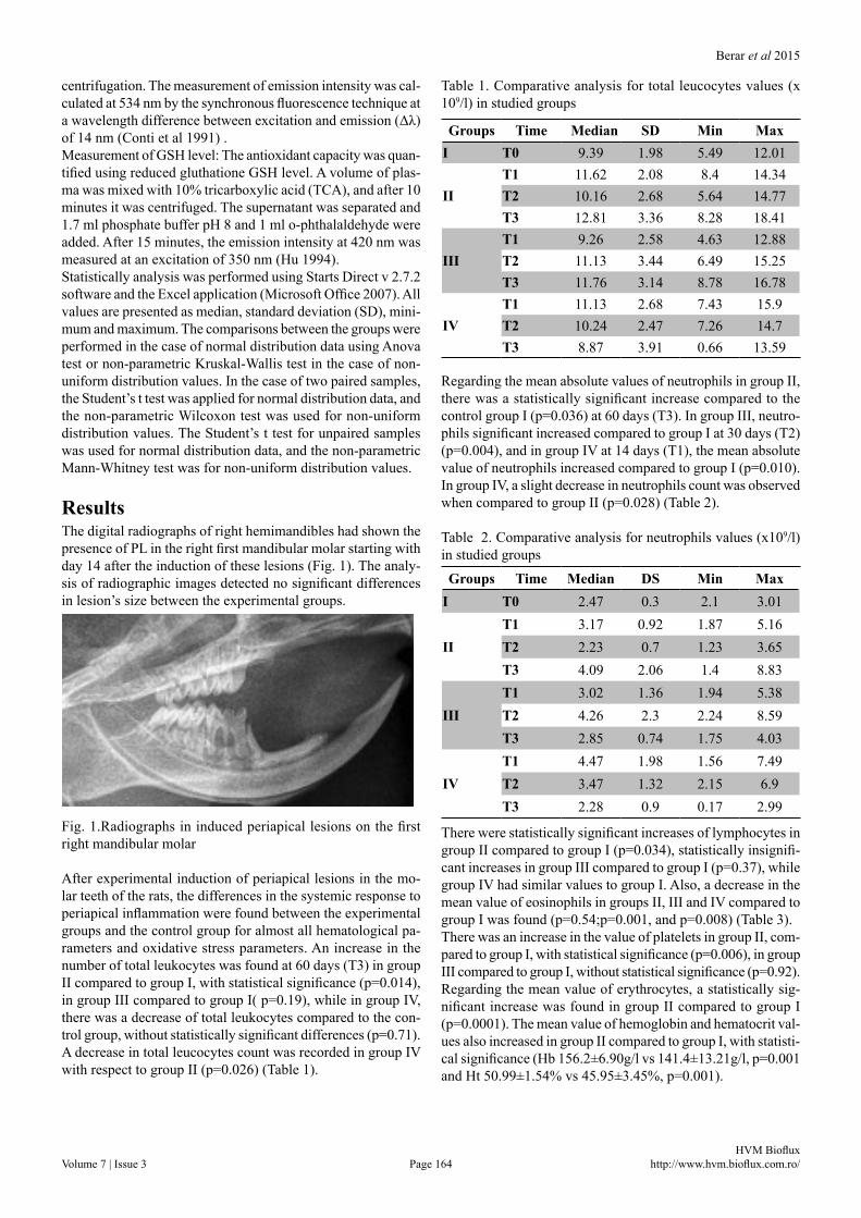
Results The digital radiographs of right hemimandibles had shown the presence of PL in the right first mandibular molar starting with day 14 after the induction of these lesions (Fig. 1). The analy-sis of radiographic images detected no significant differences in lesion’s size between the experimental groups.
Fig. 1.Radiographs in induced periapical lesions on the first right mandibular molar
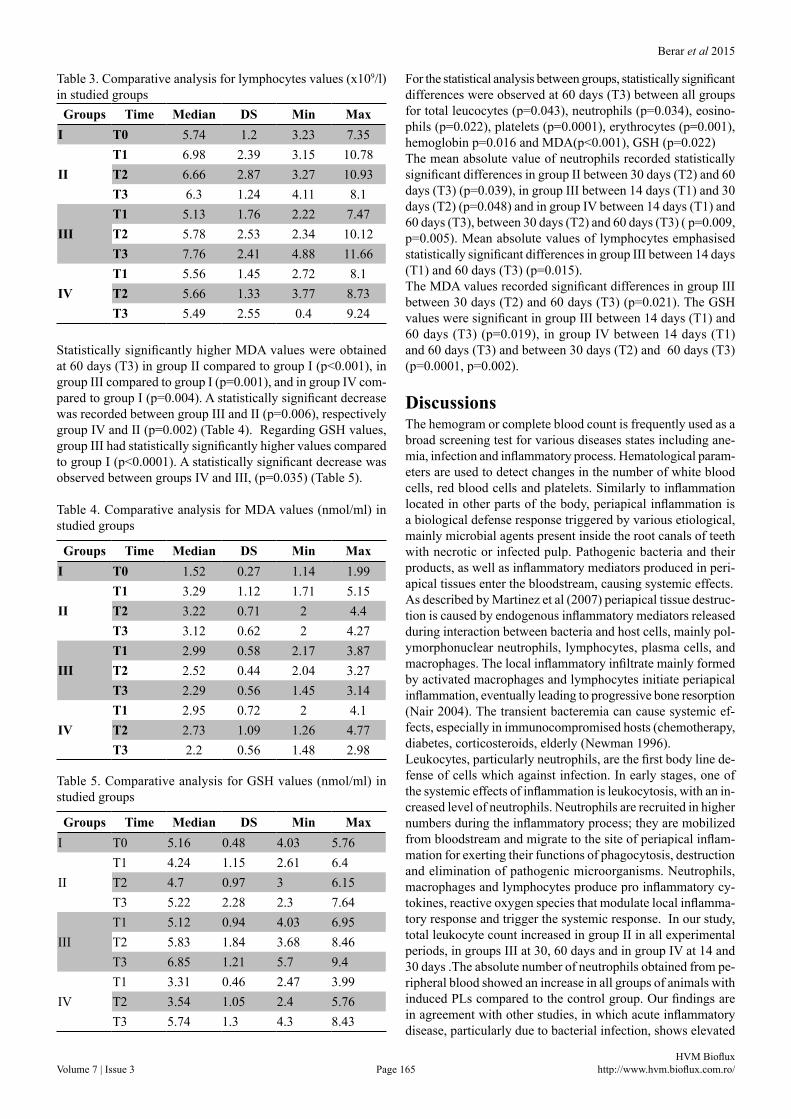
After experimental induction of periapical lesions in the mo-lar teeth of the rats, the differences in the systemic response to periapical inflammation were found between the experimental groups and the control group for almost all hematological pa-rameters and oxidative stress parameters. An increase in the number of total leukocytes was found at 60 days (T3) in group II compared to group I, with statistical significance (p=0.014), in group III compared to group I( p=0.19), while in group IV, there was a decrease of total leukocytes compared to the con-trol group, without statistically significant differences (p=0.71). A decrease in total leucocytes count was recorded in group IV with respect to group II (p=0.026) (Table 1).
Table 1. Comparative analysis for total leucocytes values (x 109/l) in studied groups
Regarding the mean absolute values of neutrophils in group II, there was a statistically significant increase compared to the control group I (p=0.036) at 60 days (T3). In group III, neutro-phils significant increased compared to group I at 30 days (T2) (p=0.004), and in group IV at 14 days (T1), the mean absolute value of neutrophils increased compared to group I (p=0.010). In group IV, a slight decrease in neutrophils count was observed when compared to group II (p=0.028) (Table 2).
Table 2. Comparative analysis for neutrophils values (x109/l) in studied groups
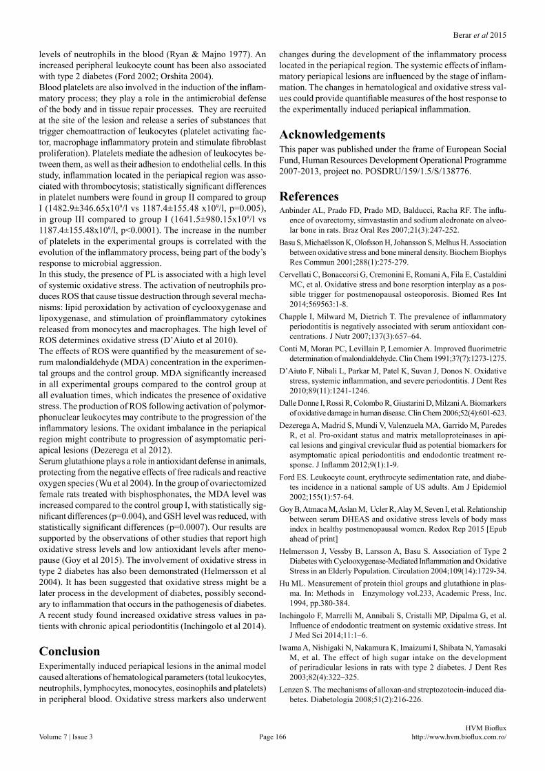
There were statistically significant increases of lymphocytes in group II compared to group I (p=0.034), statistically insignifi-cant increases in group III compared to group I (p=0.37), while group IV had similar values to group I. Also, a decrease in the mean value of eosinophils in groups II, III and IV compared to group I was found (p=0.54;p=0.001, and p=0.008) (Table 3).There was an increase in the value of platelets in group II, com-pared to group I, with statistical significance (p=0.006), in group III compared to group I, without statistical significance (p=0.92). Regarding the mean value of erythrocytes, a statistically sig-nificant increase was found in group II compared to group I (p=0.0001). The mean value of hemoglobin and hematocrit val-ues also increased in group II compared to group I, with statisti-cal significance (Hb 156.2±6.90g/l vs 141.4±13.21g/l, p=0.001 and Ht 50.99±1.54% vs 45.95±3.45%, p=0.001).
Groups Time Median SD Min MaxI T0 9.39 1.98 5.49 12.01
Table 3. Comparative analysis for lymphocytes values (x109/l) in studied groups
Statistically significantly higher MDA values were obtained at 60 days (T3) in group II compared to group I (p<0.001), in group III compared to group I (p=0.001), and in group IV com-pared to group I (p=0.004). A statistically significant decrease was recorded between group III and II (p=0.006), respectively group IV and II (p=0.002) (Table 4). Regarding GSH values, group III had statistically significantly higher values compared to group I (p<0.0001). A statistically significant decrease was observed between groups IV and III, (p=0.035) (Table 5).
Table 4. Comparative analysis for MDA values (nmol/ml) in studied groups
Table 5. Comparative analysis for GSH values (nmol/ml) in studied groups
For the statistical analysis between groups, statistically significant differences were observed at 60 days (T3) between all groups for total leucocytes (p=0.043), neutrophils (p=0.034), eosino-phils (p=0.022), platelets (p=0.0001), erythrocytes (p=0.001), hemoglobin p=0.016 and MDA(p<0.001), GSH (p=0.022) The mean absolute value of neutrophils recorded statistically significant differences in group II between 30 days (T2) and 60 days (T3) (p=0.039), in group III between 14 days (T1) and 30 days (T2) (p=0.048) and in group IV between 14 days (T1) and 60 days (T3), between 30 days (T2) and 60 days (T3) ( p=0.009, p=0.005). Mean absolute values of lymphocytes emphasised statistically significant differences in group III between 14 days (T1) and 60 days (T3) (p=0.015).The MDA values recorded significant differences in group III between 30 days (T2) and 60 days (T3) (p=0.021). The GSH values were significant in group III between 14 days (T1) and 60 days (T3) (p=0.019), in group IV between 14 days (T1) and 60 days (T3) and between 30 days (T2) and 60 days (T3) (p=0.0001, p=0.002).
DiscussionsThe hemogram or complete blood count is frequently used as a broad screening test for various diseases states including ane-mia, infection and inflammatory process. Hematological param-eters are used to detect changes in the number of white blood cells, red blood cells and platelets. Similarly to inflammation located in other parts of the body, periapical inflammation is a biological defense response triggered by various etiological, mainly microbial agents present inside the root canals of teeth with necrotic or infected pulp. Pathogenic bacteria and their products, as well as inflammatory mediators produced in peri-apical tissues enter the bloodstream, causing systemic effects. As described by Martinez et al (2007) periapical tissue destruc-tion is caused by endogenous inflammatory mediators released during interaction between bacteria and host cells, mainly pol-ymorphonuclear neutrophils, lymphocytes, plasma cells, and macrophages. The local inflammatory infiltrate mainly formed by activated macrophages and lymphocytes initiate periapical inflammation, eventually leading to progressive bone resorption (Nair 2004). The transient bacteremia can cause systemic ef-fects, especially in immunocompromised hosts (chemotherapy, diabetes, corticosteroids, elderly (Newman 1996).Leukocytes, particularly neutrophils, are the first body line de-fense of cells which against infection. In early stages, one of the systemic effects of inflammation is leukocytosis, with an in-creased level of neutrophils. Neutrophils are recruited in higher numbers during the inflammatory process; they are mobilized from bloodstream and migrate to the site of periapical inflam-mation for exerting their functions of phagocytosis, destruction and elimination of pathogenic microorganisms. Neutrophils, macrophages and lymphocytes produce pro inflammatory cy-tokines, reactive oxygen species that modulate local inflamma-tory response and trigger the systemic response. In our study, total leukocyte count increased in group II in all experimental periods, in groups III at 30, 60 days and in group IV at 14 and 30 days .The absolute number of neutrophils obtained from pe-ripheral blood showed an increase in all groups of animals with induced PLs compared to the control group. Our findings are in agreement with other studies, in which acute inflammatory disease, particularly due to bacterial infection, shows elevated
Groups Time Median DS Min MaxI T0 5.74 1.2 3.23 7.35
levels of neutrophils in the blood (Ryan & Majno 1977). An increased peripheral leukocyte count has been also associated with type 2 diabetes (Ford 2002; Orshita 2004). Blood platelets are also involved in the induction of the inflam-matory process; they play a role in the antimicrobial defense of the body and in tissue repair processes. They are recruited at the site of the lesion and release a series of substances that trigger chemoattraction of leukocytes (platelet activating fac-tor, macrophage inflammatory protein and stimulate fibroblast proliferation). Platelets mediate the adhesion of leukocytes be-tween them, as well as their adhesion to endothelial cells. In this study, inflammation located in the periapical region was asso-ciated with thrombocytosis; statistically significant differences in platelet numbers were found in group II compared to group I (1482.9±346.65x10⁹/l vs 1187.4±155.48 x10⁹/l, p=0.005), in group III compared to group I (1641.5±980.15x10⁹/l vs 1187.4±155.48x10⁹/l, p<0.0001). The increase in the number of platelets in the experimental groups is correlated with the evolution of the inflammatory process, being part of the body’s response to microbial aggression.In this study, the presence of PL is associated with a high level of systemic oxidative stress. The activation of neutrophils pro-duces ROS that cause tissue destruction through several mecha-nisms: lipid peroxidation by activation of cyclooxygenase and lipoxygenase, and stimulation of proinflammatory cytokines released from monocytes and macrophages. The high level of ROS determines oxidative stress (D’Aiuto et al 2010). The effects of ROS were quantified by the measurement of se-rum malondialdehyde (MDA) concentration in the experimen-tal groups and the control group. MDA significantly increased in all experimental groups compared to the control group at all evaluation times, which indicates the presence of oxidative stress. The production of ROS following activation of polymor-phonuclear leukocytes may contribute to the progression of the inflammatory lesions. The oxidant imbalance in the periapical region might contribute to progression of asymptomatic peri-apical lesions (Dezerega et al 2012). Serum glutathione plays a role in antioxidant defense in animals, protecting from the negative effects of free radicals and reactive oxygen species (Wu et al 2004). In the group of ovariectomized female rats treated with bisphosphonates, the MDA level was increased compared to the control group I, with statistically sig-nificant differences (p=0.004), and GSH level was reduced, with statistically significant differences (p=0.0007). Our results are supported by the observations of other studies that report high oxidative stress levels and low antioxidant levels after meno-pause (Goy et al 2015). The involvement of oxidative stress in type 2 diabetes has also been demonstrated (Helmersson et al 2004). It has been suggested that oxidative stress might be a later process in the development of diabetes, possibly second-ary to inflammation that occurs in the pathogenesis of diabetes. A recent study found increased oxidative stress values in pa-tients with chronic apical periodontitis (Inchingolo et al 2014).
ConclusionExperimentally induced periapical lesions in the animal model caused alterations of hematological parameters (total leukocytes, neutrophils, lymphocytes, monocytes, eosinophils and platelets) in peripheral blood. Oxidative stress markers also underwent
changes during the development of the inflammatory process located in the periapical region. The systemic effects of inflam-matory periapical lesions are influenced by the stage of inflam-mation. The changes in hematological and oxidative stress val-ues could provide quantifiable measures of the host response to the experimentally induced periapical inflammation.
AcknowledgementsThis paper was published under the frame of European Social Fund, Human Resources Development Operational Programme 2007-2013, project no. POSDRU/159/1.5/S/138776.
ence of ovarectomy, simvastastin and sodium aledronate on alveo-lar bone in rats. Braz Oral Res 2007;21(3):247-252.
Basu S, Michaëlsson K, Olofsson H, Johansson S, Melhus H. Association between oxidative stress and bone mineral density. Biochem Biophys Res Commun 2001;288(1):275-279.
Cervellati C, Bonaccorsi G, Cremonini E, Romani A, Fila E, Castaldini MC, et al. Oxidative stress and bone resorption interplay as a pos-sible trigger for postmenopausal osteoporosis. Biomed Res Int 2014;569563:1-8.
Chapple I, Milward M, Dietrich T. The prevalence of inflammatory periodontitis is negatively associated with serum antioxidant con-centrations. J Nutr 2007;137(3):657–64.
Conti M, Moran PC, Levillain P, Lemomier A. Improved fluorimetric determination of malondialdehyde. Clin Chem 1991;37(7):1273-1275.
D’Aiuto F, Nibali L, Parkar M, Patel K, Suvan J, Donos N. Oxidative stress, systemic inflammation, and severe periodontitis. J Dent Res 2010;89(11):1241-1246.
Dalle Donne I, Rossi R, Colombo R, Giustarini D, Milzani A. Biomarkers of oxidative damage in human disease. Clin Chem 2006;52(4):601-623.
Dezerega A, Madrid S, Mundi V, Valenzuela MA, Garrido M, Paredes R, et al. Pro-oxidant status and matrix metalloproteinases in api-cal lesions and gingival crevicular fluid as potential biomarkers for asymptomatic apical periodontitis and endodontic treatment re-sponse. J Inflamm 2012;9(1):1-9.
Ford ES. Leukocyte count, erythrocyte sedimentation rate, and diabe-tes incidence in a national sample of US adults. Am J Epidemiol 2002;155(1):57-64.
Goy B, Atmaca M, Aslan M, Ucler R, Alay M, Seven I, et al. Relationship between serum DHEAS and oxidative stress levels of body mass index in healthy postmenopausal women. Redox Rep 2015 [Epub ahead of print]
Helmersson J, Vessby B, Larsson A, Basu S. Association of Type 2 Diabetes with Cyclooxygenase-Mediated Inflammation and Oxidative Stress in an Elderly Population. Circulation 2004;109(14):1729-34.
Hu ML. Measurement of protein thiol groups and glutathione in plas-ma. In: Methods in Enzymology vol.233, Academic Press, Inc. 1994, pp.380-384.
Inchingolo F, Marrelli M, Annibali S, Cristalli MP, Dipalma G, et al. Influence of endodontic treatment on systemic oxidative stress. Int J Med Sci 2014;11:1–6.
Iwama A, Nishigaki N, Nakamura K, Imaizumi I, Shibata N, Yamasaki M, et al. The effect of high sugar intake on the development of periradicular lesions in rats with type 2 diabetes. J Dent Res 2003;82(4):322–325.
Lenzen S. The mechanisms of alloxan-and streptozotocin-induced dia-betes. Diabetologia 2008;51(2):216-226.
Berar et al 2015
Volume 7 | Issue 3 Page 167 HVM Bioflux
http://www.hvm.bioflux.com.ro/
Li X, Kolltveit KM, Tronstad L, Olsen I. Systemic Diseases Caused by Oral Infection. Clin Microbiol Rev 2000;13(4):547-558.
Martinez ZR, Naruishi K, Yamashiro K, Myokai F, Yamada T, Matsuura K, et al. Gene Profiles during Root Canal Treatment in Experimental Rat Periapical Lesions. J Endod 2007;33(8):936–943.
Munir BM, Iqbal N, Tirmazi SM, Majeed HA. Bone Healing of cyst-like periapical lesion following surgical endodontics: a case report. Pakistan Oral and Dental Journal 2011;31(2):436–438.
Nair PNR. Pathogenesis of apical periodontitis and the causes of en-dodontic failures. Crit Rev Oral Biol Med 2004;15(6):348-381.
Newman H N. Focal infection. J Dent Res 1996;75(12):1912-1919.Omi N, Ezawa I. The effect of ovariectomy on bone metabolism in
rats. Bone 1995;17(4 Suppl):163-168.Orshita K, Yamane K, Hanafusa M, Mori H, Mito K, Okubo M, Hora
H, Kohno N. Elevated white blood cell count in subjects with im-pared glucose tolerance. Diabetes Care 2004;27:491-496.
Parasuraman S, Raveendran R, Kesavan R. Blood sample collection in small laboratory animals. J Pharmacol Pharmacother 2010;1(2):87-93.
Reddy MS, Weatherford TW 3rd, Smith CA, West BD, Jeffcoat MK, Jacks TM. Jacks Alendronate Treatment of Naturally-Occurring Periodontitis in Beagle Dogs. J Periodontol 1995;66:211-217.
Roux C, Garnero P, Thomas T, Sabatier JP, Orcel P, Audran M. Recommendations for monitoring antiresorptive therapies in post-menopausal osteoporosis.Joint Bone Spine 2005;72(1):26-31.
Ryan GB, Majno G. Acute inflammation. A review. Am J Pathol 1977;86(1):183-276.
Siqueira JF, Rôças IN. Bacterial pathogenesis and mediators in apical periodontitis. Braz Dent J 2007;18:267–280.
Tani-Ishii N, Wang CY, Tanner A, Stashenko P. Changes in root canal microbiota during the development of rat periapical lesions. Oral Microbiol Immunol 1994;9(3):129-135.
Virdi HK. Hematological parameters-a diagnostic mirror for periodon-titis. Indian Journal of Dental Sciences 2003: 2(5):45-48
Wu G, Fang Y-Z, Yang S, Lupton JR, Turner ND. Glutathione metab-olism and its implications for health. J Nutr 2004;134(3):489-92.
Authors•Antonela M. Berar, Departement of Prosthodontics, ” Iuliu Hatieganu” University of Medicine and Pharmacy, 32 Clinicilor Street, 400006, Cluj-Napoca, Romania, EU, email: [email protected]
•Dragomir-Cosmin David, National Institute of Public Health-Regional Centre for Public Health, 6 Pasteur Street, 400349, Cluj-Napoca, Romania, EU, email: [email protected].
•Liana Lascu, Department of Prosthodontics “Iuliu Hatieganu” University of Medicine and Pharmacy, 32 Clinicilor Street, 400006, Cluj-Napoca, Romania, EU, email: [email protected].
•Luminita Matros, Department of Microbiology,” Iuliu Hatieganu” University of Medicine and Pharmacy,6Pasteur Street, 400349, Cluj-Napoca, Romania, EU, email: [email protected].
•Radu S. Campian, Department of Oral Rehabilitation,” Iuliu Hatieganu” University of Medicine and Pharmacy, 15 Victor Babes Street, 400012, Cluj-Napoca, Romania, EU, email: [email protected].
CitationBerar AM, David DC, Lascu L, Matros L, Campian RS. Analysis of hematological and oxidative stress parameters in the evaluation of experimentally induced periapical lesions. HVM Bioflux 2015;7(3):162-167.
Editor Stefan C. VesaReceived 9 June 2015Accepted 29 June 2015
Published Online 29 June 2015Funding None reported