Engineering DNA/FeeNeC single-atom nanozymes interface forcolorimetric biosensing of cancer cells
Liping Sun a, 1, Chao Li b, 1, Yong Yan b, Yue Yu b, Hao Zhao a, Zijue Zhou a, Feng Wang b, *,Yi Feng a, **
a School of Materials Science and Engineering, Hefei University of Technology, Hefei, 230009, Chinab School of Food and Biological Engineering, Hefei University of Technology, Hefei, 230009, China
h i g h l i g h t s
* Corresponding author.** Corresponding author. .
E-mail addresses: [email protected] (F. Wang1 Authors contributed equally to this work.
� Adenine and thymine have thehigher adsorption affinity on FeeNeCSAzymes, while cytosine has thelowest affinity.
� DNA/FeeNeC single-atom nano-zymes significantly improved theaqueous dispersion and recognitionability of SAzymes.
� DNA modification did not affect theperoxidase-like activity of FeeNeCSAzymes and the bioactivity of theadsorbed DNA [1e47].
� Diblock DNA/FeeNeC SAzymes wasdesigned for colorimetric detection ofcancer cells.
a r t i c l e i n f o
Article history:Received 12 June 2021Received in revised form11 July 2021Accepted 13 July 2021Available online 16 July 2021
Keywords:Single atom nanozymesDNAPeroxidase-like activityCancer cellsColorimetric detection
a b s t r a c t
Single atom nanozymes (SAzymes) represent the state-of-the-art technology in nanomaterial-basedcatalysis, which have attracted attentions in catalysis, cancer treatment, disinfection and biosensingfields. However, numerous SAzymes suffered from low aqueous dispersion and without recognitioncapacity, which impeded their applications in bioanalysis. Herein, we engineered DNA onto SAzymes toobtain the DNA/SAzymes conjugates, which significantly improved the aqueous dispersion and recog-nition ability of SAzymes. We synthesized iron SAzymes (FeeNeC SAzymes) as the catalytic nano-materials, and investigated the interactions between FeeNeC SAzymes and DNA. We compared A15, T15and C15 adsorption of FeeNeC SAzymes in HEPES containing 2 mM MgCl2. We found that 50 mg mL�1 FeeNeC SAzymes produced nearly 100% A15 adsorption, 90% T15 adsorption and only 69% C15 adsorption,indicating that adenine and thymine had higher adsorption affinity on FeeNeC SAzymes. Moreimportantly, DNA modification did not affect the peroxidase-like activity of FeeNeC SAzymes and thebioactivity of the adsorbed DNA. Taking the advantage of the diblock DNA with one DNA sequence(adenine) binding to FeeNeC SAzymes and the other DNA sequence (i.e., aptamer) binding to cancercells, we designed Apt/FeeNeC SAzymes for colorimetric detection of cancer cells, which offered newinsights for the use of SAzymes in biomedicine.
L. Sun, C. Li, Y. Yan et al. Analytica Chimica Acta 1180 (2021) 338856
1. Introduction
Single-atom nanozymes (SAzymes) have emerged as a new highcatalytic activity nanozyme [1e3]. Single metal atoms are welldispersed on the substrate. Each metal atom on the substrate canact as the catalytic active site and provide 100% atomic utilization intheory [4]. Fe-, Zn-, Ni-, Co-, Cu-based SAzymes have been syn-thesized and shown high oxidase or peroxidase-like activities [5,6].Compared with other nanozymes, SAzymes exhibit remarkablecatalytic activity, well-defined geometric and electronic structure[7], which have been used in the field of catalysis [8,9], cancertreatment [10], disinfection [11,12] and biosensing [13e18].
However, two disadvantages restrict the rapid spread andapplication of SAzymes. First, SAzymes display low dispersity inaqueous solution [19]. The synthesis of SAzymes requires high-temperature pyrolysis (>600 �C) in an oxygen-free environmentresulting in high-level carbonization of substrate [20]. Relativelyharsh pyrolysis pretreatment greatly increases the hydrophobicinteraction and van der Waals force within the substrate, leading toSAzymes irreversible aggregation [21]. Second, SAzymes do notcontain any specific ligand to recognize biomolecules [22]. Naturalenzymes generally conjugate with recognition biomolecules (i.e.,antibodies or aptamers) to perform detection tasks [23e25]. As thecatalytic activity of SAzymes relies on the active single atomdeposited onto the substrate, the adsorption of protein on thesurface will significantly reduce the enzyme activity, known as thepoison effect [26e28]. Therefore, it is a big challenge to endowSAzymes with specific recognition capacity without compromisingtheir catalytic activity.
DNA is the attractive biopolymer endowing nanomaterials withadditional functionality owing to the principles of programmablebase pairing [29]. Moreover, the high affinity and specificity ofaptamers make them ideal for molecular recognition, such asprotein, small molecules, cells and so on [30]. DNA-based nano-material hybrid systems have been widely explored in biosensing,DNA assembly and drug delivery [31]. As a magnetic functionalnanomaterial, Fe3O4 NPs had been applied for metal ions adsorp-tion [32e36] or DNA adsorption. Some literatures reported thatDNA adsorption on Fe3O4 NPs could remarkably affect their cata-lytic activity [37]. On one hand, DNA adsorption enhanced theperoxidase-like activity of Fe3O4 NPs by increasing substratebinding on Fe3O4 NPs [38]. The peroxidase-like activity of ATPaptamer-capped Fe3O4 NPs could be regulated by ATP [39]. On theother hand, some literatures reported that DNA modificationinhibited the peroxidase activity of magnetic nanoparticles [40].Nevertheless, the catalytic activity of DNA-engineered SAzymeswas not yet clear.
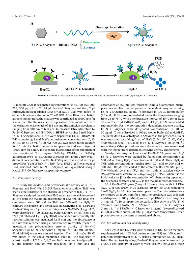
Herein, we systematically investigated the interactions betweenDNA and FeeNeC SAzymes, and the aptamer modified FeeNeCSAzymes (Apt/FeeNeC SAzymes) was designed for the colorimetricdetection of cancer cells. FeeNeC SAzymes were synthesized usingzeolitic imidazolate framework (ZIF-8) as precursors via the high-temperature pyrolysis method [41]. Due to the high carbon con-tent, FeeNeC SAzymes were prone to aggregate in the aqueoussolution. By adding a certain amount of single-stranded DNA(ssDNA), the dispersibility of FeeNeC SAzymes significantlyenhanced. Further ssDNA adsorption experiments indicated thatadenine and thymine had higher adsorption affinity to FeeNeCSAzymes in the presence of MgCl2. In particular, the introduction ofssDNA did not affect the peroxidase-like activity of FeeNeCSAzymes. As illustrated in Scheme 1, based on these findings, wedesigned diblock DNA with one DNA sequence (polyadenine)binding to FeeNeC SAzymes and the other DNA sequence(aptamer) that had the cell-targeting ability. Cancer cells that hadthe aptamer-targeting receptor could be facilely colorimetric
2
detected by Apt/FeeNeC SAzymes, proofing their potential appli-cation in bioanalysis.
2. Experimental section
2.1. Chemical and materials
Zinc nitrate hexahydrate was purchased from SinopharmChemical Reagent Co., Ltd. (Shanghai, China). 2-methylimidazole,methanol, magnesium chloride (MgCl2), 3,30,5,50-tetrame-thylbenzidine (TMB) and 4-(2-hydroxyethyl)-1-piperazineethanesulfonic acid (HEPES) were obtained fromAladdin Reagent (Shanghai, China). Iron powder was purchasedfrom Rhawn Chemical Technology Co. Ltd. (Shanghai, China).Aptamer and oligonucleotides were synthesized and purified bySangon Biotechnology Co. Ltd. (Shanghai, China) and the sequenceswere listed in Table S1.
2.2. Instruments
High-angle annular dark-field scanning transmission electronmicroscopy (ARM200F, Japan), transmission electron microscope(JEM-2100F, Japan) and scanning electron microscope (Hitachi,Japan) were performed to characterize the morphology. X-raypowder diffraction (MAX2500VL, Japan) and X-ray photoelectronspectroscopy (Thermo, US) were used to characterize the compo-sition. Inductively coupled plasma mass spectrometry (Thermo-Fisher, US) was used to determine the content of iron in SAzymes.Malvern Zetasizer Nano ZS 90 (Malvern, UK) was used to charac-terize the zeta potential. Fluorescence spectra were recorded usingF-7100 (Hitachi, Japan). The absorbance value was recorded byinfinite 200Pro microplate reader (Tecan, Switzerland). A CCK-8 cellviability kit assay was used to study the cytotoxicity of Apt/FeeNeCSAzymes in vitro. Laser scanning confocal fluorescence microscope(Leica SP8, Germany) was used to observe the intracellular locali-zation of FeeNeC SAzymes.
2.3. Synthesis of ZIF-8
ZIF-8 nanoparticles were synthesized using the methanol phasesynthesis method as described previously [11]. Briefly, 1.060 g of Zn(NO3)2$6H2O (3.56 mmol) and 1.231 g of 2-methylimidazole(15 mmol) was dissolved in 55.7 mL and 44.3 mL methanol,respectively. Both solutions were mixed together and stirred at25 �C for 2 h. Then the mixture was centrifuged at 14000 rpm for10 min to get the white precipitate, which was washed three timeswith methanol at the centrifugation speed of 14,000 rpm for 10min. Finally, ZIF-8 powder was dried in the oven overnight.
2.4. Preparation of FeeNeC SAzymes and NeC NPs
FeeNeC SAzymes were fabricated via the high-temperaturepyrolysis method [36]. Briefly, 40 mg of ZIF-8 powder and 40 mgof Fe powder were placed on either side of porcelain boat, whichwas transferred into a tubular furnace for pyrolysis at 1200 �C for6 h with a heating rate of 5 �C/min in Ar atmosphere. The as-prepared products were cooled to room temperature to obtainthe FeeNeC SAzymes. NeC NPs were also prepared in the absenceof Fe powder as the control.
2.5. DNA adsorption
To understand the adsorption of DNA by FeeNeC SAzymes andNeC NPs, FeeNeC SAzymes or NeC NPs were dispersed in 4-(2-hydroxyethyl)-1-piperazineethanesulfonic acid buffer (HEPES,
Scheme 1. Schematic illustration of preparation (A) and colorimetric detection of cancer cells (B) of Apt/FeeNeC SAzymes.
L. Sun, C. Li, Y. Yan et al. Analytica Chimica Acta 1180 (2021) 338856
10 mM, pH 7.42) at designated concentrations (0, 50, 100, 150, 200,250, 300 mg mL�1). To 98 mL FeeNeC SAzymes solution, 2 mLcarboxyfluorescein-labeled DNA (FAM-A15, 1 mM) was added toobtain a final concentration of 20 nM DNA. After 10 min incubationat room temperature, themixturewas centrifuged at 15000 rpm for5 min, then the fluorescence of supernatant was monitored withthe excitation wavelength of 485 nm and the emission wavelengthranging from 500 nm to 600 nm. To measure DNA adsorption byFeeNeC SAzymes and NeC NPs in HEPES containing 2 mM MgCl2,FeeNeC SAzymes or NeC NPs were dispersed in HEPES (10mM, pH7.42) containing 2 mM MgCl2 at designated concentrations (0, 10,20, 30, 40, 50 mg mL�1). 20 nM FAM-A15 was added to the mixturefor 10 min incubation at room temperature and centrifuged at15000 rpm for 5 min, and then the fluorescence of the supernatantwas monitored. To compare FAM-A15, FAM-T15 or FAM-C15adsorption by FeeNeC SAzymes at HEPES containing 2 mM MgCl2,different concentrations of FeeNeC SAzymes was mixed with 2 mLprobe DNA (1 mM of FAM-A15, FAM-T15 or FAM-C15), The amount ofDNA adsorbed onto FeeNeC SAzymes was quantified using aHitachi F-7100 fluorescence spectrophotometer.
2.6. Peroxidase activity
To study the oxidase- and peroxidase-like activity of FeeNeCSAzymes and NeC NPs, 3,30,5,50-Tetramethylbenzidine (TMB) wasused as the substrate in the absence or presence of H2O2. FeeNeCSAzymes catalyzed the colorless TMB substrate into a blueecoloredoxTMB with the maximum absorbance at 652 nm. The final con-centrations were 500 mM for TMB and 100 mM for H2O2. Tocompare the oxidase- and peroxidase-like activities of NeC NPs andFeeNeC SAzymes, 5 mL FeeNeC SAzymes or NeC NPs (1 mg mL�1)were dissolved in 100 mL acetate buffer (20 mM, pH 3). Then, 1 mLTMB (50 mM) and 1 mL H2O2 (10 M) were added subsequently. Thereaction solution was incubated for 5 min and the absorbance at652 nm was recorded using a fluorescence microplate reader. Toinvestigate the pH-dependent enzyme activity of FeeNeCSAzymes, 5 mL FeeNeC SAzymes (1 mg mL�1), 1 mL TMB (50 mM),93 mL Milli-Q water were mixed together. Then 1 mL H2O2 (10 M)was added to the above-mentioned solution. HAc was used toadjust the pH to 1, 2, 3, 4, 5, 6, 7, and NaOHwas used to adjust pH to9. The reaction solution was incubated for 5 min and the
3
absorbance at 652 nm was recorded using a fluorescence micro-plate reader. For the temperature dependent enzyme activity,FeeNeC SAzymes (50 mg mL�1) dissolved in 100 mL acetate buffer(20 mM, pH 3) were preincubated under the temperature rangingfrom 25 to 75 �C with a temperature interval of 10 �C for at least30 min. Then 1 mL TMB (50 mM) and 1 mL H2O2 (10 M) were addedsubsequently. For the concentration-dependent enzyme activity,FeeNeC SAzymes with designated concentration (2, 10 or50 mg mL�1) were dissolved in 100 mL acetate buffer (20 mM, pH 3).The peroxidase-like activity of Fe SAzymes in the presence of saltswas measured by adding 2 mL of NaCl (5 M), KCl (5 M), CaCl2(100 mM) or MgCl2 (100 mM) to FeeNeC SAzymes (50 mg mL�1),respectively. Other procedures were the same as those mentionedwith the temperature-dependent enzyme activity experiments.
Steady-state enzyme kinetics of FeeNeC SAzymes and A15/FeeNeC SAzymes were studied by fixing TMB concentration at500 mM or fixing H2O2 concentration at 100 mM. Then, H2O2 orTMB with concentrations ranging from 0.01 mM to 200 mM or100 mMe500 mM was added to the acetate buffer (20 mM, pH 3).The Michaelis constants (KM) and the maximal reaction velocity(Vmax) were calculated by1=n ¼ KM=Vmax½S� þ 1=Vmax, where n is theinitial velocity, [S] is the concentration of substrate, KM representsthe Michaelis constant and Vmax is the maximal reaction velocity.
20 mL FeeNeC SAzymes (5 mgmL�1) weremixed with 1 mL DNA(A15, T15 or Apt, 50 mM) in 79 mL HEPES (10 mM, pH 7.42) containing2mMMgCl2 for 10min at room temperature. Then themixturewascentrifuged at 15000 rpm for 5 min.The precipitate was collectedand dispersed in 100 mLMilli-Q water to get DNA/FeeNeC SAzymes(1 mg mL�1). To compare the peroxidase-like activity of FeeNeCSAzymes and DNA/FeeNeC SAzymes, 5 mL FeeNeC SAzymes(1 mgmL�1) or DNA/FeeNeC SAzymes (1 mgmL�1) were dissolvedin 100 mL acetate buffer (20 mM, pH 3) at room temperature. Otherprocedures were the same as mentioned above.
2.7. Cell culture and cell viability assay
The HepG2 and L02 cells were cultured in DMEM/F12 medium,supplemented with 10% fetal bovine serum (FBS) and 100 mg mL�1
penicillin/streptomycin at 37 �C using a humidified 5% CO2 incu-bator. The cytotoxicity of Apt/FeeNeC SAzymes was determined bya CCK-8 cell viability kit assay in vitro. Briefly, HepG2 cells were
L. Sun, C. Li, Y. Yan et al. Analytica Chimica Acta 1180 (2021) 338856
plated in 96-well plates with 104 cells per well and cultured inincubator for 24 h. Then, Apt/FeeNeC SAzymes were diluted inDMEM medium to get 10 mg mL�1, 50 mg mL�1 or 100 mg mL�1 ofApt/FeeNeC SAzymes, which was added into the cells and incu-bation for 24 h at 37 �C. Then the supernatant was removed. Cellswere washed twice with PBS to remove free Apt/FeeNeC SAzymes.100 mL of DMEM medium and 10 mL of CCK-8 solution were addedinto each well. After incubation for another 1 h at 37 �C, theabsorbance at 450 nm was determined using an infinite 200Promicroplate reader.
2.8. Cell imaging and colorimetric detection of cancer cells
To demonstrate the specific binding of Apt/FeeNeC SAzymeswith HepG2 live cancer cells, cells were seeded onto 14 mm cov-erslips in a 24-well plate with 100,000 cells per well and allowed togrow until 70% confluency. After washing twice with ice-cold 1xPBS, 50 mgmL�1 FAM labeled Apt/FeeNeC SAzymes or A15/FeeNeCSAzymes was added to cell culture media. After 15 min incubationfor each sample at 37 �C, the cells were fixed with 4% para-formaldehyde for 10 min at room temperature. To stain the cellnucleus, 300 nM DAPI in 1x PBS incubated with cells for 5 min atroom temperature. The coverslips were mounted on glass micro-scope slides with a drop of antifade mounting media to reducefluorescence photobleaching. Images with the cells were obtainedon a laser scanning confocal fluorescence microscope.
Colorimetric detection was used to identify the cancer cells.HepG2 cells were seeded in a 96-well plate at 20,000 per well in100 mL DMEM/F12 cell media and incubated at 37 �C overnight. Theculture media were then replaced with different concentrations ofApt/FeeNeC SAzymes (0, 100, 200, and 400 mg mL�1). After 15 minincubation at 37 �C, HepG2 cells were washed thrice with 1x PBS toremove the unbonded Apt/FeeNeC SAzymes. Then, the cells werefixed with 4% paraformaldehyde for 10 min. For the colorimetricassay, 98 mL acetate buffer (20 mM, pH 3), 1 mL TMB (50 mM) and1 mL H2O2 (10M) were added subsequently to the cells one after theother and incubated for 10 min. The absorbance at 652 nm wave-length was measured. Colorimetric detection was also used toquantify the cancer cell number. Different numbers of HepG2 andL02 cells (0, 100, 10000, 100000, 200000 cells per well) wereincubated with 100 mg mL�1 Apt/FeeNeC SAzymes. Other pro-cedures were the same with colorimetric detection mentionedabove.
3. Results and discussion
3.1. Characterization of ZIF-8, NeC NPs and FeeNeC SAzymes
The morphology, structure and composition of the FeeNeCSAzymes were characterized with TEM, HRTEM, HAADF-STEM,XRD, XPS and ICP-MS [42e46]. To obtain FeeNeC SAzymes, themethanol phase synthesis method was used to synthesize ZIF-8nanoparticles. Scanning electron microscope (SEM) and trans-mission electronmicroscopy (TEM) showed that the as-synthesizedZIF-8 exhibited a typical polyhedral structure (Figure S1A, S1B).FeeNeC SAzymes were obtained by high-temperature pyrolysis(1200 �C) method using iron powder as the source of single ironatom and ZIF-8 as the precursor. TEM image showed the as-prepared FeeNeC SAzymes with uniform size (d ¼ 80 nm)(Fig. 1A). High-resolution transmission electron microscopy(HRTEM) image showed layered graphitic carbon structures, indi-cating no metal nanoparticles or clusters existing in FeeNeCSAzymes (Fig. 1B). The atomically dispersed single iron atoms weredetected by high-angle annular dark-field scanning transmissionelectron microscopy (HAADF-STEM) identified by isolated bright
4
dots (highlighted in red circles) (Fig. 1C and D). X-ray diffraction(XRD) showed FeeNeC SAzymes had two broad diffraction peaks atz26� and 44�(Fig. 1E), corresponding to the (002) and (101) planeof graphite carbon (PDF No. 41e1487). There are no characteristicpeaks of Fe-based crystals, demonstrating that no iron nano-particles or clusters existing in the catalyst. The chemical compo-sition and elemental binding states of FeeNeC SAzymes wereinvestigated by X-ray photoelectron spectroscopy (XPS) (Fig. 1F).The atom percentages of C, N, O and Fe in FeeNeC SAzymes were87.41%, 2.54%, 9.49%, 0.56%, respectively. The C 1s spectrum wassplit into four peaks assigned to C]C, CeN, C]N and C]O groups(Figure S2A), suggesting majority of carbon belonged to sp2 state.High-resolution N 1s peak split analysis showed the existence ofpyridinic N, pyrrolic N, graphitic N, and oxidized N (Figure S2B).Inductively coupled plasma mass spectrometry (ICP-MS) was usedto determine the loading content of iron, which was calculated tobe 0.80%, which was comparable to the content measured by XPS.The morphology of NeC NPs was shown in Figure S3, which alsoshowed a polyhedral structure with an average particle size of100 nm and layered graphitic carbon structures.
3.2. Interactions of FeeNeC SAzymes and NeC NPs with singlestranded oligonucleotides
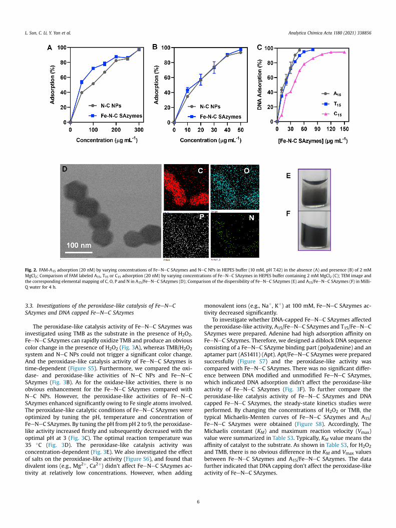
To study DNA adsorption of FeeNeC SAzymes and NeC NPs, westart with FAM-labeled 15-mer polyadenine (FAM-A15). Due to thecarbon backbone of FeeNeC SAzymes, the fluorescence of FAMwould be quenched when DNA were adsorbed on FeeNeCSAzymes. Different concentrations of FeeNeC SAzymes wereadded into FAM-A15 (20 nM) solution for 10 min incubation andcentrifuged, the fluorescence intensity of the supernatant wasmeasured to quantify DNA adsorption. As shown in Fig. 2A, A15adsorption increased by adding more FeeNeC SAzymes. A15adsorption was only 53% by adding 50 mg mL�1 FeeNeC SAzymes,at a high concentration of FeeNeC SAzymes (300 mg mL�1), A15adsorption reached nearly 100%. For comparison, A15 adsorption ofNeC NPs was also studied. Although A15 adsorption of FeeNeCSAzymes at some concentrations was higher than that of NeC NPs,300 mg mL�1 NeC NPs was also needed to reach nearly 100% A15adsorption. To further optimize the adsorption of oligonucleotideon FeeNeC SAzymes, salt was added to screen the electrostaticrepulsion of both negatively-charged oligonucleotide and FeeNeCSAzymes. A15 adsorption of FAM-A15 (20 nM) was examined byadding different concentrations of FeeNeC SAzymes in the HEPESbuffer (10 mM, pH 7.42) containing 2 mM MgCl2. As shown inFigs. 2B, 50 mg mL�1 FeeNeC SAzymes or NeC NPs was enough toreach nearly 100% A15 adsorption, which means Mg2þ was effectivefor DNA adsorption. What's more, there is no obvious difference forA15 adsorption between NeC NPs and single atom iron dopedFeeNeC SAzymes.
Next, FAM-T15 and FAM-C15 were introduced for the absorptionstudy. It was worth pointing out that poly G could form varioussecondary structures, which might complicate our analysis.Therefore, poly G was not involved in this study. DNA adsorption ofFAM-A15, FAM-T15 and FAM-C15 (20 nM) was examined by addingdifferent concentrations of FeeNeC SAzymes in HEPES buffer(10 mM, pH 7.42) containing 2 mM MgCl2. As can be seen fromFigs. 2C, 50 mg mL�1 FeeNeC SAzymes produced nearly 100% A15
adsorption, 90% T15 adsorption and only 69% C15 adsorption, whichindicated that adenine and thymine have higher adsorption affinityon FeeNeC SAzymes, while cytosine has the lowest affinity amongthe three bases.
z-potential and TEM were measured for further proving DNAadsorption on FeeNeC SAzymes. z-potential of A15/FeeNeCSAzymes decreased to �19.9 mV, suggesting a negatively charged
Fig. 1. TEM image (A), HRTEM image (B), HAADF-STEM image (C), magnified HAADF-STEM (D), XRD pattern (E) and XPS spectra (F) of FeeNeC SAzymes.
L. Sun, C. Li, Y. Yan et al. Analytica Chimica Acta 1180 (2021) 338856
A15 absorbed on FeeNeC SAzymes (Table S2). TEM image withcorresponding energy-dispersive X-ray spectroscopy (EDS)elemental mapping (Fig. 2D) showed the distribution of C, N, O andP of A15/FeeNeC SAzymes. The dispersity of single atom nano-zymes was an important feature for biomedical application. A15/FeeNeC SAzymes remained colloidally stable within 4 h incubatingduration, while bare FeeNeC SAzymes aggregated rapidly (Fig. 2Eand F).
To study the stability of A15/FeeNeC SAzymes in harsh condi-tions, urea (2 M), BSA (100 mg mL�1), mouse total blood and TritonX-100 (1 wt%) were introduced. Each sample was added into A15/FeeNeC SAzymes and incubated at room temperature for 1 h. Themixtures were centrifuged to precipitate A15/FeeNeC SAzymes.The fluorescence intensity in the supernatant was thus propor-tional to the desorbed FAM-A15 by the treatment. In all cases,fluorescence could not recover (Figure S4), suggesting that A15 was
5
adsorbed strongly onto FeeNeC SAzymes. Hydrogen bondingwasn't the main reason for adsorption. Surfactant and proteins alsocould not disrupt the A15/FeeNeC SAzymes hybrid structure,implying the potential usage of A15/FeeNeC SAzymes in vivo.
Previous paper reported that the interactions between DNAmolecules and graphene oxide were mainly the p-p stacking andhydrogen bonding resulted from the bases and carbon backbones[47]. FeeNeC SAzymes interacted with DNA strongly and stably inharsh conditions, which was different from graphene oxide, sug-gesting the interactions between DNA and FeeNeC SAzymes werenot via p-p stacking or hydrogen bonding. High temperature py-rolysis for SAzymes synthesis greatly increased the hydrophobicinteraction of FeeNeC SAzymes, which could bind to DNA basescontaining hydrophobic rings through hydrophobic interactions.Therefore, the hydrophobic interaction or the porous structure ofFeeNeC SAzymes might offer affinity to DNA.
Fig. 2. FAM-A15 adsorption (20 nM) by varying concentrations of FeeNeC SAzymes and NeC NPs in HEPES buffer (10 mM, pH 7.42) in the absence (A) and presence (B) of 2 mMMgCl2; Comparison of FAM labeled A15, T15 or C15 adsorption (20 nM) by varying concentrations of FeeNeC SAzymes in HEPES buffer containing 2 mM MgCl2 (C); TEM image andthe corresponding elemental mapping of C, O, P and N in A15/FeeNeC SAzymes (D); Comparison of the dispersibility of FeeNeC SAzymes (E) and A15/FeeNeC SAzymes (F) in Milli-Q water for 4 h.
L. Sun, C. Li, Y. Yan et al. Analytica Chimica Acta 1180 (2021) 338856
3.3. Investigations of the peroxidase-like catalysis of FeeNeCSAzymes and DNA capped FeeNeC SAzymes
The peroxidase-like catalysis activity of FeeNeC SAzymes wasinvestigated using TMB as the substrate in the presence of H2O2.FeeNeC SAzymes can rapidly oxidize TMB and produce an obviouscolor change in the presence of H2O2 (Fig. 3A), whereas TMB/H2O2system and NeC NPs could not trigger a significant color change.And the peroxidase-like catalysis activity of FeeNeC SAzymes istime-dependent (Figure S5). Furthermore, we compared the oxi-dase- and peroxidase-like activities of NeC NPs and FeeNeCSAzymes (Fig. 3B). As for the oxidase-like activities, there is noobvious enhancement for the FeeNeC SAzymes compared withNeC NPs. However, the peroxidase-like activities of FeeNeCSAzymes enhanced significantly owing to Fe single atoms involved.The peroxidase-like catalytic conditions of FeeNeC SAzymes wereoptimized by tuning the pH, temperature and concentration ofFeeNeC SAzymes. By tuning the pH from pH 2 to 9, the peroxidase-like activity increased firstly and subsequently decreased with theoptimal pH at 3 (Fig. 3C). The optimal reaction temperature was35 �C (Fig. 3D). The peroxidase-like catalysis activity wasconcentration-dependent (Fig. 3E). We also investigated the effectof salts on the peroxidase-like activity (Figure S6), and found thatdivalent ions (e.g., Mg2þ, Ca2þ) didn't affect FeeNeC SAzymes ac-tivity at relatively low concentrations. However, when adding
6
monovalent ions (e.g., Naþ, Kþ) at 100 mM, FeeNeC SAzymes ac-tivity decreased significantly.
To investigate whether DNA-capped FeeNeC SAzymes affectedthe peroxidase-like activity, A15/FeeNeC SAzymes and T15/FeeNeCSAzymes were prepared. Adenine had high adsorption affinity onFeeNeC SAzymes. Therefore, we designed a diblock DNA sequenceconsisting of a FeeNeC SAzyme binding part (polyadenine) and anaptamer part (AS1411) (Apt). Apt/FeeNeC SAzymes were preparedsuccessfully (Figure S7) and the peroxidase-like activity wascompared with FeeNeC SAzymes. There was no significant differ-ence between DNA modified and unmodified FeeNeC SAzymes,which indicated DNA adsorption didn't affect the peroxidase-likeactivity of FeeNeC SAzymes (Fig. 3F). To further compare theperoxidase-like catalysis activity of FeeNeC SAzymes and DNAcapped FeeNeC SAzymes, the steady-state kinetics studies wereperformed. By changing the concentrations of H2O2 or TMB, thetypical Michaelis-Menten curves of FeeNeC SAzymes and A15/FeeNeC SAzymes were obtained (Figure S8). Accordingly, TheMichaelis constant (KM) and maximum reaction velocity (Vmax)value were summarized in Table S3. Typically, KM value means theaffinity of catalyst to the substrate. As shown in Table S3, for H2O2and TMB, there is no obvious difference in the KM and Vmax valuesbetween FeeNeC SAzymes and A15/FeeNeC SAzymes. The datafurther indicated that DNA capping don't affect the peroxidase-likeactivity of FeeNeC SAzymes.
Fig. 3. UVevis absorption spectra of H2O2 þ TMB, NeC NPs þ H2O2 þ TMB, FeeNeC SAzymes þ H2O2 þ TMB (Inset: the photograph of the colorimetric reaction of control, NeC NPsand FeeNeC SAzymes) (A); Comparison of oxidase and peroxidase activities of NeC NPs (50 mg mL�1) and FeeNeC SAzymes (50 mg mL�1) in acetate buffer (20 mM, pH 3) (B); pH-(C), temperature- (D) and concentration-dependent (E) peroxidase-like activities of FeeNeC SAzyme; Comparison of peroxidase-like activity of FeeNeC SAzymes (50 mg mL�1) andDNA/FeeNeC SAzymes (50 mg mL�1) in acetate buffer (20 mM, pH 3) (F).
Fig. 4. Confocal fluorescence microscopy images of HepG2 cells after incubation with FAM-labeled A15/FeeNeC SAzymes (50 mg mL�1) and Apt/FeeNeC SAzymes (50 mg mL�1) for15 min. A15/FeeNeC SAzymes without HepG2 cells targeting aptamer was a negative control, Apt/FeeNeC SAzymes with a HepG2 targeting aptamer could be conjugated on thecell.
L. Sun, C. Li, Y. Yan et al. Analytica Chimica Acta 1180 (2021) 338856
7
L. Sun, C. Li, Y. Yan et al. Analytica Chimica Acta 1180 (2021) 338856
3.4. Colorimetric detection of cancer cells by Apt/FeeNeC SAzymes
Based on the recognition capability and the high peroxidase-likeactivity of Apt/FeeNeC SAzymes, a proof-of-concept colorimetricdetection of cancer cells was performed. Herein, we chose HepG2cells (human liver cancer cell) as the model due to their overex-pressed nucleolin, while L02 cells (human liver normal cell) withlower nucleolin expression were used as a control. Diblock DNAconsisting of polyadenine block and AS1411 block was modified onthe surface of FeeNeC SAzymes, which was used for targetingHepG2 cells through recognition of nucleolin by the AS1411aptamer. To demonstrate the specific binding on the HepG2 cells ofApt/FeeNeC SAzymes, laser confocal scanning microscopy (LCSM)was used by incubating HepG2 cells with FAM-labeled Apt/FeeNeCSAzymes. LCSM showed successfully binding of Apt/FeeNeCSAzymes with HepG2 cells, while no green fluorescence wasobserved with non-targeting A15/FeeNeC SAzymes (Fig. 4).
Before quantitative colorimetric detection of HepG2 cells, thecytotoxicity of Apt/FeeNeC SAzymes was evaluated by a CCK-8 cellviability kit assay. As can be seen from Fig. 5A, after incubationwithApt/FeeNeC SAzymes at concentrations of 10 mg mL�1, 50 mg mL�1
or 100 mg mL�1 for 24 h, the cell viability all remain above 85%,indicating negligible cytotoxicity of Apt/FeeNeC SAzymes for the
Fig. 5. Cell viability of HepG2 cells after incubation with different concentrations of Apt/Fcentrations of Apt/FeeNeC SAzymes (B); Absorbance values of different numbers of HepG2curve of absorbance versus HepG2 cell number (D).
8
HepG2 cells. To validate the Apt/FeeNeC SAzymes could be usedfor the quantitative colorimetric detection of cancer cells, firstly, weincubated HepG2 cells with different concentrations of Apt/FeeNeC SAzymes for 15 min. As shown in Fig. 5B, the absorbancevalue increased with a dose-dependent manner of Apt/FeeNeCSAzymes, which indicated Apt/FeeNeC SAzymes might be used forcancer cell detection. Then, the sensitivity of the colorimetric assaywith different HepG2 cell number was studied, L02 cells withdifferent numbers were also studied as a control. As shown inFig. 5C, the absorbance increased with the HepG2 cell number-dependent manner. For L02 cells, there is no obvious increase forthe absorbance value by increasing L02 cell numbers. The absor-bance value was linearly proportional to the HepG2 cell numberover the range of 100e2 � 105 cells (Fig. 5D). Our results indicatedthat the Apt/FeeNeC SAzymes could be used for colorimetric assayof cancer cells.
4. Conclusions
In summary, a simple colorimetric assay based on aptamer/FeeNeC SAzymes conjugate was developed for the detection ofcancer cells. The interaction between FeeNeC SAzymes and ssDNAwas investigated systematically. 50 mg mL�1 FeeNeC SAzymes
eeNeC SAzymes for 24 h.(A); Colorimetric detection of HepG2 cells at different con-and L02 cells treated with 100 mg mL�1 of Apt/FeeNeC SAzymes (C); .The calibration
L. Sun, C. Li, Y. Yan et al. Analytica Chimica Acta 1180 (2021) 338856
produced nearly 100% A15 adsorption, 90% T15 adsorption and only69% C15 adsorption, which indicated that adenine and thymine hadhigher adsorption affinity on FeeNeC SAzymes. DNA adsorptiondidn't affect the peroxidase-like activity of FeeNeC SAzymes.Furthermore, we designed a diblock DNA adsorbed onto FeeNeCSAzymes that could bind with cancer cells. Apt/FeeNeC SAzymescould be used for detecting cancer cells with the linear range of100e2 � 105 cells. The developed cancer cell biosensor combinedthe peroxidase-like catalysis activity of FeeNeC SAzymes and thehigh specificity of cell-specific aptamer. Inspired by this strategy, auniversal bioanalysis format for other SAzymes and molecularrecognition element may be introduced for clinical diagnostics andtherapy.
CRediT authorship contribution statement
Liping Sun: Formal analysis, Writing e original draft. Chao Li:Formal analysis, Writing e original draft, designed the research,performed experiments, analyzed the data, and wrote originaldraft. Yong Yan: Formal analysis, Writing e original draft. Yue Yu:Formal analysis, Writing e original draft, analyzed the data, dis-cussed the results, read the final manuscript. Hao Zhao: Formalanalysis, Writing e original draft. Zijue Zhou: Formal analysis,Writing e original draft, analyzed the data, discussed the results,read the final manuscript. Feng Wang: Supervision, Writing e
original draft. Yi Feng: Supervision, Writing e original draft, su-pervised the project, gave suggestions to revise the manuscript andprovided financial support.
Declaration of competing interest
There are no conflicts of interest to declare.
Acknowledgements
This work was supported by the National Natural ScienceFoundation of China (Grant 31971314), the Distinguished YouthFoundation of Anhui Province (1808085J05), the FundamentalResearch Funds for the Central Universities in China (Grant No.JZ2017HGPA0164), the Key Research and Development Plan ofAnhui Province (202104b11020015).
Appendix A. Supplementary data
Supplementary data to this article can be found online athttps://doi.org/10.1016/j.aca.2021.338856.
References
[1] L. Huang, J.X. Chen, L.F. Gan, J. Wang, S.J. Dong, Single-atom nanozymes, Sci.Adv. 5 (5) (2019).
[2] S.F. Ji, B. Jiang, H.G. Hao, Y.J. Chen, J.C. Dong, Y. Mao, Z.D. Zhang, R. Gao,W.X. Chen, R.F. Zhang, Q. Liang, H.J. Li, S.H. Liu, Y. Wang, Q.H. Zhang, L. Gu,D.M. Duan, M.M. Liang, D.S. Wang, X.Y. Yan, Y.D. Li, Matching the kinetics ofnatural enzymes with a single-atom iron nanozyme, Nat. Catal. (2021),https://doi.org/10.1038/s41929-021-00609-x.
[3] S.G. Wang, P. Zhou, L. Zhou, F. Lv, Y.J. Sun, Q.H. Zhang, L. Gu, H. Yang, S.J. Guo,A unique gas-migration, trapping, and emitting strategy for high-loadingsingle atomic Cd sites for carbon dioxide electroreduction, Nano Lett. 21(2021) 4262e4269.
[4] X. Niu, Q. Shi, W. Zhu, D. Liu, H. Tian, S. Fu, N. Cheng, S. Li, J.N. Smith, D. Du,Y. Lin, Unprecedented peroxidase-mimicking activity of single-atom nano-zyme with atomically dispersed Fe-Nx moieties hosted by MOF derivedporous carbon, Biosens, Bioelectron 142 (2019) 111495.
[5] L. Jiao, H. Yan, Y. Wu, W. Gu, C. Zhu, D. Du, Y. Lin, When nanozymes meetsingle-atom catalysis, Angew. Chem. Int. Ed. 59 (2020) 2565e2576.
[6] L. Jiao, W.Q. Xu, H.Y. Yan, Y. Wu, C.R. Liu, D. Du, Y.H. Lin, C.Z. Zhu, Fe-N-Csingle-atom nanozymes for the intracellular hydrogen peroxide detection,Anal. Chem. 91 (2019) 11994e11999.
nanozymes for biological applications, Biomater. Sci. 8 (2020) 6428e6441.[8] P. Yin, T. Yao, Y. Wu, L. Zheng, Y. Lin, W. Liu, H. Ju, J. Zhu, X. Hong, Z. Deng,
G. Zhou, S. Wei, Y. Li, Single cobalt atoms with precise N-Coordination assuperior oxygen reduction reaction catalysts, Angew. Chem. Int. Ed. 55 (2016)10800e10805.
[9] Y. Wang, K. Qi, S. Yu, G. Jia, Z. Cheng, L. Zheng, Q. Wu, Q. Bao, Q. Wang, J. Zhao,X. Cui, W. Zheng, Revealing the intrinsic peroxidase-like catalytic mechanismof heterogeneous single-atom CoeMoS2, Nano-Micro Lett. 11 (2019) 102.
[10] D. Wang, H. Wu, S.Z.F. Phua, G. Yang, W. Qi Lim, L. Gu, C. Qian, H. Wang,Z. Guo, H. Chen, Y. Zhao, Self-assembled single-atom nanozyme for enhancedphotodynamic therapy treatment of tumor, Nat. Commun. 11 (2020) 357.
[11] B. Xu, H. Wang, W. Wang, L. Gao, S. Li, X. Pan, H. Wang, H. Yang, X. Meng,Q. Wu, L. Zheng, S. Chen, X. Shi, K. Fan, X. Yan, H. Liu, A single-atom nanozymefor wound disinfection applications, Angew. Chem. Int. Ed. 58 (2019)4911e4916.
[12] R. Yan, S. Sun, J. Yang, W. Long, J. Wang, X. Mu, Q. Li, W. Hao, S. Zhang, H. Liu,Y. Gao, L. Ouyang, J. Chen, S. Liu, X.D. Zhang, D. Ming, Nanozyme-basedbandage with single-atom catalysis for brain trauma, ACS Nano 13 (2019)11552e11560.
[13] Q. Chen, S. Li, Y. Liu, X. Zhang, Y. Tang, H. Chai, Y. Huang, Size-controllable Fe-N/C single-atom nanozyme with exceptional oxidase-like activity for sensitivedetection of alkaline phosphatase, Sensor. Actuator. B Chem. 305 (2020)127511.
[14] N. Cheng, J.C. Li, D. Liu, Y. Lin, D. Du, Single-atom nanozyme based on nano-engineered Fe-N-C catalyst with superior peroxidase-like activity for ultra-sensitive bioassays, Small 15 (2019) 1901485.
[15] P.H. Li, M. Yang, Y.X. Li, Z.Y. Song, J.H. Liu, C.H. Lin, J. Zeng, X.J. Huang, Ultra-sensitive and selective detection of Arsenic(III) via electroanalysis over cobaltsingle-atom catalysts, Anal. Chem. 92 (2020) 6128e6135.
[16] Y. Wu, J.B. Wu, L. Jiao, W.Q. Xu, H.J. Wang, X.Q. Wei, W.L. Gu, G.X. Ren,N.A. Zhang, Q.H. Zhang, L. Huang, L. Gu, C.Z. Zhu, Cascade reaction systemintegrating single-atom nanozymes with abundant Cu sites for enhancedbiosensing, Anal. Chem. 92 (2020) 3373e3379.
[17] L. Yao, S. Gao, S. Liu, Y. Bi, R. Wang, H. Qu, Y. Wu, Y. Mao, L. Zheng, Single-atomenzyme-functionalized solution-gated graphene transistor for real-timedetection of mercury ion, ACS Appl. Mater. Interfaces 12 (2020) 6268e6275.
[18] X. Zhang, G. Li, G. Chen, D. Wu, X. Zhou, Y. Wu, Single-atom nanozymes: arising star for biosensing and biomedicine, Coord. Chem. Rev. 418 (2020)213376.
[19] H. Xiang, W. Feng, Y. Chen, Single-atom catalysts in catalytic biomedicine,Adv. Mater. 32 (2020) 1905994.
[20] M. Huo, L. Wang, Y. Wang, Y. Chen, J. Shi, Nanocatalytic tumor therapy bysingle-atom catalysts, ACS Nano 13 (2019) 2643e2653.
[22] L. Shen, D. Ye, H. Zhao, J. Zhang, Perspectives for single-atom nanozymes:advanced synthesis, functional mechanisms, and biomedical applications,Anal. Chem. 93 (2021) 1221e1231.
[23] M. Vargas-Montes, N. Cardona, D.M. Moncada, D.A. Molina, Y. Zhang,J.E. Gomez-Marin, Enzyme-linked aptamer assay (ELAA) for detection oftoxoplasma ROP18 protein in human serum, Front. Cell. Infect. Microbiol. 9(2019) 386.
[24] H. Xing, C.L. Zhang, G. Ruan, J. Zhang, K. Hwang, Y. Lu, Multimodal detection ofa small molecule target using stimuli-responsive liposome triggered byaptamer-enzyme conjugate, Anal. Chem. 88 (2016) 1506e1510.
[25] K.Y. Xing, J. Peng, S. Shan, D.F. Liu, Y.N. Huang, W.H. Lai, Green enzyme-linkedimmunosorbent assay based on the single-stranded binding protein-assistedaptamer for the detection of mycotoxin, Anal. Chem. 92 (2020) 8422e8426.
[26] J. Cao, M. Wang, H. Yu, Y. She, Z. Cao, J. Ye, A.M. Abd El-Aty, A. Hacimuftuoglu,J. Wang, S. Lao, An overview on the mechanisms and applications of enzymeinhibition-based methods for determination of organophosphate and carba-mate pesticides, J. Agric. Food Chem. 68 (2020) 7298e7315.
[27] Y. Chang, M. Liu, J. Liu, Highly selective fluorescent sensing of phosphitethrough recovery of poisoned nickel oxide nanozyme, Anal. Chem. 92 (2020)3118e3124.
[28] G. Sancho-Fornes, E. Peris, D. Gim�enez-Romero, S. Morais, �A. Maquieira,Enzyme inhibition microassays on blu-ray disks for drug discovery, ACSOmega 4 (2019) 5595e5600.
[29] Z. Li, J. Wang, Y. Li, X. Liu, Q. Yuan, Self-assembled DNA nanomaterials withhighly programmed structures and functions, Mater. Chem. Front. 2 (2018)423e436.
[30] X. Chen, F. Lisi, P. Bakthavathsalam, G. Longatte, S. Hoque, R.D. Tilley,J.J. Gooding, Impact of the coverage of aptamers on a nanoparticle on thebinding equilibrium and kinetics between aptamer and protein, ACS Sens. 6(2021) 538e545.
[31] Q. Hu, H. Li, L. Wang, H. Gu, C. Fan, DNA nanotechnology-enabled drug de-livery systems, Chem. Rev. 119 (2019) 6459e6506.
[32] T.A. Saleh, A.A. Al-Absi, Kinetics, isotherms and thermodynamic evaluation ofamine functionalized magnetic carbon for methyl red removal from aqueoussolutions, J. Mol. Liq. 248 (2017) 577e585.
[33] H. Javadian, M. Ruiz, T.A. Saleh, A.M. Sastre, Ca-alginate/carboxymethyl chi-tosan/Ni0.2Zn0.2Fe2.6 O4 magnetic bionanocomposite: synthesis, character-ization and application for single adsorption of Nd þ3 , Tb þ3 , and Dy þ3 rareearth elements from aqueous media, J. Mol. Liq. 306 (2020) 112760.
L. Sun, C. Li, Y. Yan et al. Analytica Chimica Acta 1180 (2021) 338856
on steel slag as an efficient magnetic adsorbent for cationic and anionic dyes,J. Environ. Chem. Eng. 9 (2021) 105126.
[35] T.A. Saleh, Trends in the sample preparation and analysis of nanomaterials asenvironmental contaminants, Trends Environ. Anal. Chem. 28 (2020), e00101.
[36] T.A. Saleh, Characterization, determination and elimination technologies forsulfur from petroleum: toward cleaner fuel and a safe environment, TrendsEnviron. Anal. Chem. 25 (2020), e00080.
[37] C. Zeng, N. Lu, Y. Wen, G. Liu, R. Zhang, J. Zhang, F. Wang, X. Liu, Q. Li, Z. Tang,M. Zhang, Engineering nanozymes using DNA for catalytic regulation, ACSAppl. Mater. Interfaces 11 (2019) 1790e1799.
[38] B. Liu, J. Liu, Accelerating peroxidase mimicking nanozymes using DNA,Nanoscale 7 (2015) 13831e13835.
[39] S. Li, X. Zhao, X. Yu, Y. Wan, M. Yin, W. Zhang, B. Cao, H. Wang, Fe3O4nanozymes with aptamer-tuned catalysis for selective colorimetric analysis ofATP in blood, Anal. Chem. 91 (2019) 14737e14742.
[40] K.S. Park, M.I. Kim, D.Y. Cho, H.G. Park, Label-Free Colorimetric detection ofnucleic acids based on target-induced shielding against the peroxidase-mimicking activity of magnetic nanoparticles, Small 7 (2011) 1521e1525.
[41] C. Zhao, C. Xiong, X. Liu, M. Qiao, Z. Li, T. Yuan, J. Wang, Y. Qu, X. Wang,F. Zhou, Q. Xu, S. Wang, M. Chen, W. Wang, Y. Li, T. Yao, Y. Wu, Y. Li,Unraveling the enzyme-like activity of heterogeneous single atom catalyst,
10
Chem. Commun. 55 (2019) 2285e2288.[42] S.O. Adio, M.H. Omar, M. Asif, T.A. Saleh, Arsenic and selenium removal from
water using biosynthesized nanoscale zero-valent iron: a factorial designanalysis, Process. Saf. Environ. 107 (2017) 518e527.
[43] J. Wang, Z. Shi, J. Fan, Y. Ge, J. Yi, G. Hu, Self-assembly of graphene into three-dimensional structures promoted by natural phenolic acids, J. Mater. Chem.22 (2012) 22459.
[44] M. Qamar, M.O. Fawakhiry, A.M. Azad, M.I. Ahmed, A. Khan, T.A. Saleh, Se-lective photocatalytic oxidation of aromatic alcohols into aldehydes bytungsten blue oxide (TBO) anchored with Pt nanoparticles, RSC Adv. 6 (2016)71108e71116.
[45] J.Y. Liu, Z.H. Wang, X.D. Yan, P.M. Jian, Metallic cobalt nanoparticles imbeddedinto ordered mesoporous carbon: a non-precious metal catalyst with excel-lent hydrogenation performance, J. Colloid Interface Sci. 505 (2017) 789e795.
[46] T.A. Saleh, M. Tuzen, A. Sarı, Magnetic activated carbon loaded with tungstenoxide nanoparticles for aluminum removal from waters, J. Environ. Chem.Eng. 5 (2017) 2853e2860.
[47] S. He, B. Song, D. Li, C. Zhu, W. Qi, Y. Wen, L. Wang, S. Song, H. Fang, C. Fan,A graphene nanoprobe for rapid, sensitive, and multicolor fluorescent DNAanalysis, Adv. Funct. Mater. 20 (2010) 453e459.


![Analytica Chimica Acta - University of California, Santa CruzAnalytica Chimica Acta 1105 (2020) 82e86 about cancer status and progression [5,6]. miR-1290 is a highly sensitive and](https://static.documents.pub/doc/80x56/60af0ef23cf3d07be8654c9c/analytica-chimica-acta-university-of-california-santa-cruz-analytica-chimica.jpg)


![Analytica Chimica Acta - download.xuebalib.comdownload.xuebalib.com/1dc8WMowcDlH.pdf · vices [10], promising organic thermoelectric materials [20], dye- sensitized solar cells [21],](https://static.documents.pub/doc/80x56/5b90021d09d3f28c298d53ca/analytica-chimica-acta-vices-10-promising-organic-thermoelectric-materials.jpg)


![Analytica Chimica Acta - bodc.ac.uk · activities of the ocean’s carbonate system [11], routine high resolution oceanic measurements using moorings, drifters, or profiling floats](https://static.documents.pub/doc/80x56/5c65ec8509d3f230488b5a3b/analytica-chimica-acta-bodcacuk-activities-of-the-oceans-carbonate-system.jpg)
![Analytica Chimica Acta - UT Arlington – UTA · 104 K.A. Schug et al. / Analytica Chimica Acta 713 (2012) 103–110 large pharmaceuticalcompanies[8,9].Infact,onlytwonewclasses of](https://static.documents.pub/doc/80x56/5c67dd2009d3f226588c984a/analytica-chimica-acta-ut-arlington-104-ka-schug-et-al-analytica.jpg)

