54
Anatomy of the Ankle Anatomy of the Ankle
| Date post: | 30-Dec-2015 |
| Category: |
Documents |
| Upload: | judith-ferguson |
| View: | 243 times |
| Download: | 3 times |
Anatomy of the AnkleAnatomy of the Ankle
AnkleAnkle
Anatomical StructuresAnatomical Structures– TibiaTibia– FibularFibular– TalusTalus
TibiaTibia
Strongest bone in lower legStrongest bone in lower leg Bears most of weightBears most of weight Bottom portion is called malleolusBottom portion is called malleolus
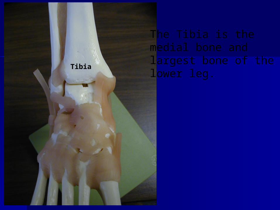
The Tibia is the medial bone and largest bone of the lower leg.
Tibia
FibulaFibula
Smaller bone of lower leg Smaller bone of lower leg Bottom portion is called malleolusBottom portion is called malleolus Primary function is muscle Primary function is muscle
attachmentattachment
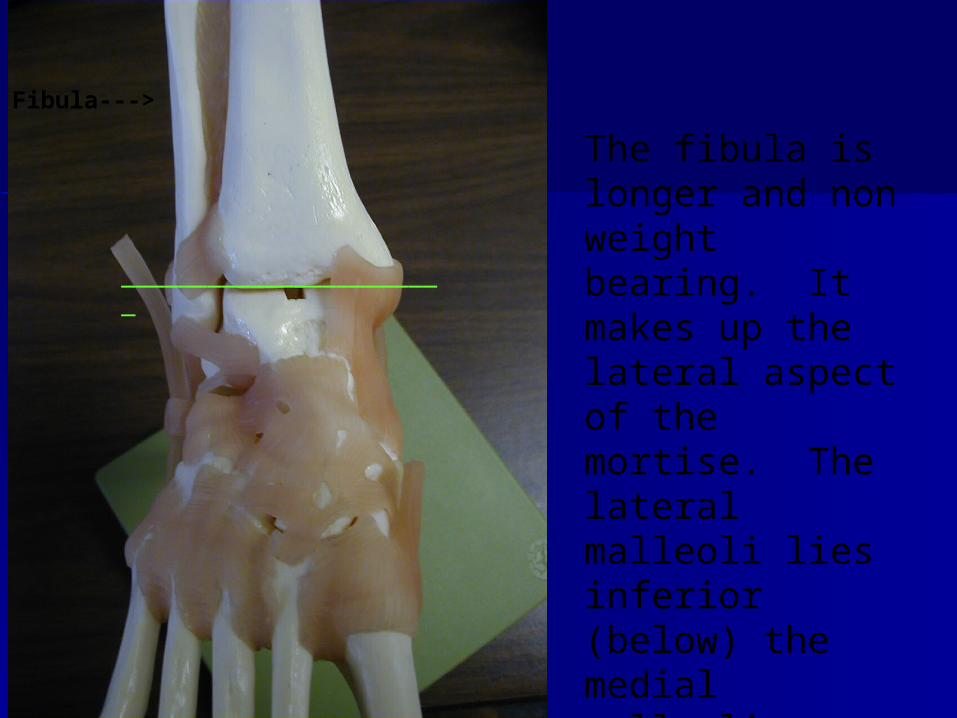
Fibula--->
The fibula is longer and non weight bearing. It makes up the lateral aspect of the mortise. The lateral malleoli lies inferior (below) the medial malleoli
_______________________
Talus Talus
Transmits forces from calcaneus Transmits forces from calcaneus to tibiato tibia
Allows for plantar flexion, Allows for plantar flexion, dorsiflexion, inversion, and dorsiflexion, inversion, and eversioneversion
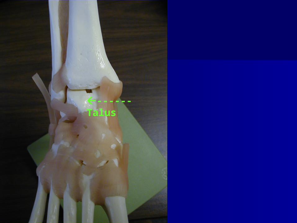
------ Talus
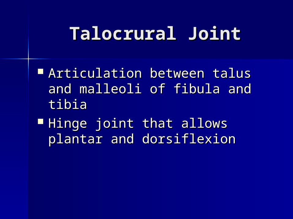
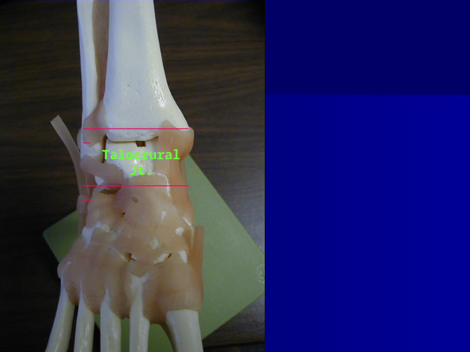
Talocrural JointTalocrural Joint
Articulation between talus and Articulation between talus and malleoli of fibula and tibiamalleoli of fibula and tibia
Hinge joint that allows plantar Hinge joint that allows plantar and dorsiflexionand dorsiflexion
________________
________________Talocrural Jt.
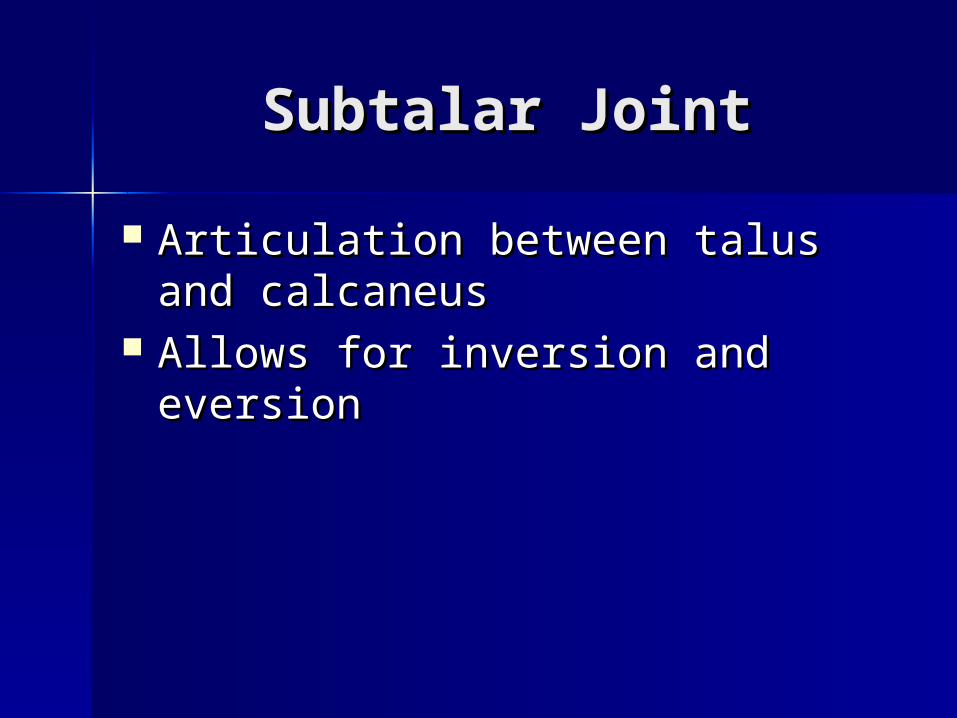
Subtalar JointSubtalar Joint
Articulation between talus and Articulation between talus and calcaneuscalcaneus
Allows for inversion and eversionAllows for inversion and eversion
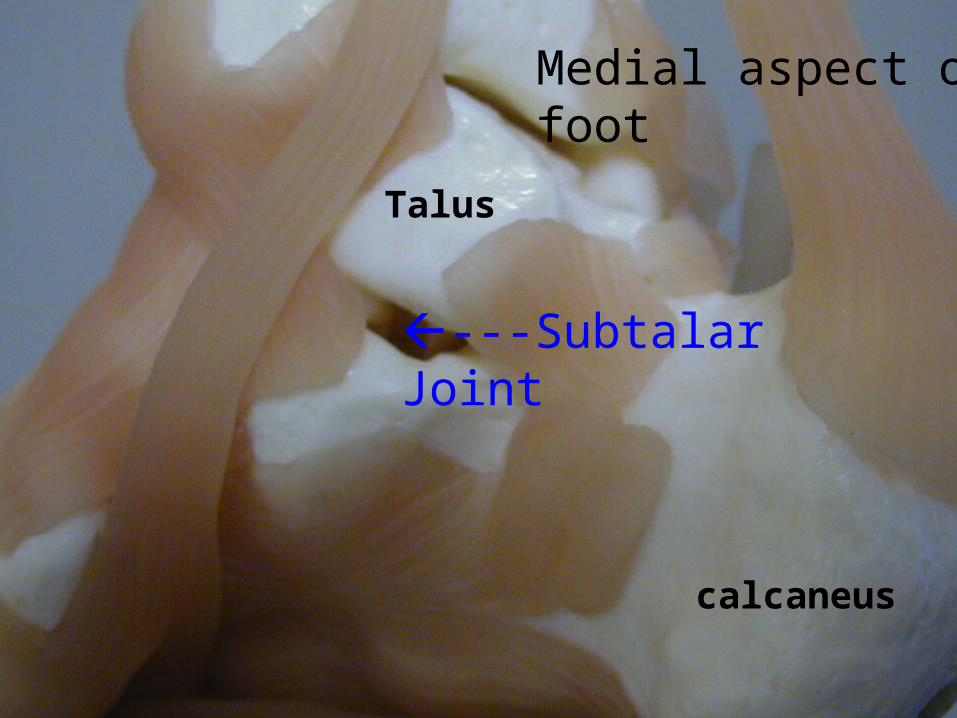
calcaneus
Talus
---Subtalar Joint
Medial aspect of foot
Ankle LigamentsAnkle Ligaments
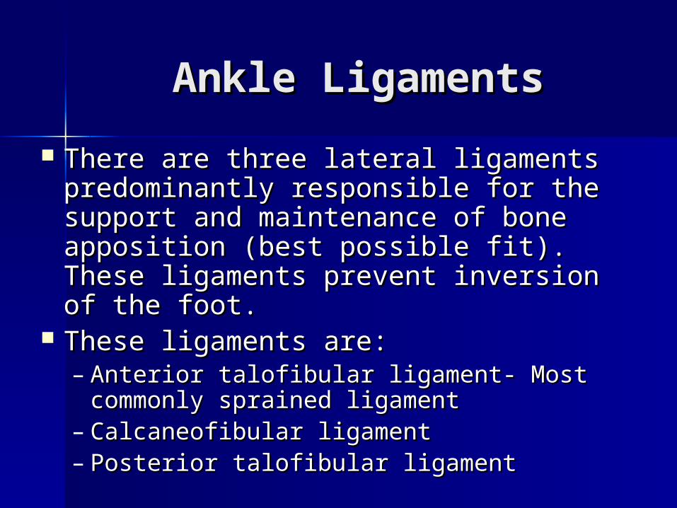
There are three lateral ligaments There are three lateral ligaments predominantly responsible for the predominantly responsible for the support and maintenance of bone support and maintenance of bone apposition (best possible fit). These apposition (best possible fit). These ligaments prevent inversion of the foot.ligaments prevent inversion of the foot.
These ligaments are:These ligaments are:– Anterior talofibular ligament- Most Anterior talofibular ligament- Most
commonly sprained ligamentcommonly sprained ligament– Calcaneofibular ligamentCalcaneofibular ligament– Posterior talofibular ligamentPosterior talofibular ligament
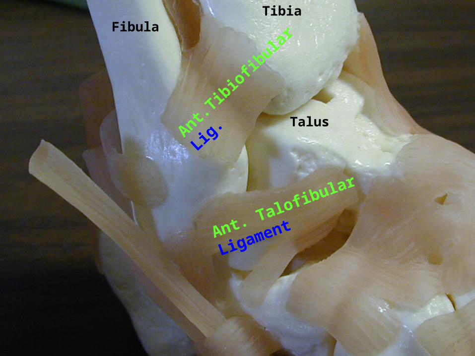
Talus
FibulaTibia
Ant. Talofibular Ligament
Ant.T
ibio
fibula
r
Lig.
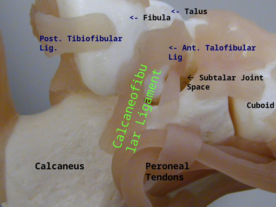
Post. Tibiofibular Lig.
<- Fibula
<- Ant. Talofibular Lig
<- Talus
Peroneal Tendons
Calc
aneofi
bula
r Li
gam
ent
Calcaneus
Subtalar Joint Space
Cuboid
calcaneus
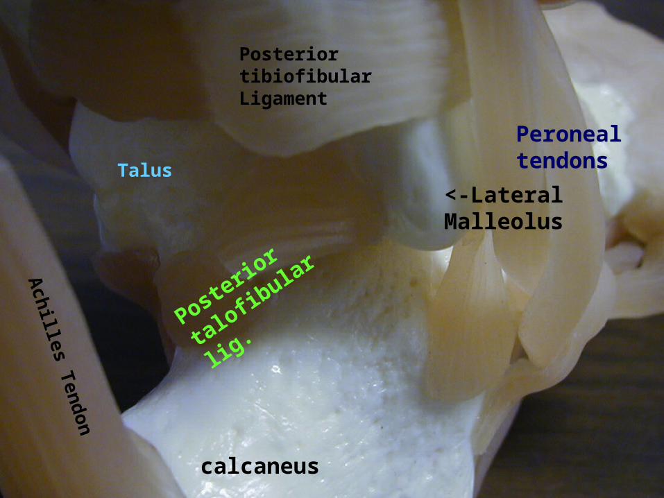
<-Lateral Malleolus
Posterior tibiofibular Ligament
Ach
illes T
endon
Talus
Posterio
r
talo
fibular lig
.
Peroneal tendons
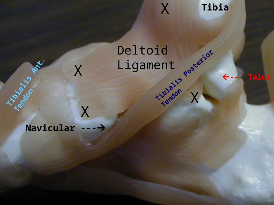
The deltoid ligamentThe deltoid ligament
Medial ligamentMedial ligament Several bands fused togetherSeveral bands fused together Prevents eversionPrevents eversion
Tibia
X
X
X
Navicular ---
-- Talus
Tibia
lis P
oste
rior T
endon
Tibi
alis
Ant
.
Tend
on
Deltoid LigamentX
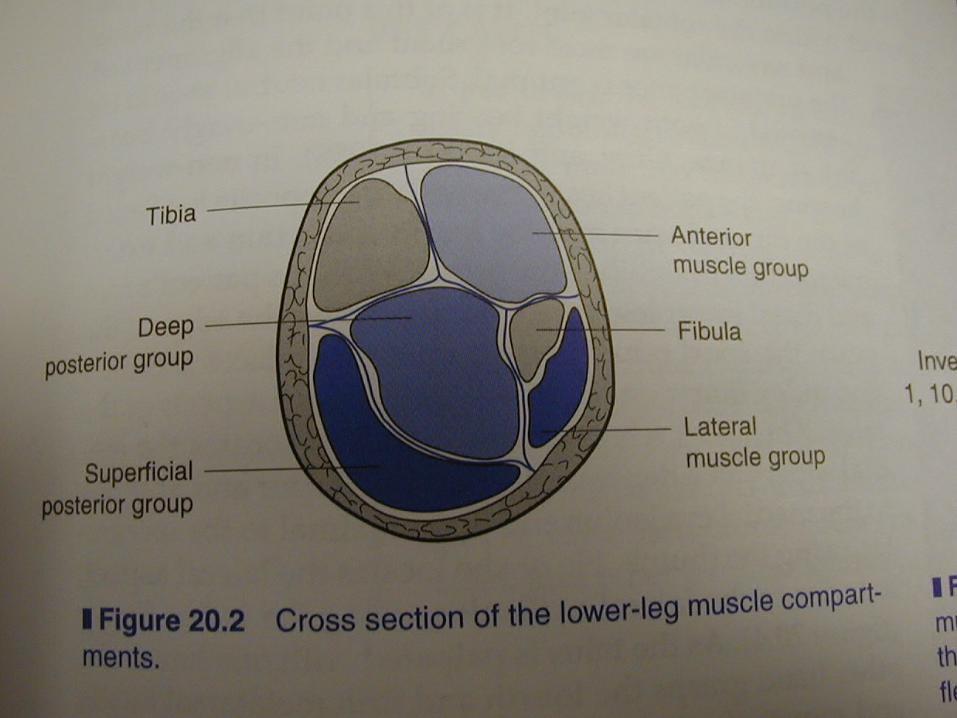
Muscles of the lower Muscles of the lower leg/ankleleg/ankle There are 4 compartments that There are 4 compartments that
make up the lower leg that make up the lower leg that operate the motions of the ankle.operate the motions of the ankle.
Injury can cause swelling inside Injury can cause swelling inside these compartments that can these compartments that can lead to tissue death or nerve lead to tissue death or nerve damage.damage.
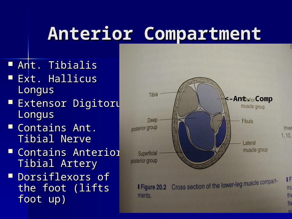
Anterior CompartmentAnterior Compartment
Ant. TibialisAnt. Tibialis Ext. Hallicus Ext. Hallicus
LongusLongus Extensor Digitorum Extensor Digitorum
LongusLongus Contains Ant. Tibial Contains Ant. Tibial
NerveNerve Contains Anterior Contains Anterior
Tibial ArteryTibial Artery Dorsiflexors of the Dorsiflexors of the
foot (lifts foot up)foot (lifts foot up)
<-Ant. Comp
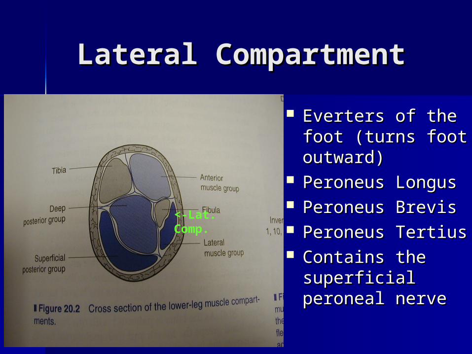
Lateral Compartment Lateral Compartment
Everters of the Everters of the foot (turns foot foot (turns foot outward)outward)
Peroneus LongusPeroneus Longus Peroneus BrevisPeroneus Brevis Peroneus TertiusPeroneus Tertius Contains the Contains the
superficial superficial peroneal nerveperoneal nerve
<-Lat. Comp.
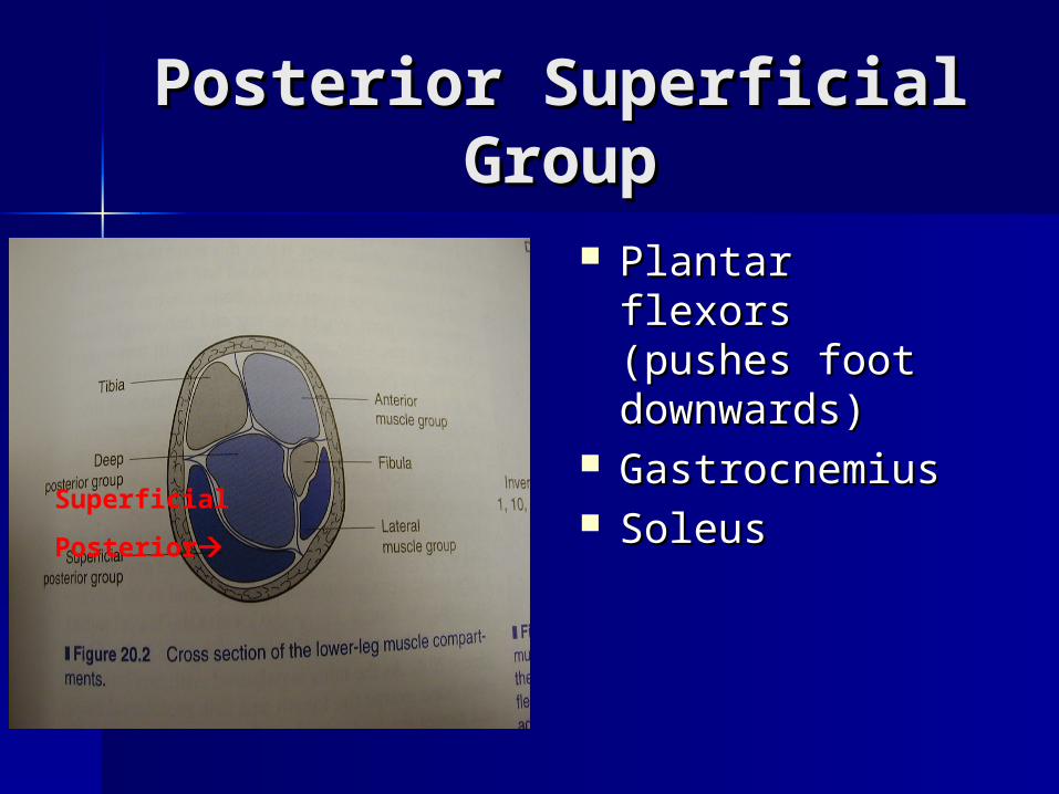
Posterior Superficial Posterior Superficial GroupGroup
Plantar flexors Plantar flexors (pushes foot (pushes foot downwards)downwards)
Gastrocnemius Gastrocnemius SoleusSoleusSuperficial
Posterior
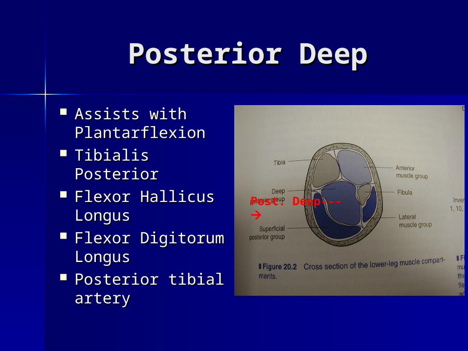
Posterior DeepPosterior Deep
Assists with Assists with PlantarflexionPlantarflexion
Tibialis PosteriorTibialis Posterior Flexor Hallicus Flexor Hallicus
LongusLongus Flexor Digitorum Flexor Digitorum
LongusLongus Posterior tibial Posterior tibial
arteryartery
Post. Deep---
Assessing the Lower Assessing the Lower Leg and AnkleLeg and Ankle HistoryHistory
– Past historyPast history– Mechanism of injuryMechanism of injury– When does it hurt?When does it hurt?– Type of, quality of, duration of pain?Type of, quality of, duration of pain?– Sounds or feelings?Sounds or feelings?– How long were you disabled?How long were you disabled?– Swelling?Swelling?– Previous treatments?Previous treatments?
ObservationsObservations– Postural deviations?Postural deviations?– Is there difficulty with walking?Is there difficulty with walking?– Deformities, asymmetries or swelling?Deformities, asymmetries or swelling?– Color and texture of skin, heat, Color and texture of skin, heat,
redness?redness?– Patient in obvious pain?Patient in obvious pain?– Is range of motion normal?Is range of motion normal?
– Percussion and compression testsPercussion and compression tests Used when fracture is suspectedUsed when fracture is suspected Percussion test is a blow to the tibia, fibula or heel to create Percussion test is a blow to the tibia, fibula or heel to create
vibratory force that resonates w/in fracture causing painvibratory force that resonates w/in fracture causing pain Compression test involves compression of tibia and fibula Compression test involves compression of tibia and fibula
either above or below site of concerneither above or below site of concern
– Thompson testThompson test Squeeze calf muscle, while foot is extended off table to test Squeeze calf muscle, while foot is extended off table to test
the integrity of the Achilles tendonthe integrity of the Achilles tendon– Positive tests results in no movement in the footPositive tests results in no movement in the foot
– Homan’s testHoman’s test Test for deep vein thrombophlebitisTest for deep vein thrombophlebitis With knee extended and foot off table, ankle is moved into With knee extended and foot off table, ankle is moved into
dorsiflexiondorsiflexion Pain in calf is a positive sign and should be referred Pain in calf is a positive sign and should be referred
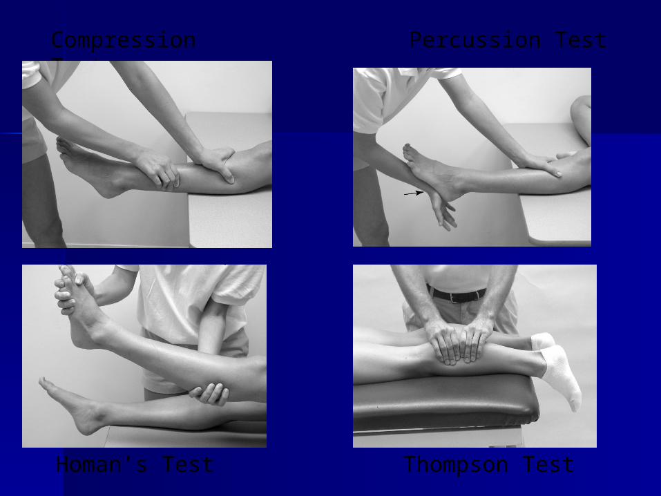
Compression Test Percussion Test
Homan’s Test Thompson Test
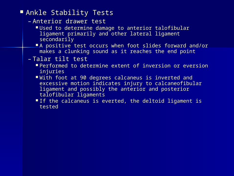
Ankle Stability TestsAnkle Stability Tests– Anterior drawer testAnterior drawer test
Used to determine damage to anterior talofibular Used to determine damage to anterior talofibular ligament primarily and other lateral ligament secondarilyligament primarily and other lateral ligament secondarily
A positive test occurs when foot slides forward and/or A positive test occurs when foot slides forward and/or makes a clunking sound as it reaches the end pointmakes a clunking sound as it reaches the end point
– Talar tilt testTalar tilt test Performed to determine extent of inversion or eversion Performed to determine extent of inversion or eversion
injuriesinjuries With foot at 90 degrees calcaneus is inverted and With foot at 90 degrees calcaneus is inverted and
excessive motion indicates injury to calcaneofibular excessive motion indicates injury to calcaneofibular ligament and possibly the anterior and posterior ligament and possibly the anterior and posterior talofibular ligamentstalofibular ligaments
If the calcaneus is everted, the deltoid ligament is testedIf the calcaneus is everted, the deltoid ligament is tested
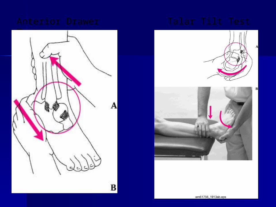
Anterior Drawer Test Talar Tilt Test
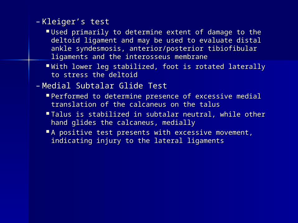
– Kleiger’s testKleiger’s test Used primarily to determine extent of damage to the deltoid Used primarily to determine extent of damage to the deltoid
ligament and may be used to evaluate distal ankle ligament and may be used to evaluate distal ankle syndesmosis, anterior/posterior tibiofibular ligaments and syndesmosis, anterior/posterior tibiofibular ligaments and the interosseus membranethe interosseus membrane
With lower leg stabilized, foot is rotated laterally to stress With lower leg stabilized, foot is rotated laterally to stress the deltoidthe deltoid
– Medial Subtalar Glide TestMedial Subtalar Glide Test Performed to determine presence of excessive medial Performed to determine presence of excessive medial
translation of the calcaneus on the talustranslation of the calcaneus on the talus Talus is stabilized in subtalar neutral, while other hand Talus is stabilized in subtalar neutral, while other hand
glides the calcaneus, mediallyglides the calcaneus, medially A positive test presents with excessive movement, A positive test presents with excessive movement,
indicating injury to the lateral ligamentsindicating injury to the lateral ligaments
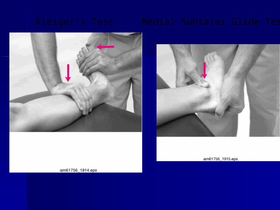
Kleiger’s Test Medial Subtalar Glide Test
Functional TestsFunctional Tests
– While weight bearing the following should be While weight bearing the following should be performedperformed
Walk on toes (plantar flexion)Walk on toes (plantar flexion) Walk on heels (dorsiflexion)Walk on heels (dorsiflexion) Walk on lateral borders of feet (inversion)Walk on lateral borders of feet (inversion) Walk on medial borders of feet (eversion)Walk on medial borders of feet (eversion) Hops on injured ankleHops on injured ankle Passive, active and resistive movements should be Passive, active and resistive movements should be
manually applied to determine joint integrity and muscle manually applied to determine joint integrity and muscle functionfunction
– If any of these are painful they should be avoidedIf any of these are painful they should be avoided
Prevention of Injury to Prevention of Injury to the Anklethe Ankle
Stretching of the Achilles tendonStretching of the Achilles tendon Strengthening of the surrounding Strengthening of the surrounding
musclesmuscles Proprioceptive training: balance Proprioceptive training: balance
exercises and agilityexercises and agility Wearing proper footwear and or Wearing proper footwear and or
tape when appropriatetape when appropriate
Specific InjuriesSpecific Injuries
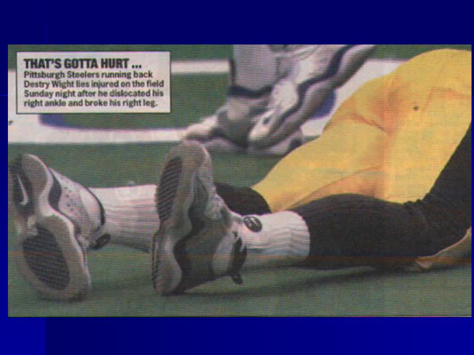
Ankle Injuries: SprainsAnkle Injuries: Sprains– Single most common injury in athletics caused by Single most common injury in athletics caused by
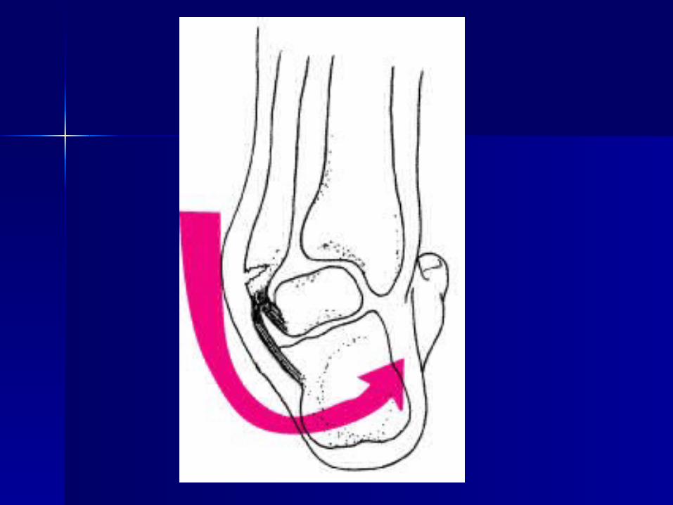
sudden inversion or eversion momentssudden inversion or eversion moments Inversion SprainsInversion Sprains
– Most common and result in injury to the lateral Most common and result in injury to the lateral ligamentsligaments
– Anterior talofibular ligament is injured with Anterior talofibular ligament is injured with inversion, plantar flexion and internal rotationinversion, plantar flexion and internal rotation
– Occasionally the force is great enough for an Occasionally the force is great enough for an avulsion fracture to occur w/ the lateral malleolusavulsion fracture to occur w/ the lateral malleolus
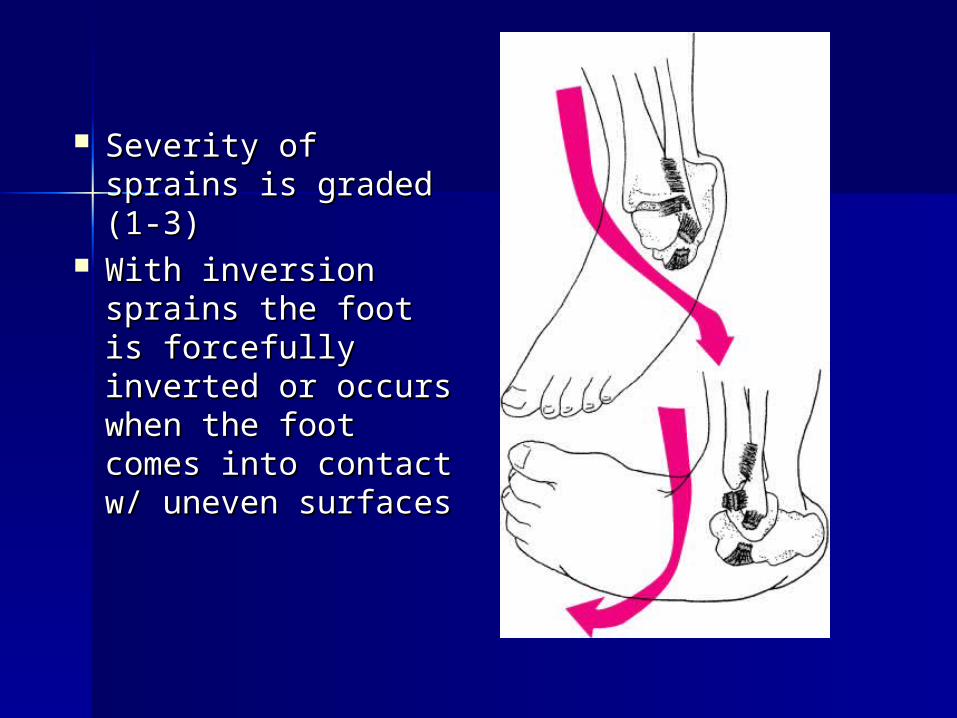
Severity of sprains Severity of sprains is graded (1-3)is graded (1-3)
With inversion With inversion sprains the foot is sprains the foot is forcefully inverted forcefully inverted or occurs when or occurs when the foot comes the foot comes into contact w/ into contact w/ uneven surfacesuneven surfaces
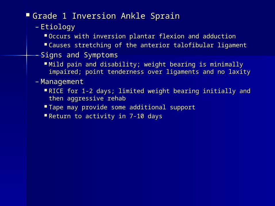
Grade 1 Inversion Ankle SprainGrade 1 Inversion Ankle Sprain– EtiologyEtiology
Occurs with inversion plantar flexion and adductionOccurs with inversion plantar flexion and adduction Causes stretching of the anterior talofibular ligamentCauses stretching of the anterior talofibular ligament
– Signs and SymptomsSigns and Symptoms Mild pain and disability; weight bearing is minimally Mild pain and disability; weight bearing is minimally
impaired; point tenderness over ligaments and no laxityimpaired; point tenderness over ligaments and no laxity
– ManagementManagement RICE for 1-2 days; limited weight bearing initially and RICE for 1-2 days; limited weight bearing initially and
then aggressive rehabthen aggressive rehab Tape may provide some additional supportTape may provide some additional support Return to activity in 7-10 daysReturn to activity in 7-10 days
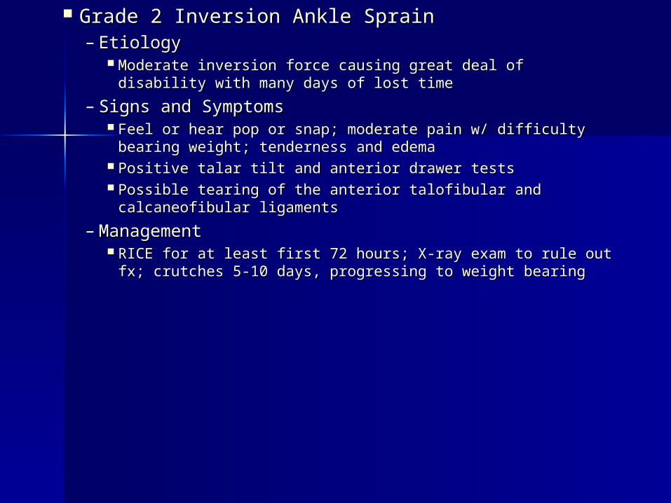
Grade 2 Inversion Ankle SprainGrade 2 Inversion Ankle Sprain– EtiologyEtiology
Moderate inversion force causing great deal of disability Moderate inversion force causing great deal of disability with many days of lost timewith many days of lost time
– Signs and SymptomsSigns and Symptoms Feel or hear pop or snap; moderate pain w/ difficulty bearing Feel or hear pop or snap; moderate pain w/ difficulty bearing
weight; tenderness and edemaweight; tenderness and edema Positive talar tilt and anterior drawer testsPositive talar tilt and anterior drawer tests Possible tearing of the anterior talofibular and Possible tearing of the anterior talofibular and
calcaneofibular ligamentscalcaneofibular ligaments
– ManagementManagement RICE for at least first 72 hours; X-ray exam to rule out fx; RICE for at least first 72 hours; X-ray exam to rule out fx;
crutches 5-10 days, progressing to weight bearingcrutches 5-10 days, progressing to weight bearing
– Management (continued)Management (continued) Will require protective immobilization but Will require protective immobilization but
begin ROM exercises early to aid in begin ROM exercises early to aid in maintenance of motion and proprioceptionmaintenance of motion and proprioception
Taping will provide support during early Taping will provide support during early stages of walking and runningstages of walking and running
Long term disability will include chronic Long term disability will include chronic instability with injury recurrence potentially instability with injury recurrence potentially leading to joint degenerationleading to joint degeneration
Must continue to engage in rehab to prevent Must continue to engage in rehab to prevent against re-injury against re-injury
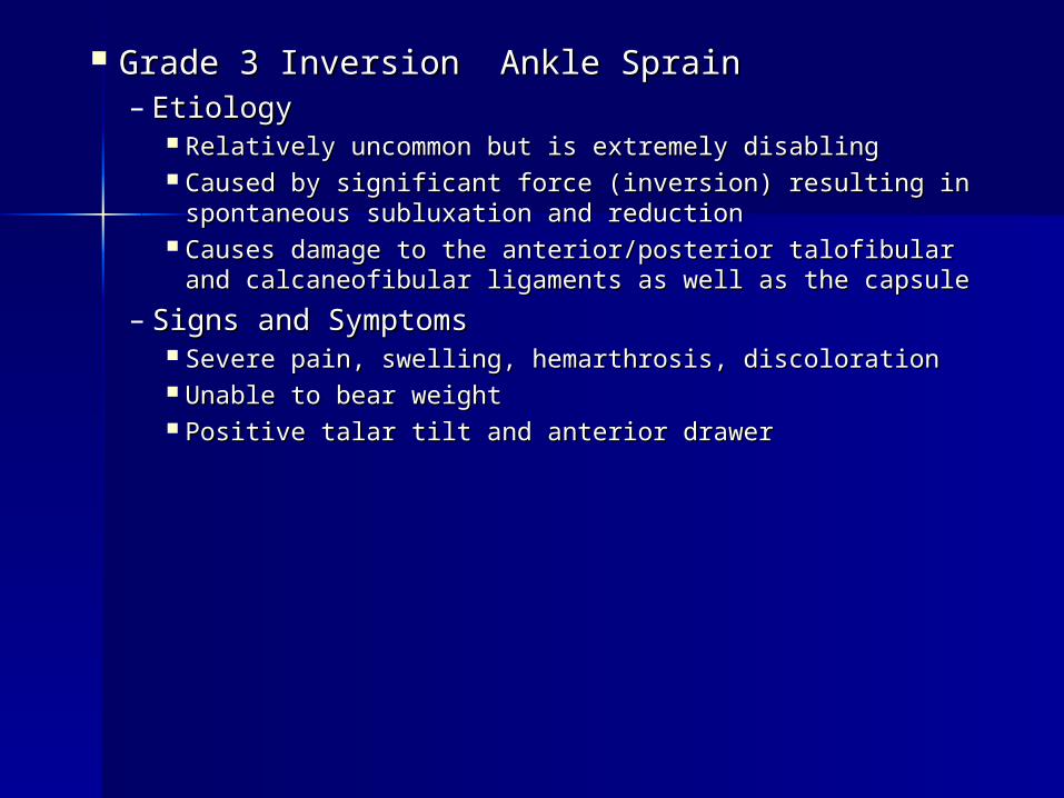
Grade 3 Inversion Ankle SprainGrade 3 Inversion Ankle Sprain– EtiologyEtiology
Relatively uncommon but is extremely disablingRelatively uncommon but is extremely disabling Caused by significant force (inversion) resulting in Caused by significant force (inversion) resulting in
spontaneous subluxation and reductionspontaneous subluxation and reduction Causes damage to the anterior/posterior talofibular Causes damage to the anterior/posterior talofibular
and calcaneofibular ligaments as well as the capsuleand calcaneofibular ligaments as well as the capsule
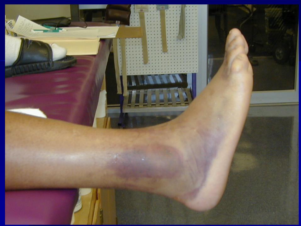
– Signs and SymptomsSigns and Symptoms Severe pain, swelling, hemarthrosis, discolorationSevere pain, swelling, hemarthrosis, discoloration Unable to bear weightUnable to bear weight Positive talar tilt and anterior drawerPositive talar tilt and anterior drawer
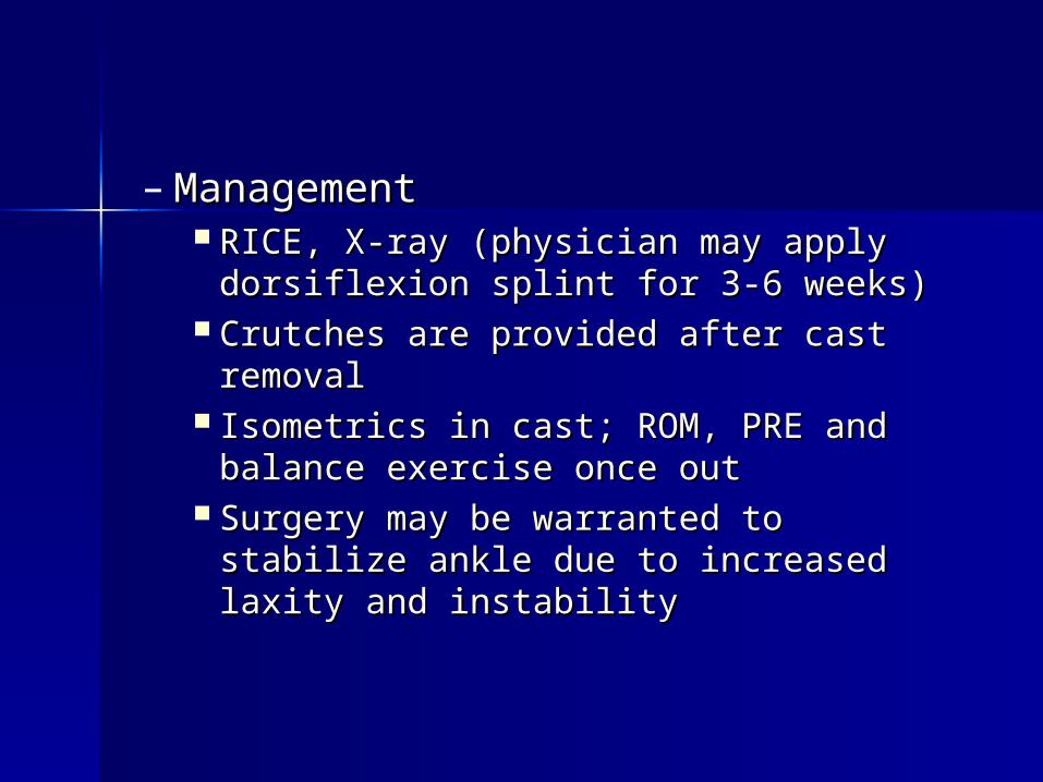
– ManagementManagement RICE, X-ray (physician may apply RICE, X-ray (physician may apply
dorsiflexion splint for 3-6 weeks)dorsiflexion splint for 3-6 weeks) Crutches are provided after cast removalCrutches are provided after cast removal Isometrics in cast; ROM, PRE and balance Isometrics in cast; ROM, PRE and balance
exercise once outexercise once out Surgery may be warranted to stabilize Surgery may be warranted to stabilize
ankle due to increased laxity and ankle due to increased laxity and instabilityinstability
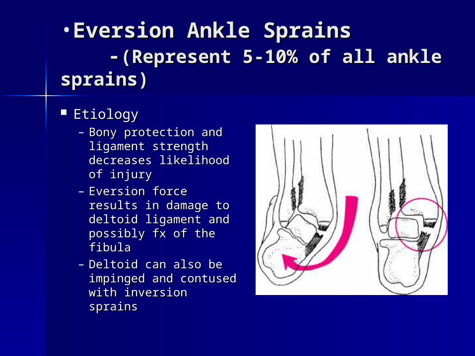
•Eversion Ankle SprainsEversion Ankle Sprains--(Represent 5-10% of all ankle (Represent 5-10% of all ankle
sprains)sprains)
Etiology Etiology – Bony protection and Bony protection and
ligament strength ligament strength decreases likelihood of decreases likelihood of injuryinjury
– Eversion force results Eversion force results in damage to deltoid in damage to deltoid ligament and possibly ligament and possibly fx of the fibulafx of the fibula
– Deltoid can also be Deltoid can also be impinged and impinged and contused with contused with inversion sprainsinversion sprains
Injury PreventionInjury Prevention
Strength training allows the Strength training allows the supporting musculature to supporting musculature to stabilize where ligaments may no stabilize where ligaments may no longer be capable of holding the longer be capable of holding the original tension between bones of original tension between bones of the joint. This will also help the joint. This will also help prevent reinjury.prevent reinjury.
Chronic Ankle Injury Chronic Ankle Injury “the vicious cycle”“the vicious cycle”
Why are some people prone to ankle Why are some people prone to ankle re-injury over and over?re-injury over and over?
Most commonly due to lack of Most commonly due to lack of rehabilitation, but more importantly rehabilitation, but more importantly lack of neuromuscular training.lack of neuromuscular training.
This means the person has not This means the person has not retrained the body to recognize where retrained the body to recognize where the ankle and foot are during motion.the ankle and foot are during motion.
This sets up the body part to be re-This sets up the body part to be re-injured due to improper feedback to injured due to improper feedback to the brain about body position.the brain about body position.
Injury PreventionInjury Prevention
Neuromuscular Control is the ability Neuromuscular Control is the ability to compensate for uneven surfaces to compensate for uneven surfaces or sudden change in surfaces. It is or sudden change in surfaces. It is retrained by using balance and retrained by using balance and agility exercises such as a BAPS agility exercises such as a BAPS board or standing on one leg with board or standing on one leg with eyes closed as well as using a eyes closed as well as using a single leg on a mini trampoline.single leg on a mini trampoline.
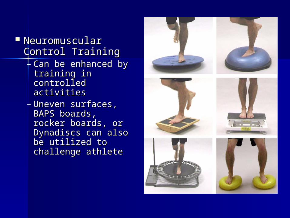
Neuromuscular Neuromuscular Control TrainingControl Training– Can be enhanced Can be enhanced
by training in by training in controlled activitiescontrolled activities
– Uneven surfaces, Uneven surfaces, BAPS boards, BAPS boards, rocker boards, or rocker boards, or Dynadiscs can also Dynadiscs can also be utilized to be utilized to challenge athletechallenge athlete
Injury preventionInjury prevention
Tight Achilles tendons can Tight Achilles tendons can predispose someone to injuring predispose someone to injuring the ankle. Tendonitis, plantar the ankle. Tendonitis, plantar fasciitis, and other disorders may fasciitis, and other disorders may occur due to a tight Achilles occur due to a tight Achilles tendon.tendon.
Injury PreventionInjury Prevention
Footwear is something often Footwear is something often overlooked but improper footwear overlooked but improper footwear can predispose someone with a can predispose someone with a foot condition such as pes planus foot condition such as pes planus (flat feet) to be more prone to (flat feet) to be more prone to having problems with their feet having problems with their feet and ankles.and ankles.
Preventative Taping and Preventative Taping and OrthosisOrthosis
Taping is often post injury treatment. Taping is often post injury treatment. Some will argue that taping will Some will argue that taping will weaken the ankle. This has not been weaken the ankle. This has not been proven without a doubt but exercise proven without a doubt but exercise and strengthening of the ankle is and strengthening of the ankle is always advised.always advised.
Othotics will help rectify conditions Othotics will help rectify conditions that are permanent and will not be that are permanent and will not be fixed by any other means.fixed by any other means.
Tape vs. BraceTape vs. Brace
Why choose one over anotherWhy choose one over another Taping may be more time consuming over Taping may be more time consuming over
bracebrace Braces may or may not allow more support Braces may or may not allow more support
over tapeover tape Tape allows more functional movement and Tape allows more functional movement and
often feels more stableoften feels more stable Tape will loosen with timeTape will loosen with time Braces will often loosen with timeBraces will often loosen with time It really is based on the quality of the brace It really is based on the quality of the brace
vs. the ability of the person to tape. Both vs. the ability of the person to tape. Both have advantages and disadvantages.have advantages and disadvantages.


