Page 1/25 In-Situ Science on Phobos with the Raman spectrometer for MMX (RAX): Preliminary Design and Feasibility of Raman Measurements Yuichiro Cho ( [email protected]) The University of Tokyo https://orcid.org/0000-0003-2749-2204 Ute Böttger DLR: Deutsches Zentrum fur Luft- und Raumfahrt Fernando Rull Universidad de Valladolid Heinz-Wilhelm Hübers Deutsches Zentrum für Luft und Raumfahrt DLR Standort Berlin: Deutsches Zentrum fur Luft- und Raumfahrt DLR Standort Berlin Tomàs Belenguer Instituto Nacional de Tecnica Aerospacial Anko Börner DLR Standort Berlin: Deutsches Zentrum fur Luft- und Raumfahrt DLR Standort Berlin Maximilian Buder DLR Berlin: Deutsches Zentrum fur Luft- und Raumfahrt DLR Standort Berlin Yuri Bunduki DLR Berlin: Deutsches Zentrum fur Luft- und Raumfahrt DLR Standort Berlin Enrico Dietz DLR Berlin: Deutsches Zentrum fur Luft- und Raumfahrt DLR Standort Berlin Till Hagelschuer DLR Berlin: Deutsches Zentrum fur Luft- und Raumfahrt DLR Standort Berlin Shingo Kameda Rikkyo University College of Science: Rikkyo Daigaku Rigakubu Daigakuin Rigaku Kekyuka Emanuel Kopp DLR Berlin: Deutsches Zentrum fur Luft- und Raumfahrt DLR Standort Berlin Matthias Lieder DLR Berlin: Deutsches Zentrum fur Luft- und Raumfahrt DLR Standort Berlin Guillermo Lopez-Reyes Universidad de Valladolid Andoni Moral Instituto Nacional de Tecnica Aerospacial
Transcript
Page 1/25
In-Situ Science on Phobos with the Ramanspectrometer for MMX (RAX): Preliminary Designand Feasibility of Raman MeasurementsYuichiro Cho ( [email protected] )
The University of Tokyo https://orcid.org/0000-0003-2749-2204Ute Böttger
DLR: Deutsches Zentrum fur Luft- und RaumfahrtFernando Rull
Universidad de ValladolidHeinz-Wilhelm Hübers
Deutsches Zentrum für Luft und Raumfahrt DLR Standort Berlin: Deutsches Zentrum fur Luft- undRaumfahrt DLR Standort BerlinTomàs Belenguer
Instituto Nacional de Tecnica AerospacialAnko Börner
DLR Standort Berlin: Deutsches Zentrum fur Luft- und Raumfahrt DLR Standort BerlinMaximilian Buder
DLR Berlin: Deutsches Zentrum fur Luft- und Raumfahrt DLR Standort BerlinYuri Bunduki
DLR Berlin: Deutsches Zentrum fur Luft- und Raumfahrt DLR Standort BerlinEnrico Dietz
DLR Berlin: Deutsches Zentrum fur Luft- und Raumfahrt DLR Standort BerlinTill Hagelschuer
DLR Berlin: Deutsches Zentrum fur Luft- und Raumfahrt DLR Standort BerlinShingo Kameda
Rikkyo University College of Science: Rikkyo Daigaku Rigakubu Daigakuin Rigaku KekyukaEmanuel Kopp
DLR Berlin: Deutsches Zentrum fur Luft- und Raumfahrt DLR Standort BerlinMatthias Lieder
DLR Berlin: Deutsches Zentrum fur Luft- und Raumfahrt DLR Standort BerlinGuillermo Lopez-Reyes
License: This work is licensed under a Creative Commons Attribution 4.0 International License. Read Full License
Version of Record: A version of this preprint was published at Earth, Planets and Space on December 1st,2021. See the published version at https://doi.org/10.1186/s40623-021-01496-z.
AbstractMineralogy is a key to understanding the origin of Phobos and its place in the context of the SolarSystem evolution. In-situ Raman spectroscopy on Phobos would be an important tool to achieve thescience objectives of the Martian Moons eXploration (MMX) mission and maximize the science merit ofsample return by characterizing the mineral composition and heterogeneity of the surface of Phobos.Conducting in-situ Raman spectroscopy under the harsh environment of Phobos requires a very sensitive,compact, lightweight, and robust Raman instrument that can be carried by the very compact MMX rover.In this context, a Raman spectrometer for MMX (RAX) is currently under development by an internationalcollaboration between teams from Japan, Germany, and Spain. To demonstrate the capability of acompact Raman system like RAX, we built an instrument that reproduces most of the opticalperformance of the �ight model using commercial off-the-shelf parts. Using this performance model, wemeasured mineral samples relevant to Phobos and Mars, such as anhydrous silicates, carbonates, andhydrous minerals. Our measurements of these samples indicate that such minerals can be measured andidenti�ed with a RAX-like Raman spectrometer with su�ciently high accuracy. We demonstrated aspectral resolution of approximately 10 cm-1 and high sensitivity of the Raman peak measurements (e.g.signal-to-noise ratios up to several 100). These results strongly suggest that the RAX instrument will becapable of determining the minerals expected on the surface of Phobos, adding valuable information toaddress the question on the moon’s origin, heterogeneity, and circum-Mars material transport.
1. IntroductionThe evolution of the Solar System is a fundamental research topic. One essential approach is thedetermination of the composition of the Solar System bodies. Their composition provides insights intothe possible origin and helps to understand the geochemical and thermal processes to which the bodyhas been exposed during its existence.
Several approaches exist to derive the composition with different kinds of spectroscopic techniques.Earth-based observations were the only one available for observations before space �ight was realized.With space missions then coming, satellites with spectrometers and cameras orbited and are orbiting thebodies taking images and spectra in the ultraviolet (UV), visible, infrared, and the spectral range beyond.By comparing these spectra with data obtained on Earth it was possible to derive the surfacecomposition on a macroscale. In-situ exploration and sample return from these bodies back to Earthprovide even more information, particularly on a microscale. In-situ measurements give an initial overviewof the general composition of the investigated target at single surface points. Detailed measurements ofreturned samples in the geologic context provide a deeper understanding of the processes on the surfaceof the investigated body. The combination of this information allows a complete picture about theprocesses the body may have experienced through its lifetime, as well as verifying existing hypotheses orestablishing alternative ones for the evolution of the body and thus that of the Solar System. Numerousexamples exist for all the approaches – here is one for illustration from JAXA’s Hayabusa mission. Duringthe Hayabusa mission the asteroid Itokawa was �rst studied using remote instrumentation from orbit
Page 5/25
(Krot et al. 2011). Later during the mission, samples were taken from the surface and brought back toEarth, where the samples were studied in the laboratories with highly sophisticated methods, such asback-scattered electron microscopy (Nakamura et al. 2011), neutron activation (Ebihara et al. 2011),synchrotron radiation X-ray tomographic microscopy (SRXTM) (Meier et al. 2013), isotopicmeasurements and mass spectroscopy (Yurimoto et al. 2011; Nagao et al. 2011; Busemann et al. 2013),and Raman microscopy (Böttger et al. 2013) (Figure 1).
In this context, mineralogy is a key discipline. Mineralogy helps to correlate the occurrence of differentminerals to the geochemical, thermal, or radiation processes that led to the formation of these minerals.Thus, techniques to derive the mineralogical composition in situ are of great interest. Ramanspectroscopy is a very appropriate method for this purpose. Raman spectroscopy is a nondestructive�ngerprint method that requires no sample preparation. It can be applied in the �eld as well as in thelaboratory. It is suitable to investigate various materials including minerals, organic and biological matter,liquids such as brines, gases, and ices.
Thus, the development of Raman spectrometers for in-situ exploration is receiving increased attention inspace research (e.g., Rull et al. 2017; Weber et al. 2017, 2018). With SuperCam on the Mars 2020 rovermission (launched in 2020) (Wiens et al. 2021) and the Raman Laser Spectrometer (RLS) on the ExoMars2022 mission (Rull et al. 2017) for the �rst time Raman instruments will be used on the surface of Marsfor mineralogical analysis and to search for signatures of past or present life. The rover that is developedfor the Martian Moons eXploration (MMX) mission by JAXA (Kawakatsu et al. 2019) will carry the Ramanspectrometer for MMX (RAX), a very compact Raman spectrometer, to investigate the surface of Phobos(Ulamec et al. 2019; Michel et al. submitted).
In this paper, we focus on the presentation of Raman spectroscopy on Phobos, the Martian moon. It isshown how Raman spectroscopy can support science to address the question of the origin of Phobosand thus add valuable input for the discussion on the evolution of the Solar System. First, the mainscienti�c questions related to Phobos are brie�y described. Second, the current instrument design toachieve the science goals is illustrated. Then, our measurements on several mineral samples relevant toPhobos and Mars are reported to demonstrate the capability of RAX.
2. Exploration Of Phobos And The Rax Instrument2.1 Science on Phobos
Phobos is one of the Martian moons orbiting Mars at less than 6000 km in 7.65 hours. One Phobos-dayis the same as the orbital period because of a tidal lock. The second moon Deimos is more than 23460km above Mars’ surface and needs 1.2624 days for one orbit. Both moons show similarities in theiralbedo and spectral behavior, but their origin is still unknown. Several models for their origin have alreadybeen discussed in detail (e.g., Pieters et al. 2014; Usui et al. 2020; Michel et al. submitted). Here we focuson how Raman spectroscopy can contribute to address the question of Phobos origin.
Page 6/25
To address the issue, we need to brie�y review the �ve most-relevant ideas about the origin of Phobos.Behind each hypothesis stands a selection of minerals that are expected to be present on Phobos. Theexpected mineralogy and geochemistry on Phobos with respect to the origin hypothesis was described byMurchie et al. (2014) and Usui et al. (2020).
These theories formulate the origin of the moons based on capture or by in-situ formation. The “capture”hypothesis includes (1) the capture of an organic- and water-rich outer Solar System body; (2) an organic-and water-poor outer Solar System body or (3) an inner Solar System body. The “in-situ formation”hypothesis either propose (4) the co-formation with Mars or (5) the formation from the ejecta after animpact of a large body onto Mars. Each of these hypotheses predicts a speci�c composition as well aselemental and mineral abundances (Table 1). The currently available re�ectance spectra measuredremotely from orbit are not conclusive because of the lack of features attributable to speci�c minerals.The low albedo of Phobos can be explained by either a carbonaceous origin or by darkening throughstrong space weathering (e.g. Shirley and Glotch, 2018 and references therein), with the correspondingmineralogy supporting the different processes. The spectra of the ‘red’ and ‘blue’ areas on Phobos bestmatch those of D-type and T-type asteroids, respectively (Rosenblatt 2011). The striking blue/red contrastcould be caused by compositional variations on Phobos or spatial variations of its physical surfaceproperties (Rosenblatt 2011; Ballouz et al. 2019). Understanding the nature of color difference and itsrelationship to mineralogy is important to resolve this. Endogenous materials, such as fragments of Marsejecta (Ramsley and Head 2013; Hyodo et al. 2019) and projectiles that hit Phobos are also expected andshould be distinguished from the original Phobos material as anomalous minerals deviated from themajority of materials found on Phobos. A measurement technique that is capable of identifying thedifferent minerals, like Raman spectroscopy, would therefore be of great bene�t.
2.2. Science with RAX
In Figure 2, Raman spectra of some representative minerals predicted by the origin theories of Phobos areshown. The spectra are well distinguishable and the �ngerprint characters are obvious. So, spatiallydistributed Raman measurements on Phobos would provide initial in-situ information on its mineralogicalcomposition and distribution, which would help narrow down the origin hypotheses.
In addition to the capability of mineral identi�cation, the need of only optical access to the sample andthe fact that no sample preparation is necessary make Raman spectroscopy a very suitable technique forspace exploration. Regardless certain technique limitations such as spectrally superimposing�uorescence or the relatively low Raman scattering e�ciency of certain materials, it is an appropriatemethod for the initial examination of an unknown surface.
To accomplish the in-situ mineralogy on Phobos, RAX is developed in Japanese-Spanish-Germancooperation to participate in the Japanese MMX Mission as part of the payload on the DLR-CNES Roverthat will be brought to Phobos’ surface during this mission (Hagelschuer et al. 2019). The MMX mission(Kawakatsu et al. 2019) and the rover are described in more detail elsewhere in this issue (Michel et al.submitted; Kuramoto et al. submitted).
Page 7/25
The science objectives are derived for RAX according to the scienti�c goals formulated for the MMXmission (Kuramoto et al. submitted). First of all, RAX shall investigate the surface mineralogy on Phobosby measuring Raman spectra of the surface and identifying mineral composition by comparing thespectra with those available in databases. The rover’s ability to move over the surface opens thepossibility to measure Raman spectra at different locations on Phobos and to study the surfaceheterogeneity. This might be used to support the characterization of a landing site under considerationand potentially to support the selection of samples for return to Earth. The obtained data can also becompared to those of Raman spectrometers on the surface of Mars to check the origin hypothesis of co-formation with Mars or accretion from ejecta after a giant impact on Mars. The measurements ofreturned samples would give a �rm con�rmation on the results of RAX. Comparing relative abundancesof minerals on the surface of Phobos with those found in the returned samples will help ensure therepresentativeness of returned samples, and thus maximize the scienti�c value of this sample returnmission.
The RAX measurements will be performed during Phobos nights or in the shadow of the rover to avoidambient light, which could be stronger than the Raman signals by orders of magnitude. Coarse laserfocusing will be achieved with raising/lowering the main body of the rover which contains the RAXinstrument. Images of the RAX footprint will be taken by the WheelCam (Michel et al. submitted). Thisfunction provides the geologic context of measured samples, including their albedo, texture, and grainsize. Measuring in the tracks of the Rover wheels and therefore sampling freshly exposed materials mightprovide information on space weathering. RAX can also be used without the laser, obtaining re�ectancespectra in a wavelength range of approximately 532-680 nm, which could be used for evaluating thealbedo and color of surface material.
2.3. Design of RAX
Albeit the high scienti�c values, conducting Raman spectroscopy on Phobos and ful�lling its scienceobjectives are a technical challenge. The instrument must endure the harsh environment on Phobos, suchas a large temperature range and fast diurnal cycles during one Phobos day (requiring RAX to withstand-55~+70℃ for storage and -40~+50℃ for operation), low surface gravity (making rover operation lessstraightforward), dust (potentially contaminating optics and actuator mechanism), vacuum (complicatingthe heat distribution within the instrument), and radiation (potentially deteriorating the transmission ofoptics and electronics). Furthermore, the RAX instrument must be particularly small and lightweight to �tin the low-mass rover. To illustrate the design to overcome these constraints, the overview of the RAXinstrument, its general speci�cation, and current status of its development are described in this section.
The RAX instrument consists of two physically separated units: the RAX Laser Assembly (RLA) and RAXSpectrometer Module (RSM) (Figure 3). The Autofocusing Subystem (AFS), dedicated to focusing thelaser on the surface of Phobos, is accommodated within the RSM. The entire RAX instrument has avolume of approximately 81 × 125 × 98 mm3 and a mass of approximately 1.4 kg. RAX is jointlydeveloped by an international collaboration among Germany, Spain, and Japan (Figure 3). The Institute
Page 8/25
of Optical Sensor Systems at Deutsches Zentrum für Luft- und Raumfahrt (DLR) develops the RSM. TheUniversity of Tokyo, JAXA, and Rikkyo University are in charge of AFS development. Instituto Nacional deTécnica Aeroespacial (INTA) and University of Valladolid, who built the RLS laser unit for the ExoMars2022 mission, provide the RLA (Figure 3).
The RLA is a compact laser module that emits a 532 nm continuous wave (CW) laser beam at a variablepower of up to 35 mW (Figure 4). This is essentially a �ight spare of the laser unit developed for theExoMars2022 mission (Rull et al. 2017). The RLA (Ribes-Pleguezuelo et al. 2019) provides laser light tothe RSM (Figure 5) through an optical �ber. The collimated laser beam is focused onto the surface ofPhobos through the Autofocusing Subsystem (AFS). The scattered light is collected and collimated by anentrance objective and sent back to the spectrometer module. A series of optics, such as a dichroic mirror,collimator lenses, slit, transmission grating, Raman edge �lter, and camera objective lenses, are mountedinside the RSM. The image of the slit is acquired by the 3D-plus CMOS sensor. The 2-dimensional imageis integrated to form a 1-D line spectrum. The electronics for controlling the laser and focus actuator areaccommodated in the RSM. Focusing the laser beam is required to maximize the intensity of Ramansignals emerging from the target surface below the rover. The AFS comprises of light-shuttle objectivelens (LSO) and actuator mechanism (Figure 6). The laser spot diameter on the sample is designed to be50 µm. The distance between the lowest tip of LSO and laser focus is 78 mm. The stroke of LSO and itsresolution are better than 13 mm and <50 µm by design. The autonomous focusing will involve a two-step procedure. In the �rst step, the re�ectance spectra of surface materials, which are illuminated by theLED placed near the entrance aperture, are used for focusing on the sample surface using the rover legsand AFS actuator. The second step is the �ne focusing using the laser and only the AFS actuator in orderto maximize the signal-to-noise-ratio (SNR) of potential Raman signals. Furthermore, the backscatteredlaser light will be measured in the RLA autofocus photodiode as well, for accurate focus distancedetermination.
3. Experimental SetupTo assess the capability of Raman spectroscopy using RAX, we built a breadboard model (BBM) fromcommercial components that simulates the performance of the actual RAX instrument aboard the Roveron Phobos. In this section, we describe the experimental setup of our BBM in comparison with the RAXinstrument.
3.1 Breadboard model
We used a �ber-fed Nd:YAG laser that emits CW radiation at 532.2 nm (JUNO532FC, SOC, Japan). Thelaser power was set at 32 mW, which results in a laser irradiance of 1.6 kW/cm2 at the target surface tosimulate the output of RLA. The laser beam was delivered to the inlet of our BBM through a multimodeoptical �ber. This �ber was used for simulating the non-Gaussian beam pattern of the �ight laser. Thespot size at the sample was designed to be 50 µm.
Page 9/25
To simulate the performance of the LSO in terms of light collection capability, the numerical aperture (NA)of the lens in our BBM was set at 0.20, which is comparable to 0.22 of the actual LSO. The lens movesvertically as the stepping motor rotates via the combination of a linear guide and lead screw (Figure 7).The stepping motor and attached gear box was identical to the one we plan to use for RAX. The objectivelens has a mass of 143 g, comparable to that of LSO (129 g). Dedicated printed circuit boards (PCBs)simulating that used for �ight model were used to verify the electrical behavior of the motor. Theresolution of vertical motion, which can be activated by one motor pulse, was measured to be < 25 µm.
A CMOS camera (MAKO G419, Allied Vision) was used as detector to simulate the actual �ight CMOSsensor. The width of a slit placed between a couple of collimator lenses was 50 µm. Because of themagni�cation of the optical system in our BBM, the slit width was imaged at 25 µm on the sensor, thesame image size as that of the RAX design. A transmission grating (1200 grooves/mm) was used. Acamera lens was connected to the commercial-off-the-shelf (COTS) CMOS camera with a custom-made�ange.
3.2 Measurement protocol
Using this BBM, bulk natural mineral samples relevant to the science objectives were selected andmeasured: anhydrous rock-forming minerals (olivine, quartz), carbonates (calcite, magnesite), sulfate(gypsum), and magnesium hydroxide (brucite). Once a sample is placed in the sample holder, theobjective lens was brought to the focus position by the actuator motion. In this study, the focus wasadjusted manually using a motor control software to maximize the intensity of the Raman signals. Themeasurements were conducted under air and at a room temperature.
The entire BBM was placed in an optical enclosure to avoid the ambient light coming into the systemthrough the objective lens. Furthermore, the optical path was covered by black plastic or anodizedaluminum to prevent strong Rayleigh light entering the optical path. The relation between CMOS sensorpixel numbers and Raman shift (cm-1) was calibrated with a Ne lamp. One pixel corresponded to 3-2 cm-1
in 0-4000 cm-1 range. Out of 2048 vertical pixels of the CMOS sensor, 500 lines containing the spectralimages were integrated in our BBM. The exposure time of the Raman measurements was set to 1 or 3 sfor each specimen. Either 100 or 50 spectra were averaged to enhance the signal-to-noise ratios of thespectra. Background (dark) spectra were measured with the same exposure time and number ofaveraging when the laser was turned off. The dark spectra were subtracted from the signal spectra toremove thermal noise. The relative spectral response of the system was corrected with a standardhalogen lamp. Nevertheless, because of a peak in the sensitivity of BBM’s CMOS sensor, a false dip/peaksometimes appeared at approximately 2500 cm-1. This wavenumber range was therefore not used for thepeak identi�cation in this study. This artifact is expected to be removed for the actual RAX instrument byfurther characterizing the spectral response function.
Each Raman feature was �tted with a Gaussian pro�le. The height S, width W, and position C of eachGaussian �t were derived. The continuum due to �uorescence was �tted simultaneously and subtracted
Page 10/25
from the spectrum. The noise level N associated with individual peaks was then de�ned by the standarddeviation of the signals over the 40 pixels in the continuum-subtracted spectra. The signal–to–noise-ratiowas calculated by dividing the peak signal S by the rms signal measured across a spectral band of 20pixels (approximately 60 cm-1) at a distance of 3 × peak width W from the Raman feature peak.
4. Results And DiscussionThe Raman spectra obtained with our RAX performance model are shown in this section to verify thedetectability of these minerals with the RAX instrument. Figure 8 shows the Raman spectra of theminerals measured with our BBM. The double peaks characteristic to olivine at 823 cm-1 and 854 cm-1
are cleary detected and resolved with our BBM. The peak positions are consistent with those of forsterite(Kuebler et al. 2006). The Raman spectrum of quartz shows a peak attributable to SiO4 stretching mode
at 467 cm-1 (Figure 8). These samples did not exhibit �uorescence. The Rayleigh light from the laser wasobserved at < 100 cm-1 with this BBM. The measurement capability of such low-wavenumber peaks willbe characterized with the engineering model of RAX using the �ight-like laser and edge �lter.
Figure 8 also shows the Raman spectra of the two carbonates: calcite (CaCO3) and magnesite (MgCO3).Both calcite and magnesite yielded unambiguous Raman peaks above the continuum due to�uorescence. For calcite, the peak at 160 cm-1 was detected, which shows the smallest Raman shiftobserved in this study. OH-related bands at wavenumber > 3000 cm-1 were seen for the gypsum (CaSO4
2H2O) and brucite (Mg(OH)2). The gypsum showed the Raman peaks characteristic to water at 3383 and
3474 cm-1, while brucite exhibits one at 3633 cm-1 (Figure 8). The signal-to-noise ratios of the individualpeaks that are currently achieved with our BBM are summarized in Table 2. The grain size, mineralmixtures, and surface roughness can in�uence the intensity of speci�c Raman features (Foucher et al.2013; Böttger et al. 2017). These aspects will be characterized in further investigation with a focus onexpected Phobos mineralogy. Nevertheless, our data suggest that a very compact Raman spectrometerlike the RAX instrument will be capable of in-situ detection of minerals expected on Phobos.
5. ConclusionFor in-situ analysis of the mineralogy on Phobos, Raman spectroscopy is very suitable to address thequestion of the origin of Phobos. RAX (Raman spectrometer for MMX) is a very compact and robustinstrument that is being developed for the rover in the scope of the MMX mission to Phobos. In this paper,we showed the main design of the instrument, particularly with respect to the challenges connected witha mission to Phobos and the constraint to �t on the small MMX rover. First results of Ramanmeasurements of Phobos-relevant minerals with a breadboard model of similar performance as expectedfor the �ight model show that RAX will ful�ll the requirements, such as the capability of resolving theolivine peaks 30 cm-1 apart and measuring with high sensitivity as required for the identi�cation ofminerals. Our results indicate that RAX will be able to obtain the Raman spectra of key mineralspotentially distributed on Phobos.
Page 11/25
AbbreviationsAFSAutofocusing subsystemBBMBreadboard modelCADComputer-aided designCMOSComplementary metal oxide semiconductorCOTSCommercial off-the-shelfCWContinuous waveDLRDeutsches Zentrum für Luft- und RaumfahrtEMEngineering modelFMFlight modelINTAInstituto Nacional de Técnica AeroespacialJAXAJapan Aerospace Exploration AgencyLEDLight emitting diodeLSOLight shuttle objectiveMMXMartian Moons ExplorationNdYAG:Neodymium-doped yttrium aluminum garnetRAXRaman spectrometer for MMXRLARAX laser assemblyRSMRAX spectrometer moduleSEMSecondary electron microscope
Page 12/25
SNRSignal-to-noise ratioSTMStructure and thermal model
DeclarationsEthics approval and consent to participate
Not applicable
Consent for publication
Not applicable
Availability of data and materials
The datasets used and/or analyzed during the current study are available from the corresponding authoron reasonable request.
Competing interests
The authors declare that they have no competing interests.
Funding
This study was supported by Japan Society for Promotion of Science Grant-in-Aid (grant numbersJP19K14778 and JP20H04607). From the Spanish side the project is partially funded by MINECO ProjectReference PID2019-107442RB-C3 and PID2019-107442RB-C2.
Authors' contributions
YC wrote the paper, with contributions from UB, FR, HWH, AMI, GLR, JAO, and IW. Conception, design, andrevision of the work: YC, HWH, FR, MB, SK, SRR, PS, SU, and TU. Conception of the instrument: UB, HWH,AB, ED, TH, CR, and FS. Data acquisition and analysis: YC, SM, JAO, MP, SR, TS, SS, KY. Interpretation ofthe data: YC, UB, FR, MP, SR, TS, and IW. TB, AB, ED, TH, MP, GP, CR, FS designed the instrument. YC, ED,SM, GP, and KY built the instrument. YB, SM, KW, KY created the software used in this study. The conceptof the autofocusing algorithm was developed by TS. EK developed the new electronics used in this study.ML, SR, and CR created the new structure models and analyzed its data. New detector models weredeveloped by CP. AMI, CCP, and PRP contributed to the conception and design of the laser. SS led thediscussion on the concept of operation. The discussion on the science of in-situ Raman spectroscopywas led by GLR, OPB, CS, and IW. MB was in charge of project management.
Acknowledgements
Page 13/25
The Raman spectra presented in Figure 1 and 2 are measured at the Raman laboratory of the DLR-Institute for Planetary Research in Berlin, Germany.
Authors' information
The authors wish it to be known that, in their opinion, the �rst 4 authors (YC, UB, FR, and HWH) should beregarded as joint First Authors.
ReferencesBallouz RL, Baresi N, Crites ST, Kawakatsu Y, Fujimoto M (2019) Surface refreshing of Martian moonPhobos by orbital eccentricity-driven grain motion. Nat Geosci 12:229-234. doi: 10.1038/s41561-019-0323-9
Böttger U, Alwmark C, Bajt S, Busemann H, Gilmour JD, Heitmann U, Hübers H-W, Meier MMM, Pavlov SG,Schade U, Spring NH and Weber I (2013) Raman Micro-spectroscopy of HAYABUSA Particles. HAYABUSA2013 Symposium of Solar Systems Materials, Tokyo, 16-18 October 2013
Böttger, U., Pavlov, S.G., Deßmann, N., Hanke, F., Weber, I., Fritz, J., Hübers, H.-W. (2017) Laser-inducedalteration of Raman spectra for micron-sized solid particles, Planetary and Space Science 138: 25-32,doi.org/10.1016/j.pss.2017.02.001.
Busemann H, Alwmark C, Bajt S, Böttger U, Gilmour JD, Heitmann U, Hübers H-W, Meier MMM, Pavlov S,Schade U, Spring NH, Weber I, Asteroid Itokawa Studied by Micro-Raman and Infrared Spectroscopy, X-Ray Tomography and High-Sensitivity Noble Gas Analysis. HAYABUSA 2013 Symposium of SolarSystems Materials, Tokyo, 16-18 October 2013
Ebihara M, Sekimoto S, Shirai N, Hamajima Y, Yamamoto M, Kumagai K, Oura Y, Ireland TR, Kitajima F,Nagao K, Nakamura T, Naraoka H, Noguchi T, Okazaki R, Tsuchiyama A, Uesugi M, Yurimoto H, ZolenskyME, Abe M, Fujimura A, Mukai T, Yada1Y (2011) Neutron Activation Analysis of a Particle Returned fromAsteroid Itokawa. Science 333:1119. doi: 10.1126/science.1207865
Foucher F, Lopez‐Reyes G, Bost N, Rull‐Perez F, Rüßmann P, Westall F (2013) Effect of grain sizedistribution on Raman analyses and the consequences for in situ planetary missions. Journal of RamanSpectroscopy 44.6: 916-925
Hagelschuer T, Belenguer T, Böttger U, Buder M, Cho Y, Dietz E, Gensch M, Hanke F, Hübers H-W, KamedaS, Kopp E, Kubitza S, Moral A, Paproth C, Pertenais M, Peter G, Rammelkamp K, Rodriguez P, Rull F, RyanC, Säuberlich T, Schrandt F, Schröder S, Ulamec S, Usui T, Vance R (2019) The Raman spectrometeronboard the MMX rover for Phobos, Proceedings of the 70th International Astronautical Congress (IAC),Walter E. Washington Convention Center, 21-25 October 2019
Page 14/25
Hyodo R, Kurosawa K, Genda Y, Usui T, Fujita K (2019) Transport of impact ejecta from Mars to its moonsas a means to reveal Martian history. Sci. Rep. 9:19833. doi: 10.1038/s41598-019-56139-x.
Kawakatsu Y, Kuramoto K, Ogawa N, Ikeda H, Ono G, et al. (2019) Mission De�nition of Martian MoonExploration (MMX). 70th International Astronautical Congress (IAC), Walter E. Washington ConventionCenter, 21-25 October 2019
Krot A (2011) Bringing Part of an Asteroid Back Home. Science 333:1098. doi: 10.1126/science.1212145
Kuebler K, Jolliff BL, Wang A, Haskin LA (2006) Extracting olivine (Fo–Fa) composition from Ramanspectral peak positions. Geochim. Cosmochim. Acta 70:6201–6222. doi: 10.1016/j.gca.2006.07.035
Kuramoto, K. et al. Martian Moons Exploration MMX: Sample Return Mission to Phobos ElucidatingFormation Processes of Habitable Planets. Earth Planets Space, this issue, submitted
Meier MMM, Alwmark C, Bajt S, Böttger U, Busemann H, Gilmour J, Heitmann U, Hübers H-W, Marone F,Pavlov S, Schade U, Spring N H, Stampanoni M., Terfelt F, Weber I (2013) Determining a Precise HE, NECosmic-Ray Exposure Age for Grains from Itokawa. HAYABUSA 2013 Symposium of Solar SystemsMaterials, Tokyo, 16-18 October 2013
Michel P, Ulamec S, Böttger U, Grott M, Murdoch N, Vernazza P, Biele J, Tardivel S, Groussin O, Jorda L,Knollenberg J, Sunday C, Zhang Y, Valette R The MMX rover: roving and performing in-situ surfaceinvestigations on Phobos. Earth Planets Space. this issue, submitted on November 24, 2020
Murchie SL, Britt DT, Pieters CM (2012) The value of Phobos sample return. Planetary and Space Science102:176–182. doi: 10.1016/j.pss.2014.04.014
Nagao K, Okazaki R, Nakamura T, Miura YN, Osawa T, Bajo Ki, Matsuda S, Ebihara M, Ireland TR, KitajimaF, Naraoka H, Noguchi T, Tsuchiyama A, Yurimoto H, Zolensky ME, Uesugi M, Shirai K, Abe M, Yada T,Ishibashi Y, Akio Fujimura A, Mukai T, Ueno M, Okada T, Yoshikawa M, Kawaguchi J (2011) IrradiationHistory of Itokawa Regolith Material Deduced from Noble Gases in the Hayabusa Samples. Science333:1128. doi: 10.1126/science.1207785
Nakamura T, Noguchi T, Tanaka M, Zolensky ME, Kimura M, Tsuchiyama A, Nakato A, Ogami T, Ishida H,Uesugi M, Yada T, Shirai K, Fujimura A, Okazaki R, Sandford SA, Ishibashi Y, Abe M, Okada T, Ueno M,Mukai T, Yoshikawa M, Kawaguchi J (2011) Itokawa Dust Particles: A Direct Link Between S-TypeAsteroids and Ordinary Chondrites. Science 333:1113. doi: 10.1126/science.1207758
Pieters CM, Murchie S, Thomas N, Britt D (2014) Composition of Surface Materials on the Moons ofMars. Planetary and Space Science 102:144-151. doi: http://dx.doi.org/10.1016/j.pss.2014.02.008
Ramsley KR, Head JW (2013) Mars impact ejecta in the regolith of Phobos: bulk concentration anddistribution. Planet. Space Sci 87:115–129. doi: 10.1016/j.pss.2013.09.005
Page 15/25
Ribes-Pleguezuelo P, Inza AM, Basset MG, Rodríguez P, Rodríguez G, Laudisio M, Galan M, Hornaff M,Beckert E, Eberhardt R, Tünnermann A (2016) Assembly processes comparison for a miniaturized laserused for the Exomars European Space Agency mission. Optical Engineering 55(11):116107. doi:10.1117/1.OE.55.11.116107
Ribes-Pleguezuelo P, Guiloht D, Basset MG, Beckert E, Eberhardt R, Tünnermann A (2019), Insights of theQuali�ed ExoMars Laser and Mechanical Considerations of Its Assembly Process. Instruments 20193(25). doi:10.3390/instruments3020025
Rosenblatt P (2011) The origin of the Martian moons revisited. Astron Astrophys Rev 19:44. doi:10.1007/s00159-011-0044-6
Rull F, Maurice S, Hutchinson I, Moral A, Perez C, Diaz C, Colombo M, Belenguer T, Lopez-Reyes G,Sansano A, Forni O, Parot Y, Striebig N, Woodward S, Howe C, Tarcea N, Rodriguez P, Seoane L, SantiagoA, Rodriguez-Prieto JA, Medina J, Gallego P, Canchal R, Santamarı ´a P, Ramos G, Vago JL, RLS Team(2017) The Raman Laser Spectrometer for the ExoMars Rover Mission to Mars. Astrobiology 17(6-7). doi:10.1089/ast.2016.1567
Shirley K, Glotch T (2018) Effects of Visible Albedo on Mid-Infrared Spectra under Simulated LunarEnvironment as Compared to Diviner Lunar Radiometer. European Planetary Science Congress (EPSC)2018, Technische Universität (TU) Berlin, 16-21 September 2018.
Ulamec S, Michel P, Grott M, Böttger U, Hübers H-W, Murdoch N, Vernazza P, Karatekin Ö, Knollenberg J,Willner K, Grebenstein M, Mary S, Chazalnoël P, Biele J, Krause C, Ho T-M, Lange C, Grundmann J-T, SasakiK, Maibaum M, Küchemann O, Reill J, Chalon M, Barthelmes S, Lichtenheldt R, Krenn R, Smisek M,Bertrand J, Moussi A, Delmas C, Tardivel S, Arrat D, F. Ijpelaan, Mélac L, Lorda L, Remetean E, Lange M,Mierheim O, Reershemius S, Usui T, Matsuoka M, Nakamura T, Wada K, Miyamoto H, Kuramoto K,LeMaitre J, Mas G, Delpech M, Celine L, Ra�egeau A, Boirard H, Schmisser R, Virmontois C, Cenac-MortheC, Besson D, Rull F (2019) A Rover for the MMX Mission to Phobos. 70th International AstronauticalCongress, (IAC), Walter E. Washington Convention Center, 21-25 October 2019
Usui T, Bajo Ki, Fujiya W. Furukawa Y, Koike M, Miura YN, Sugahara H, Tachibana S, Takano Y, KuramotoK. (2020) The Importance of Phobos Sample Return for Understanding the Mars-Moon System. Space SciRev 216:49. doi: 10.1007/s11214-020-00668-9
Yurimoto H, Abe Ki, Abe M, Ebihara M, Fujimura A, Hashiguchi M, Hashizume K, Ireland TR, Itoh S,Katayama J, Kato C, Kawaguchi J, Kawasaki N, Kitajima F, Kobayashi S, Meike T, Mukai T, Nagao K,Nakamura T, Naraoka H, Noguchi T, Okazaki R, Park C, Sakamoto N, Seto Y, Takei M, Tsuchiyama A,Uesugi M, Wakaki S, Yada T, Yamamoto K,1 Yoshikawa M, Zolensky ME (2011) Oxygen IsotopicCompositions of Asteroidal Materials Returned from Itokawa by the Hayabusa Mission. Science333:1116. doi: 10.1126/science.1207776
Page 16/25
Weber I, Böttger U, Pavlov SG, Hübers H-W, Hiesinger H, Jessberger EK (2017) Laser alteration on ironsul�des under various environmental conditions. J Raman Spectrosc 48(11):1509-1517. doi:10.1002/jrs.5083
Weber I, Böttger U, Pavlov SG, Stojic A, Hübers H-W, Jessberger EK (2018) Raman spectra of hydrousminerals investigated under various environmental conditions in preparation for planetary spacemissions. J Raman Spectrosc 49(11):1830-1839. doi: 10.1002/jrs.5463
Wiens RC, Maurice S, Robinson SH, Nelson AE, Cais P, Bernardi P, Newell RT, Clegg S, Sharma SK, StormsS, Deming J, Beckman D, Ollila AN, Gasnault O, Anderson RB, Andre Y, Angel SM, Arana G, Auden E, Beck P,Becker J, Benzerara K, Bernard S, Beyssac O, Borges L, Bousquet B, Boyd K, Caffrey M, Carlson J, CastroK, Celis J, Chide B, Clark K, Cloutis E, Cordoba EC, Cousin A, Dale M, De�ores L, Delapp D, Deleuze M,Dirmyer M, Donny C, Dromart G, Duran MG, Egan M, Ervin J, Fabre C, Fau A, Fischer W, Forni O, Fouchet T,Fresquez R, Frydenvang J, Gasway D, Gontijo I, Grotzinger J, Jacob X, Jacquinod S, Johnson JR,Klisiewicz RA, Lake J, Lanza1 N, Laserna J, Lasue J, Mouélic SL, Legett C, Leveille R, Lewin E, Lopez-Reyes G, Lorenz R, Lorigny E, Love SP, Lucero B, Madariaga JM, Madsen M, Madsen S, Mangold N,Manrique JA, Martinez JP, Martinez-Frias J, McCabe KP, McConnochie TH, McGlown JM, McLennan SM,Melikechi N, Meslin P-Y, Michel JM, Mimoun D, Misra A, Montagnac G, Montmessin F, Mousset V,Murdoch N, Newsom H, Ott LA, Ousnamer ZR, Pares L, Parot Y, Pawluczyk R, Peterson1 CG, Pilleri P, PineP, Pont G, Poulet F, Provost C, Quertier B, Quinn H, Rapin W, Reess J-M, Regan AH, Reyes-Newell AL,Romano PJ, Royer C, Rull F, Sandoval B, Sarrao JH, Sautter V, Schoppers MJ, Schröder S, Seitz D,Shepherd T, Sobron P, Dubois B, Sridhar V, Toplis MJ, Torre-Fdez I, Trettel IA, Underwood M, Valdez A,Valdez J, Venhaus D, Willis P (2021) The SuperCam Instrument Suite on the NASA Mars 2020 Rover:Body Unit and Combined System Tests. Space Sci Rev 217:4. doi:10.1007/s11214-020-00777-5
TablesTable 1 Origin hypotheses for Phobos and the predicted mineral abundances (Murchie et al. 2014).
Hypotheses for Origin Mineral abundances
1. Capture of organic- and water-richouter solar system body
Abundant phyllosilicates; carbonates and organic phases;anhydrous silicate phases rare
2. Capture of organic- and water-poorouter solar system body
Anhydrous, med.Fe (20–40%) pyroxene + olivine; abundantamorphous carbon or graphite?
3. Capture of inner solar system body Low carbonates, phyllosilicates; pyroxene, olivine probablyin range of known meteorites
4. Co-accretion with Mars Anhydrous silicates with Fe, Mg of bulk Mars; lowabundance of C-bearing phases
5. Giant impact on Mars Evolved, basaltic mineralogy consistent with manydatasets for Mars
Page 17/25
Table 2 Identi�ed Raman peaks and their signal-to-noise ratios.
Sample Sampletype
Peakposition(cm− 1)
Signal-to-noiseratio
Number of spectraused for averaging
Exposure time for asingle spectrum (s)
Olivine Crystalizedbulk
823 102 100 1
854 77
Quartz Bulk 467 582 100 1
Calcite Bulk 160 14 50 1
284 45
711 9
1078 72
Magnesite Bulk 333 12 100 1
1088 22
Gypsum Bulk 1001 43 100 1
1124 10
3383 4
3474 7
Brucite Bulk 3633 98 100 3
Figures
Page 18/25
Figure 1
(a) Microscopy image of a cut, sputtered and polished Hayabusa Particle RA-QD02-0051 embedded inepoxy. (b) Color composite of mineral composition derived from Raman spectral imaging at DLR Berlin.(c) Raman spectra of typical minerals found on #51. The colors of the spectra correspond to the colors ofthe Raman spectral image. Plagioclase is a Na, Ca-feldspar (Böttger et al. 2013).
Page 19/25
Figure 2
Raman spectra of some representative minerals predicted for the origin hypotheses of Phobos.
Page 20/25
Figure 3
Block diagram of the RAX instrument. RSM, RLA, and AFS are developed respectively by Germany, Spain,and Japan.
Page 21/25
Figure 4
Engineering model of the RLA. Its mass is 112 g including the laser module, thermo electrical module(TEM), interface plate, cables, and connectors. A new laser assembly batch, based on RLS (Ribes-Pleguezuelo et al. 2016) design, will be manufactured for the use in RAX. (credit: RLS project).
Page 22/25
Figure 5
(a) CAD model of the RAX spectrometer module (RSM). The AFS is seen in the frame. (b) Structural andthermal model (STM) being assembled at DLR.
Page 23/25
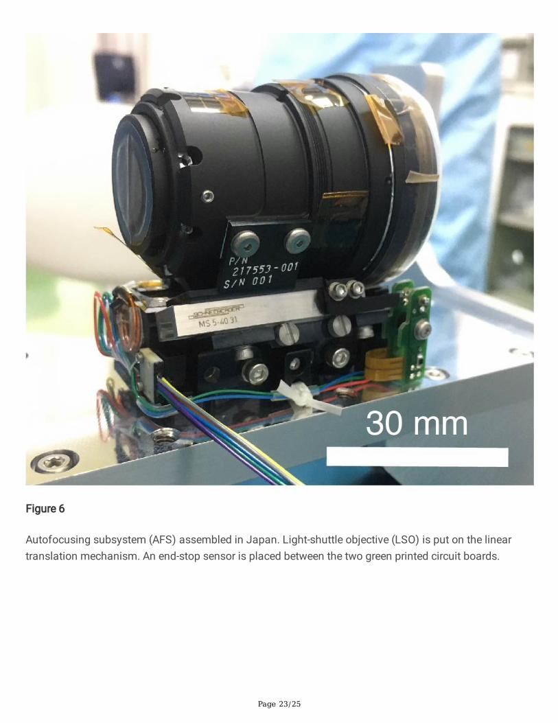
Figure 6
Autofocusing subsystem (AFS) assembled in Japan. Light-shuttle objective (LSO) is put on the lineartranslation mechanism. An end-stop sensor is placed between the two green printed circuit boards.
Page 24/25
Figure 7
Breadboard model of RAX. (a) Entire breadboard. (b) Focusing unit. Actuator rotations are converted tovertical motion with a lead screw and linear guide. The actuator and linear guide are the COTScomponent used for the RAX �ight model after screening. Mass of the lens is comparable to �ight lensbarrel.
Page 25/25
Figure 8
Raman spectra of minerals obtained with a RAX BBM. Anhydrous minerals (olivine and quartz),carbonates (calcite and magnesite), hydrated sulfate (gypsum) and magnesium hydroxide (brucite). Eachspectrum is offset for clarity.
Supplementary Files
This is a list of supplementary �les associated with this preprint. Click to download.