Análisis genómico al azar de Edwardsiella tarda ETSJ54: anotación de genes relacionados con virulencia 69 ARTÍCULO ORIGINAL/ORIGINAL ARTICLE Análisis genómico al azar de Edwardsiella tarda ETSJ54: anotación de genes relacionados con virulencia A random genome analysis of Edwardsiella tarda ETSJ54: annotation of putative virulence- related genes A análise do genoma aleatória de Edwardsiella tarda ETSJ54: anotação de genes de virulência relacionados ao putativos Noel Verjan - García 1,2,3 , Carlos A. Iregui - Castro 2 , Ikuo Hirono 3 1 MVZ, MSc, PhD, Grupo de Investigación Inmunobiología y Patogénesis, Departamento de Sanidad Animal, Facultad de Medicina Veterinaria y Zootecnia, Universidad del Tolima, Ibagué Colombia. 2 MV, PhD, Laboratorio de Patobiología, Facultad de Medicina Veterinaria y Zootecnia, Universidad Nacional de Colombia, Bogotá Colombia. 3 PhD, Laboratory of Genome Science, Graduate School of Marine Science and Technology, Tokyo University of Marine Science and Technology, Konan 4-5-7, Minato, Tokyo 108-8477, Japan Email: [email protected]Recibido: marzo 5 de 2012 Aceptado: mayo 11 de 2013 Resumen Como un paso inicial para comprender los mecanismos de patogenicidad usados por Edwardsiella tarda durante la infec- ción en peces, se llevo a cabo un secuenciamiento genómico parcial y al azar de librerias de ADN construidas en vectores cosmido y plasmido generadas a partir de una cepa (ETSJ54) virulenta de E. tarda para identificar genes presumiblemente relacionados con su virulencia. Los genes relacionados con virulencia de acuerdo a la semejanza en las secuencias de nucleotides con otras especies bacterianas fueron agrupados en nueve categorías que incluyeron quimiotaxis y motilidad, endotoxina (LPS), secreción de toxinas por los sistemas scretorios I y III, adquisición de hierro, proteasas y sobreviviencia dentro de macrófagos. Los resultados indican que E. tarda posee un amplio rango de genes involucrados en la virulencia y en la patogenicidad de generos bacterianos diversos y especies como Salmonella, Yersinia and Vibrios. Los resultados también indican que existe un alto flujo de genes en el genoma de E. tarda que podrían explicar en algún grado su potencial de infectar y causar enfermedad en varias especies animales. Palabras clave: Edwardsiellosis, secuenciamiento genómico, virulencia, patogénesis. Abstract As an initial step to understand the pathogenic mechanisms displayed by Edwardsiella tarda during infection in fish, we conducted a random genome sequencing of cosmid and plasmid DNA libraries generated from a virulent E. tarda strain (ETSJ54) to identify putative virulence-related genes. The assumed virulence-related genes of E. tarda were grouped into nine categories including chemotaxis and motility, adhesion and invasion, endotoxin (LPS), toxin secretion by type I and type III secretion systems, iron uptake, proteases, and intra-macrophage survival. The results reveal that E. tarda is equipped with a wide range of genes involved in virulence and pathogenesis of diverse bacterial genera and species including Salmo- ORINOQUIA 17-1 AGOSTO 2013.indd 69 19/09/2013 02:28:50 p.m.
Transcript
Análisis genómico al azar de Edwardsiella tarda ETSJ54: anotación de genes relacionados con virulencia 69
ARTÍCULO ORIGINAL/ORIGINAL ARTICLE
Análisis genómico al azar de Edwardsiella tarda ETSJ54: anotación
de genes relacionados con virulencia
A random genome analysis of Edwardsiella tarda ETSJ54: annotation of putative virulence- related genes
A análise do genoma aleatória de Edwardsiella tarda ETSJ54: anotação de genes de virulência relacionados ao putativos
Noel Verjan - García1,2,3, Carlos A. Iregui - Castro2, Ikuo Hirono3
1 MVZ, MSc, PhD, Grupo de Investigación Inmunobiología y Patogénesis, Departamento de Sanidad Animal, Facultad de Medicina Veterinaria y Zootecnia, Universidad del Tolima, Ibagué Colombia.2 MV, PhD, Laboratorio de Patobiología, Facultad de Medicina Veterinaria y Zootecnia, Universidad Nacional de Colombia, Bogotá Colombia.3 PhD, Laboratory of Genome Science, Graduate School of Marine Science and Technology, Tokyo University of Marine Science and Technology, Konan 4-5-7, Minato, Tokyo 108-8477, Japan Email: [email protected]
Recibido: marzo 5 de 2012 Aceptado: mayo 11 de 2013
Resumen
Como un paso inicial para comprender los mecanismos de patogenicidad usados por Edwardsiella tarda durante la infec-ción en peces, se llevo a cabo un secuenciamiento genómico parcial y al azar de librerias de ADN construidas en vectores cosmido y plasmido generadas a partir de una cepa (ETSJ54) virulenta de E. tarda para identificar genes presumiblemente relacionados con su virulencia. Los genes relacionados con virulencia de acuerdo a la semejanza en las secuencias de nucleotides con otras especies bacterianas fueron agrupados en nueve categorías que incluyeron quimiotaxis y motilidad, endotoxina (LPS), secreción de toxinas por los sistemas scretorios I y III, adquisición de hierro, proteasas y sobreviviencia dentro de macrófagos. Los resultados indican que E. tarda posee un amplio rango de genes involucrados en la virulencia y en la patogenicidad de generos bacterianos diversos y especies como Salmonella, Yersinia and Vibrios. Los resultados también indican que existe un alto flujo de genes en el genoma de E. tarda que podrían explicar en algún grado su potencial de infectar y causar enfermedad en varias especies animales.
As an initial step to understand the pathogenic mechanisms displayed by Edwardsiella tarda during infection in fish, we conducted a random genome sequencing of cosmid and plasmid DNA libraries generated from a virulent E. tarda strain (ETSJ54) to identify putative virulence-related genes. The assumed virulence-related genes of E. tarda were grouped into nine categories including chemotaxis and motility, adhesion and invasion, endotoxin (LPS), toxin secretion by type I and type III secretion systems, iron uptake, proteases, and intra-macrophage survival. The results reveal that E. tarda is equipped with a wide range of genes involved in virulence and pathogenesis of diverse bacterial genera and species including Salmo-
ORINOQUIA 17-1 AGOSTO 2013.indd 69 19/09/2013 02:28:50 p.m.
70 ORINOQUIA - Universidad de los Llanos - Villavicencio, Meta. Colombia Vol. 17 - No 1 - Año 2013
Introduction
Edwardsiellosis is a systemic suppurative disease cau-sed by the Gram-negative bacterium Edwardsiella tar-da, a member of the family enterobacteriaceae (Ewing et al., 1965). E. tarda is usually found in water-living ani-mals, causing disease in cultured marine and fresh-wa-ter fishes around the world (Miyazaki and Kaige 1985). The bacterium may also cause sporadic infections in birds, frogs, reptiles, marine and terrestrial mammals including humans (Verjan et al., 2012). The infection in man often occurs accidentally during manipulation of aquatic animals and range from self-limited gastrointes-tinal and extraintestinal infections up to lethal septice-mia (Wang et al., 2005; Spencer et al., 2008).
Multiple proteins appear to be involved in the viru-lence and pathogenesis of E. tarda infections, some of them are hemolysins (Hirono et al., 1997), siderophore production, resistance to serum killing, motility media-ted by the flagella, and phosphate uptake (Mathew et al., 2001), a sialidase Nan A that increase coloniza-tion of fish tissues (Jin et al., 2012), a type III secretion system that allow survival and replication of E. tarda within macrophages (Okuda et al., 2006), a DNA ade-nine methylase (Dam) that reduce UV radiation and H2O2 sensibility (Sun et al., 2010), an iron-cofactored superoxide dismutase (FeSOD) that inhibits macropha-ge-mediated immune responses (Cheng et al., 2010), and plasmids coding antibiotic resistance genes, trans-posases and conjugal transfer genes have also been associated with E. tarda virulence (Yu et al., 2012).
The above studies have contributed substantially to un-derstand the pathogenic mechanisms used by E. tarda during the infection process in fish, and the information gathered from the whole genome sequence of E. tar-
da EIB202 strain showed that a substantial proportion of the genome is devoted to the growth and survival under diverse conditions including intracellular niches (Wang et al., 2009). We initially reported the identifica-tion of seven antigenic protein coding genes of E. tar-da ETSJ54 strain (Verjan et al., 2005), and subsequent studies by others reported the usefulness and protecti-ve effects of some of those proteins in vaccinated fish (Hou et al., 2009). Our group also performed a partial genome sequencing of the E. tarda ETSJ54 genome and deposited in the Gene Bank database a number of vi-rulent-related genes (Verjan, 2005). Here, we present the annotation and a discussion of the putative roles of those genes that were available since 2005, before the whole genome of E. tarda was published. By that time there were no many sequenced genes of E. tarda available and by using the basic local alignment search tool (BlastX, version 2.2.28+), the obtained nucleotide sequences resembled those from many Gram-negative enteropathogens, however, an up-to-date BlastP (BlastP, version 2.2.28+) results is presented here and indicate that almost all the coded proteins of the E. tarda ETSJ54 genome correspond to those of the E. tarda EIB202 stra-in (Wang et al., 2009). The results shows that E. tarda ETSJ54, is equipped with the genes coding for major surface structures involved in motility, lipopolysaccha-rides and capsular polysaccharides, endo and exo-toxin secretion, iron uptake, intramacrophage survival and proteases among others. The presence of a variety of insertion sequence elements not only indicates a high genetic flux in the E. tarda genome but also suggests this bacterium has a highly dynamic and potentially rapidly evolving genome that could explain in some extent its potential to infect and to cause disease in a number of animal species.
nella, Yersinia and Vibrios species. The results also indicate a high genetic flux in the E. tarda genome that could explain in some extent its potential to infect and to cause disease in a number of animal species.
Como um passo inicial para entender os mecanismos patogenéticos expostos por Edwardsiella tarda durante a infecção no peixe, conduzimos uma genoma sequencing de cosmid e plasmad ADN bibliotecas geradas de um virulento E. tarda tensão (ETSJ54) para identificar genes putativos relacionados com virulência. Os genes relacionados com virulência assumidos de E. tarda foram agrupados em ocho categorias inclusive chemotaxis e motility, endotoxin (LPS), tipo I e tipo III sistemas de substância segreda, compreensão de ferro, procaçoadores, e intra-macrophage sobrevivência. Os resultados revelam que E. tarda é equipado com uma larga variedade de genes implicados na virulência e pathogenesis de gêneros bacterianos diversos e espécie inclusive Salmonella, Yersinia e espécie Vibrios. Os resultados também indicam um alto fluxo genético no E. tarda genoma que pode explicar em alguma extensão o seu potencial para infeccionar e causar a doença em um número de espécie dos animais.
ORINOQUIA 17-1 AGOSTO 2013.indd 70 19/09/2013 02:28:50 p.m.
Análisis genómico al azar de Edwardsiella tarda ETSJ54: anotación de genes relacionados con virulencia 71
Material and methods
Bacterial strains and culture conditions
E. tarda SJ54 (ETSJ54) was isolated from an outbreak of disease in Japanese flounder (Paralichthys olivaceus) in Shizuoka, Japan. The bacterium was grown on heart in-fusion medium (Difco Laboratories, Detroit, MI, USA) at 30 oC. All bacterial strains and plasmids used in this stu-dy are described in Table 1. Escherichia coli strains XL1-Blue MR and JM109 were grown in Luria-Bertani (LB) or 2 × YT medium at 37 oC and when required, ampicillin at concentrations of 50 µg/ml and chloramphenicol at 20 µg/ml were added (Sambrook and Russell 2001).
Construction of genomic DNA libraries
Genomic DNA from ETSJ54 was isolated by the method of Ausubel (Ausubel et al., 1994), and partially digested with a fixed concentration of Sau3A1 enzyme at the indicated time-lapses (30s, 60s, 90s, 2’, 3’, 5’, 7’, 10’
and 15’). Genomic DNA fragments obtained at each digestion period were separated in 1% agarose by pul-sed field gel electrophoresis (PFGE), with pulse times of 5s to 20s at 6 volts for 8 hr. Genomic DNA fragments in the 20-40 Kbp range (Figure 1) were dephosphoryla-ted with calf intestinal alkaline phosphatase (Promega, Madison, WI, USA) and ligated into the BamHI site of Supercos I vector (Stratagene, La Jolla, CA, U.S.A). The recombinant molecules were packaged into lambda (λ) phage particles (Epicentre Technologies, Madison, WI, USA) and used to infect E. coli XL1-Blue MR. Geno-mic DNA from ETSJ54 was also subjected to random mechanical shearing by using an ultrasonic disrupter UD-21 (Tomy Digital Biology Co, Tokyo Japan), cou-pled with a micro tip to produce small DNA fragments (0.5-2 kbp) by ultrasounds. The DNA fragments were ligated into the plasmid vector puC118 (Takara, Oht-su, Japan) to generate a plasmid DNA library. E. coli JM109 was transformed with recombinant plasmids by the heat shock method and all DNA, cosmid and plasmid preparations were carried out using standard
Table 1. Strains and plasmids used in this work
Bacterial strains and plasmids
Genotype, phenotype or characteristicsSource or reference
ORINOQUIA 17-1 AGOSTO 2013.indd 71 19/09/2013 02:28:50 p.m.
72 ORINOQUIA - Universidad de los Llanos - Villavicencio, Meta. Colombia Vol. 17 - No 1 - Año 2013
procedures (Sambrook and Russell 2001). Cosmid and plasmid DNA libraries were amplified and stored at -80 °C until use.
Subcloning and nucleotide sequence determination
Cosmid and plasmid libraries were cultured in LB agar plates with ampicillin and single colonies were ran-domly isolated and grown in 2 x YT broth for cos-mid or plasmid DNA isolation. Sequencing of the terminal ends of cosmid DNA was performed with T3, 5´-(ATTAACCCTCACTAAAGGGA)-3´ and T7, 5´-TAATACGACTCACTATAGGG 3´primers sets to identify putative ORF flanking the E. tarda DNA frag-ments. Cosmid DNA was digested with EcoRI res-triction enzyme to estimate the size of the inserted DNA fragment, followed by digestion with several restriction enzymes (i.e, BamHI, EcoRI, EcoRV, HincII, HindIII, PstI, SacI, or SacII) and the DNA fragments ligated into plasmid vectors (pUC118, pBluescript, or pHSG399) for sequencing (Figure 2). Plasmid DNA were sequenced with M13F (5′-GTAAAACGACGGC-CAGTACG-3′) and M13R (5′-ACTATCTAGAGCGGC-CGCTT-3′) primer sets. The nucleotide sequences were determined by the cycle sequencing method using Thermo sequenase fluorescent-labeled primer cycle sequencing kit (Amersham Pharmacia Biotech, Little Chalfont Buckinghamshire, UK). Specific oligo-nucleotides primers were designed to amplify some of the putative open reading frames (ORFs). The PCR
products were ligated into pGEM-T Easy vector (Pro-mega, Madison, WI, USA) and sequenced. Details for any technique will be provided if required.
Gene annotation and classification
The DNA sequence data of ETSJ54 were compared with those in the GenBank (www.ncbi.nlm.nih.gov) database using the BLASTX (Version 2.2.28+) soft-ware (Zhang et al., 2000) of the National Center for Biotechnology Information, to identify DNA sequen-ces that resemble our query sequence based on si-milarity of the nucleotide sequence. The identified closest homologous gene sequence in other bacte-rial species allowed predicting its putative function or the potential origin of the DNA sequence and its classification. The functional classification of E. tarda DNA sequences followed that used for other pathogens such as Yersinia and Salmonella species database of the Sanger Institute (www.sanger.ac.uk/Projects/Microbes/), or those reported in the Mi-crobial Genome Database (http://mbgd.genome.ad.jp). The putative virulence-related genes of E. tar-da ETSJ54 were submitted to the GenBank data base and the data included the closest original hits ob-tained when no E. tarda genome was known. Here, we provide an updated comparison of the predicted amino acid sequence of the ETSJ54 ORFs using the BLASTP (Version 2.2.28+) software (Altschul et al., 1997).
Figure 1. Flowchart of Edwardsiella tarda ETSJ54 cosmid DNA library construction. Genomic DNA of E. tarda ETSJ54 was isolated and digested with Sau3AI restriction enzyme for 30s, 60s, 90s, 2’, 3’, 5’, 7’, 10’ and 15’ and analyzed in 1% agarose by pulsed field gel electrophoresis (Lanes 2-9). Lane 1: undigested genomic DNA. PFG-M: PFG DNA ladder marker. M: HindIII digested lambda DNA marker. B. Digested genomic DNA was dephosphorylated and ligated into the BamHI of Super Cos I vector. C. E. coli XL-1BlueMR cells were infected with lambda phage particles carrying the recombinant cosmid molecules.
ORINOQUIA 17-1 AGOSTO 2013.indd 72 19/09/2013 02:28:50 p.m.
Análisis genómico al azar de Edwardsiella tarda ETSJ54: anotación de genes relacionados con virulencia 73
Results
Functional classification of E. tarda ETSJ54 open reading frames (ORFs)
One thousand and one hundred fifty eight (1,158) pu-tative ORFs of the Edwardsiella tarda ETSJ54 genome were identified from a total of 1,382 sequenced clones (1,056 cosmid and 326 plasmid clones). The number of putative ORFs and the coded genes revealed that there was not significant redundancy in the sequenced clones, and indicates these libraries are unique and represent an important tool for further studies. The functional classification of E. tarda ETSJ54 ORFs (Table
2) shows 5 major categories as follows: small molecule metabolism (256 ORFs), which constitute 22% from total ORFs and contain protein-coding genes involved in degradation of carbon compound and amino acids, energy metabolism, central intermediary metabolism, amino acid biosynthesis, polyamine synthesis, nucleo-sides and nucleotides biosynthesis, cofactors and fatty acid biosynthesis. The other four major categories are the broad regulatory function-related genes (65 ORFs), macromolecule metabolism (219 ORFs), cell proces-ses (179 ORFs) and others (439 ORFs), which include insertion sequence elements and hypothetical prote-ins. The percentages of E. tarda ETSJ54 ORFs in each subcategory are shown in Figure 3.
Figure 2. Subcloning and sequencing of plasmid DNA clones carrying E. tarda ETSJ54 genomic DNA fragments. Cosmid DNA was isolated and the 5’ and 3’ regions of the inserted DNA were sequenced using T3 and T7 primers (A). The cosmid DNA was digested with several restriction enzymes (B), and the DNA fragments ligated into pUC118 or pHSG398 plasmid vectors (C). The nucleotide sequence of the inserted DNA fragments were sequenced with M13 primers sets (D), and generated sequence data was compared with those in the Gene Bank database (See Materials and Methods).
ORINOQUIA 17-1 AGOSTO 2013.indd 73 19/09/2013 02:28:50 p.m.
74 ORINOQUIA - Universidad de los Llanos - Villavicencio, Meta. Colombia Vol. 17 - No 1 - Año 2013
Table 2. Functional classification of Edwardsiella tarda ETSJ54 ORFs
Functional categoryNo.
ORFs% Functional category
No. ORFs
%
1 Small molecule metabolism 2 Broad regulatory functions [250] 65
1.A Degradation 4 2.1 Signal transduction 0
1.A.1 Carbon compounds 33 Total 65 5,65
1.A.2 Amino acids 7 3 Macromolecule metabolism
1.B Energy metabolism 3.A Synthesis and modification
1.B.1 Glycolysis 5 of macromolecules
1.B.2 Pyruvate dehydrogenase 2 3.A.1 rRNA and stable RNAs 2
1.B.3 Tricarboxylic acid cycle 3 3.A.2 Ribosomal protein synthesis and modification 4
ORINOQUIA 17-1 AGOSTO 2013.indd 74 19/09/2013 02:28:50 p.m.
Análisis genómico al azar de Edwardsiella tarda ETSJ54: anotación de genes relacionados con virulencia 75
Virulence-related genes in the E. tarda ETSJ54 strain
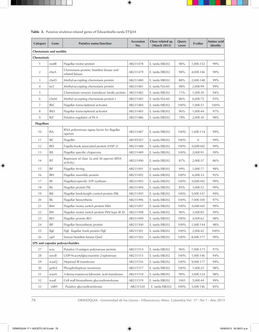
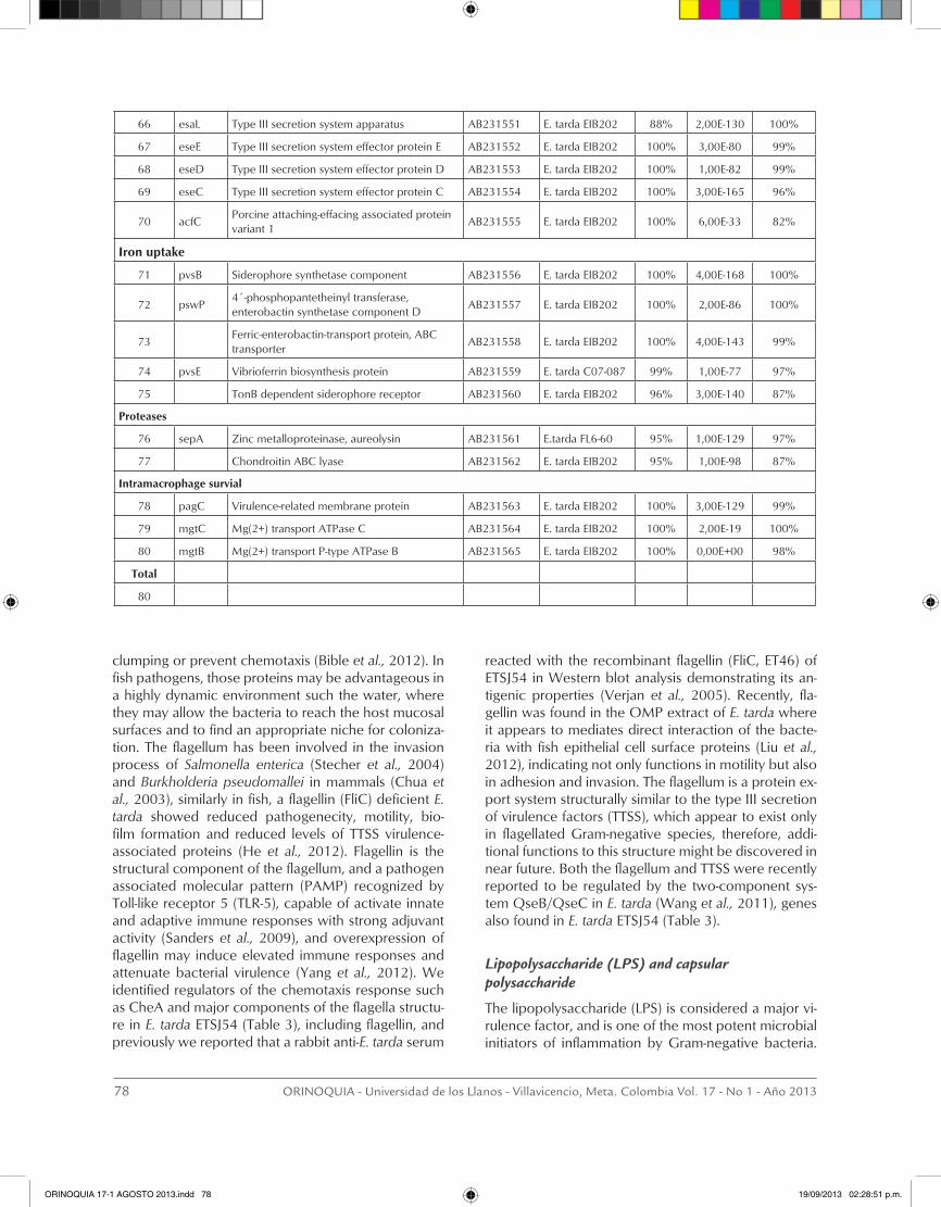
A total of one hundred and five (105) putative viru-lence-related genes of E. tarda ETSJ54 were annotated and deposited in the Gene Bank database. Identifi-cation was made by comparison of their nucleotide sequence with those in other bacterial pathogens, in which virulence-related genes and the coding protein have been characterized in some extent. Eighty (80) putative virulence-related genes were grouped into 8 subcategories and the GeneBank accession numbers are presented in Table 3. The subcategories in which the E. tarda ETSJ54 ORFs fall into were chemotaxis and motility conferred by the flagellum, capsular polysac-charide and endotoxin production, exotoxin secretion by type I and type III secretion systems, iron uptake, proteases and intramacrophage survival. A wide ran-ge of membrane proteins, lipoproteins and proteins involved in peptidoglycan biosynthesis are also com-ponents of the bacterial cell wall, and may play diffe-rent roles in the pathogenesis of the disease, they were
classified as ¨other virulence-related genes¨ and not included in this report. The predicted amino acid se-quences coded by 80 of the ETSJ54 ORFs were com-pared to those in the protein sequence database and show that almost all coded proteins resemble those recently reported in E. tarda EIB202 (Wang et al., 2009) and the E. tarda C07-087 (Tekedar et al., 2013), howe-ver, there still differences between E. tarda strains and the amino acid identity may varies from 48% to 100 %. These differences may support further studies of its characterization.
Discussion
Chemotaxis and motility conferred by the flagellum
Bacteria are able to sense, respond and adapt to envi-ronmental signals that may be useful or detrimental to cell survival. Chemotaxis proteins and the flagellum are coupled to various signal transduction pathways that modulate gene expression to drive motility, cell-to-cell
78 pagC Virulence-related membrane protein AB231563 E. tarda EIB202 100% 3,00E-129 99%
79 mgtC Mg(2+) transport ATPase C AB231564 E. tarda EIB202 100% 2,00E-19 100%
80 mgtB Mg(2+) transport P-type ATPase B AB231565 E. tarda EIB202 100% 0,00E+00 98%
Total
80
clumping or prevent chemotaxis (Bible et al., 2012). In fish pathogens, those proteins may be advantageous in a highly dynamic environment such the water, where they may allow the bacteria to reach the host mucosal surfaces and to find an appropriate niche for coloniza-tion. The flagellum has been involved in the invasion process of Salmonella enterica (Stecher et al., 2004) and Burkholderia pseudomallei in mammals (Chua et al., 2003), similarly in fish, a flagellin (FliC) deficient E. tarda showed reduced pathogenecity, motility, bio-film formation and reduced levels of TTSS virulence-associated proteins (He et al., 2012). Flagellin is the structural component of the flagellum, and a pathogen associated molecular pattern (PAMP) recognized by Toll-like receptor 5 (TLR-5), capable of activate innate and adaptive immune responses with strong adjuvant activity (Sanders et al., 2009), and overexpression of flagellin may induce elevated immune responses and attenuate bacterial virulence (Yang et al., 2012). We identified regulators of the chemotaxis response such as CheA and major components of the flagella structu-re in E. tarda ETSJ54 (Table 3), including flagellin, and previously we reported that a rabbit anti-E. tarda serum
reacted with the recombinant flagellin (FliC, ET46) of ETSJ54 in Western blot analysis demonstrating its an-tigenic properties (Verjan et al., 2005). Recently, fla-gellin was found in the OMP extract of E. tarda where it appears to mediates direct interaction of the bacte-ria with fish epithelial cell surface proteins (Liu et al., 2012), indicating not only functions in motility but also in adhesion and invasion. The flagellum is a protein ex-port system structurally similar to the type III secretion of virulence factors (TTSS), which appear to exist only in flagellated Gram-negative species, therefore, addi-tional functions to this structure might be discovered in near future. Both the flagellum and TTSS were recently reported to be regulated by the two-component sys-tem QseB/QseC in E. tarda (Wang et al., 2011), genes also found in E. tarda ETSJ54 (Table 3).
Lipopolysaccharide (LPS) and capsular polysaccharide
The lipopolysaccharide (LPS) is considered a major vi-rulence factor, and is one of the most potent microbial initiators of inflammation by Gram-negative bacteria.
ORINOQUIA 17-1 AGOSTO 2013.indd 78 19/09/2013 02:28:51 p.m.
Análisis genómico al azar de Edwardsiella tarda ETSJ54: anotación de genes relacionados con virulencia 79
Three components structure the LPS molecule, the hy-drophobic lipid moiety or lipid A, an oligosaccharide core attached to the lipid A, and the O-antigen (Gyorfy et al., 2013). LPS mediates cell activation by a signaling pathway involving the LPS binding protein (LBP) that transfer LPS to CD14 and then to the MD-2 and TLR-4 complex (Ohto et al., 2007), that form a multimeric complex on the surface of monocytic cells that lead to cytokine production (such as TNF-α, IL-1, IL-6) and a systemic inflammatory reaction that can result in mul-tiple organ failure, shock and death (Gyles 2011). The structure of the O-polysaccharide of E. tarda was repor-ted (Vinogradov et al., 2005) and gives insights into the differences and relationships with other LPS molecules and their differential immunostimulatory activities. We identified a number of genes involved in the synthe-sis and assembly of the LPS and the capsular polysac-charide of E. tarda ETSJ54; however, the mechanisms of action in fish are yet to be recognized. Fish were reported to be low responders or insensitivity to the effects of LPS (Iliev et al., 2005), although, there had been some reports on the immunomodulatory capa-city of various LPS preparations (Sampath et al., 2009; Nayak et al., 2011), today the hemodynamics and vas-cular changes that can be induced in mammals upon LPS administration are considered absent in fish. It is accepted that LPS could induce a differential immune response in fish that appears to depend on its structure and source (Hang et al., 2013; Kadowaki et al., 2013), and it become necessary to evaluate the role of the LPS in the fish model of Gram-negative sepsis, as this might be different to that known in mammals. LPS and the capsular polysaccharide in E. tarda may also be in-volved in conferring additional properties to the bacte-rium such as serum resistance (complement mediated killing), intramacrophage survival or even have another roles not yet described.
Secretion of toxins: Type I secretion system
The bacterial type I secretion system (T1SS) is involved in the secretion of various cell toxins and adhesins such as the giant nonfimbrial adhesin of Salmonella (Griessl et al., 2013). The pore forming toxin hemolysin (HlyA) from E. coli is the example of toxins inserted into the host cell membrane to form a pore or channel that leads to lysis of the host cell (Chen et al., 1996). The E. tarda hemolysin (EthA) was characterized in early studies (Hi-rono et al., 1997). The protein was associated with lysis of the phagocytic vacuole within macrophages (Janda et al., 1991), cytotoxicity in HEp-2 cells (Strauss et al., 1997), and most recently required for cell invasion and internalization of E. tarda by epithelial papilloma of carp (EPC) cells (Wang et al., 2010).
Another toxin that may be involved in the pathogene-sis of E. tarda infections, but not yet described is the leukotoxin or RTX (repeats in the structural toxin), an initially described cytotoxic pore-forming toxin that appears to have a broad spectrum of biological and biochemical activities (Linhartova et al., 2010). It has been well characterized in Mannheimia haemolytica, where it shows dose-dependent activity ranging from activation, increases respiratory burst and degranula-tion of leukocytes at low dose of toxin, up to apoptosis and necrosis at high doses (Narayanan et al., 2002). In this study, we identified in the E. tarda ETSJ54 geno-me the genes coding for the hemolysin A and the he-molysin activator protein hlyB, and a gene coding for the Salmonella typhimurium large repetitive protein, also called hemagglutinin/hemolysin related protein in Ralstonia solanacearum (Salanoubat et al., 2002) or RTX family exoprotein of E. coli (Perna et al., 2001). A functional characterization of this protein in E. tarda will allow us to understand more about the pathogenic mechanisms displayed by the bacterium during the in-duction of disease.
Type III secretion system
Plant and animal bacterial pathogens possess a type III secretion system (TTSS) that secretes bacterial virulen-ce proteins into the host cells, capable of modulating a variety of cellular pathways (Hicks et al., 2011), to generate a differential antigen-specific T cell responses (Lee et al., 2012). This system consists of a secretion apparatus, regulatory proteins, toxins (effector prote-ins) and chaperone proteins which protect and guide the effector proteins to the TTS apparatus (Ehrbar and Hardt 2005). The TTSS is used for different purposes including attachment, internalization, invasion, multi-plication within the host cells and systemic spreading (Abe et al., 2005), and appear to be switched off in vitro, when the bacteria is not in contact with host cells (Gaillard et al., 2011). In E. coli this system may induce effacement of the microvilli from intestinal epithelial cells, leading to the formation of attaching/effacing (A/E) lesions (Abe et al., 2005; He et al., 2004). Yersinia species and Pseudomonas aeruginosa effector proteins mediate inhibition of phagocytosis by interfering with the host cell signaling, perturbing the dynamics of the cytoskeleton, and blocking the production of proin-flammatory cytokines (Navarro et al., 2005; Sodhi et al., 2005), whereas in Salmonella typhimurium, TTSS ap-pear to mediates irreversible adhesion and invasion in vitro (Misselwitz et al., 2012), as well as invasion to the intestinal epithelial cells and trafficking to the basolate-ral side in vivo (Muller et al., 2012). A type III secretion system was previously identified and characterized in
ORINOQUIA 17-1 AGOSTO 2013.indd 79 19/09/2013 02:28:51 p.m.
80 ORINOQUIA - Universidad de los Llanos - Villavicencio, Meta. Colombia Vol. 17 - No 1 - Año 2013
virulent strains of E. tarda (Rao et al., 2004; Zheng et al., 2005), and in the course of this study we also found several components of the E. tarda type III secretion system (Table 3), however its relevance in fish cell/tis-sue damage needs further studies.
Genes associated with the iron acquisition system
The genome of E. tarda ETSJ54 like other enteropatho-gens possess a gene cluster that encode proteins in-volved in biosynthesis and utilization of siderophores, proteins that mediates iron uptake (Sudheesh et al., 2012), an element involved in many biological pro-cesses such as respiration, tricarboxylic acid cycle, oxygen transport, gene regulation and DNA biosynthe-sis (Krewulak and Vogel 2008). The concentration of iron within the host under normal conditions is too low to permit growth of bacteria, and the pathogens are forced to express highly efficient mechanisms for iron acquisition. In fact, bacteria can acquire ferrous iron (Fe2+) and accessible host iron-binding proteins (hemoglobin, transferrin, lactoferrin) by using recep-tor-mediated transport systems such as the FeoA-in-teracting G-protein-like transporter FeoB (Kim et al., 2012). However, the main mechanism that contribu-tes to the virulence is the production of iron-chelating compounds (siderophores) also called enterobactin (catecholate) and ferrichrome (hydroxamate), charac-terized by their high specificity and affinity towards ferric (Fe3+) iron (Andrews et al., 2003; Miethke and Marahiel 2007). Siderophore production appear to be regulated by the iron-responsive transcriptional repres-sor fur and by small RNA molecules such as RyhB (Sal-vail et al., 2010). This study identified genes involved in the synthesis and transport of siderophores through the bacterial cell wall in E. tarda ETSJ54 (Table 3), that gives support to preliminary observations that sugges-ted the presence of this iron acquisition system in this bacterium (Kokubo et al., 1990), however, its role in the pathogenesis of edwardsiellosis remains to be elu-cidated.
Proteases
Pathogenic microorganism secretes proteolytic en-zymes that mediate tissue destruction and facilitate colonization and infection. Proteases have cytotoxic activities, activate cytolitic toxins, stimulate the produc-tion of inflammatory mediators enhancing vascular per-meability, promote uptake of nutrients by pathogens, and particularly, they appear to process and degrade vital molecules of the innate inmune system, including the proteins of the coagulation intrinsic pathway and complement proteins (Potempa and Pike 2009), thus
proteolytic cleavage appears to be a mechanisms of antibacterial activities inactivation (Potempa and Po-tempa 2012). The metalloproteinase produced by Staphylococcus aureus (Aureolysin) is an example of zinc-dependent metalloproteinases produced as pre-cursor (proAur) with autocatalytic activation properties (Nickerson et al., 2008), and involved in the cleavage of host-plasma proteins and modulation of immuno-logical reactions (Laarman et al., 2011). We identified two proteases genes in the E. tarda genome, one with nucleotide sequence identity to the zinc metalloprotei-nase of S. epidermidis and the other had identity the chondroitin ABC lyase of Proteus vulgaris, an enzyme that has beneficial effects in reducing the chondroitin sulphate proteoglycans-mediated inhibition of central nervous system repair, following spinal cord injury (Bradbury and Carter 2011). The involvement of these proteins in the pathogenesis of the disease in fish ne-eds specific studies of their biological function.
Intramacrophage survival
Bacterial pathogens evolved mechanism to circumvent the hostile environment within phagocytic cells, avoi-ding phagosome-lysosome fusion, conferring survival and an intracellular lifestyle (Grabenstein et al., 2006) or enabling the bacteria to adapt to intramacrophage stresses (Thompson et al., 2011). S. typhimurium, Yer-sinia pestis and Y. pseudotuberculosis survive within macrophages by regulating the expression of several genes of the two-component regulatory PhoP/PhoQ system. The gene products mediate survival to the bactericide cationic peptides, inhibit antigen proces-sing and presentation and therefore, inhibit induction of specific immunity (Pujol and Bliska 2005). E. tarda is an intracellular pathogen, and virulent strains of E. tarda proliferate and increase in number inside the ma-crophages since 9 hr after phagocytosis, which is not observed with low virulent strains (Ishibe et al., 2008). The intracellular life style and replication of E. tarda within murine macrophages depend on the expres-sion of the type III secretion system, which induces an NFκB-mediated anti-apoptotic response in the infected macrophages (Okuda et al., 2006). Mutations in the TTSS apparatus, chaperones, effectors and regulators of E. tarda were found to have decreased survival and growth within fish phagocytes (Tan et al., 2005). In addition to the genes involved in survival of E. tarda within macrophage reported previously (Srinivasa Rao et al., 2001), we identified mgtC, mgtB, molecules in-volved in intramacrophage survival and growth under Mg2+ deprived media (Alix and Blanc-Potard 2007), and pagC, another molecule regulated by the PhoP-PhoQ two-component system, found to be required
ORINOQUIA 17-1 AGOSTO 2013.indd 80 19/09/2013 02:28:51 p.m.
Análisis genómico al azar de Edwardsiella tarda ETSJ54: anotación de genes relacionados con virulencia 81
to serum resistance in Salmonella enterica (Nishio et al., 2005), that may also contribute, although at lower levels, to this particular life style (Alix et al., 2008).
Conclusions
Preliminary studies reported that E. tarda produ-ce several virulence-related factors involved in the pathogenesis of edwardsiellosis. Some of the above virulence related factors were corroborated in recent studies using transposon mutagenesis. Moreover, in this study, we contribute to the understanding of the pathogenesis of Edwardsiella tarda infections by anno-tating a number of genes coding for several virulen-ce-related factors, supporting previous observations about its virulence. This preliminary study reveals this bacterium possess a number of putative virulence-re-lated genes associated with mobile genetic elements that mirror a high genetic flux and horizontal gene transfer, and pathogenic mechanisms similar to those displayed by Salmonella and Yersinia species in mam-mals. This information will be useful to initiate spe-cific studies on the role of each gene-protein in the pathogenesis induced by this bacterium in fish and mammals.
Acknowledgements
This study was supported in part by Grants-in-Aid for Scientific Research from the Ministry of Education, Science, Sports and Culture of Japan.
ReferencesEwing WH, McWhorter AC, Escobar MR,Lubin AH. Edwardsiella, a
new genus of Enterobacteriaceae based on a new species, E. tarda. International Bulletin of Bacterial Nomenclature and Ta-xonomy, 1965;15:33–8.
Miyazaki T,Kaige N. Comparative histopathology of edwardsiellosis in fishes. Fish Pathology, 1985; 20:219-227
Verjan N, Iregui CA,Hirono I. Edwardsiellosis, common and novel manifestations of the disease: A review. Revista Colombiana de Ciencia Animal, RCCA. 2012; 5:73-82.
Wang IK, Kuo HL, Chen YM, Lin CL, Chang HY, Chuang FR, et al. Ex-traintestinal manifestations of Edwardsiella tarda infection. Inter J Clinic Pract, 2005; 59:917-21.
Spencer JD, Hastings MC, Rye AK, English BK,Ault BH. Gastroente-ritis caused by Edwardsiella tarda in a pediatric renal transplant recipient Pediatr Transplant, 2008; 12:238-41.
Hirono I, Tange N,Aoki T. Iron-regulated haemolysin gene from Ed-wardsiella tarda. Mol Microbiol, 1997;24:851-6.
Mathew JA, Tan YP, Srinivasa Rao PS, Lim TM,Leung KY. Edward-siella tarda mutants defective in siderophore production,
motility, serum resistance and catalase activity. Microbiol, 2001;147:449-57.
Jin RP, Hu YH, Sun BG, Zhang XH,Sun L. Edwardsiella tarda sialidase: pathogenicity involvement and vaccine potential. Fish Shellfish Immunology. 2012; 33:514-21.
Okuda J, Arikawa Y, Takeuchi Y, Mahmoud MM, Suzaki E, Kataoka K, et al. Intracellular replication of Edwardsiella tarda in murine macrophage is dependent on the type III secretion system and induces an up-regulation of anti-apoptotic NF-kappaB target genes protecting the macrophage from staurosporine-induced apoptosis. Microbial Pathogenesis, 2006; 41:226-40.
Sun K, Jiao XD, Zhang M,Sun L. DNA adenine methylase is invol-ved in the pathogenesis of Edwardsiella tarda. Vet Microbiol, 2010;141:149-54.
Cheng S, Zhang M,Sun L. The iron-cofactored superoxide dismuta-se of Edwardsiella tarda inhibits macrophage-mediated innate immune response. Fish Shellfish Immunology, 2010; 29:972-8.
Yu JE, Cho MY, Kim JW,Kang HY. Large antibiotic-resistance plasmid of Edwardsiella tarda contributes to virulence in fish. Microbial Pathogenesis, 2012; 52:259-66.
Wang Q, Yang M, Xiao J, Wu H, Wang X, Lv Y, et al. Genome se-quence of the versatile fish pathogen Edwardsiella tarda pro-vides insights into its adaptation to broad host ranges and intracellular niches. PLoS One, 2009; 4: 7646.
Verjan N. Genetic loci of major antigenic protein genes of Edwardsi-ella tarda. Applied Environ Microbiol, 71:5654-8.
Hou JH, Zhang WW,Sun L. 2009. Immunoprotective analysis of two Edwardsiella tarda antigens. J Gen Applied Microbiol, 2005; 55:57-61.
Verjan N. 2005b. Virulence-related and antigenic protein genes of Edwardsiella tarda. PhD thesis, Tokyo University of Marine Sci-ence and Technology, Tokyo, Japan
Sambrook J,Russell DW. 2001. Molecular cloning. A Laboratory Manual. Third edition, Cold Spring Harbor Laboratory Press, Cold Spring Harbor, New York.
Ausubel FH, Brent R, Kingston E, Moore DD, Seidman JG, Smith JA, et al. 1994. Current protocols in Molecular Biology. John Wiley and Son.
Zhang Z, Schwartz S, Wagner L, Miller W. A greedy algorithm for aligning DNA sequences. J Comput Biol, 2000; 7:203-14.
Altschul SF, Madden TL, Schaffer AA, Zhang J, Zhang Z, Miller W, et al. Gapped BLAST and PSI-BLAST: a new generation of protein database search programs. Nucl Acids Res, 1997; 25:3389-402.
Tekedar HC, Karsi A, Williams ML, Vamenta S, Banes MM, Duke M, et al. 2013. Genome sequence of the fish pathogen Edwarsiella tarda C07-087. Published only in data base.
Bible A, Russell MH,Alexandre G. The Azospirillum brasilense Che1 chemotaxis pathway controls swimming velocity, which affects transient cell-to-cell clumping. J Bacteriol, 2012; 194:3343-55.
Stecher B, Hapfelmeier S, Muller C, Kremer M, Stallmach T,Hardt WD. Flagella and chemotaxis are required for efficient induc-
ORINOQUIA 17-1 AGOSTO 2013.indd 81 19/09/2013 02:28:51 p.m.
82 ORINOQUIA - Universidad de los Llanos - Villavicencio, Meta. Colombia Vol. 17 - No 1 - Año 2013
tion of Salmonella enterica serovar Typhimurium colitis in streptomycin-pretreated mice. Infection and Immunity, 2004; 72:4138-50.
Chua KL, Chan YY,Gan YH. Flagella are virulence determinants of Burkholderia pseudomallei. Infection and Immunity, 2003; 71:1622-9.
He Y, Xu T, Fossheim LE,Zhang XH. FliC, a flagellin protein, is essen-tial for the growth and virulence of fish pathogen Edwardsiella tarda. PLoS One. 2012; 7: 45070.
Sanders CJ, Franchi L, Yarovinsky F, Uematsu S, Akira S, Nunez G, et al. Induction of adaptive immunity by flagellin does not requi-re robust activation of innate immunity. Eur J Immunol, 2009; 39:359-71.
Yang X, Thornburg T, Suo Z, Jun S, Robison A, Li J, et al. Flagella overexpression attenuates Salmonella pathogenesis. PLoS One. 2012; 7: 46828.
Liu Y, Zhang H, Liu Y, Li H,Peng X. Determination of the heteroge-neous interactome between Edwardsiella tarda and fish gills. J Proteomics, 2012; 75:1119-28.
Wang X, Wang Q, Yang M, Xiao J, Liu Q, Wu H, et al. QseBC con-trols flagellar motility, fimbrial hemagglutination and intracel-lular virulence in fish pathogen Edwardsiella tarda. Fish and Shellfish Immunology, 2011; 30:944-53.
Gyorfy Z, Duda E,Vizler C. Interactions between LPS moieties and macrophage pattern recognition receptors. Vet Immunol Im-munopathol, 2013;152:28-36.
Ohto U, Fukase K, Miyake K,Satow Y. Crystal structures of human MD-2 and its complex with antiendotoxic lipid IVa. Science, 2007; 316:1632-4.
Gyles CL. Relevance in pathogenesis research Veterinary Microbiol-ogy. 2011; 153:2-12.
Vinogradov E, Nossova L, Perry MB, Kay WW. Structural character-ization of the O-polysaccharide antigen of Edwardsiella tarda MT 108. Carbohydrate Research, 2005; 340:85-90.
Sampath V, Radish AC, Eis AL, Broniowska K, Hogg N,Konduri GG. Attenuation of lipopolysaccharide-induced oxidative stress and apoptosis in fetal pulmonary artery endothelial cells by hypoxia. Free Radic Biol Med, 2009; 46:663-71.
Nayak SK, Swain P, Nanda PK, Mohapatra D,Behera T. Immuno-modulating potency of lipopolysaccharides (LPS) derived from smooth type of bacterial pathogens in Indian major carp. Vet Microbiol, 2011;151:413-7.
Hang BT, Milla S, Gillardin V, Phuong NT,Kestemont P. In vivo effects of Escherichia coli lipopolysaccharide on regulation of immune response and protein expression in striped catfish (Pangasianodon hypophthalmus). Fish Shellfish Immunology, 2013; 34:339-47.
Kadowaki T, Yasui Y, Nishimiya O, Takahashi Y, Kohchi C, Soma GI, Inagawa H. Orally administered LPS enhances head kidney ma-crophage activation with down-regulation of IL-6 in common carp (Cyprinus carpio). Fish and Shellfish Immunology, 2013; 34(6): 1569-1575.
Griessl MH, Schmid B, Kassler K, Braunsmann C, Ritter R, Barlag B, et al. 2013. Structural Insight into the Giant Ca-Binding Adhesin SiiE: Implications for the Adhesion of Salmonella enterica to Po-larized Epithelial Cells. Structure.
Chen JD, Lai SY,Huang SL. Molecular cloning, characterization, and sequencing of the hemolysin gene from Edwardsiella tarda. Ar-chiv Microbiol, 1996;165:9-17.
Janda JM, Abbott SL,Oshiro LS. Penetration and replication of Ed-wardsiella spp. in HEp-2 cells. Infection and Immunity, 1991; 59:154-61.
Strauss EJ, Ghori N,Falkow S. An Edwardsiella tarda strain contain-ing a mutation in a gene with homology to shlB and hpmB is defective for entry into epithelial cells in culture. Infection and Immunity, 1997; 65:3924-32.
Wang X, Wang Q, Xiao J, Liu Q, Wu H,Zhang Y. Hemolysin EthA in Edwardsiella tarda is essential for fish invasion in vivo and in vitro and regulated by two-component system EsrA-EsrB and nucleoid protein HhaEt. Fish and Shellfish Immunology, 2010; 29:1082-91.
Linhartova I, Bumba L, Masin J, Basler M, Osicka R, Kamanova J, et al. RTX proteins: a highly diverse family secreted by a common mechanism. FEMS Microbiology Reviews, 2010; 34:1076-112.
Narayanan SK, Nagaraja TG, Chengappa MM,Stewart GC. Leukoto-xins of gram-negative bacteria. Vet Microbiol, 2002; 84:337-56.
Salanoubat M, Genin S, Artiguenave F, Gouzy J, Mangenot S, Arlat M, et al. Genome sequence of the plant pathogen Ralstonia solanacearum. Nature, 2002; 415:497-502.
Perna NT, Plunkett G, 3rd, Burland V, Mau B, Glasner JD, Rose DJ, et al. Genome sequence of enterohaemorrhagic Escherichia coli O157:H7. Nature, 2001; 409:529-33.
Hicks SW, Charron G, Hang HC,Galan JE. Subcellular targeting of Salmonella virulence proteins by host-mediated S-palmi-toylation Cell Host and Microbes. 2011;10:9-20.
Lee SJ, McLachlan JB, Kurtz JR, Fan D, Winter SE, Baumler AJ, et al. 2012. Temporal expression of bacterial proteins instructs host CD4 T cell expansion and Th17 development. PLoS Pathogens, 8:e1002499. doi:10.1371/journal.ppat.1002499
Ehrbar K,Hardt WD. Bacteriophage-encoded type III effectors in Salmonella enterica subspecies 1 serovar Typhimurium. Infect Genet Evol, 2005; 5:1-9.
Abe A, Matsuzawa T, Kuwae A. Type-III effectors: sophisticated bacterial virulence factors. Comptes Rendus Biologies, 2005; 328:413-28.
Gaillard ME, Bottero D, Castuma CE, Basile LA,Hozbor D. Labora-tory adaptation of Bordetella pertussis is associated with the loss of type three secretion system functionality. Infect Immun, 2011; 79:3677-82.
ORINOQUIA 17-1 AGOSTO 2013.indd 82 19/09/2013 02:28:51 p.m.
Análisis genómico al azar de Edwardsiella tarda ETSJ54: anotación de genes relacionados con virulencia 83
He SY, Nomura K,Whittam TS. Type III protein secretion mechanism in mammalian and plant pathogens. Biochim et Biophys Acta, 2004; 1694:181-206.
Navarro L, Alto NM,Dixon JE. Functions of the Yersinia effector pro-teins in inhibiting host immune responses. Curr Opin Microbiol, 2005; 8:21-7.
Sodhi A, Sharma RK,Batra HV. Yersinia rLcrV and rYopB inhibits the activation of murine peritoneal macrophages in vitro. Immuno-logy Letters, 2005; 99:146-52.
Misselwitz B, Barrett N, Kreibich S, Vonaesch P, Andritschke D, Rout S, et al. 2012. Near surface swimming of Salmonella Typhimuri-um explains target-site selection and cooperative invasion. PLoS Pathogens, 8(7):e1002810. doi:10.1371/journal.ppat.1002810
Muller AJ, Kaiser P, Dittmar KE, Weber TC, Haueter S, Endt K, et al. Salmonella gut invasion involves TTSS-2-dependent epithelial traversal, basolateral exit, and uptake by epithelium-sampling lamina propria phagocytes. Cell Host and Microbe, 2012; 11:19-32.
Rao PS, Yamada Y, Tan YP,Leung KY. Use of proteomics to identify novel virulence determinants that are required for Edwardsiella tarda pathogenesis. Mol Microbiol, 2004; 53:573-86.
Zheng J, Tung SL,Leung KY. Regulation of a type III and a putative secretion system in Edwardsiella tarda by EsrC is under the con-trol of a two-component system, EsrA-EsrB. Infect Imm, 2005; 73:4127-37.
Sudheesh PS, Al-Ghabshi A, Al-Mazrooei N,Al-Habsi S. 2012. Com-parative pathogenomics of bacteria causing infectious diseases in fish. International Journal of Evolutionary Biology. 2012; 457264.
Krewulak KD,Vogel HJ. Structural biology of bacterial iron uptake. Biochim et Biophys Acta, 2008; 1778:1781-804.
Kim H, Lee H,Shin D. The FeoA protein is necessary for the FeoB transporter to import ferrous iron. Biochem Biophysl Res Com-mun, 2012; 423:733-8.
Andrews SC, Robinson AK,Rodriguez-Quinones F. Bacterial iron ho-meostasis. FEMS Microbiology Reviews. 2003; 27:215-37.
Miethke M,Marahiel MA. Siderophore-based iron acquisition and pathogen control. Microbiol Mol Biol Rev, 2007; 71:413-51.
Salvail H, Lanthier-Bourbonnais P, Sobota JM, Caza M, Benjamin JA, Mendieta ME, et al. A small RNA promotes siderophore production through transcriptional and metabolic remodeling. Proceedings of National Academy of Sciences of the United States of America. 2010; 107:15223-8.
Kokubo T, Lida T, Wakabayashi H. Production of siderophore by Edwardsiella tarda. Fish Pathol, 1990; 25:237-241.
Potempa J,Pike RN. Corruption of innate immunity by bacterial pro-teases. J Innate Immun, 2009; 1:70-87.
Potempa M,Potempa J. Protease-dependent mechanisms of comple-ment evasion by bacterial pathogens. Biological Chemistry, 2012; 393:873-88.
Nickerson NN, Joag V,McGavin MJ. Rapid autocatalytic activation of the M4 metalloprotease aureolysin is controlled by a con-served N-terminal fungalysin-thermolysin-propeptide domain. Mol Microbiol, 2008; 69:1530-43.
Laarman AJ, Ruyken M, Malone CL, van Strijp JA, Horswill AR,Rooij-akkers SH. Staphylococcus aureus metalloprotease aureolysin cleaves complement C3 to mediate immune evasion. J Immu-nol, 2011; 186:6445-53.
Bradbury EJ,Carter LM. Manipulating the glial scar: chondroitinase ABC as a therapy for spinal cord injury. Brain Res Bull, 2011; 84:306-16.
Grabenstein JP, Fukuto HS, Palmer LE,Bliska JB. Characterization of phagosome trafficking and identification of PhoP-regulated genes important for survival of Yersinia pestis in macrophages. Infect Immun, 2006; 74:3727-41.
Thompson JA, Liu M, Helaine S,Holden DW. Contribution of the PhoP/Q regulon to survival and replication of Salmonella en-terica serovar Typhimurium in macrophages. Microbiology, 2011, 157:2084-93.
Pujol C,Bliska JB. Turning Yersinia pathogenesis outside in: subver-sion of macrophage function by intracellular yersiniae. Clin Im-munol, 2005; 114:216-26.
Ishibe K, Osatomi K, Hara K, Kanai K, Yamaguchi K,Oda T. Compari-son of the responses of peritoneal macrophages from Japanese flounder (Paralichthys olivaceus) against high virulent and low virulent strains of Edwardsiella tarda. Fish Shellfish Immunol, 2008; 24:243-51.
Tan YP, Zheng J, Tung SL, Rosenshine I,Leung KY. Role of type III secretion in Edwardsiella tarda virulence. Microbiology, 2005; 151:2301-13.
Srinivasa Rao PS, Lim TM, Leung KY. Opsonized virulent Edwardsi-ella tarda strains are able to adhere to and survive and replicate within fish phagocytes but fail to stimulate reactive oxygen in-termediates. Infect Immun, 2001; 69:5689-97.
Alix E,Blanc-Potard AB. MgtC: a key player in intramacrophage survi-val. Trends Immunol, 2007; 15:252-6.
Nishio M, Okada N, Miki T, Haneda T,Danbara H. Identification of the outer-membrane protein PagC required for the serum resis-tance phenotype in Salmonella enterica serovar Choleraesuis. Microbiology, 2005; 151:863-73.
Alix E, Miki T, Felix C, Rang C, Figueroa-Bossi N, Demettre E, et al. In-terplay between MgtC and PagC in Salmonella enterica serovar Typhimurium. Microb Pathog, 2008; 45:236-40.
ORINOQUIA 17-1 AGOSTO 2013.indd 83 19/09/2013 02:28:52 p.m.