32
Antenatal Fetal Assessment Gregory L Goyert, MD Division Head, MFM Women’s Health Services Henry Ford Health System
Antenatal Fetal Assessment
Gregory L Goyert, MD
Division Head, MFM
Women’s Health Services
Henry Ford Health System
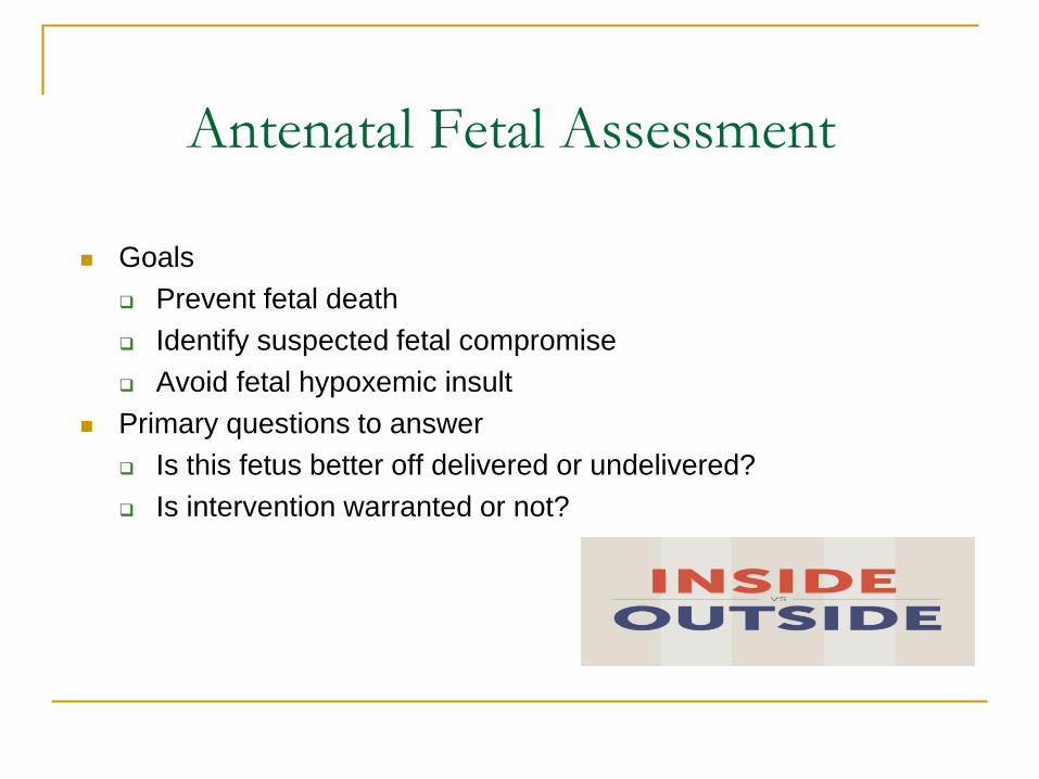
Antenatal Fetal Assessment
Goals
Prevent fetal death
Identify suspected fetal compromise
Avoid fetal hypoxemic insult
Primary questions to answer
Is this fetus better off delivered or undelivered?
Is intervention warranted or not?
Antenatal Fetal Assessment
Evidence of Efficacy
No RCT’s demonstrate decreased risk of fetal death
Incorporated into routine practice without benefit
of rigorous RCT’s
Unlikely such a study will ever be conducted
Too widely ingrained into current practice
Multiple retrospective trials suggest
decreased risk fetal death in tested patients
Versus untested (low-risk) patients and
Untested historical (at-risk) patients
ACOG Practice Bulletin #145, Reaffirmed 2016
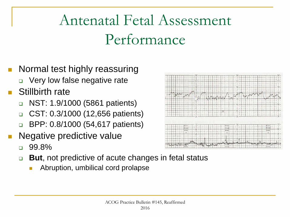
Antenatal Fetal Assessment
Performance
Normal test highly reassuring
Very low false negative rate
Stillbirth rate
NST: 1.9/1000 (5861 patients)
CST: 0.3/1000 (12,656 patients)
BPP: 0.8/1000 (54,617 patients)
Negative predictive value
99.8%
But, not predictive of acute changes in fetal status
Abruption, umbilical cord prolapse
ACOG Practice Bulletin #145, Reaffirmed
2016
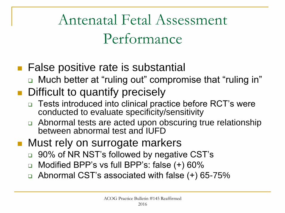
Antenatal Fetal Assessment
Performance
False positive rate is substantial Much better at “ruling out” compromise that “ruling in”
Difficult to quantify precisely Tests introduced into clinical practice before RCT’s were
conducted to evaluate specificity/sensitivity
Abnormal tests are acted upon obscuring true relationship between abnormal test and IUFD
Must rely on surrogate markers 90% of NR NST’s followed by negative CST’s
Modified BPP’s vs full BPP’s: false (+) 60%
Abnormal CST’s associated with false (+) 65-75%
ACOG Practice Bulletin #145 Reaffirmed
2016
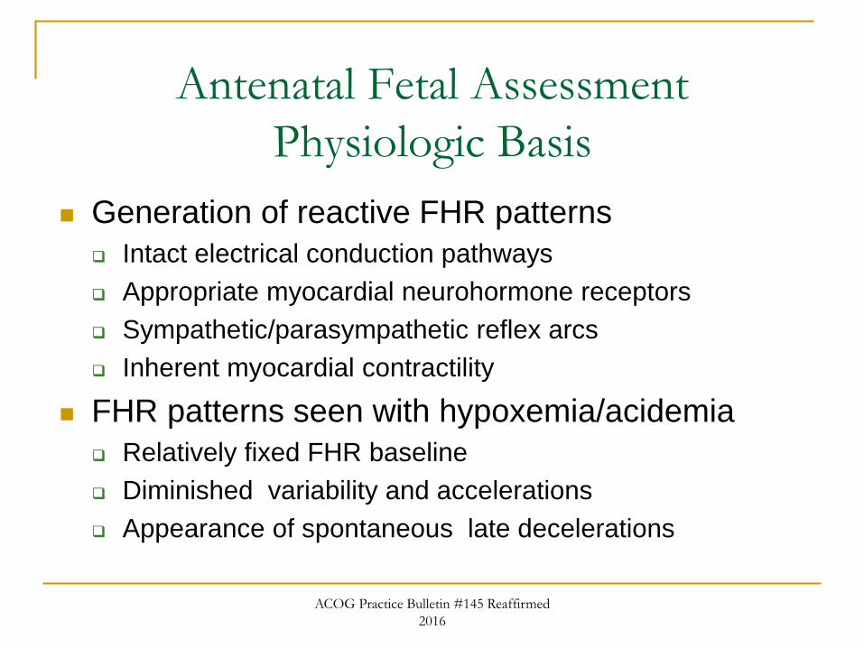
Antenatal Fetal Assessment
Physiologic Basis
Generation of reactive FHR patterns
Intact electrical conduction pathways
Appropriate myocardial neurohormone receptors
Sympathetic/parasympathetic reflex arcs
Inherent myocardial contractility
FHR patterns seen with hypoxemia/acidemia
Relatively fixed FHR baseline
Diminished variability and accelerations
Appearance of spontaneous late decelerations
ACOG Practice Bulletin #145 Reaffirmed
2016
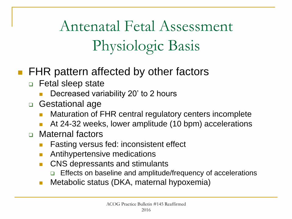
Antenatal Fetal Assessment
Physiologic Basis
FHR pattern affected by other factors Fetal sleep state
Decreased variability 20’ to 2 hours
Gestational age Maturation of FHR central regulatory centers incomplete
At 24-32 weeks, lower amplitude (10 bpm) accelerations
Maternal factors Fasting versus fed: inconsistent effect
Antihypertensive medications
CNS depressants and stimulants Effects on baseline and amplitude/frequency of accelerations
Metabolic status (DKA, maternal hypoxemia)
ACOG Practice Bulletin #145 Reaffirmed
2016
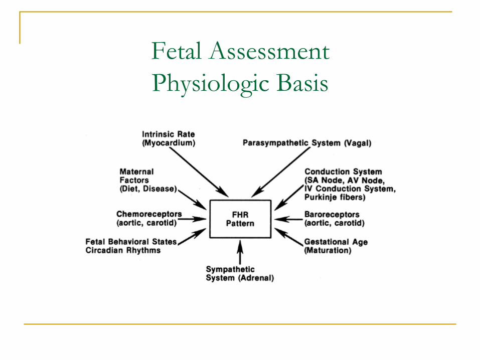
Fetal Assessment
Physiologic Basis
Antenatal Fetal Assessment
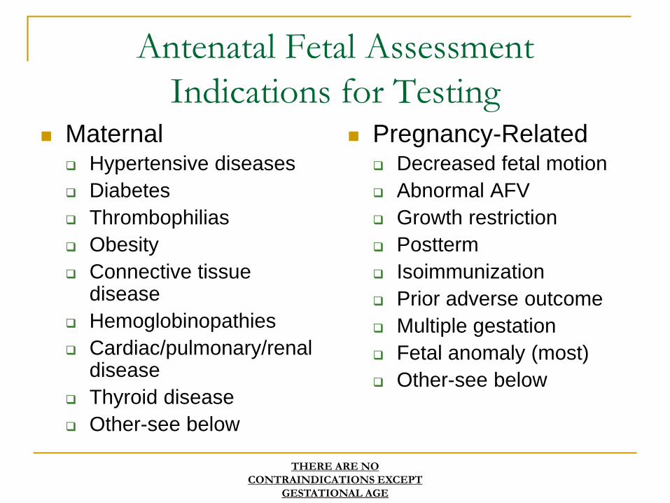
Indications for Testing Maternal
Hypertensive diseases
Diabetes
Thrombophilias
Obesity
Connective tissue disease
Hemoglobinopathies
Cardiac/pulmonary/renal disease
Thyroid disease
Other-see below
Pregnancy-Related Decreased fetal motion
Abnormal AFV
Growth restriction
Postterm
Isoimmunization
Prior adverse outcome
Multiple gestation
Fetal anomaly (most)
Other-see below
THERE ARE NO
CONTRAINDICATIONS EXCEPT
GESTATIONAL AGE
Antenatal Fetal Assessment
Initiation of Testing
Don’t start until prepared to intervene
Balance multiple factors
Neonatal survival prognosis
Severity of underlying MF disease
Must account for entire clinical picture
Example: PPROM at 25 weeks’
Iatrogenic prematurity
1.5% of women tested preterm were
delivered for false positive antenatal
surveillance tests
Miller, et al. Am J Obstet Gynecol
174(3):812,1996
Antenatal Fetal assessment
Initiation of Testing
32-34 weeks gestation appropriate point to
start most at-risk patients in testing
Consider testing earlier
Particularly severe pathophysiology
Multiple maternal/fetal issues
Individualize management plan
Must be prepared to act on abnormal results
ACOG Practice Bulletin #145 Reaffirmed
2016
Antenatal Fetal Assessment
-Fetal Movement-
Decreased movement sometimes, but not
always, perceived prior to fetal death
Simplest technique for monitoring well being
No RCT’s demonstrating reduced risk IUFD
Perception of a vigorous fetus reassuring
Further fetal assessment indicated with
maternal perception of diminished fetal
activity
Antenatal Fetal Assessment
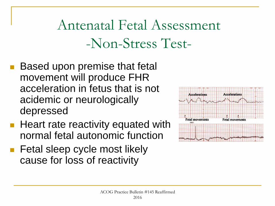
-Non-Stress Test-
Based upon premise that fetal movement will produce FHR acceleration in fetus that is not acidemic or neurologically depressed
Heart rate reactivity equated with normal fetal autonomic function
Fetal sleep cycle most likely cause for loss of reactivity
ACOG Practice Bulletin #145 Reaffirmed
2016
Antenatal Fetal Assessment
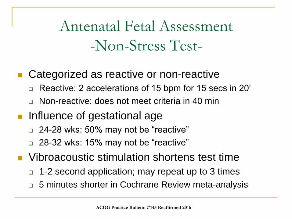
-Non-Stress Test-
Categorized as reactive or non-reactive
Reactive: 2 accelerations of 15 bpm for 15 secs in 20’
Non-reactive: does not meet criteria in 40 min
Influence of gestational age
24-28 wks: 50% may not be “reactive”
28-32 wks: 15% may not be “reactive”
Vibroacoustic stimulation shortens test time
1-2 second application; may repeat up to 3 times
5 minutes shorter in Cochrane Review meta-analysis
ACOG Practice Bulletin #145 Reaffirmed 2016
Antenatal Fetal Assessment
-Contraction Stress Test-
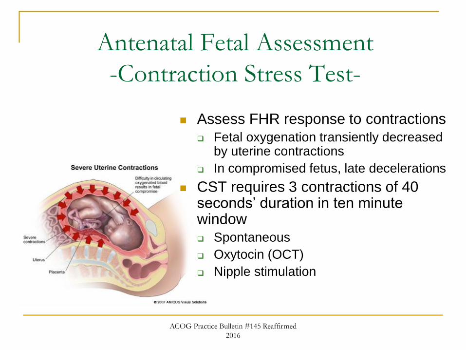
Assess FHR response to contractions
Fetal oxygenation transiently decreased by uterine contractions
In compromised fetus, late decelerations
CST requires 3 contractions of 40 seconds’ duration in ten minute window
Spontaneous
Oxytocin (OCT)
Nipple stimulation
ACOG Practice Bulletin #145 Reaffirmed
2016
Antenatal Fetal Assessment
-Contraction Stress Test-
Classified on basis of late decelerations Negative
No late or significant variable decelerations
Positive
Late decelerations after 50% or more of contractions
Equivocal-suspicious
Late decelerations after < 50% of contractions
Equivocal-tachysystole
Decelerations associated with contractions more frequent than every 2 minutes or lasting more than 90 secs
Unsatisfactory
Fewer than 3 contractions in 10 minutes
ACOG Practice Bulletin #145 Reaffirmed
2016

Antenatal Fetal Assessment
-Contraction Stress Test-
Under most circumstances, delivery indicated
when CST is positive
Route of delivery not dictated
Contraindications to CST
Preterm labor
PPROM
Placenta previa; vasa previa
ACOG Practice Bulletin #145 Reaffirmed
2016
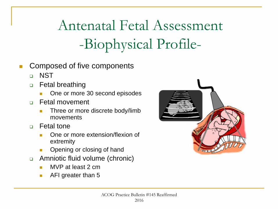
Antenatal Fetal Assessment
-Biophysical Profile-
Composed of five components
NST
Fetal breathing
One or more 30 second episodes
Fetal movement
Three or more discrete body/limb movements
Fetal tone
One or more extension/flexion of extremity
Opening or closing of hand
Amniotic fluid volume (chronic)
MVP at least 2 cm
AFI greater than 5
ACOG Practice Bulletin #145 Reaffirmed
2016
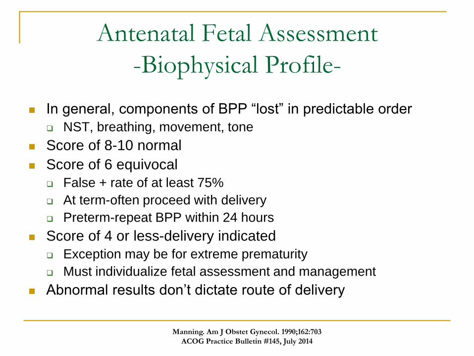
Antenatal Fetal Assessment
-Biophysical Profile-
In general, components of BPP “lost” in predictable order
NST, breathing, movement, tone
Score of 8-10 normal
Score of 6 equivocal
False + rate of at least 75%
At term-often proceed with delivery
Preterm-repeat BPP within 24 hours
Score of 4 or less-delivery indicated
Exception may be for extreme prematurity
Must individualize fetal assessment and management
Abnormal results don’t dictate route of delivery
Manning. Am J Obstet Gynecol. 1990;162:703
ACOG Practice Bulletin #145, July 2014
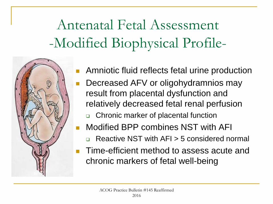
Antenatal Fetal Assessment
-Modified Biophysical Profile-
Amniotic fluid reflects fetal urine production
Decreased AFV or oligohydramnios may
result from placental dysfunction and
relatively decreased fetal renal perfusion
Chronic marker of placental function
Modified BPP combines NST with AFI
Reactive NST with AFI > 5 considered normal
Time-efficient method to assess acute and
chronic markers of fetal well-being
ACOG Practice Bulletin #145 Reaffirmed
2016
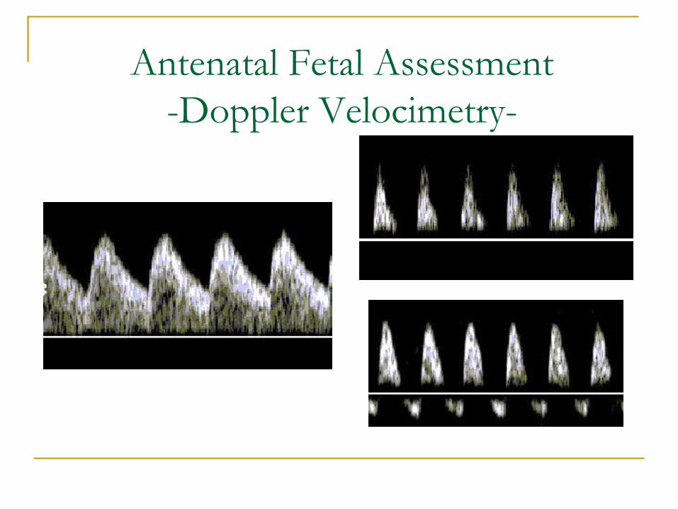
Antenatal Fetal Assessment
-Doppler Velocimetry-
Antenatal Fetal Assessment
-Doppler Velocimetry-
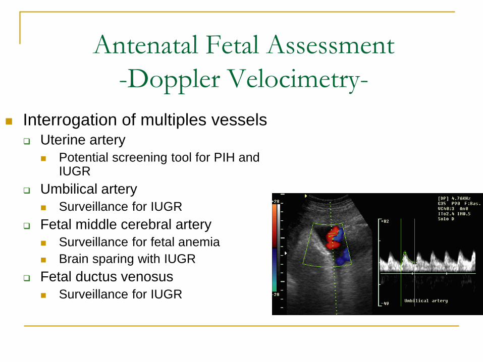
Interrogation of multiples vessels Uterine artery
Potential screening tool for PIH and IUGR
Umbilical artery
Surveillance for IUGR
Fetal middle cerebral artery
Surveillance for fetal anemia
Brain sparing with IUGR
Fetal ductus venosus
Surveillance for IUGR
Antenatal Fetal Assessment
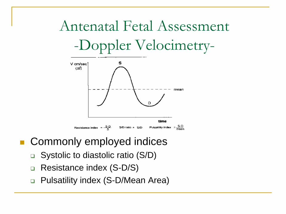
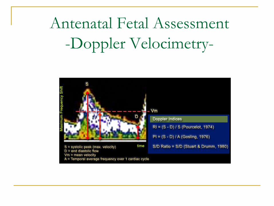
-Doppler Velocimetry-
Commonly employed indices
Systolic to diastolic ratio (S/D)
Resistance index (S-D/S)
Pulsatility index (S-D/Mean Area)
Antenatal Fetal Assessment
-Doppler Velocimetry-
Antenatal Fetal Assessment
Doppler Velocimetry: IUGR
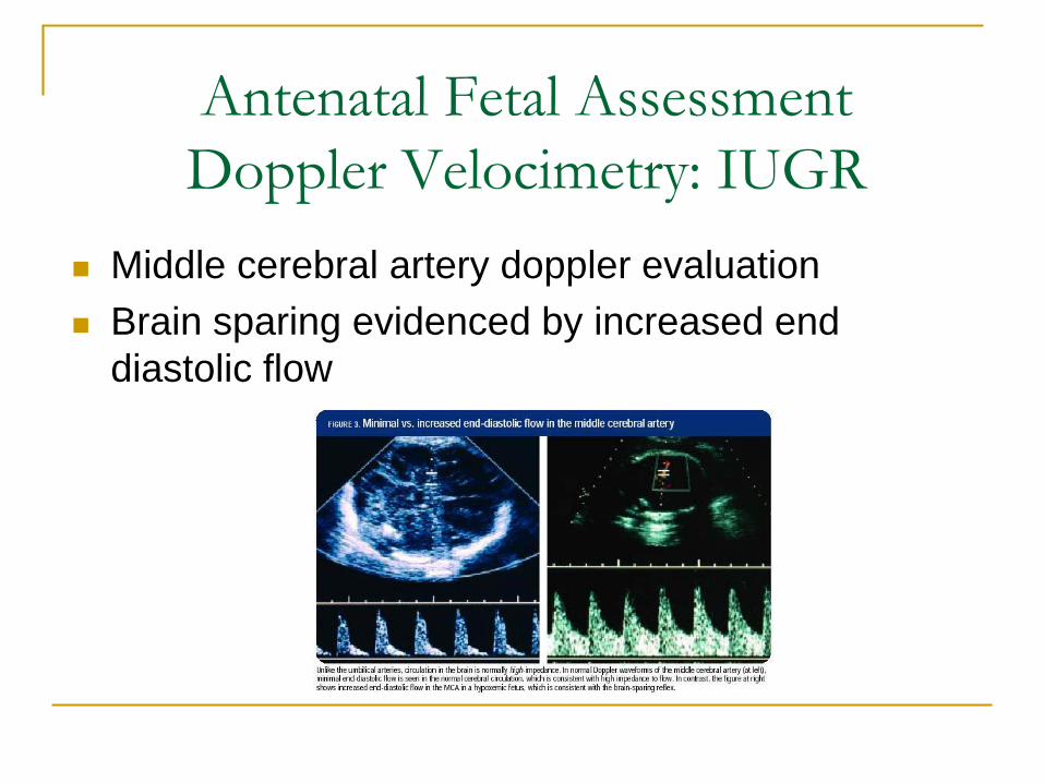
Middle cerebral artery doppler evaluation
Brain sparing evidenced by increased end
diastolic flow
Antenatal Fetal Assessment
Doppler Velocimetry: IUGR
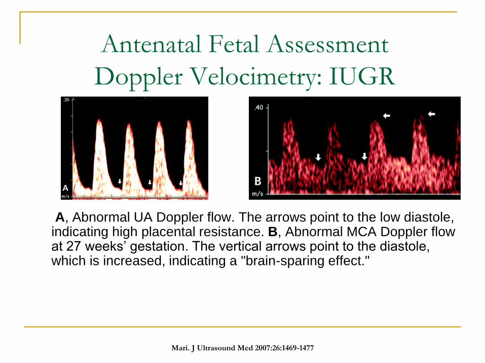
A, Abnormal UA Doppler flow. The arrows point to the low diastole, indicating high placental resistance. B, Abnormal MCA Doppler flow at 27 weeks’ gestation. The vertical arrows point to the diastole, which is increased, indicating a "brain-sparing effect."
Mari. J Ultrasound Med 2007:26:1469-1477
Antenatal Fetal Assessment
-Doppler Velocimetry-
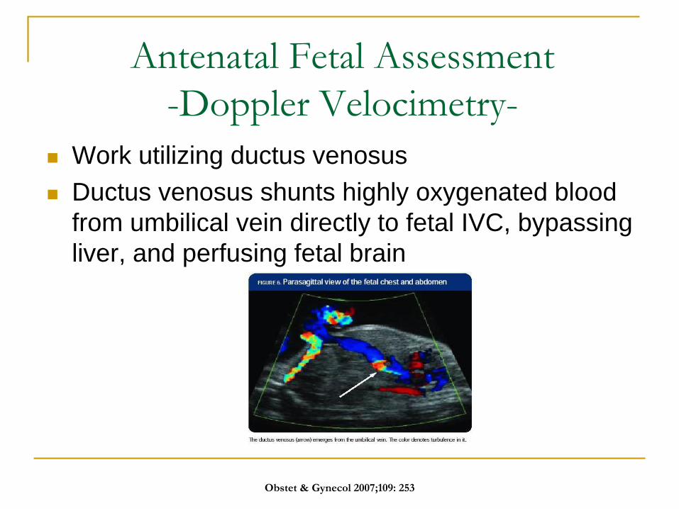
Work utilizing ductus venosus
Ductus venosus shunts highly oxygenated blood
from umbilical vein directly to fetal IVC, bypassing
liver, and perfusing fetal brain
Obstet & Gynecol 2007;109: 253
Antenatal Fetal Assessment
Doppler Velocimetry: IUGR
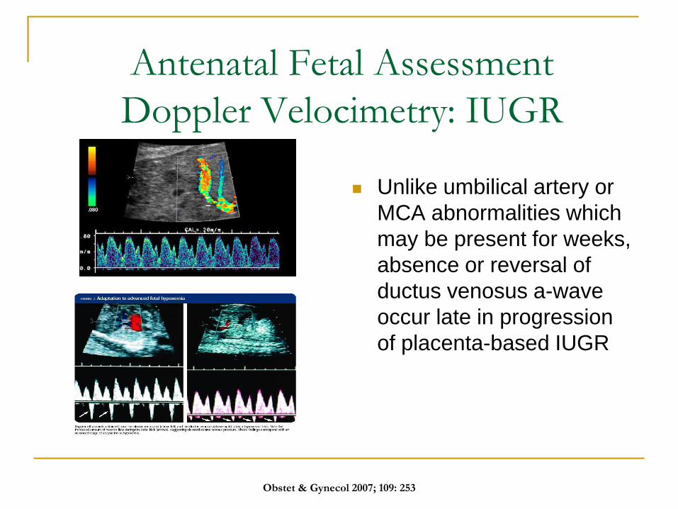
Unlike umbilical artery or
MCA abnormalities which
may be present for weeks,
absence or reversal of
ductus venosus a-wave
occur late in progression
of placenta-based IUGR
Obstet & Gynecol 2007; 109: 253
Antenatal Fetal Assessment
-Doppler Velocimetry-
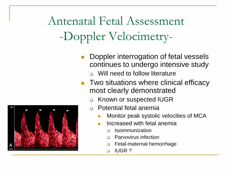
Doppler interrogation of fetal vessels continues to undergo intensive study
Will need to follow literature
Two situations where clinical efficacy most clearly demonstrated
Known or suspected IUGR
Potential fetal anemia
Monitor peak systolic velocities of MCA
Increased with fetal anemia
Isoimmunization
Parvovirus infection
Fetal-maternal hemorrhage
IUGR ?
Antenatal Fetal Assessment
ACOG Summary Level A: Good and consistent scientific evidence
Use of MVP < 2 cm, not AFI < 5 cm, to diagnose oligohydramnios associated with reduced unnecessary interventions
For IUGR fetuses, umbilical artery Doppler velocimetry with NST/ BPP’s associated with improved outcomes
Level B: Limited/inconsistent scientific evidence
Abnormal results from NST should be followed up with additional testing (BPP or CST)
Level C: Consensus and expert opinion
Start testing at 32 weeks, but individualize
Once started, repeat testing 1-2 times/ week
Acute changes in status require reassessment
ACOG Practice Bulletin #145 Reaffirmed
2016
Antenatal Fetal Assessment
ACOG Summary
Level C: Consensus and expert opinion
Delivery of fetus with abnormal testing may be attempted by labor induction
Repetitive late decelerations usually dictate cs
With isolated persistent oligohydramnios, MVP < 2 cm, delivery at 36-37 weeks recommended
If < 36 weeks, individualize management
ACOG Practice Bulletin #145 Reaffirmed
2016
Antenatal Fetal Assessment
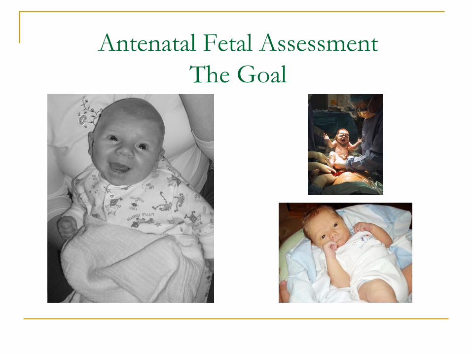
The Goal


