Harrasser et al. AMB Expr (2015) 5:77 DOI 10.1186/s13568-015-0162-z
ORIGINAL ARTICLE
Antibacterial efficacy of titanium-containing alloy with silver-nanoparticles enriched diamond-like carbon coatingsNorbert Harrasser1, Sebastian Jüssen1, Ingo J. Banke1, Ralf Kmeth2, Ruediger von Eisenhart‑Rothe1, Bernd Stritzker2, Hans Gollwitzer1,3 and Rainer Burgkart1*
Abstract
Silver ions (Ag+) have strong bactericidal effects and Ag‑coated medical devices proved their effectiveness in reduc‑ing infections in revision total joint arthroplasty. We quantitatively determined the antimicrobial potency of different surface treatments on a titanium alloy (Ti), which had been conversed to diamond‑like carbon (DLC‑Ti) and doped with high (Ag:PVP = 1:2) and low (Ag:PVP = 1:10 and 1:20) concentrations of Ag (Ag‑DLC‑Ti) with a modified tech‑nique of ion implantation. Bacterial adhesion and planktonic growth of clinically relevant bacterial strains (Staphylo-coccus epidermidis, Staphylococcus aureus, and Pseudomonas aeruginosa) on Ag‑DLC‑Ti were compared to untreated Ti by quantification of colony forming units on the adherent surface and in the growth medium as well as semiquan‑titatively by determining the grade of biofilm formation by scanning electron microscopy. (1) A significant (p < 0.05) antimicrobial effect could be found for all Ag‑DLC‑Ti samples (reduced growth by 5.6–2.5 logarithmic levels). (2) The antimicrobial effect was depending on the tested bacterial strain (most for P. aeruginosa, least for S. aureus). (3) Antimi‑crobial potency was positively correlated with Ag concentrations. (4) Biofilm formation was decreased by Ag‑DLC‑Ti surfaces. This study revealed potent antibacterial effects of Ag‑DLC‑Ti. This may serve as a promising novel approach to close the gap in antimicrobial protection of musculoskeletal implants.
IntroductionWith a constantly rising demand for orthopedic surgery with approximately 2.6 million orthopedic implants per-formed annually in the United States, the frequency of associated infections is bound to increase (Liu et al. 2012; Zimmerli and Ochsner 2003; Kurtz et al. 2008). Preven-tion of periprosthetic joint infections (PJI) has therefore an important impact not only on patient morbidity but also on the cost effectiveness of hospital care (Gosheger et al. 2004). Management of PJI often requires multiple
staged surgeries and the use of antibiotics as a support-ive therapy (Giulieri et al. 2004; Zimmerli and Ochsner 2003). A major problem in septic revision surgery is the formation of biofilm on implanted foreign materials (Schrenzel et al. 2004). These biofilms contain 5–50 µm thick glycoprotein matrices that protect the bacteria through a diffusion limitation process, and increase their resistance to antibodies, macrophages, and antibiotics (Ceri et al. 1999). Once a significant amount of biofilm has formed, eradication of infection is nearly impossible without removing the implant (Zimmerli and Ochsner 2003).
In order to decrease the infection and especially rein-fection rate several methods have been developed, at which rising occurrences of antibiotic resistances among
Open Access
*Correspondence: [email protected] 1 Clinic of Orthopedics and Sports Orthopedics, Klinikum rechts der Isar, Technical University of Munich, Ismaninger Str. 22, 81675 Munich, GermanyFull list of author information is available at the end of the article
bacteria make antibiotic-based strategies more and more questionable (Jamsen et al. 2010; Schmidmaier et al. 2006; Hetrick and Schoenfisch 2006; Poelstra et al. 2000). In this context alternative methods are favored. Promis-ing could be the use of non-antibiotic “active” antibacte-rial coatings which release antibacterial agents, e.g. silver ions (Ag+) (Gosheger et al. 2004; Hardes et al. 2007; Har-rasser et al. 2015), copper ions (Cu++) (Shirai et al. 2009; Baena et al. 2006), nitric oxide (Holt et al. 2011; Nablo et al. 2005), chlorhexidine/chloroxylenol (Darouiche et al. 1998) or chitosan (Bumgardner et al. 2003). Compared to antibiotics these agents act more broadly against a wide range of bacteria. In addition, at least proven for the use of silver (Ag), microbes without intrinsic resistance can-not gain resistance (Lee et al. 2005). Information on the use of these bactericidal coatings on wear surfaces and direct bone contact is lacking since Ag-coatings have been used so far only on surfaces without direct bone or joint contact. This fact is important, given that, e.g. in total knee replacement roughly 50 % of the surface is exposed to synovial fluid and in main parts tribologically active. On the other hand, revision prostheses are usually composed of large stems applied intramedullary which additionally represent a vulnerable surface area for bac-terial contamination. To summarize, in septic revision surgery a relevant portion of the susceptible prosthesis is not protected against bacterial reinfection. Antibacterial-agent-enriched diamond-like carbon (DLC) coatings may solve this dilemma. The term DLC is used to describe hydrogen-free carbon solids that contain an amorphous network of tetrahedrally and trigonally hybridized car-bon atoms with physical properties tending to be inter-mediate between those of graphite and those of diamond (Dearnaley 1993). DLC is an ideal surface coating for prosthetic joints, because it is wear resistant, atomi-cally smooth, and corrosion resistant, has a low friction coefficient, and is immune to scratching by third body wear particles (Morrison et al. 2006). Therefore, if DLC coating is applied on titanium (Ti), a material not used for wear surfaces due to its high sensitivity to contact wear and fretting corrosion, it improves its wear resist-ance and makes it suitable for tribologically loaded joint parts (Firkins et al. 1998). Additionally, DLC coating can improve the osseointegration of titanium making it even more valuable for orthopedic applications (Mändl et al. 2001). Another property of this coating is the ability to use it as a carrier for ions, e.g. Ag+. By release of Ag+, DLC coatings on Ti can act as local antibacterial agents, and therefore extend its medical implications to septic revision surgery (Cloutier et al. 2014; Katsikogianni et al. 2006; Dwivedi et al. 2013). DLC coatings are not without limitations since structural properties can vary widely depending on the deposition techniques employed. One
of the greatest challenges of DLC technology is adhesion and internal stresses (Xu and Pruitt 1999). Several DLC technologies are described, at which DLC films usually are deposited on surfaces. The bonding strength between coating and surface seems to be the weakest point (Wal-ter et al. 1997). If the DLC layers tend to dissolve from the surface positive tribological features can quickly deterio-rate leading to high grade wear (Roy and Lee 2007). This could be conflicting if DLC coated Ti was used on wear surfaces. On the other hand, methods of DLC coating with strong linking to the sample surface are described (Liu et al. 2004). Thus, the risk of detachment of the DLC coating from the surface, even under shear forces is mini-mized (Schwarz and Stritzker 2010; Popa et al. 2013). In this context, among the techniques described for the synthesis of DLC coatings a modified method of plasma immersion ion implantation (PIII) has proven promising advantages (Schwarz and Stritzker 2010).
We have used a new technique to incorporate silver (Ag) DLC thin films on Ti samples (Ag-DLC-Ti) in order to provide these otherwise inert medical-devices with antimicrobial properties. Bactericidal potency is stud-ied on the sample’s surface and the surrounding growth medium. This study provides valuable information for determining the suitability of Ag-DLC-Ti as antibacterial materials for septic revision implants.
MethodsStudy substratesWe used cylindrical substrates (diameter: 10 mm, height: 2 mm) of corundum-blasted medical TiAl6V4 (Ra ~ 5 µm; Goodfellow GmbH, Nauheim, Germany). The samples were coated with Ag-doped DLC, incu-bated for 24 h and afterwards tested for their antimicro-bial effects on the surface (bacterial sessile growth) and the surrounding growth medium (bacterial planktonic growth).
DLC doping with Ag was carried out with polyvi-nylpyrrolidone (PVP) as a stabilizing agent. The first testing series was conducted with high concentrations of Ag within the DLC coating (Ag:PVP = 1:2) and three different bacterial strains (Staphylococcus epidermidis, Staphylococcus aureus, Pseudomonas aeruginosa). For reduction of potential toxic side-effects of Ag on eukar-yotic cells a reduction of Ag+ would be advantageous (Hardes et al. 2007). Hence, detection of minimal nec-essary Ag+ concentration in DLC-Ti samples was evalu-ated in a further testing series with low concentrations of Ag within the DLC coating (Ag:PVP = 1:10 and 1:20). This series was only conducted with the most resistant strain against Ag from testing series one (S. aureus). Sample features and used bacterial strains are summa-rized in Table 1.
Page 3 of 11Harrasser et al. AMB Expr (2015) 5:77
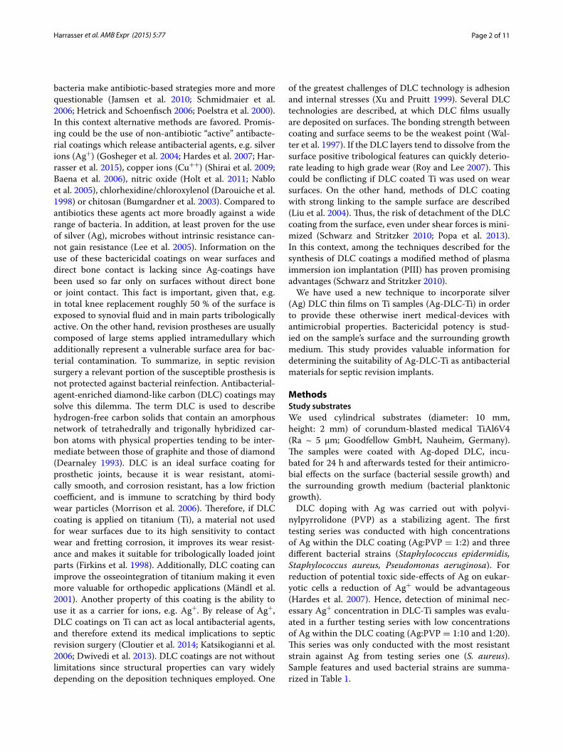
DLC film depositionDLC-processing of all plates was performed at the department of experimental physics IV (University of Augsburg, Germany) according to a modified technic of ion irradiation of polymers. The coating process is described fully elsewhere (Schwarz and Stritzker 2010). Briefly summarized, an ethanol-based solution of sil-ver nitrate, benzoin and polyvinylpyrrolidone (PVP) was prepared and exposed to UV light to initiate pho-tochemical reduction. This results in a colloidal solution of silver nanoparticles stabilized by PVP, with amount and size of the particles depending on the initial PVP-Ag and Ag-Benzoin ratios. The solution was then applied to the substrate via dip-coating. The resulting silver nano-particle-containing polymer layer is subsequently trans-ferred to amorphous carbon by plasma immersion ion implantation (PIII). A big advantage of this procedure is the fact that setting of amount and size of silver nano-particles is independent from DLC-formation. In sum-mary, the following surfaces have been investigated: Ti (untreated) = corundum-blasted medical TiAl6V4 alloy; DLC-Ti = corundum-blasted medical TiAl6V4 with DLC surface coating; Ag-DLC-Ti = corundum-blasted medical TiAl6V4 with DLC surface coating containing nanocol-loidal silver in different molar ratios (high concentrated: Ag:PVP = 1:2; low concentrated: Ag:PVP = 1:10 and 1:20; Note: Amount of Ag+ is higher in 1:10 compared to 1:20). A TEM (transmission electron microscopy)-image of the Ag-DLC coating on Ti is given in Fig. 1.
Sterilization of samples and sealing of uncoated surface with paraffin waxSamples were rinsed with distilled water for 10 min, air-dried in a laminar flow cabinet and thereafter sterilized with gamma-beam with the dose of 26.5 kGy (Isotron Deutschland GmbH, Allershausen, Germany). All manip-ulations of the samples were conducted by holding the lower surface. As a consequence these parts of the sam-ples were not surface treated and needed protection from the testing environment. Hence, paraffin wax was first
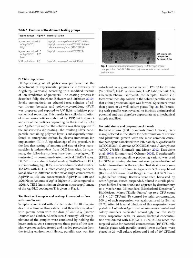
autoclaved in a glass container with 120 °C for 20 min (Varioklav®, H+P Labortechnik, H+P Labortechnik AG, Oberschleißheim, Germany), the samples’ lower sur-faces were then dip-coated in the solvent paraffin wax so that a thin protection layer was formed. Specimens were then placed in 24-well culture plates (Fig. 2a, b). Pretest-ing with paraffin wax revealed no intrinsic antimicrobial potential and was therefore appropriate as a mechanical sample stabilizer.
Bacterial strains and preparation of inoculaBacterial strains (LGC Standards GmbH, Wesel, Ger-many) selected in the study for determination of surface and planktonic growth were the most common causa-tive pathogens associated with PJI, namely S. epidermidis (ATCC35984), S. aureus (ATCC25923) and P. aeruginosa (ATCC 27835) (Zimmerli and Moser 2012; Darouiche et al. 1998; Zimmerli and Ochsner 2003). S. epidermidis (RP62a), as a strong slime producing variant, was used for SEM (scanning electron microscopy)-evaluation of biofilm formation on the samples. Test strains were rou-tinely cultured in Columbia Agar with 5 % sheep blood (Becton–Dickinson, Heidelberg, Germany) at 37 °C over-night before testing. Bacteria were then harvested by centrifugation, rinsed, suspended, diluted in sterile phos-phate buffered saline (PBS) and adjusted by densitometry to a MacFarland 0.5 standard (MacFarland Densimat™, BioMérieux, Marcy l’Etoile, France) up to a CFU count of 1 × 10⁵ CFU/ml. To control bacterial concentration, 100 μl of each suspension was again cultured for 24 h at 37 °C. After 24 h serial dilutions of this suspension were plated on Colombia-Agar. The colonies were counted and colony numbers calculated accordingly. For the study every suspension with its known bacterial concentra-tion was diluted with DMEM + 10 % FCS to reach the targeted value for bacterial concentration (105 CFU/ml). Sample plates with paraffin-coated lower surfaces were placed in 24-well culture plates and 1 ml of 105 CFU/ml
Fig. 1 Transmission electron microscopy (TEM) image of silver nanoparticles of AG‑DLC‑Ti (note: major nanoparticles are marked with arrows)
Page 4 of 11Harrasser et al. AMB Expr (2015) 5:77
bacterial suspensions were added. Incubation of the well plates was conducted for 24 h at 37 °C.
AnalysisBacterial surface adhesion was evaluated by determining bacterial concentration on the specimen. Bacterial plank-tonic growth was measured in the growth medium. For every group four independent testing runs with four dif-ferent samples were conducted. Therefore, altogether 16 samples were tested for every group.
Determination of bacterial growth on sample surfacesColonized sample plates were removed from the wells with a sterile forceps, carefully rinsed twice with sterile PBS, transferred to vials containing 3 ml of sterile PBS and sonicated for 7 min (Elmasonic S60H, Elma, Singen, Germany) to remove adhering bacteria. 100 μl of the fluid were aspirated, plated on Colombia Agar at 37 °C for 24 h and quantified after incubation (CFU/ml).
SEM-analysis was conducted semiquantitatively to evaluate inhibition of biofilm formation. SEM-images were compiled of native Ti, DLC-Ti and Ag-DLC-Ti (Ag:PVP = 1:2). Biofilm formation was quantified in five categories: (1) no biofilm formation, (2) biofilm cov-ering less than 25 % of the surface, (3) biofilm covering between 25 and 75 % of the surface, (4) biofilm covering more than 75 % of the surface, (5) biofilm formation cov-ering the entire surface.
Determination of bacterial planktonic growthA 700-μl volume of each well was supplemented with 700 μl neutralizing solution as described by Tilton (Tilton and Rosenberg 1978) (1.0 g sodium thioglycolate + 1.46 g sodium thiosulfate in 1000 ml deionized water). The neu-tralizing solution acts as an inhibitor for reminiscent metal toxicity on bacteria. The suspension was plated on Columbia Agar after serial dilutions and incubated
at 37 °C for 24 h. Thereafter, CFU were quantified and extrapolated to CFU/ml.
StatisticsAll results are presented as mean ± standard devia-tion. Statistical significance was computed using non-parametric methods and the method of closed testing procedure (Kruskal–Wallis and Mann–Whitney U test). P < 0.05 was considered statistically significant. Statisti-cal tests were performed with use of SPSS (version 20.0; Chicago, Illinois). Statistical analysis was conducted per consultation with the Institute of Medical Statistics and Epidemiology (Klinikum rechts der Isar, Technische Uni-versität München, Munich, Germany).
ResultsAntimicrobial effect of high concentrated (Ag:PVP = 1:2) Ag‑DLC‑Ti on S. epidermidis ATCC35984, S. aureus ATCC25923 and P. aeruginosa ATCC27835Average viable counts of bacteria recovered from the samples and in the supernatant growth medium are sum-marized in Table 2.
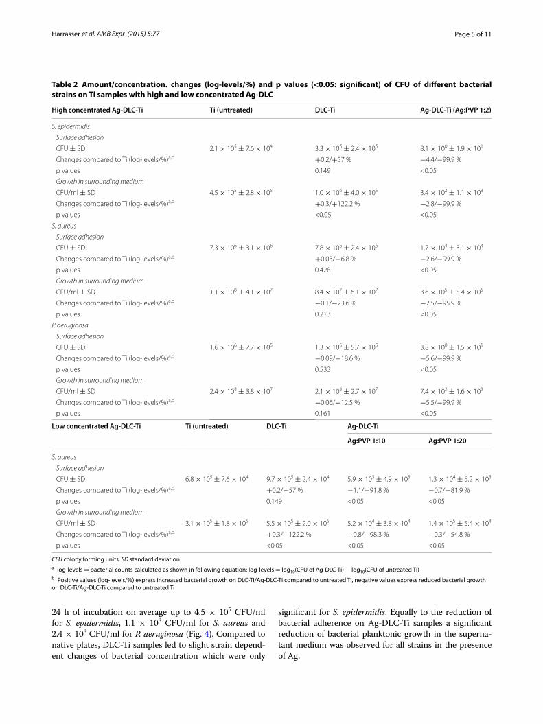
Analysis of bacterial surface adhesion showed strain dependent differences in growth in the absence of Ag. On untreated Ti plates on average 2.1 × 10⁵ CFU of S. epi-dermidis, 7.3 × 10⁶ CFU of S. aureus and 1.6 × 10⁶ CFU of P. aeruginosa adhered after 24 h of incubation (Fig. 3). Compared to native plates, DLC-Ti samples showed not significant strain dependent changes of bacterial amounts. On all Ag-DLC-Ti surfaces (Ag:PVP = 1:2), compared to native plates, a significant reduction of bac-terial amount was detected.
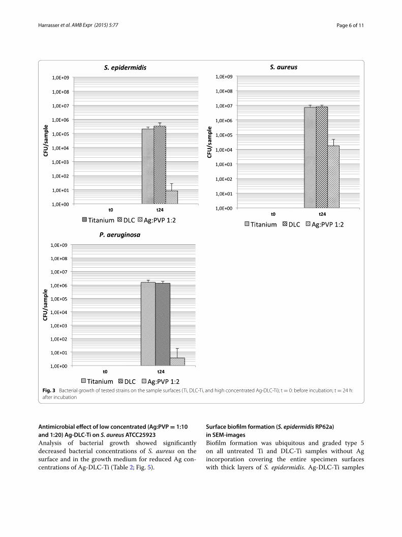
Analysis of planktonic growth in the growth medium showed, in accordance to the results of surface growth, strain dependent differences. Initial bacterial con-centration (t = 0) of all strains was approximately 1 × 10⁵ CFU/ml and increased in the absence of Ag after
Fig. 2 Sample preparation; ion irradiation of samples with missing irradiation of the sample’s lower surfaces (a), placement of samples in well culture plates with paraffin wax (arrow) covering the sample’s lower surface (b)
Page 5 of 11Harrasser et al. AMB Expr (2015) 5:77
24 h of incubation on average up to 4.5 × 10⁵ CFU/ml for S. epidermidis, 1.1 × 10⁸ CFU/ml for S. aureus and 2.4 × 10⁸ CFU/ml for P. aeruginosa (Fig. 4). Compared to native plates, DLC-Ti samples led to slight strain depend-ent changes of bacterial concentration which were only
significant for S. epidermidis. Equally to the reduction of bacterial adherence on Ag-DLC-Ti samples a significant reduction of bacterial planktonic growth in the superna-tant medium was observed for all strains in the presence of Ag.
Table 2 Amount/concentration. changes (log-levels/%) and p values (<0.05: significant) of CFU of different bacterial strains on Ti samples with high and low concentrated Ag-DLC
CFU colony forming units, SD standard deviationa log-levels = bacterial counts calculated as shown in following equation: log-levels = log10(CFU of Ag-DLC-Ti) − log10(CFU of untreated Ti)b Positive values (log-levels/%) express increased bacterial growth on DLC-Ti/Ag-DLC-Ti compared to untreated Ti, negative values express reduced bacterial growth on DLC-Ti/Ag-DLC-Ti compared to untreated Ti
High concentrated Ag‑DLC‑Ti Ti (untreated) DLC‑Ti Ag‑DLC‑Ti (Ag:PVP 1:2)
Changes compared to Ti (log‑levels/%)a,b +0.3/+122.2 % −0.8/−98.3 % −0.3/−54.8 %
p values <0.05 <0.05 <0.05
Page 6 of 11Harrasser et al. AMB Expr (2015) 5:77
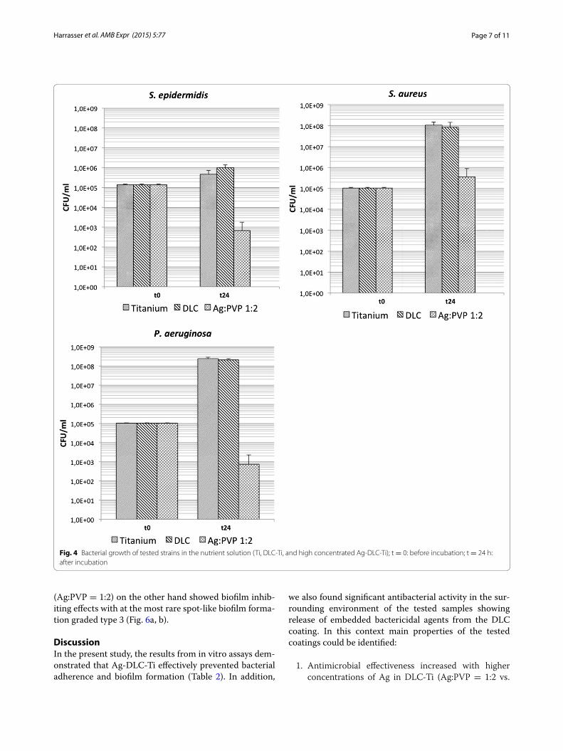
Antimicrobial effect of low concentrated (Ag:PVP = 1:10 and 1:20) Ag‑DLC‑Ti on S. aureus ATCC25923Analysis of bacterial growth showed significantly decreased bacterial concentrations of S. aureus on the surface and in the growth medium for reduced Ag con-centrations of Ag-DLC-Ti (Table 2; Fig. 5).
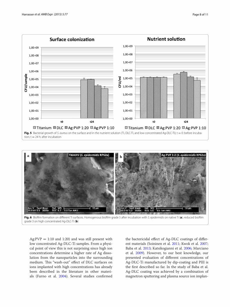
Surface biofilm formation (S. epidermidis RP62a) in SEM‑imagesBiofilm formation was ubiquitous and graded type 5 on all untreated Ti and DLC-Ti samples without Ag incorporation covering the entire specimen surfaces with thick layers of S. epidermidis. Ag-DLC-Ti samples
Fig. 3 Bacterial growth of tested strains on the sample surfaces (Ti, DLC‑Ti, and high concentrated Ag‑DLC‑Ti); t = 0: before incubation; t = 24 h: after incubation
Page 7 of 11Harrasser et al. AMB Expr (2015) 5:77
(Ag:PVP = 1:2) on the other hand showed biofilm inhib-iting effects with at the most rare spot-like biofilm forma-tion graded type 3 (Fig. 6a, b).
DiscussionIn the present study, the results from in vitro assays dem-onstrated that Ag-DLC-Ti effectively prevented bacterial adherence and biofilm formation (Table 2). In addition,
we also found significant antibacterial activity in the sur-rounding environment of the tested samples showing release of embedded bactericidal agents from the DLC coating. In this context main properties of the tested coatings could be identified:
1. Antimicrobial effectiveness increased with higher concentrations of Ag in DLC-Ti (Ag:PVP = 1:2 vs.
Fig. 4 Bacterial growth of tested strains in the nutrient solution (Ti, DLC‑Ti, and high concentrated Ag‑DLC‑Ti); t = 0: before incubation; t = 24 h: after incubation
Page 8 of 11Harrasser et al. AMB Expr (2015) 5:77
Ag:PVP = 1:10 and 1:20) and was still present with low concentrated Ag-DLC-Ti samples. From a physi-cal point of view this is not surprising since high ion concentrations determine a higher rate of Ag disso-lution from the nanoparticles into the surrounding medium. This “wash-out” effect of DLC surfaces on ions implanted with high concentrations has already been described in the literature in other materi-als (Furno et al. 2004). Several studies confirmed
the bactericidal effect of Ag-DLC coatings of differ-ent materials (Soininen et al. 2011; Kwok et al. 2007; Baba et al. 2013; Katsikogianni et al. 2006; Marciano et al. 2009). However, to our best knowledge, our presented evaluation of different concentrations of Ag-DLC-Ti manufactured by dip-coating and PIII is the first described so far. In the study of Baba et al. Ag-DLC coating was achieved by a combination of magnetron sputtering and plasma source ion implan-
Fig. 5 Bacterial growth of S. aureus on the surface and in the nutrient solution (Ti, DLC‑Ti, and low concentrated Ag‑DLC‑Ti); t = 0: before incuba‑tion; t = 24 h: after incubation
Fig. 6 Biofilm formation on different Ti surfaces. Homogenous biofilm grade 5 after incubation with S. epidermidis on native Ti (a), reduced biofilm grade 3 on high concentrated Ag‑DLC‑Ti (b)
Page 9 of 11Harrasser et al. AMB Expr (2015) 5:77
tation. Different concentrations of Ag were used, but no significant differences of bactericidal effects from high and low concentrated Ag-DLC coatings were found (Baba et al. 2013). This emphasizes the impor-tance of the manufacturing process of DLC onto the dissolution of Ag+ from the coating. Nevertheless, bactericidal effects can also be explained by inter-actions of bacteria with the surface and not only by release of silver-nanoparticles. Contradicting state-ments exist whether DLC coating alone exhibits bac-tericidal effects or not. Marciano et al. (2009) found a significant bactericidal potency of DLC, other inves-tigators found no antibacterial effect (Soininen et al. 2011; Baba et al. 2013). In our study DLC coating alone had no significant bactericidal effect compared to untreated Ti. Similar to the findings of Soininen et al. (2011), who used the same bacterial strains, S. epidermidis’ and S. aureus’ adhesion on DLC coat-ings were slightly higher compared to uncoated Ti samples (0.2 and 0.03 log-levels; p > 0.05). Adhesion of P. aeruginosa on the other hand was slightly lower (0.09 log-levels; p > 0.05). This proves the statement that bactericidal effects in the present study derived mainly from the release of Ag+ from the coating.
2. Another main finding of our study is the differ-ent susceptibility against Ag-doped DLC coatings depending on the bacterial strain. P. aeruginosa showed the highest susceptibility (reduced surface growth by 5.6 log-levels; p < 0.05) followed by S. epi-dermidis (reduced surface growth by 4.4 log-levels; p < 0.05) and S. aureus (reduced surface growth by 2.6 log-levels; p < 0.05). In general, several studies confirmed higher bactericidal potency of Ag against Gram-negative compared to Gram-positive strains (Flores et al. 2013; Taglietti et al. 2012; Kim et al. 2007). Ag acts by binding to membranes, enzymes and nucleic acids. Consequently the respiratory chain is inhibited and therefore the aerobe metabolism of microorganisms disturbed (Gosheger et al. 2004). Bacteria are quite susceptible to Ag with only neg-ligible possibility of intrinsic resistance (Kumar and Munstedt 2005). Nevertheless, the effect observed in the present study is not fully understood. Differ-ent cell morphology or generation time of the bacte-ria may only be two of several reasons for our find-ings (Morones et al. 2005). However, this finding is important for the future use of Ag-doped DLC sur-faces since PJI involve a variety of different bacterial strains with different susceptibilities against Ag.
This study involves several limitations. Ion concentra-tions of Ag in the surrounding medium were not assessed. Antibacterial effects in the surrounding medium and on
the sample surface could be caused or at least supported by antiadhesive surface features of DLC alone. Even though, compared to untreated Ti, no significant reduc-tion of growth of S. aureus in the growth medium and P. aeruginosa on the surface and in the growth medium was detected for DLC-Ti, a tendency to diminished bac-terial amounts of these bacteria was observed (Table 2). Another limitation is that only S. aureus was investigated for low concentrated Ag-DLC-Ti. It can be estimated that the bactericidal effect on P. aeruginosa and S. epidermidis would be also evident if low concentrated Ag-DLC coat-ings were used, since S. aureus was the least susceptible strain of the three in the high concentrated testing group. Additionally, no influence of Ag-DLC-Ti on osseointe-gration was investigated. It is known, that DLC coat-ing of Ti can lead to increased osseointegration (Mändl et al. 2001). If this effect of DLC coatings is inhibited in the presence of Ag is unknown. Further investigations are needed in order to clear whether the concentration and duration of delivery of the released Ag+ of DLC coat-ings is sufficient to avoid implant infection in vivo and how they interact with eukaryotic cells. However, this was not the scope of this proof of principle investigation. Lastly, we did not evaluate the tribological behavior of this coating. DLC coatings of Ti exhibited good results in experimental wear studies, if Ag-DLC coatings do also is unknown (Xu and Pruitt 1999; Brizuela et al. 2002) but is the aim of further studies.
In summary, our findings show that Ag-DLC-Ti manu-factured by a modified technique of dip-coating and ion implantation has considerable effects as an antibacterial coating. Thus, Ag-DLC-Ti can be considered a promis-ing material for next generation orthopedic devices. The suitability of this coating for biomedical applications will be confirmed by wear tests and in vitro biocompatibility assessments.
AbbreviationsAg: silver; Ag+: silver ions; Ag‑DLC: silver incorporated diamond‑like carbon coating; Ag‑DLC‑Ti: silver incorporated diamond‑like carbon coating on tita‑nium; CFU: colony forming units; DLC: diamond‑like carbon; DLC‑Ti: diamond‑like carbon coating on titanium; PIII: plasma immersion ion implantation; PJI: periprosthetic joint infections; SD: standard deviation; Ti: corundum‑blasted medical titanium (TiAl6V4) alloy.
Authors’ contributionsNH, SJ carried out the microbiological testing and drafted the manuscript. RK and BS provided DLC‑processing of samples. HG, RB, RE and IB conceived of the study, and participated in its design and coordination and helped to draft the manuscript. All authors read and approved the final manuscript.
Author details1 Clinic of Orthopedics and Sports Orthopedics, Klinikum rechts der Isar, Technical University of Munich, Ismaninger Str. 22, 81675 Munich, Ger‑many. 2 Experimental Physics IV, Institut für Physik, Augsburg University, Universitätsstr. 1, 86135 Augsburg, Germany. 3 ATOS Clinic, Effnerstr.38, 81925 Munich, Germany.
Page 10 of 11Harrasser et al. AMB Expr (2015) 5:77
AcknowledgementsWe thank PD Dr. Thomas Grupp for providing the PE discs and Jutta Tübel for excellent technical help and advice. This work was supported by the “Deutsche Forschungsgemeinschaft (DFG)” within the interdisciplinary project “Quantitative Evaluation der statischen und dynamischen Zelladhäsion und –aktivität an antibakteriellen DLC‑Schichten für den biomedizinischen Einsatz” (BU 1154/2‑1 and GO 1906/2‑1, STR 361/18‑1).
Competing interestsThe authors declare that they have no competing interests.
Received: 4 September 2015 Accepted: 11 November 2015
Morimura T (2013) Preparation and antibacterial properties of Ag‑containing diamond‑like carbon films prepared by a combination of magnetron sputtering and plasma source ion implantation. Vacuum 89(89):179–184
Baena MI, Marquez MC, Matres V, Botella J, Ventosa A (2006) Bactericidal activity of copper and niobium‑alloyed austenitic stainless steel. Curr Microbiol 53(6):491–495. doi:10.1007/s00284‑006‑0193‑4
Brizuela M, Garcia‑Luis A, Viviente JL, Braceras I, Onate JI (2002) Tribological study of lubricious DLC biocompatible coatings. J Mater Sci Mater Med 13(12):1129–1133
Bumgardner JD, Wiser R, Elder SH, Jouett R, Yang Y, Ong JL (2003) Contact angle, protein adsorption and osteoblast precursor cell attachment to chitosan coatings bonded to titanium. J Biomater Sci Polym Ed 14(12):1401–1409
Ceri H, Olson ME, Stremick C, Read RR, Morck D, Buret A (1999) The Calgary Biofilm Device: new technology for rapid determination of antibiotic susceptibilities of bacterial biofilms. J Clin Microbiol 37(6):1771–1776
Cloutier M, Tolouei R, Lesage O, Levesque L, Turgeon S, Tatoulian M, Mantovani D (2014) On the long term antibacterial features of silver‑doped diamon‑dlike carbon coatings deposited via a hybrid plasma process. Biointer‑phases 9(2):029013. doi:10.1116/1.4871435
Darouiche RO, Farmer J, Chaput C, Mansouri M, Saleh G, Landon GC (1998) Anti‑infective efficacy of antiseptic‑coated intramedullary nails. J Bone Joint Surg Am 80(9):1336–1340
Dearnaley G (1993) Diamond‑like carbon: a potential means of reducing wear in total joint replacements. Clin Mater 12(4):237–244
Dwivedi N, Kumar S, Carey JD, Tripathi RK, Malik HK, Dalai MK (2013) Influence of silver incorporation on the structural and electrical properties of dia‑mond‑like carbon thin films. ACS Appl Mater Interfaces 5(7):2725–2732. doi:10.1021/am4003183
Firkins P, Hailey JL, Fisher J, Lettington AH, Butter R (1998) Wear of ultra‑high molecular weight polyethylene against damaged and undamaged stainless steel and diamond‑like carbon‑coated counterfaces. J Mater Sci Mater Med 9(10):597–601
Flores CY, Minan AG, Grillo CA, Salvarezza RC, Vericat C, Schilardi PL (2013) Citrate‑capped silver nanoparticles showing good bactericidal effect against both planktonic and sessile bacteria and a low cytotoxicity to osteoblastic cells. ACS Appl Mater Interfaces 5(8):3149–3159. doi:10.1021/am400044e
Furno F, Morley KS, Wong B, Sharp BL, Arnold PL, Howdle SM, Bayston R, Brown PD, Winship PD, Reid HJ (2004) Silver nanoparticles and polymeric medi‑cal devices: a new approach to prevention of infection? J Antimicrob Chemother 54(6):1019–1024. doi:10.1093/jac/dkh478
Giulieri SG, Graber P, Ochsner PE, Zimmerli W (2004) Management of infection associated with total hip arthroplasty according to a treatment algorithm. Infection 32(4):222–228. doi:10.1007/s15010‑004‑4020‑1
Gosheger G, Hardes J, Ahrens H, Streitburger A, Buerger H, Erren M, Gunsel A, Kemper FH, Winkelmann W, Von Eiff C (2004) Silver‑coated megaen‑doprostheses in a rabbit model—an analysis of the infection rate and toxicological side effects. Biomaterials 25(24):5547–5556. doi:10.1016/j.biomaterials.2004.01.008
Hardes J, Ahrens H, Gebert C, Streitbuerger A, Buerger H, Erren M, Gunsel A, Wedemeyer C, Saxler G, Winkelmann W, Gosheger G (2007) Lack of toxicological side‑effects in silver‑coated megaprostheses in humans. Biomaterials 28(18):2869–2875. doi:10.1016/j.biomaterials.2007.02.033
Harrasser N, Jussen S, Banke IJ, Kmeth R, von Eisenhart‑Rothe R, Stritzker B, Gollwitzer H, Burgkart R (2015) Antibacterial efficacy of ultrahigh molecu‑lar weight polyethylene with silver containing diamond‑like surface layers. AMB Express 5(1):64. doi:10.1186/s13568‑015‑0148‑x
Holt J, Hertzberg B, Weinhold P, Storm W, Schoenfisch M, Dahners L (2011) Decreasing bacterial colonization of external fixation pins through nitric oxide release coatings. J Orthop Trauma 25(7):432–437. doi:10.1097/BOT.0b013e3181f9ac8a
Jamsen E, Furnes O, Engesaeter LB, Konttinen YT, Odgaard A, Stefansdottir A, Lidgren L (2010) Prevention of deep infection in joint replacement surgery. Acta Orthop 81(6):660–666. doi:10.3109/17453674.2010.537805
Katsikogianni M, Spiliopoulou I, Dowling DP, Missirlis YF (2006) Adhesion of slime producing Staphylococcus epidermidis strains to PVC and diamond‑like carbon/silver/fluorinated coatings. J Mater Sci Mater Med 17(8):679–689. doi:10.1007/s10856‑006‑9678‑8
Kim JS, Kuk E, Yu KN, Kim JH, Park SJ, Lee HJ, Kim SH, Park YK, Park YH, Hwang CY, Kim YK, Lee YS, Jeong DH, Cho MH (2007) Antimicrobial effects of silver nanoparticles. Nanomedicine 3(1):95–101. doi:10.1016/j.nano.2006.12.001
Kumar R, Munstedt H (2005) Silver ion release from antimicrobial polyam‑ide/silver composites. Biomaterials 26(14):2081–2088. doi:10.1016/j.biomaterials.2004.05.030
Kurtz SM, Lau E, Schmier J, Ong KL, Zhao K, Parvizi J (2008) Infection burden for hip and knee arthroplasty in the United States. J Arthroplasty 23(7):984–991. doi:10.1016/j.arth.2007.10.017
Kwok SCH, Zhang W, Wan GJ, McKenzie DR, Bilek MMM, Chu PK (2007) Hemo‑compatibility and anti‑bacterial properties of silver doped diamond‑like carbon prepared by pulsed filtered cathodic vacuum arc deposition. Diam Relat Mater 16:1353–1360
Lee D, Cohen RE, Rubner MF (2005) Antibacterial properties of Ag nanoparticle loaded multilayers and formation of magnetically directed antibacterial microparticles. Langmuir 21(21):9651–9659. doi:10.1021/la0513306
Liu XY, Chu PK, Ding CX (2004) Surface modification of titanium, titanium alloys, and related materials for biomedical applications. Mater Sci Eng R Rep. 47:49–121
Liu Y, Zheng Z, Zara JN, Hsu C, Soofer DE, Lee KS, Siu RK, Miller LS, Zhang X, Carpenter D, Wang C, Ting K, Soo C (2012) The antimicrobial and oste‑oinductive properties of silver nanoparticle/poly (DL‑lactic‑co‑glycolic acid)‑coated stainless steel. Biomaterials 33(34):8745–8756. doi:10.1016/j.biomaterials.2012.08.010
Mändl S, Krause D, Thorwarth G, Sader R, Zeilhofer F, Horch HH, Rauschen‑bach B (2001) Plasma immersion ion implantation treatment of medical implants. Surf Coat Technol 142–144:1046–1050
Marciano FR, Bonetti LF, Santos LV, Da‑Silva NS, Corat EJ, Trava‑Airoldi VJ (2009) Antibacterial activity of DLC and Ag, ÄìDLC films produced by PECVD technique. Diam Relat Mater 18:1010–1014
Morones JR, Elechiguerra JL, Camacho A, Holt K, Kouri JB, Ramirez JT, Yacaman MJ (2005) The bactericidal effect of silver nanoparticles. Nanotechnology 16(10):2346–2353. doi:10.1088/0957‑4484/16/10/059
Morrison ML, Buchanan RA, Liaw PK, Berry CJ, Brigmon RL, Riester L, Abernathy H, Jin C, Narayan RJ (2006) Electrochemical and antimicrobial proper‑ties of diamondlike carbon‑metal composite films. Diam Relat Mater 15:138–146
Nablo BJ, Rothrock AR, Schoenfisch MH (2005) Nitric oxide‑releasing sol‑gels as antibacterial coatings for orthopedic implants. Biomaterials 26(8):917–924. doi:10.1016/j.biomaterials.2004.03.031
Poelstra KA, Barekzi NA, Slunt JB, Schuler TC, Grainger DW (2000) Surgi‑cal irrigation with pooled human immunoglobulin G to reduce post‑operative spinal implant infection. Tissue Eng 6(4):401–411. doi:10.1089/107632700418100
Popa AC, Stan GE, Husanu MA, Pasuk I, Popescu ID, Popescu AC, Mihailescu IN (2013) Multi‑layer haemocompatible diamond‑like carbon coatings obtained by combined radio frequency plasma enhanced chemical vapor deposition and magnetron sputtering. J Mater Sci Mater Med 24(12):2695–2707. doi:10.1007/s10856‑013‑5026‑y
Page 11 of 11Harrasser et al. AMB Expr (2015) 5:77
Roy RK, Lee KR (2007) Biomedical applications of diamond‑like carbon coatings: a review. J Biomed Mater Res B Appl Biomater 83(1):72–84. doi:10.1002/jbm.b.30768
Schmidmaier G, Lucke M, Wildemann B, Haas NP, Raschke M (2006) Prophylaxis and treatment of implant‑related infections by antibiotic‑coated implants: a review. Injury 37(Suppl 2):S105–S112. doi:10.1016/j.injury.2006.04.016
Schrenzel J, Harbarth S, Schockmel G, Genne D, Bregenzer T, Flueckiger U, Petignat C, Jacobs F, Francioli P, Zimmerli W, Lew DP, Swiss Staphylococ‑cal Study G (2004) A randomized clinical trial to compare fleroxacin‑rifampicin with flucloxacillin or vancomycin for the treatment of staphy‑lococcal infection. Clin Infect Dis 39(9):1285–1292. doi:10.1086/424506
Schwarz FS, Stritzker B (2010) Plasma immersion ion implantation of polymers and silver–polymer nano composites. Surf Coat Technol 204:1875–1879
Shirai T, Tsuchiya H, Shimizu T, Ohtani K, Zen Y, Tomita K (2009) Prevention of pin tract infection with titanium‑copper alloys. J Biomed Mater Res B Appl Biomater 91(1):373–380. doi:10.1002/jbm.b.31412
Soininen A, Levon J, Katsikogianni M, Myllymaa K, Lappalainen R, Konttinen YT, Kinnari TJ, Tiainen VM, Missirlis Y (2011) In vitro adhesion of staphylococci to diamond‑like carbon polymer hybrids under dynamic flow conditions. J Mater Sci Mater Med 22(3):629–636. doi:10.1007/s10856‑011‑4231‑9
Taglietti A, Diaz Fernandez YA, Amato E, Cucca L, Dacarro G, Grisoli P, Nec‑chi V, Pallavicini P, Pasotti L, Patrini M (2012) Antibacterial activity of glutathione‑coated silver nanoparticles against Gram positive and Gram negative bacteria. Langmuir 28(21):8140–8148. doi:10.1021/la3003838
Tilton RC, Rosenberg B (1978) Reversal of the silver inhibition of microorgan‑isms by agar. Appl Environ Microbiol 35(6):1116–1120
Walter KC, Nastasi M, Munson C (1997) Adherent diamond‑like carbon coat‑ings on metals via plasma source ion implantation. Surf Coat Technol 93:287–291
Xu T, Pruitt L (1999) Diamond‑like carbon coatings for orthopaedic applica‑tions: an evaluation of tribological performance. J Mater Sci Mater Med 10(2):83–90
Zimmerli W, Moser C (2012) Pathogenesis and treatment concepts of ortho‑paedic biofilm infections. FEMS Immunol Med Microbiol 65(2):158–168. doi:10.1111/j.1574‑695X.2012.00938.x
Zimmerli W, Ochsner PE (2003) Management of infection associated with prosthetic joints. Infection 31(2):99–108. doi:10.1007/s15010‑002‑3079‑9


![Near-Net-Shape Production of Hollow Titanium Alloy ... · titanium and titanium alloy components rather than the development of new alloys with enhanced properties.[9] The results](https://static.documents.pub/doc/80x56/602cb612dd533f5ba6321945/near-net-shape-production-of-hollow-titanium-alloy-titanium-and-titanium-alloy.jpg)

