+ All Categories
Home > Documents > Antimicrobial peptides To membranes and beyond.pdf

Antimicrobial peptides To membranes and beyond.pdf

Date post: 10-Feb-2017
Category:
Upload: ngokien
View: 222 times
Download: 1 times
Share this document with a friend
42
Antimicrobial peptides: To membranes and beyond 1 2 Jose F. Marcos*, and Mónica Gandía 3 4 Departamento de Ciencia de los Alimentos, Instituto de Agroquímica y 5 Tecnología de Alimentos (IATA), CSIC, Apartado de Correos 73, Burjassot, 6 46100 Valencia, Spain 7 8 * Corresponding author: 9 Dr. Jose F. Marcos 10 Instituto de Agroquímica y Tecnología de Alimentos (IATA). 11 Apartado de Correos 73. Burjassot. E-46100 Valencia. Spain. 12 e-mail: [email protected] 13 Tel: 34-96-3900022 14 Fax: 34-96-3636301 15 16 17
Transcript

Antimicrobial peptides: To membranes and beyond 1

2

Jose F. Marcos*, and Mónica Gandía 3

4

Departamento de Ciencia de los Alimentos, Instituto de Agroquímica y 5

Tecnología de Alimentos (IATA), CSIC, Apartado de Correos 73, Burjassot, 6

46100 Valencia, Spain 7

8

* Corresponding author: 9

Dr. Jose F. Marcos 10

Instituto de Agroquímica y Tecnología de Alimentos (IATA). 11

Apartado de Correos 73. Burjassot. E-46100 Valencia. Spain. 12

e-mail: [email protected] 13

Tel: 34-96-3900022 14

Fax: 34-96-3636301 15

16

17

2 of 41

1. Abstract 1

2

Background: Antimicrobial peptides (AMP) are widely recognized as promising 3

alternatives to the current use of antibiotics and fungicides. Amino acid 4

sequences of a vast majority of AMP share cationic and amphipathic 5

biophysical properties that allow their insertion into lipid bilayers, and can lead 6

to alteration of biological membrane functions. Initial characterization studies 7

linked these properties to antimicrobial killing activity. However, additional data 8

indicate that this is not the sole mode of action and that more subtle 9

mechanisms might mediate the interaction with and effect to target microbes, as 10

well as the specificity and toxicity of peptides. As such, antimicrobial peptides 11

are increasingly viewed as powerful multifunctional drugs. 12

13

Objective: This review will summarize findings on these alternative non-lytic 14

modes of antimicrobial action that go beyond membrane disruption, with an 15

emphasis on the specific interaction with microbial cell wall/membrane 16

components, signaling of AMP exposure, and intracellular targets of peptide 17

action. We will also explore how novel technologies can help to reveal, 18

characterize and exploit these antimicrobial properties. 19

20

Conclusion: Detailed knowledge on non-lytic modes of action of antimicrobial 21

peptides will help in the design and discovery of novel antibacterial and 22

antifungal compounds. 23

24

3 of 41

Keywords: Antibacterial peptide, antifungal peptide, antimicrobial mechanism, 1

apoptosis, cell-penetrating peptides, chaperone, glycan, MAPK signaling, 2

membrane permeation, two-component system. 3

4 of 41

2. Introduction 1

2

Antimicrobial peptides and/or small antimicrobial proteins (AMP) have been 3

characterized from a vast number of organisms, from bacteria to insects, plants 4

and humans [1]. The increasing problem of antibiotic resistance in clinic [2,3] 5

and the pressure to reduce antibiotic and fungicide use in agricultural and food 6

industry [4-7] has put AMP at the edge front as promising compounds to fight 7

microbial infections and contaminations. Over 1000 natural AMP are currently 8

known to their amino acid sequence. Moreover, the advent of amenable 9

synthetic procedures, wide offer of biotech companies with peptide synthesis 10

facilities, and high throughput (HT) approaches to synthesize and screen large 11

collections and libraries of peptides have increased substantially the number 12

and diversity of non-natural synthetic peptides with antimicrobial activity. Thus 13

an enormous amount of peptides endowed with antimicrobial activity are 14

currently known. However, only a minor proportion of them have been 15

characterized in detail and studied in relation to their antimicrobial mode of 16

action. 17

18

AMP share common biophysical properties. They are small, from just 5-6 amino 19

acid residues in some synthetic peptides to about 50 -and even up to 100- in 20

natural ones; most of them (although not all) are cationic having positive charge 21

at physiological conditions due to the presence of arginine and lysine residues, 22

also have a high proportion (up to 50%) of hydrophobic residues, and are 23

capable to fold or arrange into a variety of amphipathic structures and 24

conformations. Cationic charge and amphipathic arrangement are on the basis 25

5 of 41

of their propensity of in vitro interaction with anionic lipid bilayers. In fact, initial 1

studies after discovery of the first AMP were coincident in concluding that 2

antimicrobial activity was a primary consequence of the capacity of cationic 3

amphipathic peptides to interact and disrupt biological membranes, thus 4

resulting in direct cell killing. Treatment of microorganisms with above minimal 5

inhibitory AMP concentration resulted in microbial cell permeation that 6

correlated with microbicidal potency. As consequence, many of the 7

contributions that dealt with AMP mechanism relied on models of peptide 8

interaction with -and disruption of- lipid bilayers. 9

10

However, a key weakness point was that AMP cationic charge led to the early 11

recognition that salts -at concentrations close to physiological- blocked AMP 12

interaction with negatively charged microbe surface groups, and also 13

diminished the in vitro inhibitory activity of microorganism growth. It was difficult 14

to reconciliate this apparent limitation with the evidence that the high number 15

and diversity of AMP in living organisms is indicative of an important 16

physiological role, presumably in host defense. 17

18

A way out of this dilemma derives from the recognition of multilayer roles of 19

peptides in the regulation of host response, also acting on specific cells as 20

effectors of the adaptive immune system [8]. Representative examples are 21

human defensins and the cathelicidin LL-37 [9,10]. In particular, human 22

defensins have emerged as an evolutionary link that bridges innate and 23

adaptive immune responses [10]. But there are also additional alternatives to 24

justify the prevalence of AMP in nature. 25

6 of 41

1

If specific AMP can participate in complex processes such as regulation of the 2

mammal immune response, it is not senseless that can also exert multifaceted 3

lethal actions on microbes. In fact, alternative mechanisms have been 4

increasingly considered as part of AMP action against microbes [11-13]. 5

Reports have analyzed peptide action towards microbes (not under in vitro 6

membrane mimicking lipid environments) in greater detail, and accumulated 7

convincing evidence that specific peptides do not exert antimicrobial activity by 8

primarily permeabilizing cell membranes. Among these are apidaecin [14] or 9

dermicidin [15] acting on bacterial cells, and specific human defensins and 10

histatin-5 [16,17] or synthetic peptides [18,19] on fungal cells. Therefore, 11

alternative non-lytic modes of action are recognized. Recent reviews have 12

addressed this question in the case of antibacterial peptides [20,21] or the 13

antifungal action of defensins [22]. This contribution aims to provide an updated 14

and broader view to these alternative AMP modes of action or interactions with 15

microbes, to the approaches to further investigate the extent and significance of 16

these, as well as to discuss how this knowledge can be incorporated to the 17

design of novel and improved AMP with higher antimicrobial potency and lower 18

unspecific toxicity. The use of (model) microorganisms for which genomic-scale 19

tools are available and the identification of genes that modulate the microbial 20

susceptibility will be pivotal in the understanding of peptide antimicrobial action. 21

We will not address, however, the responses of microbes to counteract peptide 22

action, that recent reviews summarize in detail [23]. 23

24

3. Interaction with microbial surface. 25

7 of 41

1

Before reaching cell membranes, the first layer of contact for surrounding 2

peptides is the outer microbial surface. There was no evidence of specific and 3

canonical receptors linked to the interaction of peptides with target 4

microorganism and microbial killing. Early reports on the similar activity of 5

stereoisomeric AMP sustained the conclusion that interaction was not stereo-6

specific [24]. Recently, non-chiral interaction has even been shown for peptides 7

acting intracellularly [25]. However, there were also examples for which 8

stereospecificity was shown [26], and in some cases correlated with an 9

antimicrobial mechanism that was not pore-forming [14]. 10

11

Several AMP bind bacterial lipopolysaccharide (LPS), and such binding is linked 12

with the differential antimicrobial activity of peptide analogs with lower affinity for 13

LPS [27,28] (Table 1). On the cell side, changes in cell wall/lipid bilayer 14

composition can alter interaction and thus activity of peptides. A mechanism of 15

bacterial resistance to peptide exposure involves the alteration of cell 16

envelope/membrane, to increase the net charge thus reducing the electrostatic 17

interaction with cationic antimicrobial peptides [23,29-31] (Table 2). More 18

intriguingly, biophysical properties of specific synthetic peptides [32], or distinct 19

combinations of temporins acting synergistically [33] can indeed modulate the 20

translocation of peptides across bacterial outer envelopes and allow their 21

access to and interaction with bacterial membranes. 22

23

In addition to electrostatic attraction, specific cell membrane/wall components 24

have been shown to promote the interaction with peptides. A significant 25

8 of 41

example is the antibacterial peptidic lantibiotic nisin, for which it is established 1

that the membrane-bound peptidoglycan precursor Lipid II acts as a docking 2

moiety to attract the peptide to the bacterial membrane and promote peptide 3

insertion into membrane and cell permeation [34-36]. Interestingly, nisin is 4

active at nanomolar range against bacteria containing Lipid II, roughly three 5

orders of magnitude more active than peptides that only act through 6

permeation. Nisin is not active against yeast or filamentous fungi; however, 7

yeast spheroplasts are rapidly lysed when incubated in the presence of nisin at 8

concentrations which do not affect intact cells [37]. This latter activity is 9

presumed to be a consequence of the intrinsic capacity of nisin to disturb lipid 10

bilayers. Thus, even in the case of true membrane-perturbing peptides, 11

additional factors such as their propensity to interact with specific compounds 12

might modulate (and enhance) their (membrane) activity. Interestingly, the 13

related peptidic lantibiotic mersacidin also binds Lipid II, albeit at its terminal N-14

acetylglucosamine, and inhibits bacterial growth with no cell permeation 15

[38,39]. Because of these binding capacities nisin, as well as mersacidin, also 16

interfere with the peptidoglycan biosynthesis of bacterial envelope [35] (Table 17

1), being one clear example of multilayer actions among AMP. 18

19

Nisin is a well known example, but analogous situations may occur with specific 20

defensins, whose antifungal activity is linked to the presence of distinct classes 21

of membrane glycolipids. Hence, the activity of the plant defensin DmAMP1 and 22

the cyclic lipopeptide syringomycin E are dependent on the biosynthesis of the 23

acidic sphingolipid mannosyl diinositolphosphoryl ceramide, since the presence 24

of functional IPT1 and SKN1 genes were linked to an enhanced resistance 25

9 of 41

phenotype of the yeast S. cerevisiae [40-42] (see Table 1 and Table 2). Also, 1

the plant RsAFP2 or the insect heliomycin are coincident in requiring the 2

presence of the neutral sphingolipid glucosyl ceramide [43,44]. The F. 3

graminearum GCS1 gene involved in the biosynthesis of glucosyl ceramide also 4

mediates the susceptibility to the plant MsDef1 defensin but, interestingly, not to 5

the related MtDef4 [43]. It has been postulated that sphingolipid-rich lipid rafts 6

could promote interaction with membranes of a subclass of defensin AMP [22]. 7

However, other related defensins do not specifically require such membrane 8

lipids, and in fact additional defensin-interacting molecules have been 9

demonstrated [45]. On the other hand, plant PSD1 seems to operate through a 10

distinct mechanism since it has been shown to be internalized and act on 11

intracellular targets (see below) [46]. Therefore, although structurally related, 12

the broad class of defensin peptides do comprise AMP with distinct interacting 13

partners and effects on target cells. 14

15

A repeated issue in several of these examples is that sugar moieties (mostly as 16

part of more complex molecules) seem to act as interacting/docking partners for 17

a variety of distinct AMP. In addition to the examples described above, 18

phosphomannans of yeast cell wall mannoproteins increase toxicity of the 19

antifungal osmotin, probably by serving also as docking structures that facilitate 20

the interaction and diffusion across the cell wall [47] and numerous antifungal 21

proteins are known for their ability to bind chitin [48,49]. Mutants of filamentous 22

fungi with specific chitin synthase genes disrupted show alterations in their 23

sensitivity to antifungal proteins [48] (Table 2). In this regard, specific cell wall 24

components, including glycans and glycoproteins, have been recognized as 25

10 of 41

potential targets for selective antifungals [50-54]. It would be desirable the 1

detailed characterization of the interaction of selected AMP with such glycan 2

structures, in order to obtain information on both the potential microbial targets 3

and the peptide structural requirements for activity. 4

5

Additional studies report on the involvement of cell wall proteins on the fungal 6

susceptibility to AMP. In several examples, the relevance of specific cell wall 7

proteins is likely due to a general strengthening of cell wall that increases 8

thickness or reinforces its structural resistance after exposure to peptides, as is 9

the case of yeast PIR proteins in the interaction with tobacco osmotin [55] or 10

other CWP proteins with nisin [37]. 11

12

A remarkable example studied in detail is that of SSA1 and SSA2, cell surface 13

proteins from Candida that mediate the activity of distinct AMP as human β-14

defensins and histatin, but not of human neutrophil defensins [45,56]. These 15

proteins are highly conserved in organism from distinct phylogenetic scale as 16

part of the ATPase heat shock protein 70 (HSP70) family. In Saccharomyces, 17

they have been localized to the nucleus, cytoplasm and cell wall, and are 18

chaperones pivotal in active protein refolding as response to stress. Binding of 19

histatin-5 to Candida cell wall SSA1/2 proteins was demonstrated and also 20

linked to fungicidal activity [56,57]. This binding is necessary but not sufficient 21

for the cell killing activity of the peptide [57], which also requires peptide cell 22

internalization (see below). Histatin-5 binding has been recently mapped to the 23

ATPase domain of SSA2 protein and showed to be enhanced by protein-bound 24

nucleotides [58]. 25

11 of 41

1

Finally, there are peptides whose effect is expressed at the microbial 2

surface/membrane but are known as non-lytic. The synthetic BM0 was identified 3

as an inhibitor of the activity of a yeast plasma membrane ATPase (Pma1) that 4

impairs in vitro growth with no permeation at growth-inhibitory concentrations 5

[18]. The peptide locates and is reported to be active at the cell surface. 6

7

4. Signaling of peptide exposure 8

9

An antimicrobial action solely based on permeation is expected to be quick, not 10

allowing a coordinated cell response to peptide exposure. However, a variety of 11

different observations indicate that microorganism respond to antimicrobial 12

peptides in different ways. For instance, transcriptome analyses have shown 13

global changes in gene expression after exposure to distinct AMP in both 14

bacteria [59-61] and fungi [62-64] (see below). These studies underline a 15

response of microorganism that might be related to peptide mechanism of 16

antimicrobial action and/or be part of the mechanisms to counteract peptide 17

deleterious effect. In any case, some of these reports also indicate the 18

existence of cell signaling components that coordinate such responses. 19

20

In bacteria, two-component and related sensor systems participate in AMP 21

recognition and induction of transduction cascades that modulate the bacterial 22

response to peptide exposure [59,65]. The two-component system PhoPQ is a 23

determinant of virulence in a number of Gram-negative bacteria and mediates 24

the interaction with AMP [30,66,67], and the membrane bound sensor kinase 25

12 of 41

PhoQ is activated by cationic AMP binding to an acidic surface domain [65]. 1

The additional PmrAB two-component system also responds and mediates the 2

response and resistance to AMP [61,68,69]. An analogous three-component 3

system could operate in Gram-positives and is involved in the signaling and 4

coordinated expression of different responses to AMP [59]. These sensor 5

systems are part of the bacterial defensive armor and a general response 6

mediated by them is the modification of the anionic charge of the bacterial 7

surface to reduce peptide interaction (see below). Therefore, they would not be 8

directly linked with the killing mechanism of AMP. However, the detailed 9

knowledge of their involvement in the response to peptides and the structural 10

characterization of the interaction could lead to the design of AMP capable of 11

by-passing or disturbing these bacterial surveillance systems. 12

13

In yeast, a transmembrane receptor-like protein is required for sensitivity to 14

osmotin, and functions upstream of RAS2 in a signaling pathway that induces 15

apoptosis after exposure to the AMP [70,71]. Different fungal protein kinase 16

signaling cascades mediate the response to distinct antimicrobial peptides and 17

proteins, and mutations in the corresponding genes resulted in increased 18

sensitivity [63,72,73] (Table 2). At least in some examples, the involvement of 19

each pathway seems to be dependent of the specific peptide as was nicely 20

demonstrated in the case of two related plant defensins [73]. The increased 21

sensitivity of fungal cells deleted in components of these signaling cascades is 22

indicative that they are part of the microbial response to peptide exposure and 23

damage, and not necessarily linked to the peptide antimicrobial action. 24

25

13 of 41

On the other hand, independent studies indicate that specific AMP (such as frog 1

dermaseptins, human lactoferrin, tobacco osmotin or fungal AFP) induce 2

apoptosis markers in yeast [71,74,75] or filamentous fungi [76]. Contrary to the 3

above discussed data, mutation of involved genes enhance resistance to 4

peptides, which supports that induced microbial suicide is indeed part of the 5

fungal killer mechanism in these examples [71,74,76]. Reactive oxygen species 6

(ROS) are known as markers of cell suicide [77], and additional studies broaden 7

the number of AMP for which their effect is associated with intracellular ROS 8

production, which also suggest an induction of intracellular signaling pathways 9

[71,78,79]. However, the role of ROS in antimicrobial action remains 10

controversial for specific peptides such as histatin [80,81]. 11

12

5. Cell internalization 13

14

Distinct AMP have been demonstrated to translocate across the cell membrane 15

in a non-disruptive mechanism, and examples exist in bacteria and fungi. Insect 16

apidaecin was one of the first AMP for which a non-pore forming mechanism 17

was invoked [14], and early uptake assays with radiolabeled peptide indicated 18

retention by E. coli cells [82]. The use of fluorescently labeled peptides and 19

proteins coupled to confocal microscopy has boosted the demonstration of cell 20

internalization of this and other AMP [19,46,83-89]. Additional techniques such 21

as immunodetection were used to show that antimicrobial proteins enter fungal 22

hyphae [87]. In several of these reports, peptide internalization was shown at 23

peptide concentrations and/or times of exposure at which no obvious growth 24

alteration or cell membrane damage could be observed. For instance, the 25

14 of 41

synthetic hexapeptide PAF26 was internalized at sub-inhibitory concentrations 1

by fungal hyphae and caused severe morphological alterations in the absence 2

of mycelium permeation, which was finally produced at higher completely 3

inhibitory concentrations [19]. Another noteworthy example is that of the 4

synthetic NK-2 peptide, which is selectively internalized by Plasmodium infected 5

red blood cells due to the increase in the negative charge of the membrane that 6

occurs in infected cells, and subsequently kills the protozoan pathogen 7

intracellulary [90]. 8

9

Targeting of peptides to specific cell compartments has also been shown and in 10

some cases linked to the antimicrobial activity. Thus, the pea defensin PSD1 11

was shown to locate inside the nucleus of the model fungus Neurospora crassa, 12

wherein it might alter cell cycle progression [46]. Also, histatin-5 at low (inactive) 13

concentrations is directed towards yeast vacuoles, while inhibitory higher 14

concentrations locate in the cytosol [17]. 15

16

A paradigmatic example of cell penetration is that of antimicrobial peptides 17

derived from bovine lactoferrin, which have been shown to be internalized by 18

both bacterial and yeast cells [86,91]. It remains to be determined, however, 19

whether cell internalization share common mechanism for both classes of 20

microorganisms. 21

22

The capability to penetrate target cells is therefore increasingly viewed as a 23

common property of distinct cationic AMP (Table 1). AMP having cell 24

translocation activity share biophysical properties with the so-called cell 25

15 of 41

penetrating peptides (CPP), which has brought into question the differences 1

between antimicrobial and cell penetrating peptides [92]. CPP have been 2

proposed as shuttle vehicles for the delivery of killing or therapeutical molecules 3

into (mammalian) cells [93]. Peptides initially known as CPP onto mammalian 4

cells have been later demonstrated to have antimicrobial effects on distinct 5

microorganisms, and this activity correlated with internalization into microbial 6

cells [94,95]. An attractive hypothesis is therefore that this class of peptides are 7

in fact dual molecules in which internalization determinants do not necessarily 8

overlap with antimicrobial [21]. Interestingly, for selected AMP such as the 9

proline-rich apidaecin the microbial internalization and killing activities seem to 10

be separated, and peptide uptake was necessary but not sufficient for 11

antibacterial activity of selected analogs [82]. Experiments should address the 12

identification of these (separate) determinants in additional model AMP, in order 13

to determine the minimum amino acid sequence requirements for these 14

activities, if any, and help in the design of “modular” domains with distinct 15

functional capabilities. 16

17

An still unsolved question relates to the actual mechanism(s) of non-lytic 18

peptide internalization [96,97]. Despite numerous efforts, the underlying 19

mechanism of AMP/CPP uptake is still unclear and controversy exists, which 20

likely reflects the involvement of distinct pathways and processes depending on 21

the peptide and cells under study. Several peptides have been demonstrated to 22

be internalized in an active, energy-dependent process [82,88]. Internalization 23

may follow multiple and simultaneous endocytic pathways, and even at high 24

peptide concentrations a non-lytic endocytosis-independent uptake [97]. Initial 25

16 of 41

interaction seems to depend on cell surface complex glycans [98]. Deciphering 1

the mechanism of peptide cell penetration including the related 2

similarities/differences among CPP acting on mammalian cells and AMP on 3

microbes, will undoubtedly help to design improved antimicrobial peptides 4

endowed with higher specificity. 5

6

AMP sequence analogs will surely help to characterize the above described 7

open questions such as the internalization mechanism/pathways, and 8

determine structure activity relationships [84]. An study showed the importance 9

of a single proline residue at the hinge region of buforin, required for bacterial 10

membrane translocation [83]. This residue is also critical for maintaining both 11

antibacterial and antifungal activities of the peptide. Interestingly, buforin 12

analogs with the proline substituted did not penetrate bacterial cells but rather 13

remained at the surface and seemed to kill bacteria through permeation. 14

15

6. Intracellular targets 16

17

It is expected that AMP that translocate into cells might disturb cell homeostasis 18

in different ways determined by their intrinsic properties as well as their 19

targeting/interaction with cellular organelles. Due to their cationic nature, most 20

AMP readily bind nucleic acids (DNA and RNA) in vitro which might result in a 21

broad inhibition of DNA synthesis, transcription and/or mRNA translation inside 22

cells [82,85,99-101]. Even for some peptides known to cause permeation, an 23

inhibition of DNA, RNA and/or protein synthesis was reported [102,103], 24

indicating that disruption of cell membranes might be combined with inhibition of 25

17 of 41

intracellular targets. However, it is obvious for nearly all the examples analyzed, 1

that nucleic acid binding by known AMP is quite unspecific, at least in vitro. 2

Therefore, specificity of inhibition of such peptides would be derived from the 3

interaction with outer microbial envelopes or cell membrane components that 4

enable internalization. 5

6

More specific intracellular mechanisms have also been proposed. Distinct 7

approaches have been used to identify host protein partners of AMP. Affinity 8

purifications identified S. cerevisiae DNA binding proteins involved in DNA 9

repair, as partners of the AMP dermaseptin S3 [74], and contributed to the 10

understanding that its mode of action is related to the induction of apoptosis in 11

yeast. Similarly, immunoaffinity purification of proline-rich AMP incubated with 12

E. coli proteins identified the DnaK and GroEL bacterial chaperones with the 13

ability to bind AMP, and DnaK binding was shown to be related to bacterial 14

killing [104,105] (Table 1). DnaK is an ATPase HSP that is similar to the class of 15

SSA proteins from yeast that bind to histatin (see above), and in fact the AMP 16

pyrrhocoricin binding inhibits ATPase activity and protein refolding by the 17

chaperone, which would led to accumulation of misfolded proteins and lethality 18

[104]. DnaK and related chaperones are also overexpressed in Lactococcus 19

lactis strains with enhanced resistance to nisin [106]. These chaperones are 20

biologically selected for recognition and binding of aberrantly folded 21

proteins/peptides, and as such are critical for the cell response and survival to 22

various types of stress (for instance, heat stress). Therefore, it seems that 23

distinct classes of AMP are prone to interact with specific cell chaperones. It 24

18 of 41

remains to be determined whether this is a behavior that extends to other AMP, 1

and how this is related to cell killing. 2

3

A unique example of intracellular target is that of the pea defensin PSD1, which 4

in a yeast two hybrid approach interacted with several fungal nuclear proteins, 5

including cyclin F with which also in vitro binding was demonstrated [46]. The 6

defensin translocated to the fungal nucleus (see above) and further analysis 7

indicated that affects normal cell cycle progression. 8

9

Several members of the mammalian superfamily A of RNases have been 10

shown to have a direct antimicrobial function, and be involved in the innate 11

immune system [107]. Plant antimicrobial PR-10 proteins also have 12

ribonuclease-like properties. The peanut RNase AhPR-10 has been shown to 13

locate inside hyphae and kill susceptible fungi [108]. Interestingly, a point 14

mutation devoid of RNase activity internalizes into hyphae but does not inhibit 15

fungal growth or disrupt membrane permeability, thus separating the cell 16

penetration from the ribonuclease activity and, further, linking this and the 17

antimicrobial properties. Although not related with nuclease activity, there are 18

previous examples in which cell penetration has been separated from peptide 19

antimicrobial activity [82], thus confirming that penetration and killing can be 20

separated steps of the antimicrobial mechanism of peptides. 21

22

Also, there are examples of AMP that are not internalized although elicit an 23

intracellular action, as specific plant defensins that induce extended 24

19 of 41

citosqueleton disorganization despite being retained at the cell surface of yeast 1

cells [72]. 2

3

7. Genome-wide analyses of microbial response to peptide exposure. 4

5

The use of genomic tools is expected to help in the characterization of 6

alternative modes of AMP action, including the effects on microorganisms, the 7

determinants of susceptibility to peptide action and the identification of potential 8

microbial targets. These approaches will lead to the identification of microbial 9

genes that modulate sensitivity to peptides (Table 2), as a critical part of the 10

detailed knowledge of AMP action. However, few studies have been reported 11

yet, so as to draw general conclusions. Most of these have been conducted by 12

exposing model organisms such as E. coli or S. cerevisiae to sub-lethal peptide 13

concentrations, and analyzing the transcriptomic response or testing the 14

susceptibility of genome-wide mutant collections. An alternative approach is to 15

compare the transcriptional profile of AMP-resistant bacterial strains with that of 16

the corresponding parentals [106]. 17

18

These types of studies have been carried out both in Gram-negative (i.e., E. 19

coli) and Gram-positive bacteria [59-61,106]. A major transcriptional change 20

relates to stress response, and also to the attempt to reduce peptide interaction 21

and exposure, namely by increasing the net charge of the bacterial surface 22

(explained above), strengthening of exopolysaccharide envelope, and inducing 23

extrusion/transporter systems [59,61,106,109]. Interestingly, it was found that 24

selected E. coli genes respond differently to distinct AMP as occurs with the 25

20 of 41

sensor BasS of the AMP responsive two-component BasRS [61] (see also 1

above), reflecting a specificity in AMP sensing and/or action that would depend 2

on the peptide sequence. This latter work also showed that non-active 3

sequence analogs do not exert the same transcriptional response as the 4

parental antimicrobial peptide. In any case, it is noteworthy that most of the 5

genes that respond significantly to AMP correspond to proteins of unknown 6

function [60,61], implying the existence of a part of the microbial response that 7

clearly needs to be characterized in detail and might be related to the 8

activity/specificity of the peptides. 9

10

There is also a limited number of reports on the analysis at a genome scale of 11

the effect of AMP on fungi, and these have been focused on unicellular yeasts 12

[62-64]. Conclusions need to be critically tested for their relevance to 13

filamentous fungi. For instance, a common report in yeast is the induction by 14

distinct AMP of the osmotic stress response regulated by the HOG MAPK 15

pathway, involved in osmotic tolerance and cell wall maintenance [62,63]. 16

Indeed, it seems to be a cross-tolerance phenotype between AMP and osmotic 17

stress [62]. However, the HOG pathway does not participate in the response of 18

F. graminearum to specific defensins [73]. 19

20

A direct comparison of a S. cerevisiae genome-wide screen between two 21

unrelated AMP that had been previously known to kill by permeation -a 22

dermaseptin derivative and magainin 2- indicate that their actions are more 23

complex than membrane disruption, and also demonstrated common and -24

interestingly- unique effects of each peptide [64]. Thus, treatment with either 25

21 of 41

peptide elicits responses related to DNA damage that would be part of a 1

general reaction to damage/stress. Likewise, a significant functional annotation 2

related with ribosome assembly and rRNA processing was found among genes 3

repressed by both peptides. However, and considering changes in gene 4

expression and alterations of susceptibility of deletant strains, only a minor 5

proportion of the nearly 5,000 genes analysed showed common behaviours 6

among the two peptides. For instance, no gene deletions were found that 7

conferred increased resistance to both peptides. Deletion of genes involved in 8

vacuolar transport and protein targeting to the vacuole increased sensitivity to 9

the dermaseptin peptide [64]. An independent work showed that over-10

expression of a different vacuolar H+-ATPase increased resistance to a distinct 11

AMP [110], and targeting of histatin-5 to vacuoles from the surrounding 12

cytoplasm seems to be a survival mechanism in Candida [17]. Thus, different 13

approaches that include genomic screens identified transport to fungal vacuoles 14

as a common mechanism of defence to AMP. 15

16

Also, comparisons among genomic screens based on collections of mutant 17

strains or on transcriptomic data indicate that both approaches are 18

complementary and required for a deep characterization of AMP mechanisms, 19

since they do not necessarily led to the identification of the same gene sets [64]. 20

An additional approach is also the screening of cDNA expression libraries to 21

identify genes whose over-expression can influence the sensitivity of fungi to 22

AMP [110], which permitted the identification of genes that led to increased 23

either resistance or susceptibility to a plant defence peptide. Authors report that 24

these would have not been identified in deletant mutant screens since the 25

22 of 41

corresponding knockout strains did not show alterations of sensitivity to 1

peptides. 2

3

A limitation of the above studies on fungi is that they use unicellular yeasts, 4

namely S. cerevisiae. Although it has obvious advantages, S. cerevisiae is also 5

limited as a model organism. AMP active against laboratory yeast strains may 6

have no effect on pathogenic yeast or filamentous fungi and viceversa. Also, S. 7

cerevisiae has undergone gene duplication during evolution that may produce 8

function redundancy and bias the analysis of peptide antifungal activity. Despite 9

these disadvantages, the use of yeast as model for drug (antimicrobial peptide) 10

characterization and development has a wide potential, albeit similar studies 11

should be extended/confirmed onto filamentous fungi and must be encouraged. 12

13

A significant conclusion in all these genome-wide studies with both bacteria and 14

fungi is that genes involved in known resistances to nowadays antibiotics and 15

fungicides are not usually identified, indicating that AMP are indeed compounds 16

of novel modes of action that could aid in the development of novel 17

antimicrobial strategies. 18

19

8. Expert opinion 20

21

Researchers have agreed over the last years that non-lytic modes of AMP 22

action exist, and even co-exist with membrane disrupting properties of well 23

known lytic peptides [11,20-22]. In our view such alternative mechanisms go 24

beyond membrane interaction and in a broad sense might explain the specific 25

23 of 41

properties of selected peptides, including their specificity towards certain 1

microbes or cells and also their potency not always correlated with their 2

membrane permeation capacity. As summary, Figure 1 shows a conceptual 3

model of the steps of AMP action onto microbes that includes, but also goes 4

beyond, their interaction with biological membranes. Three major steps are 5

envisioned as relevant to accommodate the current knowledge of AMP 6

mechanisms in a general model of peptide antimicrobial action: (1) Interaction 7

with outer microbial structures, (2) interaction with cell membrane that can result 8

in peptide sensing that signals peptide exposure, internalization/translocation to 9

cell interior, and/or disruption of lipid bilayer, and (3) intracellular targets that are 10

related to/explain peptide activity. In this review we have discussed examples 11

on how specific the activity of selected peptides can be, regarding all these 12

steps. Previously not foreseen examples are the significance of stress-related 13

chaperones observed with distinct microorganisms and peptides at the level of 14

protein:peptide interactions [57,58,105], inhibition of activity [104], and 15

responsive genes [106]. Also, the importance of vacuolar transport and peptide 16

targeting to the vacuole as suggested by overexpression/deletion of related 17

genes [64,110], and localization data [17]. Detailed knowledge of the molecular 18

and cellular bases of each one of these steps in an increasing number of 19

peptides/microbes (including clinical and agronomically relevant pathogens) 20

might allow the design of AMP with increased potency and lower unspecific 21

toxicity. 22

23

In our view, studies on antimicrobial peptide action must be shifted from the 24

peptide structural requirements/biophysical properties to the cell determinants 25

24 of 41

of sensitivity to peptides. The use of model organisms, genomic approaches, 1

and screening/selection of collections of mutants will be pivotal to unravel the 2

mechanism of action of selected peptides. We have given some relevant 3

examples of this [61,64,106], but they are still scarce. These types of studies 4

are identifying relevant genes modulating sensitivity to AMP (Table 2). With an 5

increasing number of reports we will be in good position to ask the fundamental 6

question on whether there are non-lytic modes of AMP action “common” to 7

distinct peptides and whether each peptide class has specific properties not 8

shared by others. Also related, we will be able to classify AMP in relation to their 9

effect on target cells, as well as to identify those cell targets that are more 10

promising in terms of potency and specificity. 11

12

The use of synthetic peptides and their sequence analogs will be critical in 13

establishing structure/activity relationships between peptide sequence, 14

antimicrobial activity and effects on microorganisms, with the potential for 15

extrapolation to the genetic/molecular determinants of peptide susceptibility of 16

the latter. It is intriguing, for instance, that highly related plant defensins seem to 17

interact with distinct cell components/signaling cascades [43,73]. A 18

consequence is that minor amino acid changes in AMP could be responsible for 19

differences in the specific modes of action [35,57]. Due to their small size and 20

feasibility of synthetic production, small AMP can be used to dissect the 21

molecular basis of such differences and specificities. 22

23

It is conceivable that an holistic combination of detailed knowledge of modes of 24

action, genome screens, high-throughput peptide identification technologies, 25

25 of 41

and amino acid sequence requirements, could lead to the development of novel 1

rationally designed AMP to be used as drugs against functionally important 2

microbial targets. 3

4

Among the very interesting potential of AMP, the elucidation of specific modes 5

of action constrained in relatively small peptide molecules might allow the 6

combination of more than one killing mechanisms in one single AMP, as 7

multitarget drugs, and could potentiate the antimicrobial activity while 8

diminishing the probability of developing resistance in the susceptible microbes 9

[35]. 10

11

9. Acknowledgements 12

13

We apologize to all the investigators whose research could not be appropriately 14

cited due to space limitations. Our work on antimicrobial peptides has been 15

supported by grants BIO2003-00927 and BIO2006-09523 (Spain). M.G. is 16

recipient of a research contract from the JAE-DOC Program (CSIC, Spain). 17

18

26 of 41

Figure Legend 1

2

Figure 1. 3

General model of the antimicrobial mechanism of AMP. The figure shows a 4

schematic view of an eukaryotic cell, although the conceptual framework can be 5

also applied to bacterial cells. A major difference would rely on peptide import to 6

nucleus, which has been demonstrated for specific AMP in fungal cells (see text 7

for details). Three major steps are depicted in the antimicrobial mechanism of 8

AMP (blue line): (1) Interaction with outer microbial structures, (2) interaction 9

with cell membrane components that results in disruption of lipid bilayer/pore-10

formation (2a), internalization/translocation to cell interior (2b), and/or signaling 11

of peptide exposure (2c); and (3) intracellular targets (as detailed, 3a-3d) that 12

have been demonstrated in distinct examples as related to peptide activity (see 13

text for details). The relevance of glycan structures for peptide interaction is 14

highlighted by hexagons within cell wall/envelope glycans, membrane lipids, 15

and glycoproteins. AMP folding (thick blue line) may change upon each step. 16

17

27 of 41

Table 1. Selected examples of non-lytic antimicrobial peptide mechanisms

Peptide Microorganism Interaction/Binding Cell Uptake Mechanism References

Nisin Bacteria Lipid II Inhibition of peptidoglycan synthesis [35,36]

Buforin II Bacteria Yes Binding DNA/RNA [83,85]

Magainin 2 Bacteria LPS Yes [91,111]

Indolicidin Bacteria LPS Yes Binding to nucleic acids/Inhibition of proteins and nucleic acid synthesis [99,112]

Pyrrhocoricin Bacteria Yes HSP (DnaK) binding / Prevention of chaperone protein folding [84,104]

Lactoferricin B Bacteria LPS Yes Binding to nucleic acids / Inhibition of macromolecular synthesis [28,91,113]

DmAMP1 Fungi/yeast M(IP)2C sphingolipid [42]

RsAFP2 Fungi/yeast GlcCer sphingolipid ROS [79]

Psd1 Fungi Yes Binding to Cyclin F / Cell cycle impairment [46]

Osmotin PR5 Yeast Membrane Receptors / Phosphomannoproteins Apoptosis [47,70]

BM0 Yeast No Inhibition of Plasma membrane H+-ATPase [18]

Dermaseptin S3 Yeast Apoptosis, ROS and DNA damage [74]

Histatin-5 Yeast SSA1/2 proteins Yes ROS (¿?), Subcellular targeting [17,58,80]

28 of 41

Table 2. Representative examples of genes that modulate the sensitivity of microorganisms to antimicrobial peptides.

Peptide Microorganism Genes Functional annotation/category References

Different AMP S. typhimurium pagP Lipid A acylation [30]

Different AMP S. aureus Dlt Operon D-alanine esterification of LTA [29]

Different AMP S. aureus MprF Lys modification of PG [31]

Nisin L. lactis DltD D-alanine esterification of LTA [106]

AhrC Transcriptional repressor of Arc operon (Arginine catabolism) [106]

Protamine S. meliloti ExoT, ExoU, NdvB Exopolysaccharide and glucan biosynthesis [109]

Y01361, HutU, UreF Nitrogen metabolism [109]

Y01826 ABC membrane transporter [109]

DmAMP1 S. cerevisiae IPT1 Sphingolipid biosynthesis [42]

SKN1 Sphingolipid biosynthesis [40]

Syringomicyn E S. cerevisiae IPT1, ELO2, ELO3, CSG1, CSG2 Sphingolipid biosynthesis [41]

MiAMP1 S. cerevisiae YGL191W Mitochondrial Cyt. c ox. sub. [110]

VMA11 Vacuolar H+-ATPase sub. c [110]

29 of 41

SKS1 Serine/Threonine protein kinase [110]

PTC7 Protein phosphatase type 2C [110]

Dermaseptin S3 S. cerevisiae Izh2, Izh3, Stm1, Aif1 Regulation of Apoptosis [74]

MsDef1 F. graminearum GCS1 Glucosylceramide synthase [43]

MsDef1 / MtDef2 / RsAFP2 F. graminearum STE11, STE7, GPMK1, MGV1 MAPK signaling cascade [73]

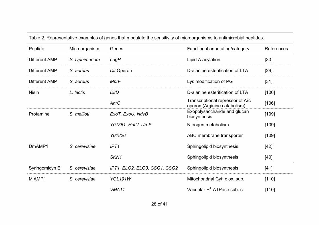
AFP A. oryzae ChsB, CsmA Chitin synthases, classes III and V [48]

AFP F. oxysporum ChsV Chitin synthase, class V [48]

1

30 of 41

References 1

2

1. Zasloff M. Antimicrobial peptides of multicellular organisms. Nature 3 2002;415:389-395 4

2. Hancock REW, Sahl HG. Antimicrobial and host-defense peptides as 5 new anti-infective therapeutic strategies. Nat Biotechnol 2006;24:1551-6 1557 7

3. Toke O. Antimicrobial peptides: New candidates in the fight against 8 bacterial infections. Biopolymers 2005;80:717-735 9

4. Marcos JF, Muñoz A, Pérez-Payá E, Misra S, López-García B. 10 Identification and rational design of novel antimicrobial peptides for plant 11 protection. Annu Rev Phytopathol 2008;46:273-301 12

5. Montesinos E. Antimicrobial peptides and plant disease control. FEMS 13 Microbiol Lett 2007;270:1-11 14

6. Papagianni M. Ribosomally synthesized peptides with antimicrobial 15 properties: Biosynthesis, structure, function, and applications. Biotechnol 16 Adv 2003;21:465-499 17

7. Rydlo T, Miltz J, Mor A. Eukaryotic antimicrobial peptides: Promises and 18 premises in food safety. J Food Sci 2006;71:R125-R135 19

8. Brown KL, Hancock REW. Cationic host defense (antimicrobial) 20 peptides. Curr Opin Immunol 2006;18:24-30 21

9. Braff MH, Hawkins MA, Di Nardo A, López-García B, Howell MD, Wong 22 C, Lin K, Streib JE, Dorschner R, Leung DYM, Gallo RL. Structure-23 function relationships among human cathelicidin peptides: Dissociation of 24 antimicrobial properties from host immunostimulatory activities. J 25 Immunol 2005;174:4271-4278 26

●● This work characterizes the role of human cathelicidin LL37 on immune 27 defense, indicating that antimicrobial activity against microbes resides 28 within specific domains/motifs of the peptide, which do not directly 29 correlate with immunomodulatory functions 30

10. Selsted ME, Ouellette AJ. Mammalian defensins in the antimicrobial 31 immune response. Nat Immunol 2005;6:551-557 32

11. Brogden KA. Antimicrobial peptides: Pore formers or metabolic inhibitors 33 in bacteria? Nat Rev Microbiol 2005;3:238-250 34

12. Yeaman MR, Yount NY. Mechanisms of antimicrobial peptide action and 35 resistance. Pharmacol Rev 2003;55:27-55 36

31 of 41

13. Jenssen H, Hamill P, Hancock REW. Peptide antimicrobial agents. Clin 1 Microbiol Rev 2006;19:491-511 2

14. Casteels P, Tempst P. Apidaecin-type peptide antibiotics function 3 through a non-poreforming mechanism involving stereospecificity. 4 Biochem Biophys Res Commun 1994;199:339-345 5

15. Steffen H, Rieg S, Wiedemann I, Kalbacher H, Deeg A, Sahl HG, 6 Peschel A, Gotz F, Garbe C, Schittek B. Naturally processed dermcidin-7 derived peptides do not permeabilize bacterial membranes and kill 8 microorganisms irrespective of their charge. Antimicrob Agents 9 Chemother 2006;50:2608-2620 10

16. Vylkova S, Nayyar N, Li WS, Edgerton M. Human beta-defensins kill 11 Candida albicans in an energy-dependent and salt-sensitive manner 12 without causing membrane disruption. Antimicrob Agents Chemother 13 2007;51:154-161 14

17. Mochon AB, Liu HP. The antimicrobial peptide Histatin-5 causes a 15 spatially restricted disruption on the Candida albicans surface, allowing 16 rapid entry of the peptide into the cytoplasm. PLoS Pathog 17 2008;4:e1000190 18

● A detailed description of the non-lytic mode of action for Histatin 5 by 19 using fluorescent labeling and confocal microscopic techniques that 20 show a sequential differential peptide targeting to different cell locations 21 and effects, depending on peptide concentration and time of exposure. 22

18. Monk BC, Niimi K, Lin S, Knight A, Kardos TB, Cannon RD, Parshot R, 23 King A, Lun D, Harding DRK. Surface-active fungicidal D-peptide 24 inhibitors of the plasma membrane proton pump that block azole 25 resistance. Antimicrob Agents Chemother 2005;49:57-70 26

19. Muñoz A, López-García B, Marcos JF. Studies on the mode of action of 27 the antifungal hexapeptide PAF26. Antimicrob Agents Chemother 28 2006;50:3847-3855 29

20. Hale JD, Hancock RE. Alternative mechanisms of action of cationic 30 antimicrobial peptides on bacteria. Expert Rev Anti Infect Ther 31 2007;5:951-959 32

21. Otvos LJr. Antibacterial peptides and proteins with multiple cellular 33 targets. J Pept Sci 2005;11:697-706 34

22. Aerts AM, Francois IEJA, Cammue BPA, Thevissen K. The mode of 35 antifungal action of plant, insect and human defensins. Cell Mol Life Sci 36 2008;65:2069-2079 37

23. Peschel A, Sahl HG. The co-evolution of host cationic antimicrobial 38 peptides and microbial resistance. Nat Rev Microbiol 2006;4:529-536 39

32 of 41

24. Wade D, Boman A, Wahlin B, Drain CM, Andreu D, Boman HG, 1 Merrifield RB. All-D amino acid-containing channel-forming antibiotic 2 peptides. Proc Natl Acad Sci USA 1990;87:4761-4765 3

25. Luque-Ortega JR, Van't Hof W, Veerman ECI, Saugar JM, Rivas L. 4 Human antimicrobial peptide histatin 5 is a cell-penetrating peptide 5 targeting mitochondrial ATP synthesis in Leishmania. FASEB J 6 2008;22:1817-1828 7

26. Bulet P, Urge L, Ohresser S, Hetru C, Otvos L. Enlarged scale chemical 8 synthesis and range of activity of drosocin, an O-glycosylated 9 antibacterial peptide of Drosophila. Eur J Biochem 1996;238:64-69 10

27. Piers KL, Brown MH, Hancock REW. Improvement of outer membrane-11 permeabilizing and lipopolysaccharide-binding activities of an 12 antimicrobial cationic peptide by C-terminal modification. Antimicrob 13 Agents Chemother 1994;38:2311-2316 14

28. Farnaud S, Spiller C, Moriarty L, Patel A, Gant V, Odell EW, Evans R. 15 Interactions of lactoferricin-derived peptides with LPS and antimicrobial 16 activity. FEMS Microbiol Lett 2004;233:193-199 17

29. Peschel A, Otto M, Jack RW, Kalbacher H, Jung G, Gotz F. Inactivation 18 of the dlt operon in Staphylococcus aureus confers sensitivity to 19 defensins, protegrins, and other antimicrobial peptides. J Biol Chem 20 1999;274:8405-8410 21

30. Guo L, Lim KB, Poduje CM, Daniel M, Gunn JS, Hackett M, Miller SI. 22 Lipid A acylation and bacterial resistance against vertebrate antimicrobial 23 peptides. Cell 1998;95:189-198 24

31. Peschel A, Jack RW, Otto M, Collins LV, Staubitz P, Nicholson G, 25 Kalbacher H, Nieuwenhuizen WF, Jung G, Tarkowski A, van Kessel 26 KPM, van Strijp JAG. Staphylococcus aureus resistance to human 27 defensins and evasion of neutrophil killing via the novel virulence factor 28 MprF is based on modification of membrane lipids with L-lysine. J Exp 29 Med 2001;193:1067-1076 30

32. Papo N, Shai Y. A molecular mechanism for lipopolysaccharide 31 protection of gram-negative bacteria from antimicrobial peptides. J Biol 32 Chem 2005;280:10378-10387 33

33. Rosenfeld Y, Barra D, Simmaco M, Shai Y, Mangoni ML. A synergism 34 between temporins toward gram-negative bacteria overcomes resistance 35 imposed by the lipopolysaccharide protective layer. J Biol Chem 36 2006;281:28565-28574 37

34. Hsu STD, Breukink E, Tischenko E, Lutters MAG, de Kruijff B, Kaptein R, 38 Bonvin AMJJ, van Nuland NAJ. The nisin-lipid II complex reveals a 39 pyrophosphate cage that provides a blueprint for novel antibiotics. Nat 40 Struct Mol Biol 2004;11:963-967 41

33 of 41

●● A detailed presentation of the structural characterization of the interaction 1 of Nisin with its docking partner: the bacterial wall precursor Lipid II. 2

35. Wiedemann I, Breukink E, van Kraaij C, Kuipers OP, Bierbaum G, de 3 Kruijff B, Sahl HA. Specific binding of nisin to the peptidoglycan 4 precursor lipid II combines pore formation and inhibition of cell wall 5 biosynthesis for potent antibiotic activity. J Biol Chem 2001;276:1772-6 1779 7

●● A reference study that demonstrates multiple killing mechanisms of the 8 model AMP Nisin, in permeating microbial cells but also blocking 9 envelope peptidoglycan synthesis. 10

36. Breukink E, Wiedemann I, van Kraaij C, Kuipers OP, Sahl HG, de Kruijff 11 B. Use of the cell wall precursor lipid II by a pore-forming peptide 12 antibiotic. Science 1999;286:2361-2364 13

37. Dielbandhoesing SK, Zhang H, Caro LH, van der Vaart JM, Klis FM, 14 Verrips CT, Brul S. Specific cell wall proteins confer resistance to Nisin 15 upon yeast cells. Appl Environ Microbiol 1998;64:4047-4052 16

38. Breukink E, de Kruijff B. Lipid II as a target for antibiotics. Nat Rev Drug 17 Discov 2006;5:321-323 18

39. Brotz H, Bierbaum G, Leopold K, Reynolds PE, Sahl HG. The lantibiotic 19 mersacidin inhibits peptidoglycan synthesis by targeting lipid II. 20 Antimicrob Agents Chemother 1998;42:154-160 21

40. Thevissen K, Idkowiak-Baldys J, Im YJ, Takemoto J, François IEJA, 22 Ferket KKA, Aerts AM, Meert EMK, Winderickx J, Roosen J, Cammue 23 BPA. SKN1, a novel plant defensin-sensitivity gene in Saccharomyces 24 cerevisiae, is implicated in sphingolipid biosynthesis. FEBS Lett 25 2005;579:1973-1977 26

41. Stock SD, Hama H, Radding JA, Young DA, Takemoto JY. Syringomycin 27 E inhibition of Saccharomyces cerevisiae: Requirement for biosynthesis 28 of sphingolipids with very-long-chain fatty acids and mannose- and 29 phosphoinositol-containing head groups. Antimicrob Agents Chemother 30 2000;44:1174-1180 31

42. Thevissen K, Cammue BPA, Lemaire K, Winderickx J, Dickson RC, 32 Lester RL, Ferket KK, Van Even F, Parret AH, Broekaert WF. A gene 33 encoding a sphingolipid biosynthesis enzyme determines the sensitivity 34 of Saccharomyces cerevisiae to an antifungal plant defensin from dahlia 35 (Dahlia merckii). Proc Natl Acad Sci USA 2000;97:9531-9536 36

43. Ramamoorthy V, Cahoon EB, Jia L, Thokala M, Minto RE, Shah DM. 37 Glucosylceramide synthase is essential for alfalfa defensin-mediated 38 growth inhibition but not for pathogenicity of Fusarium graminearum. Mol 39 Microbiol 2007;66:771-786 40

34 of 41

● This work demonstrates the importance of the gene GCS1, involved in 1 glucosylceramide biosynthesis, for the sensitivity to the antifungal 2 defensin MsDef1, but strikingly not to the related MtDef4. 3

44. Thevissen K, Warnecke DC, François IEJA, Leipelt M, Heinz E, Ott C, 4 Zahringer U, Thomma BPHJ, Ferkel KKA, Cammue BPA. Defensins from 5 insects and plants interact with fungal glucosylceramides. J Biol Chem 6 2004;279:3900-3905 7

45. Vylkova S, Li XS, Berner JC, Edgerton M. Distinct antifungal 8 mechanisms: β-Defensins require Candida albicans Ssa1 protein, while 9 Trk1p mediates activity of cysteine-free cationic peptides. Antimicrob 10 Agents Chemother 2006;50:324-331 11

46. Lobo DS, Pereira IB, Fragel-Madeira L, Medeiros LN, Cabral LM, Faria J, 12 Bellio M, Campos RC, Linden R, Kurtenbach E. Antifungal Pisum 13 sativum defensin 1 interacts with Neurospora crassa cyclin F related to 14 the cell cycle. Biochemistry 2007;46:987-996 15

●● This work demonstrates the interaction of the plant defensin PSD1 with 16 nuclear Cyclin F, its targeting to fungal nucleus, and indications that it 17 might alter cell cycle progression, by using distinct experimental 18 approaches. It is a significant demonstration of AMP specific nuclear 19 targets in fungi. 20

47. Ibeas JI, Lee H, Damsz B, Prasad DT, Pardo JM, Hasegawa PM, 21 Bressan RA, Narasimhan ML. Fungal cell wall phosphomannans 22 facilitate the toxic activity of a plant PR-5 protein. Plant J 2000;23:375-23 383 24

48. Hagen S, Marx F, Ram AF, Meyer V. The antifungal protein AFP from 25 Aspergillus giganteus inhibits chitin synthesis in sensitive fungi. Appl 26 Environ Microbiol 2007;73:2128-2134 27

49. Fujimura M, Ideguchi M, Minami Y, Watanabe K, Tadera K. Amino acid 28 sequence and antimicrobial activity of chitin-binding peptides, Pp-AMP 1 29 and Pp-AMP 2, from Japanese bamboo shoots (Phyllostachys 30 pubescens). Biosci Biotechnol Biochem 2005;69:642-645 31

50. Georgopapadakou NH. Update on antifungals targeted to the cell wall: 32 Focus on β-1,3-glucan synthase inhibitors. Expert Opin Investig Drugs 33 2001;10:269-280 34

51. Thevissen K, François IEJA, Aerts AM, Cammue BPA. Fungal 35 sphingolipids as targets for the development of selective antifungal 36 therapeutics. Curr Drug Targets 2005;6:923-928 37

52. Odds FC, Brown AJP, Gow NAR. Antifungal agents: mechanisms of 38 action. Trends Microbiol 2003;11:272-279 39

53. Selitrennikoff CP, Nakata M. New cell wall targets for antifungal drugs. 40 Curr Opin Investig Drugs 2003;4:200-205 41

35 of 41

54. Heinisch J. Baker's yeast as a tool for the development of antifungal 1 drugs which target cell integrity − an update. Expert Opin Drug Discov 2 2008;3:931-943 3

55. Yun DJ, Zhao Y, Pardo JM, Narasimhan ML, Damsz B, Lee H, Abad LR, 4 Durzo MP, Hasegawa PM, Bressan RA. Stress proteins on the yeast cell 5 surface determine resistance to osmotin, a plant antifungal protein. Proc 6 Natl Acad Sci USA 1997;94:7082-7087 7

56. Li XS, Reddy MS, Baev D, Edgerton M. Candida albicans Ssa1/2p Is the 8 cell envelope binding protein for human salivary Histatin 5. J Biol Chem 9 2003;278:28553-28561 10

57. Jang WS, Li XWS, Sun JNN, Edgerton M. The P-113 fragment of Histatin 11 5 requires a specific peptide sequence for intracellular translocation in 12 Candida albicans, which is independent of cell wall binding. Antimicrob 13 Agents Chemother 2008;52:497-504 14

●● By using peptide fragments and sequence analogs the authors 15 demonstrate the separation in the AMP histatin 5 of the binding cell wall 16 protein Ssa2 from translocation into the cytosol, thus dissociating two 17 necessary steps in the peptide interaction with target microorganisms. 18

58. Sun JNN, Li WS, Jang WS, Nayyar N, Sutton MD, Edgerton M. Uptake of 19 the antifungal cationic peptide Histatin 5 by Candida albicans Ssa2p 20 requires binding to non-conventional sites within the ATPase domain. 21 Mol Microbiol 2008;70:1246-1260 22

●● A remarkable characterization and mapping to a limited protein domain 23 of the binding of the yeast Ssa2 HSP to the AMP Histatin 5, which is 24 required for efficient cell uptake. 25

59. Li M, Lai YP, Villaruz AE, Cha DJ, Sturdevant DE, Otto M. Gram-positive 26 three-component antimicrobial peptide-sensing system. Proc Natl Acad 27 Sci USA 2007;104:9469-9474 28

●● This work extends to Gram-positive bacteria the involvement of (three-29 component) sensor systems to signal and control the response to AMP, 30 thus highlighting the existence of common signaling mechanisms along 31 distinct microbial organism. 32

60. Hong RW, Shchepetov M, Weiser JN, Axelsen PH. Transcriptional profile 33 of the Escherichia coli response to the antimicrobial insect peptide 34 Cecropin A. Antimicrob Agents Chemother 2003;47:1-6 35

61. Tomasinsig L, Scocchi M, Mettulio R, Zanetti M. Genome-wide 36 transcriptional profiling of the Escherichia coli response to a proline-rich 37 antimicrobial peptide. Antimicrob Agents Chemother 2004;48:3260-3267 38

● Characterization of the bacterial global transcriptional response to a 39 specific AMP. The expression changes of selected genes to a sequence 40

36 of 41

derivative that is devoid of antimicrobial activity and to other unrelated 1 AMP were also studied, and indicate distinctive responses. 2

62. Gamberi T, Cavalieri D, Magherini F, Mangoni ML, De Filippo C, Borro 3 M, Gentile G, Simmaco M, Modesti A. An integrated analysis of the 4 effects of Esculentin 1-21 on Saccharomyces cerevisiae. Biochim 5 Biophys Acta 2007;1774:688-700 6

63. Vylkova S, Jang WS, Li WS, Nayyar N, Edgerton M. Histatin 5 initiates 7 osmotic stress response in Candida albicans via activation of the Hog1 8 mitogen-activated protein kinase pathway. Eukaryot Cell 2007;6:1876-9 1888 10

64. Morton CO, Hayes A, Wilson M, Rash BM, Oliver SG, Coote P. Global 11 phenotype screening and transcript analysis outlines the inhibitory 12 mode(s) of action of two amphibian-derived, alpha-helical, cationic 13 peptides on Saccharomyces cerevisiae. Antimicrob Agents Chemother 14 2007;51:3948-3959 15

●● A reference study to determine the mechanism of action of antifungal 16 peptides by using distinct/complementary functional genomic 17 approaches. In addition, two different peptides were compared. 18 Transcriptomic data and analysis of a genome-wide collection of yeast 19 strains with gene deletions indicated that Magainin 2 and Dermaseptin 20 S3 have both common and unique effects to fungal cells. 21

65. Bader MW, Sanowar S, Daley ME, Schneider AR, Cho US, Xu WQ, 22 Klevit RE, Le Moual H, Miller S. Recognition of antimicrobial peptides by 23 a bacterial sensor kinase. Cell 2005;122:461-472 24

●● This work reveals an acidic surface domain from the PhoQ sensor kinase 25 that participates in cationic antimicrobial peptide binding, as a first step in 26 signal transduction across the bacterial membrane. 27

66. Fields PI, Groisman EA, Heffron F. A Salmonella locus that controls 28 resistance to microbicidal proteins from phagocytic cells. Science 29 1989;243:1059-1062 30

67. Miller SI, Pulkkinen WS, Selsted ME, Mekalanos JJ. Characterization of 31 defensin resistance phenotypes associated with mutations in the phoP 32 virulence regulon of Salmonella typhimurium. Infect Immun 33 1990;58:3706-3710 34

68. McPhee JB, Lewenza S, Hancock REW. Cationic antimicrobial peptides 35 activate a two-component regulatory system, PmrA-PmrB, that regulates 36 resistance to polymyxin B and cationic antimicrobial peptides in 37 Pseudomonas aeruginosa. Mol Microbiol 2003;50:205-217 38

● Identification of an additional two-component system in Gram-negative 39 bacteria (PmrAB) that regulates resistance to AMP. 40

37 of 41

69. Gunn JS, Lim KB, Krueger J, Kim K, Guo L, Hackett M, Miller SI. PmrA-1 PmrB-regulated genes necessary for 4-aminoarabinose lipid A 2 modification and polymyxin resistance. Mol Microbiol 1998;27:1171-1182 3

70. Narasimhan ML, Coca MA, Jin JB, Yamauchi T, Ito Y, Kadowaki T, Kim 4 KK, Pardo JM, Damsz B, Hasegawa PM, Yun DJ, Bressan RA. Osmotin 5 is a homolog of mammalian adiponectin and controls apoptosis in yeast 6 through a homolog of mammalian adiponectin receptor. Mol Cell 7 2005;17:171-180 8

71. Narasimhan ML, Damsz B, Coca MA, Ibeas JI, Yun DJ, Pardo JM, 9 Hasegawa PM, Bressan RA. A plant defense response effector induces 10 microbial apoptosis. Mol Cell 2001;8:921-930 11

72. Koo JC, Lee B, Young ME, Koo SC, Cooper JA, Baek D, Lim CO, Lee 12 SY, Yun DJ, Cho MJ. Pn-AMP1, a plant defense protein, induces actin 13 depolarization in yeasts. Plant Cell Physiol 2004;45:1669-1680 14

73. Ramamoorthy V, Zhao XH, Snyder AK, Xu JR, Shah DM. Two mitogen-15 activated protein kinase signalling cascades mediate basal resistance to 16 antifungal plant defensins in Fusarium graminearum. Cell Microbiol 17 2007;9:1491-1506 18

● The authors extend here on the differential requirements to respond to 19 the two related plant defensins MsDef1 and MtDef4, with an emphasis on 20 the cell-signaling mediated by protein kinases cascades. 21

74. Morton CO, dos Santos SC, Coote P. An amphibian-derived, cationic, 22 alpha-helical antimicrobial peptide kills yeast by caspase-independent 23 but AIF-dependent programmed cell death. Mol Microbiol 2007;65:494-24 507 25

● A detailed study that combines molecular genetics and cell biology 26 methods to show that a specific AMP induces cell suicide/apoptosis in 27 yeast 28

75. Andrés MT, Viejo-Díaz M, Fierro JF. Human lactoferrin induces 29 apoptosis-like cell death in Candida albicans: Critical role of K+ channel-30 mediated K+ efflux. Antimicrob Agents Chemother 2008;52:4081-4088 31

76. Leiter E, Szappanos H, Oberparleiter C, Kaiserer L, Csernoch L, 32 Pusztahelyi T, Emri T, Posci I, Salvenmoser W, Marx F. Antifungal 33 protein PAF severely affects the integrity of the plasma membrane of 34 Aspergillus nidulans and induces an apoptosis-like phenotype. 35 Antimicrob Agents Chemother 2005;49:2445-2453 36

77. Madeo F, Herker E, Wissing S, Jungwirth H, Eisenberg T, Frohlich KU. 37 Apoptosis in yeast. Curr Opin Microbiol 2004;7:655-660 38

78. Lupetti A, Paulusma-Annema A, Senesi S, Campa M, van Dissel JT, 39 Nibbering PH. Internal thiols and reactive oxygen species in candidacidal 40

38 of 41

activity exerted by an N-terminal peptide of human lactoferrin. Antimicrob 1 Agents Chemother 2002;46:1634-1639 2

79. Aerts AM, François IEJA, Meert EMK, Li QT, Cammue BPA, Thevissen 3 K. The antifungal activity of RsAFP2, a plant defensin from Raphanus 4 sativus, involves the induction of reactive oxygen species in Candida 5 albicans. J Mol Microbiol Biotechnol 2007;13:243-247 6

80. Helmerhorst EJ, Troxler RF, Oppenheim FG. The human salivary peptide 7 histatin 5 exerts its antifungal activity through the formation of reactive 8 oxygen species. Proc Natl Acad Sci USA 2001;98:14637-14642 9

81. Veerman ECI, Nazmi K, van Hof W, Bolscher JGM, den Hertog AL, 10 Amerongen AVN. Reactive oxygen species play no role in the 11 candidacidal activity of the salivary antimicrobial peptide histatin 5. 12 Biochem J 2004;381:447-452 13

82. Castle M, Nazarian A, Yi SS, Tempst P. Lethal effects of apidaecin on 14 Escherichia coli involve sequential molecular interactions with diverse 15 targets. J Biol Chem 1999;274:32555-32564 16

83. Park CB, Yi KS, Matsuzaki K, Kim MS, Kim SC. Structure-activity 17 analysis of buforin II, a histone H2A-derived antimicrobial peptide: The 18 proline hinge is responsible for the cell-penetrating ability of buforin II. 19 Proc Natl Acad Sci USA 2000;97:8245-8250 20

● A study that highlights the potential of using AMP sequence analogs with 21 specific amino acid residue substitutions to characterize the interaction of 22 peptides with microbes. The work demonstrated that the proline hinge is 23 a key structural factor for the cell-penetrating ability of peptide buforin II. 24

84. Kragol G, Hoffmann R, Chattergoon MA, Lovas S, Cudic M, Bulet P, 25 Condie BA, Rosengren KJ, Montaner LJ, Otvos LJr. Identification of 26 crucial residues for the antibacterial activity of the proline-rich peptide, 27 pyrrhocoricin. Eur J Biochem 2002;269:4226-4237 28

85. Park CB, Kim HS, Kim SC. Mechanism of action of the antimicrobial 29 peptide buforin II: Buforin II kills microorganisms by penetrating the cell 30 membrane and inhibiting cellular functions. Biochem Biophys Res 31 Commun 1998;244:253-257 32

86. van der Kraan MIA, van Marle J, Nazmi K, Groenink J, Van't Hof W, 33 Veerman ECI, Bolscher JGM, Arnerongen AVN. Ultrastructural effects of 34 antimicrobial peptides from bovine lactoferrin on the membranes of 35 Candida albicans and Escherichia coli. Peptides 2005;26:1537-1542 36

87. van der Weerden NL, Lay FT, Anderson MA. The plant defensin, NaD1, 37 enters the cytoplasm of Fusarium oxysporum hyphae. J Biol Chem 38 2008;283:14445-14452 39

39 of 41

88. Gyurko C. Killing of Candida albicans by histatin 5: Cellular uptake and 1 energy requirement. Antonie Van Leeuwenhoek Int J Gen Molec 2 Microbiol 2001;79:297-309 3

89. Powers JPS, Martin MM, Goosney DL, Hancock REW. The antimicrobial 4 peptide polyphemusin localizes to the cytoplasm of Escherichia coli 5 following treatment. Antimicrob Agents Chemother 2006;50:1522-1524 6

90. Gelhaus C, Jacobs T, Andra J, Leippe M. The antimicrobial peptide NK-7 2, the core region of mammalian NK-lysin, kills intraerythrocytic 8 Plasmodium falciparum. Antimicrob Agents Chemother 2008;52:1713-9 1720 10

91. Haukland HH, Ulvatne H, Sandvik K, Vorland LH. The antimicrobial 11 peptides lactoferricin B and magainin 2 cross over the bacterial 12 cytoplasmic membrane and reside in the cytoplasm. FEBS Lett 13 2001;508:389-393 14

92. Henriques ST, Melo MN, Castanho MARB. Cell-penetrating peptides and 15 antimicrobial peptides: how different are they? Biochem J 2006;399:1-7 16

93. Foged C, Nielsen HM. Cell-penetrating peptides for drug delivery across 17 membrane barriers. Expert Opin Drug Deliv 2008;5:105-117 18

94. Palm C, Netzerea S, Hallbrink M. Quantitatively determined uptake of 19 cell-penetrating peptides in non-mammalian cells with an evaluation of 20 degradation and antimicrobial effects. Peptides 2006;27:1710-1716 21

95. Jung HJ, Park Y, Hahm KS, Lee DG. Biological activity of Tat (47-58) 22 peptide on human pathogenic fungi. Biochem Biophys Res Commun 23 2006;345:222-228 24

96. Jones AT. Gateways and tools for drug delivery: Endocytic pathways and 25 the cellular dynamics of cell penetrating peptides. Int J Pharm 26 2008;354:34-38 27

97. Duchardt F, Fotin-Mleczek M, Schwarz H, Fischer R, Brock R. A 28 comprehensive model for the cellular uptake of cationic cell-penetrating 29 peptides. Traffic 2007;8:848-866 30

98. Console S, Marty C, García-Echeverría C, Schwendener R, Ballmer-31 Hofer K. Antennapedia and HIV transactivator of transcription (TAT) 32 "protein transduction domains" promote endocytosis of high molecular 33 weight cargo upon binding to cell surface glycosaminoglycans. J Biol 34 Chem 2003;278:35109-35114 35

99. Subbalakshmi C, Sitaram N. Mechanism of antimicrobial action of 36 indolicidin. FEMS Microbiol Lett 1998;160:91-96 37

100. Patrzykat A, Friedrich CL, Zhang LJ, Mendoza V, Hancock REW. 38 Sublethal concentrations of pleurocidin-derived antimicrobial peptides 39

40 of 41

inhibit macromolecular synthesis in Escherichia coli. Antimicrob Agents 1 Chemother 2002;46:605-614 2

101. Friedrich CL, Rozek A, Patrzykat A, Hancock REW. Structure and 3 mechanism of action of an indolicidin peptide derivative with improved 4 activity against gram-positive bacteria. J Biol Chem 2001;276:24015-5 24022 6

102. Lehrer RI, Barton A, Daher KA, Harwig SS, Ganz T, Selsted ME. 7 Interaction of human defensins with Escherichia coli. Mechanism of 8 bactericidal activity. J Clin Invest 1989;84:553-561 9

103. Boman HG, Agerberth B, Boman A. Mechanisms of action on 10 Escherichia coli of cecropin P1 and PR-39, two antibacterial peptides 11 from pig intestine. Infect Immun 1993;61:2978-2984 12

104. Kragol G, Lovas S, Varadi G, Condie BA, Hoffmann R, Otvos LJr. The 13 antibacterial peptide pyrrhocoricin inhibits the ATPase actions of DnaK 14 and prevents chaperone-assisted protein folding. Biochemistry 15 2001;40:3016-3026 16

●● The initial characterization of the binding of pyrrhocoricin to Escherichia 17 coli heat shock protein DnaK, that results in the impairment of the protein 18 activity and thus 19

105. Otvos LJr, O I, Rogers ME, Consolvo PJ, Condie BA, Lovas S, Bulet P, 20 Blaszczyk-Thurin M. Interaction between heat shock proteins and 21 antimicrobial peptides. Biochemistry 2000;39:14150-14159 22

106. Kramer NE, Van Hijum SAFT, Knol J, Kok J, Kuipers OP. Transcriptome 23 analysis reveals mechanisms by which Lactococcus lactis acquires nisin 24 resistance. Antimicrob Agents Chemother 2006;50:1753-1761 25

● A unique example of an alternative approach to the characterization of 26 the bacterial resistance to AMP, by using transcriptomic approaches. The 27 authors compared the global gene expression profile of a bacterial strain 28 resistant to Nisin with that of the parental strain. A set of general 29 responses is proposed. 30

107. Boix E, Nogues MV. Mammalian antimicrobial proteins and peptides: 31 overview on the RNase A superfamily members involved in innate host 32 defence. Mol Biosyst 2007;3:317-335 33

108. Chadha P, Das RH. A pathogenesis related protein, AhPR10 from 34 peanut: an insight of its mode of antifungal activity. Planta 2006;225:213-35 222 36

● Demonstration of the linkage between the RNase activity of a plant PR10 37 and its antimicrobial properties; these latter require, although are not 38 linked to, internalization into fungal cells. 39

41 of 41

109. Nogales J, Muñoz S, Olivares J, Sanjuán J. Sinorhizobium meliloti genes 1 involved in tolerance to the antimicrobial peptide protamine. FEMS 2 Microbiol Lett 2006;264:160-167 3

110. Stephens C, Harrison SJ, Kazan K, Smith FWN, Goulter KC, Maclean 4 DJ, Manners JM. Altered fungal sensitivity to a plant antimicrobial 5 peptide through over-expression of yeast cDNAs. Curr Genet 6 2005;47:194-201 7

111. Matsuzaki K, Sugishita K, Miyajima K. Interactions of an antimicrobial 8 peptide, magainin 2, with lipopolysaccharide-containing liposomes as a 9 model for outer membranes of gram-negative bacteria. FEBS Lett 10 1999;449:221-224 11

112. Hsu CH, Chen CP, Jou ML, Lee AYL, Lin YC, Yu YP, Huang WT, Wu 12 SH. Structural and DNA-binding studies on the bovine antimicrobial 13 peptide, indolicidin: evidence for multiple conformations involved in 14 binding to membranes and DNA. Nucleic Acids Res 2005;33:4053-4064 15

113. Ulvatne H, Samuelsen O, Haukland HH, Kramer M, Vorland LH. 16 Lactoferricin B inhibits bacterial macromolecular synthesis in Escherichia 17 coli and Bacillus subtilis. FEMS Microbiol Lett 2004;237:377-384 18

19 20

Signaling

2c

1

2a

2b

3a

3d

• Resistance Response• Apoptosis

• Nuc. Ac. Bind.• DNA/RNA Biosynt.• Ribosome biogen.• DNA damage• Cell cycle

3b

3c

• Protein Trans.

• Chaperone folding

• Vacuole Transp.

Figure 1


Recommended