Page 1
RESEARCH ARTICLES
Arachidonic Acid: An Evolutionarily Conserved SignalingMolecule Modulates Plant Stress Signaling Networks C W
Tatyana Savchenko,a Justin W. Walley,a,b E. Wassim Chehab,a,1 Yanmei Xiao,a Roy Kaspi,a Matthew F. Pye,c
Maged E. Mohamed,d,2 Colin M. Lazarus,d Richard M. Bostock,c and Katayoon Dehesha,3
a Department of Plant Biology, University of California, Davis, California 95616b Department of Biological Sciences, University of California, San Diego, California 92093-0380c Department of Plant Pathology, University of California, Davis, California 95616d School of Biological Sciences, University of Bristol, Bristol BS8 1UG, United Kingdom
Fatty acid structure affects cellular activities through changes in membrane lipid composition and the generation of a
diversity of bioactive derivatives. Eicosapolyenoic acids are released into plants upon infection by oomycete pathogens,
suggesting they may elicit plant defenses. We exploited transgenic Arabidopsis thaliana plants (designated EP) producing
eicosadienoic, eicosatrienoic, and arachidonic acid (AA), aimed at mimicking pathogen release of these compounds. We
also examined their effect on biotic stress resistance by challenging EP plants with fungal, oomycete, and bacterial
pathogens and an insect pest. EP plants exhibited enhanced resistance to all biotic challenges, except they were more
susceptible to bacteria than the wild type. Levels of jasmonic acid (JA) were elevated and levels of salicylic acid (SA) were
reduced in EP plants. Altered expression of JA and SA pathway genes in EP plants shows that eicosapolyenoic acids
effectively modulate stress-responsive transcriptional networks. Exogenous application of various fatty acids to wild-type
and JA-deficient mutants confirmed AA as the signaling molecule. Moreover, AA treatment elicited heightened expression
of general stress-responsive genes. Importantly, tomato (Solanum lycopersicum) leaves treated with AA exhibited reduced
susceptibility to Botrytis cinerea infection, confirming AA signaling in other plants. These studies support the role of AA, an
ancient metazoan signaling molecule, in eliciting plant stress and defense signaling networks.
INTRODUCTION
In animals and plants, fatty acids (FAs) are key molecules that
participate in various biological processes. Structural properties
of FAs, such as their chain length and their degree of desatura-
tion, largely determine the nature of these processes. In unicel-
lular organisms, such as Escherichia coli and yeast, FAs have
been shown to regulate gene transcription (Black et al., 2000). In
mammals, the expression of many genes is modulated positively
or negatively by FAs through changes in rate of transcription or
posttranscriptional modifications (Duplus et al., 2000; Huang
et al., 2004; Pegorier et al., 2004). In plants, FAs and/or their
derived metabolites are also recognized as signaling molecules
central to various biological processes.
Exogenous and endogenous unsaturated FAs and FA me-
tabolites can significantly alter plant gene expression and me-
tabolism to influence the outcome of plant–microbe and
plant–herbivore interactions (Upchurch, 2008). Mounting evi-
dence from studies with Arabidopsis thaliana plants defective in
stearoyl-acyl carrier protein-desaturase, the enzyme responsible
for conversion of stearic acid (18:0) to oleic acid (18:1 D9), has
established that the levels of 18:1 are a critical regulator of
salicylic acid (SA)- and jasmonic acid (JA)-mediated defense
signaling in the plant (A. Kachroo et al., 2003; P. Kachroo et al.,
2003, 2005; Chandra-Shekara et al., 2007; Venugopal et al.,
2009). Specifically, reduction of the 18:1 levels leads to consti-
tutive expression of PR genes and enhanced resistance to
Hyaloperonospora arabidopsidis (formerly Peronospora para-
sitica), in concert with reduced expression of a subset of JA-
dependent response genes and decreased resistance toBotrytis
cinerea (Kachroo et al., 2001). An important role for linoleic acid
(LA; 18:2 D9,12) in conidiation, development, and aflatoxin syn-
thesis has been described in the interaction between toxigenic
Aspergillus flavus and its hosts, with apparent reciprocity in the
ability of the plant and pathogen to influence oxylipin profiles
during the interaction (Brodhagen et al., 2008).
Plants also respond to exogenous treatment with eicosapoly-
enoic acids and to pathogens containing them during infection.
Specifically, eicosapentaenoic acid (EPA; 20:5 D5,8,11,14,17) and
arachidonic acid (AA; 20:4 D5,8,11,14) are potent elicitors of
1Current address: Biochemistry and Cell Biology, Rice University,Houston, TX 77005-1892.2 Current address: School of Pharmacy, University of Zagazig, Zagazig44519, Egypt.3 Address correspondence to [email protected] author responsible for distribution of materials integral to thefindings presented in this article in accordance with the policy describedin the Instructions for Authors (www.plantcell.org) is: Katayoon Dehesh([email protected] ).CSome figures in this article are displayed in color online but in blackand white in the print edition.WOnline version contains Web-only data.www.plantcell.org/cgi/doi/10.1105/tpc.110.073858
The Plant Cell, Vol. 22: 3193–3205, October 2010, www.plantcell.org ã 2010 American Society of Plant Biologists
Dow
nloaded from https://academ
ic.oup.com/plcell/article/22/10/3193/6094857 by guest on 20 Septem
ber 2021
Page 2
programmed cell death and defense responses in Solanaceous
plants (Bostock et al., 1981; Knight et al., 2001; Garcia-Pineda
et al., 2004) and are reported to induce resistance to viruses in
potato (Solanum tuberosum) and tobacco (Nicotiana tabacum)
(Rozhnova et al., 2003; Ozeretskovskaya et al., 2004). Treatment
of potato with AA and EPA results in the coordinate activation of
defense-related responses (Bostock et al., 1986) and a dramatic
and immediate shift in terpenoid metabolism, redirecting the
pathway from higher terpenoids (i.e., steroid glycoalkaloids) to
sesquiterpenoid phytoalexins (Tjamos and Kuc, 1982; Choi et al.,
1992, 1994). FAs not commonly found in plants, such as EPA
and AA that are abundant in lipids of Phytophthora species
and related oomycetes, are released into plant tissue from
spores during early stages of infection (Ricker and Bostock,
1992). Metabolic studies in potato have shown that AA is
relatively stable compared with naturally occurring LA (18:2
D9,12), with a portion of the AA oxidized to hydroxy acids and
other products and a larger portion rapidly incorporated into
phospho- and glycero-lipids (Preisig and Kuc, 1988). A critical
structural feature for their elicitor activity in plants is cis-unsatu-
ration at theD5-position, and evidence supports the participation
of plant lipoxygenases in this activity (Bostock et al., 1981, 1992;
Preisig and Kuc, 1985). Based on these observations, it has been
proposed that eicosapolyenoic acids have transorganismal sig-
naling activity (Rozhnova et al., 2003; Ozeretskovskaya et al.,
2004).
Plants and animals possess parallel, analogous FA signaling
systems. Perhaps the best example are the parallel roles played
by FA oxidation products oxylipins (plants) and eicosanoids
(animals) in mediating stress signaling cascades. Plant oxylipins,
also referred to as octadecanoids, are the oxidation products of
LA or linolenic acid (ALA; 18:3 D9,12,15), and a major product of
this pathway is JA (Farmer et al., 1998; Blee, 2002). JA and its
derivatives canmodulate a wide range of biological processes in
plants, among them plant resistance to insects and pathogens
(Gardner, 1995; Blee, 2002). Animal lipidmediators, eicosanoids,
include prostaglandins, leukotrienes, lipoxins, and other related
compounds derived primarily from AA (Schultz, 2002). Similar to
oxylipins, eicosanoids play multiple biological roles in animals,
including regulating wound responses, inflammation, and im-
mune responses (Magnan and Vervloet, 1999; van Ryn et al.,
2000). The structures and biosynthesis of JA and its precursors
and derivatives are analogous to those of animal eicosanoids.
The parallels between these molecules is further reinforced by
their functional similarities, as they are synthesized and released
for immediate and local, as well as systemic, responses to
stresses (Straus and Glass, 2001; Blee, 2002).
Most plants do not contain eicosapolyenoic acids. However,
because of the abundance of these FAs in oomycete pathogens
and their potential signaling activity in plants when delivered by
external challenge, we questioned whether endogenous pro-
duction of eicosapolyenoic acids in plants also would modulate
plant defense responses. To address this question, we exam-
ined the resistance of transgenic lines that produce eicosa-
polyenoic acids, mainly eicosadienoic acid (EDA; 20:2 D11,14),
eicosatrienoic acid (ETrA; 20:3D11,14,17), and AA, to a wide range
of biotic challengers. The outcome of these studies, together
with data obtained from external application of various FAs,
advance the notion that as in animal systems, AA in plants can
function as a signaling molecule that not only triggers FA-
mediated defense responses, but also elicits general stress
signaling networks.
RESULTS
EDA, ETrA, andAAAre theDominant Eicosapolyenoic Acids
in EP Leaves
Two different strategies were employed to generate independent
transgenic Arabidopsis lines producing eicosapolyenoic acids
via the D8-desaturation pathway. In one approach, an eicosa-
polyenoic acid–producing transgenic line (designated as EP1)
was generated by sequential introduction of three sets of con-
stitutively expressed genes (C18-D9-elongase, D8-desaturase,
and D5-desaturase) (Qi et al., 2004). In the second approach, line
EP2was generated by a single transformation step using a binary
vector containing these same genes in tandem (described in
Methods). In the D8-desaturation pathway, LA and ALA are first
elongated by a C18-D9-elongase to EDA (20:2 D11,14) and ETrA
(20:3 D11,14,17), respectively. A D8-desaturase introduces a
double bond at the D8 position of the carbon chain to produce
dihomo-g-linolenic acid (20:3 D8,11,14) and eicosatetraenoic acid
(20:4 D8,11,14,17), the substrates used by a D5-desaturase to
produce AA and EPA. These FAs esterified predominantly to
phosphatidylcholine cause no visible effect on plant morphology
(Fraser et al., 2004). Monitoring EP plants at all developmental
stages established that these plants are phenotypically indistin-
guishable from the wild type. Analysis of FA composition of
leaves determined that EP plants contain easily detectable levels
of eicosapolyenoic acids, predominantly EDA, ETrA, and AA
(Table 1).
Table 1. EP Plants Contain Eicosapolyenoic Acids, Mainly EDA, EtrA,
and AA
FA
mol % of Total Wild Type EP1 EP2
16:0 14.50 6 0.403 13.99 6 0.285 12.61 6 0.053
16:1D7 2.02 6 0.169 2.28 6 0.156 2.43 6 0.112
16:2D7,10 0.61 6 0.063 0.63 6 0.168 0.68 6 0.023
16:3D7,10,13 15.11 6 0.186 15.82 6 0.545 16.34 6 0.069
18:0 1.41 6 0.243 1.15 6 0.136 2.07 6 0.029
18:1D9 1.33 6 0.192 1.04 6 0.017 0.78 6 0.086
18:2D9,12 14.18 6 0.139 12.89 6 0.476 10.33 6 0.144
18:3D9,12,15 50.72 6 0.705 49.79 6 0.232 40.38 6 0.066
20:0 0.12 6 0.015 0.09 6 0.012 0.12 6 0.015
20:2D11,14 (EDA) 0.77 6 0.057 6.20 6 0.043
20:3D11,14,17 (ETrA) 0.95 6 0.055 7.34 6 0.053
20:3D8,11,14 (DGLA) 0.02 6 0.01 0.16 6 0.081
20:4D8,11,14,17 (ETA) 0.16 6 0.039 0.24 6 0.052
20:4D5,8,11,14 (AA) 0.42 6 0.067 0.25 6 0.016
20:5D5,8,11,14,17 (EPA) 0.01 6 0.006 0.08 6 0.017
Total C20 PUFA 2.33 14.27
FA analyses were performed on leaf tissue and each value is the mean6
SD of six samples. DGLA, dihomo-g-linolenic acid; ETA, eicosatetrae-
noic acid
3194 The Plant Cell
Dow
nloaded from https://academ
ic.oup.com/plcell/article/22/10/3193/6094857 by guest on 20 Septem
ber 2021
Page 3
EP Plants Are Differentially Resistant to Biotic Stresses
In response to a change in quantity or type of fatty acid compo-
sition of membrane lipids, plants display a number of alterations
in their defenses against stresses. To examine whether endog-
enous production of eicosapolyenoic acids would alter plant
defense responses, we exposed EP and wild-type plants to a
range of biotic challengers, including a piercing-sucking insect
(aphid [Myzus persicae]), two isolates of the fungal pathogen
B. cinerea (Grape and B05.10), an oomycete pathogen (Phytoph-
thora capsici), and a bacterial pathogen (Pseudomonas syringae
pv tomatoDC3000 [Pst]). EP1 and EP2 plants responded similarly
to the biotic stresses examined. These data indicated that EP
plants are significantly more resistant to all biotic challengers,
except Pst (Figures 1A to 1E; see Supplemental Figures 1A and
1B online). Specifically, dual choice assays showed that aphid
nymph deposition is significantly lower on EP than on wild-type
plants (Figure 1A). Furthermore, infection of leavesby two isolates
of B. cinerea resulted in a significantly smaller lesion size on EP
than wild-type leaves (Figures 1B; see Supplemental Figure 1A
online). EP plants also displayed enhanced resistance to P.
capsici, as reflected by the number of P. capsici sporangia and
by the amount of pathogen DNA present on these plants com-
pared with the corresponding wild type (Figures 1C and 1D). By
contrast, EP plants are notably more susceptible to Pst infection
(Figure 1E; see Supplemental Figure 1B online). These data
collectively demonstrate that the in vivo perturbation of FA
composition of membrane lipids significantly and differentially
altered the tolerance of EP plants to these biotic challengers.
The Levels of JA and SAMetabolites and Their
Corresponding Transcripts Are Altered in EP Plants
The lifestyle of a plant pathogen can influence which defense
signal transduction pathway is activated. In Arabidopsis, the
SA signaling pathway predominantly mediates the response
to biotrophic pathogens (McDowell and Dangl, 2000), while JA-
dependent defense responses are more closely associated with
response to infection with necrotrophic pathogens that acquire
nutrients from dead or dying host cells (Glazebrook, 2005). In
addition to necrotrophic pathogens, the JA signaling pathway is
also important for the activation of defense cascades in response
to wounding and herbivores, including piercing-sucking insects
(Staswick et al., 1998; Halim et al., 2006; Chehab et al., 2008).
To determine whether changes in the levels of SA and JA
underlie the observed altered tolerance of EP plants to the biotic
challengers examined, we measured the levels of these metab-
olites in EP andwild-type plants. These data demonstrate that JA
levels, specifically in wounded EP1 and EP2 plants, are approx-
imately twofold higher than their corresponding wild-type levels
(Figure 2A; see Supplemental Figure 2A online). The similarity
between elevated levels of JA in response to wounding in EP1
and EP2 plants is despite the ;20% reduction in the levels of
18:3, the primary FA precursor for the JA biosynthetic pathway,
in EP2 compared with the wild-type and EP1 plants (Table 1).
These elevated levels of JA are consistent with the enhanced
resistance of EP plants to aphids, B. cinerea, and P. capsici
(Figures 1A to 1D; see Supplemental Figure 1A online). In
contrast with JA levels, there is an ;40% reduction in the SA
levels in unwounded plants with a concomitant enhanced sus-
ceptibility of EP plants to Pst (Figures 1E and 3A; see Supple-
mental Figures 1B and 4A online).
To address themolecular basis of the altered JA andSA levels,
we examined the relative expression levels of JA andSApathway
genes via real-time quantitative PCR. Specifically, we examined
relative expression levels of a subset of JA biosynthetic and JA-
inducible genes (Figures 2B and 2C; see Supplemental Figure 2B
online). Among the biosynthetic genes, we focused on genes
encoding a phospholipase A (DONGLE [DGL]), six LIPOXYGE-
NASEs (LOX1-6), and ALLENE OXIDE SYNTHASE (AOS).DGL is
a member of the PLA1-I family that has galactolipase as well as
phospholipase A1 (PLA1) enzymatic activity and is known to be
required for the production of basal andwound-induced levels of
JA (Hyun et al., 2008). The activity of DGL leads to the release of
unesterified FAs that are then oxygenated by LOXs and con-
verted to their corresponding hydroperoxides. These hydroper-
oxides are substrates for the several competing oxylipin branch
pathway genes, among them AOS, the major control point in the
JA biosynthetic pathway (Feussner and Wasternack, 2002). We
also examined the expression levels of VEGETATIVE STORAGE
PROTEIN2 (VSP2), a JA-inducible marker gene (Lorenzo and
Solano, 2005). These data collectively demonstrate that, with the
exception of LOX1 and LOX4, which are not significantly altered,
the expression levels of all the other genes examined are signif-
icantly higher in EP than the corresponding wild-type plants
(Figure 2C; see Supplemental Figure 2B online). Thus, these data
support the notion that even minor perturbation of FA compo-
sition of membrane lipids leads to modulation of transcriptional
networks underlying JA production, thereby enhancing plant
resistance to a range of biotic stresses.
We also examined the relative expression levels of the HY-
DROPEROXIDE LYASE (HPL), the main gene in the HPL branch
of the oxylipin pathway responsible for production of aldehydes
and oxoacids (Chehab et al., 2008). As shown (see Supplemental
Figure 3 online), the expression of HPL is also notably enhanced
in wounded EP1 compared with the wild-type plants. We were
unable to measure the levels of HPL-derived product since the
Arabidopsis accession Columbia-0 is a natural loss-of-function
mutant in HPL and thus C6-aldehydes, although it produces the
HPL truncated transcript (Duan et al., 2005; Chehab et al., 2008).
This finding shows the broad regulatory impact of perturbation of
fatty acid composition of membrane lipids in modulating the JA
and HPL oxylipin transcriptional networks.
To determine if there is a correlation between SA metabolite
levels and the expression of SA pathway transcripts, we ana-
lyzed the relative expression of SA biosynthetic and SA-inducible
genes by real-time quantitative PCR (Figures 3B and 3C; see
Supplemental Figure 4B online). For these analyses, we selected
genes required for the biosynthesis and transport of SA, EN-
HANCED DISEASE SUSCEPTIBILITY5 (EDS5), PHYTOALEXIN
DEFICIENT4 (PAD4), and ISOCHORISMATESYNTHASE1 (ICS1).
EDS5 (previously named sid1) encodes a protein with sequence
similarity to the multidrug and toxin extrusion family of trans-
porter proteins, suggested to transport SA or its precursor out of
the plastid after synthesis (Nawrath et al., 2002). PAD4 encodes
a lipase/esterase-like protein that is involved in a positive
Arachidonic Acid Modulates Transcriptional Networks 3195
Dow
nloaded from https://academ
ic.oup.com/plcell/article/22/10/3193/6094857 by guest on 20 Septem
ber 2021
Page 4
regulatory loop that increases SA levels (Jirage et al., 1999), and
ICS1 encodes the enzyme for SA synthesis (Wildermuth et al.,
2001). To examine the SA-inducible genes, we focused on
NONEXPRESSOR OF PR GENE1 (NPR1) (Cao et al., 1994), the
transcription factor WRKY70 as a key regulator of JA-SA cross-
talk (Li et al., 2004), and PATHOGENESIS-RELATED1 (PR1), a
marker for intact SA signaling (Shah, 2003). These data show that
the basal expression levels of all the genes, except that of EDS5,
are significantly lower in EP compared with wild-type plants
(Figure 3C; see Supplemental Figure 4B online). The reduction in
the expression levels of these genes is consistent with a reduced
level of SA and enhanced susceptibility of EP plants to Pst (Fig-
ures 1E, 3A, and 3C; see Supplemental Figures 1B and 4 online).
AA-Mediated Modulation of Expression of JA and SA
Pathway Genes and Metabolites
To identify the FA responsible for the modulation of JA and SA
pathway gene transcripts and their respective metabolites, we
exogenously treated wild-type leaves with the FA substrates of
the oxylipin pathway, namely, LA and ALA, as well as the
predominant 20-carbon-long FAs absent in the wild type but
present in EP plants, namely, EDA, ETrA, and AA. These exper-
iments established AA as the only FA capable of increasing basal
levels of JA (Figure 4A; see Supplemental Figure 5A online).
Furthermore, we demonstrated that the effect of AA on JA
production is concentration dependent and that the peak of JA
levels was detected in plants treated with 10 mM AA with no
additional increase in these levels in plants treated with higher
concentrations of AA (100 mM) (see Supplemental Figure 5B
online). These data fully supports those obtained from transgenic
lines where the JA levels were similarly increased in EP1 and EP2
plants that contain 0.45 and 0.25 mol % AA, respectively. These
data collectively illustrate that increasing concentrations of ex-
ogenously applied or endogenously produced AA beyond the
basal threshold levels is ineffective for further enhancement of JA
levels. In addition, in agreement with the data obtained from EP
lines, this increase in JA may be due to heightened expression
levels of JA biosynthetic pathway genes in response to AA
treatment. As expected, the expression levels of the JA biosyn-
thetic gene AOS and the JA marker gene VSP2 are increased in
response to AA treatment (Figure 4B). Consistent with the data
obtained from the EP transgenic lines, exogenous application of
Figure 1. EP Plants Exhibit Altered Tolerance to a Range of Biotic
Challengers.
(A) Choice bioassays performed on pairs of wild-type and EP plants that
were caged. A single female aphid (M. persicae) was released in each
cage. The initial nymph deposition preference was determined within 2 d
of aphid release, in four independent experiments. Bar graphs represent
the actual numbers of aphids. One-tailed binomial tests were used to
determine significance (P = 0.0012).
(B) Visual symptoms 3 d after spot inoculation with conidia of B. cinerea
(grape isolate). Lesion size determined 3 d after inoculation. Data are
means of 48 independent biological replicates 6 SE. Asterisks denote a
significant difference from the wild type (P < 0.005) as determined by
t tests.
(C) and (D) Hydroponically growing plants were inoculated with zoo-
spores of P. capsici. Disease development was monitored at 48 h after
inoculation by sporangia counts on roots in 10 microscope fields (C) and
by colonization as measured by real-time quantitative PCR analysis of
P. capsici DNA (D). Asterisks denote a significant difference from the wild
type (x2 = 5.45, P = 0.002 for sporangial counts; x2 = 14.4, P = 0.0002 for
colonization) as determined by Wilcoxon rank sums test. Data are the
means of 40 independent biological replicates 6 SE.
(E) Bacterial growth in wild-type and EP1 plants inoculated with Pst. The
bioluminescence was recorded 2 d after inoculation as photon counts
per second (Cps). Asterisks denote a significant difference from the wild
type (P = 0.001) as determined by t test. Data are means of four
independent experiments 6 SE.
[See online article for color version of this figure.]
3196 The Plant Cell
Dow
nloaded from https://academ
ic.oup.com/plcell/article/22/10/3193/6094857 by guest on 20 Septem
ber 2021
Page 5
AA reduced SA levels and decreased the relative expression
levels of PAD4, ICS1, WRKY70, and PR1 (Figures 4C and 4D).
A substantial body of evidence has established that the SA and
JA signaling pathways are mutually antagonistic and that this
regulatory crosstalk may have evolved as a mechanism for fine-
tuning the induction of defenses in response to different plant
pathogens (Kunkel and Brooks, 2002; Bostock, 2005). To es-
tablish whether AA plays a direct role in modulating SA pathway
transcripts and metabolite levels or an indirect role mediated
through an antagonistic effect of JA on the SA pathway, we
employed a JA-deficient line generated by a T-DNA insertion in
AOS resulting in loss of aos function (Park et al., 2002). The aos
loss of function mutant plants were treated with exogenous AA
and subsequently analyzed for the SA metabolite pools and the
expression levels of SA pathway genes. These analyses clearly
established that application of AA in the absence of JA neither
reduces SA metabolite levels nor significantly modifies the
expression of SA pathway genes (Figures 5A and 5B).
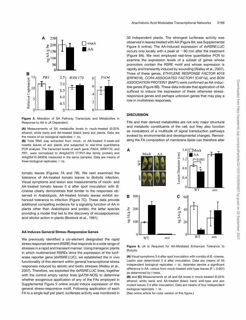
AA-Mediated Enhanced Tolerance to Botrytis Is
JA Dependent
We next examined whether exogenous application of AA alters
tolerance of wild-type and aosmutant plants toBotrytis infection.
These experiments were performed using different application
strategies to rule out the possibility that direct contact with AA
affected germination of conidia. In one approach, AA and Bo-
trytis conidia were simultaneously spotted on single leaves per
plant on the same and/or on two different spots. In the other
approach, Botrytis conidia were spotted at a distance from the
AA application site ;30 min after AA treatment, when the AA
containing droplet was no longer visible. Visual symptoms to-
gether with lesion size measurements of mock- and AA-treated
wild-type and aos mutant plants 3 d after inoculation with B.
cinerea determined that, irrespective of the inoculation strate-
gies, AA-treated wild-type leaves exhibit enhanced tolerance
to infection, whereas mock-treated leaves do not (Figure 6A).
Furthermore, in contrast with the wild-type plants, AA treatment
did not alter the susceptibility of aos mutant leaves to infection,
indicating that AA-mediated enhanced tolerance toBotrytis is JA
dependent (Figure 6A). We next analyzed the JA and SA levels in
these infected leaves and detected a twofold increase in JA
levels in AA-treated wild-type leaves (Figure 6B). As expected,
the JA levels in aos mutant leaves were below the detection
levels under all the experimental conditions (Figure 6B). In con-
trast with heightened JA levels, the SA levels were reduced in
AA-treated wild-type leaves infected with Botrytis (Figure 6C).
These data are consistent with those obtained from AA-treated,
uninfected leaves (Figures 4A). However, AA treatment did not
alter the SA levels in aos leaves, suggesting that the AA-medi-
ated signaling network underlying alteration of SA levels is JA
dependent.
Figure 2. Levels of JA Pathway Transcripts and Metabolites Are En-
hanced in EP Plants.
(A) Measurements of JA metabolite levels in the wild type (white bars)
and EP1 (black bars) before (NW) and 90 min after wounding (W) show
enhanced basal- and wound-induced levels of JA in EP plants. Data are
means of six independent experiments 6 SD.
(B) Simplified representation of JA-pathway genes.
(C) Total RNA was extracted from 4-week-old rosette leaves before (NW)
and 90 min after wounding (W) and subjected to real-time quantitative
PCR analysis. The transcript levels of each gene (DGL, LOX1 through 6,
AOS, and VSP2) were normalized to At4g34270 (T1P41-like family
protein) and At4g26410 (M3E9) measured in the same samples. Data
are means of three biological replicates 6 SE.
Arachidonic Acid Modulates Transcriptional Networks 3197
Dow
nloaded from https://academ
ic.oup.com/plcell/article/22/10/3193/6094857 by guest on 20 Septem
ber 2021
Page 6
AA Signaling Function(s) Is Not Limited to Arabidopsis
To determine whether exogenous application of the FAs em-
ployed in Supplemental Figure 5 online would modulate JA and
SA metabolite levels in plants other than Arabidopsis, tomato
(Solanum lycopersicum) leaves were treated with LA, ALA, EDA,
ETrA, and AA. Subsequent measurements of these metabolites
clearly showed that, similar to Arabidopsis, only AA treatment
leads to enhanced levels of JA and reduced levels of SA in
Figure 3. Expression of SA Pathway Genes and SA Metabolite Levels
Are Reduced in EP Plants.
(A)Measurements of SA metabolite levels in unwounded wild type (white
bars) and EP1 (black bars) show reduced SA levels in EP1 plants. Data
are means of six independent experiments 6 SD.
(B) Simplified representation of SA pathway genes.
(C) Total RNA was extracted from 4-week-old rosette leaves and
subjected to real-time quantitative PCR analysis. The transcript levels
of each gene, EDS5, PAD4, ICS1, NPR1, WRKY70, and PR1, were
normalized to At4g34270 (T1P41-like family protein) and At4g26410
(M3E9) measured in the same samples. Data are means of three
biological replicates 6 SE.
Figure 4. AA Coordinates Events Underlying Alteration in Expression of
JA and SA Pathway Genes and Metabolites.
Measurements were performed on leaves exogenously treated with 10
mM AA (black bars) or mock treated with 0.02% ethanol (white bars).
(A) and (C) Measurements of JA and SA metabolite levels in AA- and
mock-treated plants.
(B) and (D) Total RNA was extracted from AA- or mock-treated 4-week-
old rosette leaves and subjected to real-time quantitative PCR analysis.
The transcript levels of each gene, AOS, VSP2, PAD4, ICS1, WRKY70,
and PR1, were normalized to At4g34270 (T1P41-like family protein) and
At4g26410 (M3E9) measured in the same samples. Data are means of
three biological replicates 6 SE.
3198 The Plant Cell
Dow
nloaded from https://academ
ic.oup.com/plcell/article/22/10/3193/6094857 by guest on 20 Septem
ber 2021
Page 7
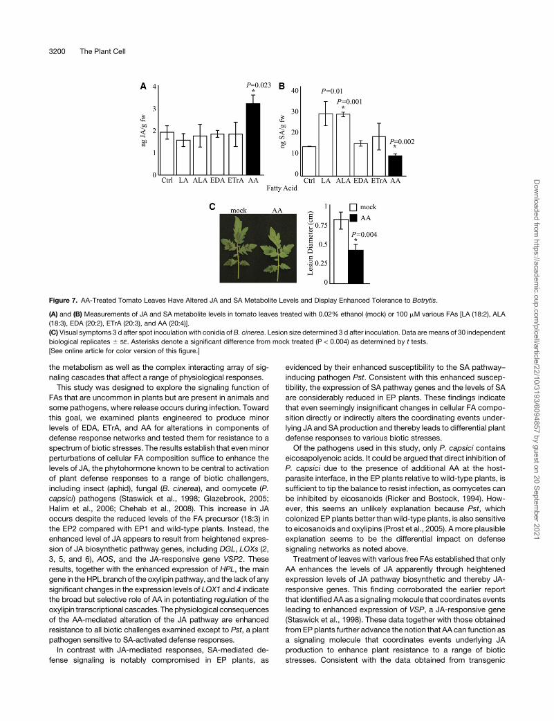
tomato leaves (Figures 7A and 7B). We next examined the
tolerance of AA-treated tomato leaves to Botrytis infection.
Visual symptoms and lesion size measurements of mock- and
AA-treated tomato leaves 3 d after spot inoculation with B.
cinerea clearly demonstrate that similar to the responses ob-
served in Arabidopsis, AA-treated tomato leaves exhibit en-
hanced tolerance to infection (Figure 7C). These data provide
additional compelling evidence for a signaling function of AA in
plants other than Arabidopsis and potato, the latter species
providing a model that led to the discovery of eicosapolyenoic
acid elicitor action in plants (Bostock et al., 1981).
AA Induces General Stress-Responsive Genes
We previously identified a cis-element designated the rapid
stress response element (RSRE) that responds to awide range of
stresses in a rapid and transient manner. Using transgenic plants
in which multimerized RSREs drive the expression of the lucif-
erase reporter gene (4xRSRE:LUC), we established the in vivo
functionality of this element within general transcriptional stress
responses induced by abiotic and biotic stresses (Walley et al.,
2007). Therefore, we exploited the 4xRSRE:LUC lines, together
with the control empty vector lines (pATM-NOS) to determine
whether exogenous application of any of the FAs employed in
Supplemental Figure 5 online would induce expression of this
general stress-responsive motif. Following application of each
FA to a single leaf per plant, luciferase activity was monitored in
30 independent plants. The strongest luciferase activity was
observed in leaves treated with AA (Figure 8A; see Supplemental
Figure 6 online). The AA-induced expression of 4xRSRE:LUC
occurs only locally with a peak at ;90 min after the treatment
(Figure 8A). We next employed real-time quantitative PCR to
examine the expression levels of a subset of genes whose
promoters contain the RSRE motif and whose expression is
rapidly and transiently induced by wounding (Walley et al., 2007).
Three of these genes, ETHYLENE RESPONSE FACTOR #018
(ERF#018), CCR4-ASSOCIATED FACTOR1 (CAF1a), and BON
ASSOCIATION PROTEIN1 (BAP1) were confirmed as AA-induc-
ible genes (Figure 8B). These data indicate that application of AA
sufficed to induce the expression of these otherwise stress-
responsive genes and perhaps unknown genes that may play a
role in multistress responses.
DISCUSSION
FAs and their derived metabolites are not only major structural
and metabolic constituents of the cell, but they also function
as modulators of a multitude of signal transduction pathways
evoked by environmental and developmental changes. Remod-
eling the FA composition of membrane lipids can therefore alter
Figure 5. Alteration of SA Pathway Transcripts and Metabolites in
Response to AA is JA Dependent.
(A) Measurements of SA metabolite levels in mock-treated (0.02%
ethanol; white bars) and AA-treated (black bars) aos plants. Data are
the means of six biological replicates 6 SD.
(B) Total RNA was extracted from mock- or AA-treated 4-week-old
rosette leaves of aos plants and subjected to real-time quantitative
PCR analysis. The transcript levels of each gene, PAD4, WRKY70, and
PR1, were normalized to At4g34270 (T1P41-like family protein) and
At4g26410 (M3E9) measured in the same samples. Data are means of
three biological replicates 6 SE.
Figure 6. JA Is Required for AA-Mediated Enhanced Tolerance to
Botrytis.
(A) Visual symptoms 3 d after spot inoculation with conidia of B. cinerea.
Lesion size determined 3 d after inoculation. Data are means of 45
independent biological replicates 6 SE. Asterisks denote a significant
difference in AA- versus from mock-treated wild-type leaves (P < 0.001)
as determined by t tests.
(B) and (C) Measurements of JA and SA levels in mock-treated (0.02%
ethanol; white bars) and AA-treated (black bars) wild-type and aos
mutant leaves 3 d after inoculation. Data are means of four independent
biological replicates 6 SE.
[See online article for color version of this figure.]
Arachidonic Acid Modulates Transcriptional Networks 3199
Dow
nloaded from https://academ
ic.oup.com/plcell/article/22/10/3193/6094857 by guest on 20 Septem
ber 2021
Page 8
the metabolism as well as the complex interacting array of sig-
naling cascades that affect a range of physiological responses.
This study was designed to explore the signaling function of
FAs that are uncommon in plants but are present in animals and
some pathogens, where release occurs during infection. Toward
this goal, we examined plants engineered to produce minor
levels of EDA, ETrA, and AA for alterations in components of
defense response networks and tested them for resistance to a
spectrum of biotic stresses. The results establish that evenminor
perturbations of cellular FA composition suffice to enhance the
levels of JA, the phytohormone known to be central to activation
of plant defense responses to a range of biotic challengers,
including insect (aphid), fungal (B. cinerea), and oomycete (P.
capsici) pathogens (Staswick et al., 1998; Glazebrook, 2005;
Halim et al., 2006; Chehab et al., 2008). This increase in JA
occurs despite the reduced levels of the FA precursor (18:3) in
the EP2 compared with EP1 and wild-type plants. Instead, the
enhanced level of JA appears to result from heightened expres-
sion of JA biosynthetic pathway genes, including DGL, LOXs (2,
3, 5, and 6), AOS, and the JA-responsive gene VSP2. These
results, together with the enhanced expression of HPL, the main
gene in theHPL branchof the oxylipin pathway, and the lack of any
significant changes in the expression levels of LOX1 and 4 indicate
the broad but selective role of AA in potentiating regulation of the
oxylipin transcriptional cascades. Thephysiological consequences
of the AA-mediated alteration of the JA pathway are enhanced
resistance to all biotic challenges examined except to Pst, a plant
pathogen sensitive to SA-activated defense responses.
In contrast with JA-mediated responses, SA-mediated de-
fense signaling is notably compromised in EP plants, as
evidenced by their enhanced susceptibility to the SA pathway–
inducing pathogen Pst. Consistent with this enhanced suscep-
tibility, the expression of SA pathway genes and the levels of SA
are considerably reduced in EP plants. These findings indicate
that even seemingly insignificant changes in cellular FA compo-
sition directly or indirectly alters the coordinating events under-
lying JA and SA production and thereby leads to differential plant
defense responses to various biotic stresses.
Of the pathogens used in this study, only P. capsici contains
eicosapolyenoic acids. It could be argued that direct inhibition of
P. capsici due to the presence of additional AA at the host-
parasite interface, in the EP plants relative to wild-type plants, is
sufficient to tip the balance to resist infection, as oomycetes can
be inhibited by eicosanoids (Ricker and Bostock, 1994). How-
ever, this seems an unlikely explanation because Pst, which
colonized EP plants better than wild-type plants, is also sensitive
to eicosanoids and oxylipins (Prost et al., 2005). Amore plausible
explanation seems to be the differential impact on defense
signaling networks as noted above.
Treatment of leaves with various free FAs established that only
AA enhances the levels of JA apparently through heightened
expression levels of JA pathway biosynthetic and thereby JA-
responsive genes. This finding corroborated the earlier report
that identified AA as a signalingmolecule that coordinates events
leading to enhanced expression of VSP, a JA-responsive gene
(Staswick et al., 1998). These data together with those obtained
fromEPplants further advance the notion that AA can function as
a signaling molecule that coordinates events underlying JA
production to enhance plant resistance to a range of biotic
stresses. Consistent with the data obtained from transgenic
Figure 7. AA-Treated Tomato Leaves Have Altered JA and SA Metabolite Levels and Display Enhanced Tolerance to Botrytis.
(A) and (B) Measurements of JA and SA metabolite levels in tomato leaves treated with 0.02% ethanol (mock) or 100 mM various FAs [LA (18:2), ALA
(18:3), EDA (20:2), ETrA (20:3), and AA (20:4)].
(C) Visual symptoms 3 d after spot inoculation with conidia of B. cinerea. Lesion size determined 3 d after inoculation. Data are means of 30 independent
biological replicates 6 SE. Asterisks denote a significant difference from mock treated (P < 0.004) as determined by t tests.
[See online article for color version of this figure.]
3200 The Plant Cell
Dow
nloaded from https://academ
ic.oup.com/plcell/article/22/10/3193/6094857 by guest on 20 Septem
ber 2021
Page 9
lines, exogenous application of AA to both uninfected as well as
Botrytis-infected wild-type plants reduced the expression of
SA pathway genes and the levels of SA. The employment of a
JA-deficient mutant line enabled us to uncouple the direct versus
the indirect AA-mediated signaling function in reduction of SA
and SA pathway genes. Collectively, these experiments un-
equivocally demonstrate that the reduced expression level of SA
pathway genes and lowered SA metabolite levels are not medi-
ated directly by AA, but indirectly through antagonist effects of
the JApathway, awell-establishedmechanism that allows plants
to fine-tune the induction of their defense responses to different
pathogens (Kunkel andBrooks, 2002; Bostock, 2005). Moreover,
enhanced tolerance of AA-treated tomato leaves to Botrytis
infection provides compelling evidence for signaling function of
AA in plants other than Arabidopsis and potato.
To determine the regulatory role of FA in altering the general
stress transcriptional network, we treated 4xRSRE:LUC trans-
genic lines with a range of FAs. These experiments corroborated
the earlier finding that AA is the signaling molecule that coordi-
nates events underlying JA production and demonstrated that
AA also potentiates the local and rapid activation of the RSRE.
Furthermore, real-time quantitative PCR analysis of a selected
group of genes that contain RSREs in their promoters showed
that AA treatment also leads to enhanced expression levels of
ERF#018,CAF1a, andBAP1. These findings clearly illustrate that
AA not only elicits expression of the synthetic 4xRSRE:LUC but
also induces expression of genes with the native RSRE motif in
their promoters. These data collectively advance the notion that
as in animal systems, AA in plants can function as a signaling
molecule that triggers both oxylipin-mediated defense re-
sponses and elicits general stress signaling networks.
This study indicates that although most plants do not produce
AA, they nevertheless have evolved a capacity to perceive and
respond to this ancient signaling molecule as part of their global
defense-responsive network against biotic invaders. This signal
acts reciprocally on the JA and SA pathways, concomitantly
enhancing JA levels through enhanced expression of JA biosyn-
thetic genes and suppressing SA levels indirectly through the
well-known antagonism between these two pathways. In addi-
tion, more generally, this signal induces expression of genes
bearing theRSRE regulatory element in their promoters. Our data
thus expand the repertoire of signaling molecules known to
trigger plant defenses and provide evidence that AA acts via a
mechanism that regulates the general stress transcriptional
network in addition to the JA-biosynthetic pathway.
METHODS
Generation of Eicosapolyenoic Acid Producing Arabidopsis Lines
The generation of the eicosapolyenoic acid–producing line referred to
here as EP1, by sequential transformation with constitutively expressed
C18-D9-elongase, C20-D8-desaturase, and C20-D5-desaturase genes,
Figure 8. AA Elicits Expression of 4xRSRE:LUC and Genes with the
RSRE Motif.
(A) Image of individual 4xRSRE:LUC transgenic plants before and 90 min
after treatment with either 0.02% ethanol (mock) or different FAs (LA,
ALA, EDA, EtrA, and AA). Images of empty vector lines, pATM-NOS (Ctrl)
before and after AA treatment, are representative of the data obtained
from mock experiments as well as exogenous application of other FAs.
(B) Total RNA was extracted from AA- (black bars) or mock-treated
(white bars) 4-week-old rosette leaves and subjected to real-time quan-
titative PCR analysis. The transcript levels of CAF1a, ERF#018, and
BAP1 genes, which contain RSRE motifs in their promoter sequences,
were normalized to At4g34270 and At4g26410 measured in the same
samples. Data are means of three biological replicates 6 SE.
Arachidonic Acid Modulates Transcriptional Networks 3201
Dow
nloaded from https://academ
ic.oup.com/plcell/article/22/10/3193/6094857 by guest on 20 Septem
ber 2021
Page 10
was described previously (Qi et al., 2004). Line EP2 was generated
by a single step of transformation, using a plasmid made by adding the
D8- and D5-desaturase genes to pCB302.3D9, a binary plasmid with the
C18-D9-elongase coding region in the cauliflower mosaic virus 35S
promoter-nos terminator expression cassette of pCB302.3 (Fraser
et al., 2004) as follows. pCB302D9 was first converted to destination
vector pCB302D9G by insertion of a GATEWAY cassette (Invitrogen) into
a uniqueHindIII site. Plasmid pENTR3FECwasmade by cloning the PCR-
amplified polylinker of pNEB193 (New England Biolabs; primers M13Fg,
59-CACCGTAAAACGACGGCCAG-39, and M13R, 59-CAGGAAACAGC-
TATGAC-39) in pENTR/SD/D-TOPO (Invitrogen), then ligating a cauli-
flower mosaic virus 35S promoter-nos terminator expression cassette
between the HindIII and EcoRI sites. The D8-desaturase coding region
was inserted into the expression cassette as a BamHI-KpnI fragment
isolated from pBlueBacD8 (Qi et al., 2004) to create pENTRD8. The D5-
desaturase gene was PCR amplified from the pCAMBIA construct made
by Qi et al. (2004) with primers (P35Sbeg, 59-CACCAAGCTTGCATGC-
CTGCA-39; TnosEndH, 59-AAGCTTCCCGATCTAGTAACAT-39) that al-
lowed the product to be cloned in pENTR/D-TOPO (Invitrogen), and then
excised as a HindIII fragment for insertion into pENTRD8. The D8- and
D5-desaturase genes from the resultant pENTRD8D5 were transferred to
pCB302D9G by recombination in vitro using LR Clonase II (Invitrogen).
The resultant plasmid, pCB302D9D8D5, was transferred to Agrobacte-
rium tumefaciens strain GV3101 and used to transform Arabidopsis
thaliana Columbia to BASTA resistance as described previously (Fraser
et al., 2004; Qi et al., 2004).
Plant Growth andWounding Treatments
Arabidopsis plants were grown in a 16-h-light/8-h-dark cycle at 228C for
most experiments, except plants used for bioassays. The latter were
grown in a 12-h-light/12-h-dark photoperiod. Mechanical wounding of
leaves was performed with a hemostat as previously described (Walley
et al., 2007). Tissues harvested before and 90 min after wounding were
immediately frozen in liquid nitrogen and stored at2808C until use. Most
experiments were performed on 4-week-old plants, unless stated other-
wise.
To generate the tomato (Solanum lycopersicum) seedlings, tomato
seeds were first surfaced sterilized with the following treatment se-
quence: 50% HCl (10 min) and rinsed with sterile deionized water, 10%
trisodium phosphate (15 min), and rinsed three times in sterile deionized
water and 70% ethanol (10 min) for 30 min and rinsed with sterile
deionized water and 50% bleach (20 min) followed by sterile deionized
water rinse (three times). Surface-sterilized seeds were started in germi-
nation paper in beakers containing sterile deionized water. Seedlings
were then transferred to flats of potting soil and grown for an additional
2 weeks in a climate-controlled greenhouse with supplemental lighting
until at least two true leaves had developed on each plant.
FA Analysis
Fatty acid methyl esters (FAMEs) were prepared from leaves using the
previously describedmethod (Browse et al., 1986). FAMEswere analyzed
by gas chromatography–mass spectrometry using a Hewlett Packard
6890 series gas chromatograph coupled to an Agilent Technologies 5973
Network mass selective detector. Heptadecanoic acid (17:0) (Cayman
Chemical) was added to the leaf extracts during preparation of FAMEs
and provided an internal standard.
Plant Hormone Extraction and Quantification
Extraction of JAs (methyl jasmonate and JA) and SA was performed as
previously described (Engelberth et al., 2004; Chehab et al., 2008).
Dihydro-JA and deuterated SA (C/D/N Isotopes) were used as internal
standards. The methyl ester derivatives were analyzed by gas chroma-
tography–mass spectrometry operated in electronic ionization mode.
Mass spectral analysis was done in selective ion monitoring mode.
Fragment ions monitored were as follows: JA-ME 224, 151, 83; dihydro-
JA-ME 96, 83; SA-ME 152; SA-D4-ME 156. Quantification calibration
curves were generated by derivatization and analysis of known quantities
of pure JA and SA (Sigma-Aldrich).
Expression Analysis
Total RNA from rosette leaves was isolated by TRIzol extraction (Life
Technologies) and further purified using the Qiagen RNeasy kit with on
column DNase treatment (Qiagen) to eliminate DNA contamination. RNA
was reverse transcribed using Superscript III (Invitrogen). Real-time
quantitative PCR was conducted in 50-mL reactions containing cDNA
synthesized from 10 ng of total RNA, 13 iQ SYBR Green Supermix (Bio-
Rad Laboratories), and 200 nM for each primer. Amplification and data
analysis were performed as previously described (Walley et al., 2008).
At4g34270 and At4g26410 were used as reference genes for the internal
controls as previously described for transcript normalization (Walley et al.,
2008). Primers are listed in Supplemental Table 1 online.
Fungal Pathogenicity Tests
Botrytis cinerea ‘Grape’ and ‘B05.10’ isolates were used in the assay.
Preparation of inoculum and infection of Arabidopsis plants were per-
formed as previously described (Rowe and Kliebenstein, 2007). Mature
rosette leaves excised from 4-week-old Arabidopsis plants were placed
in Petri dishes containing 1% agar. Leaves were inoculated with 5-mL
droplets of 53 104 spores/mL in half-strength filtered organic grape juice
and incubated at room temperature under ambient light conditions.
Lesion diameter was measured from digital images of at least 30 infected
leaves using Image J with scale objects included in images as previously
described (Chehab et al., 2008; Walley et al., 2008).
Phytophthora capsici Culture and Inoculation
A pepper isolate of Phytophthora capsici (from Yolo County, CA; Bostock
lab collection) wasmaintained on V-8 agar plates.We found this isolate to
be capable of readily infecting Arabidopsis roots. Two-week-old plants
maintained hydroponically in aerated half-strength Hoagland solution
were inoculated by transferring the plants to a 24-well Cellstar multiwall
plate (Greiner Bio-One) and placing the roots in 1 mL of half-strength
Hoagland solution containing 105 zoospores of P. capsici per milliliter.
Disease development was monitored at 48 h after inoculation and
assessed as sporangia counts on roots in 10 microscope fields at 3200
magnification and by plant colonization asmeasured by quantitative PCR
of P. capsici DNA.
P. capsici DNA Quantitation in Infected Host Tissue
Arabidopsis roots were flash frozen in liquid nitrogen and were used for
DNA extraction using DNeasy Plant Mini kits (Qiagen). Pathogen DNA
was quantifiedwith real-timePCRusing established primers forP. capsici
(Cap-FW 59-TTTAGTTGGGGGTCTTGTACC-39 and Cap-RV1 59-CCT-
CCACAACCAGCAACA-39 [Silvar et al., 2005]). Standard curves were
verified for this system for pure P. capsici DNA (R2 = 0.99) and P. capsici
DNA amended with 100 ng Arabidopsis DNA (R2 = 0.99).
Aphid Dual-Choice Assay
The choice assays were performed as previously described (Chehab
et al., 2008). Briefly, green peach aphid (Myzus persicae) colonies were
maintained on cabbage seedlings (Brassica oleracea var capitata) at
3202 The Plant Cell
Dow
nloaded from https://academ
ic.oup.com/plcell/article/22/10/3193/6094857 by guest on 20 Septem
ber 2021
Page 11
laboratory conditions (23 6 38C, 50% 6 20% relative humidity, and 16 h
light). Choice bioassays were performed to identify the plants on which
aphids prefer to deposit their nymphs. Female aphids were transferred
using a fine hair brush and released into the center of a soil-containing
pot, in which 11 plants were kept at laboratory conditions and examined
every 24 h for two successive days. The location of the first deposited
nymphs was recorded. The bioassays were performed with ;49 repli-
cates and four trials.
Bacterial Pathogenicity Test
A bioluminescent strain of Pseudomonas syringae pv tomato DC3000
(Pst) was used to analyze susceptibility of wild-type and EP plants to a
bacterial pathogen. Arabidopsis leaves were hand infiltrated with bacte-
rial inocula of OD600 = 0.002 as previously described (Fan et al., 2008).
Leaf discs (0.7 cm in diameter) were excised from inoculated leaves 2
days after inoculation. Bioluminescence was measured with a Micro-
Lumat LB96P luminometer (EG and G Berthold) and recorded as photon
counts per second. Thirty plants were analyzed in four independent
experiments.
Application of Exogenous FAs
FAs were purchased from Cayman Chemical Co. FA stock solutions (300
mM) were prepared in 80% ethanol. Working solutions of indicated
concentrations (see Supplemental Figures 3 and 4 online) were prepared
in DI water as sonicated emulsion with ethanol content adjusted to
0.02%. For each measurement, 5 mL of emulsion with the required
concentration was applied on leaves of growing plants.
Luciferase Imaging
Luciferase imaging of 4xRSRE:LUC and control empty vector lines
(pATM-NOS) were performed as previously described (Walley et al.,
2007). Briefly, 10- to 14-d-old plants grown on soil were sprayed with 2.5
mM luciferin (Promega) in 0.001% Triton X-100 ;16 to 20 h prior to
treatment. Subsequently, 5mL of FAwas applied to a single leaf per plant.
For each treatment, at least 30 plants were imaged using an Andor
DU434-BV CCD camera (Andor Technology). Images were acquired
every 5 min over a 4-h period. Luciferase activity was quantified for a
defined area as mean counts pixel21 exposure time21 using Andor Solis
image analysis software (Andor Technology).
Statistical Analyses
To determine statistical significance of treatment effects or the effects of
genotype when comparing wild-type versus EP plants, t tests were
performed in most cases using Sigma Stat v3.5. To determine treatment
effects on P. capsici colonization as measured by quantitative PCR and
by sporangia counts on colonized roots, theWilcoxon rank sums test was
used to compare means because the data did not satisfy the analysis of
variance criterion for normality. Statistical analyses were performed using
JMP software version 8.0 (The SAS Institute). To investigate the effect of
the trials and aphid types on probability of plant preference (wild type
versus EP), Pearson x2 tests were performed. One-tailed binomial tests
were performed to test the significance of the aphids’ choices for nymph
deposition (Zar, 1999).
Accession Numbers
Sequence data from this article can be found in the Arabidopsis Genome
Initiative or GenBank/EMBL databases under the following accession
numbers: PAD4 (At3g52430), ICS1 (At1g74710), NPR1 (At1g64280),
WRKY70 (At3g56400), PR1 (At2g14610), PLA1 (DGL) (At1g05800),
LOX1 (At1g55020), LOX2 (At3g45140), LOX3 (At1g17420), LOX4
(At1g72520), LOX5 (At3g22400), LOX6 (At1g67560), AOS (At5g42650),
VSP2 (At5g24770), HPL (At4g15440), CAF1a (At3g44260), ERF#018
(At1g74930), BAP1 (At3g61190), and EDS5 (At4g39030).
Supplemental Data
The following materials are available in the online version of this article.
Supplemental Figure 1. EP Plants Exhibit Altered Tolerance to Biotic
Challengers.
Supplemental Figure 2. Levels of JA Pathway Transcripts and
Metabolites Are Enhanced in EP Plants.
Supplemental Figure 3. Enhanced Expression Levels of HPL in EP
Plants.
Supplemental Figure 4. Expression of SA Pathway Genes and SA
Metabolite Levels Are Reduced in EP2 Plants.
Supplemental Figure 5. JA Production Is Induced by Exogenous
Application of AA in a Concentration-Dependent Manner.
Supplemental Figure 6. Relative Luciferase Activity of 4xRSRE:LUC
Plants Treated with 10 mM of Various FAs.
Supplemental Table 1. List of Primers Used for Quantitative RT-PCR
Analyses.
ACKNOWLEDGMENTS
We thank Peter Quail for his critical review of this manuscript. This
work was supported by National Science Foundation Grant 0543904
to K.D.
Received January 5, 2010; revised September 16, 2010; accepted
September 22, 2010; published October 8, 2010.
REFERENCES
Black, P.N., Faergeman, N.J., and DiRusso, C.C. (2000). Long-chain
acyl-CoA-dependent regulation of gene expression in bacteria, yeast
and mammals. J. Nutr. 130: 305S–309S.
Blee, E. (2002). Impact of phyto-oxylipins in plant defense. Trends Plant
Sci. 7: 315–322.
Bostock, R.M. (2005). Signal crosstalk and induced resistance: Strad-
dling the line between cost and benefit. Annu. Rev. Phytopathol. 43:
545–580.
Bostock, R.M., Kuc, J.A., and Laine, R.A. (1981). Eicosapentaenoic
and arachidonic acids from Phytophthora infestans elicit fungitoxic
sesquiterpenes in the potato. Science 212: 67–69.
Bostock, R.M., Schaeffer, D.A., and Hammerschmidt, R. (1986).
Comparison of elicitor activities of arachidonic acid, fatty acids and
glucans from Phytophthora infestans in hypersensitivity expression in
potato tuber. Physiol. Mol. Plant Pathol. 29: 349–360.
Bostock, R.M., Yamamoto, H., Choi, D., Ricker, K.E., and Ward, B.L.
(1992). Rapid stimulation of 5-lipoxygenase activity in potato by the
fungal elicitor arachidonic acid. Plant Physiol. 100: 1448–1456.
Brodhagen, M., Tsitsigiannis, D.I., Hornung, E., Goebel, C., Feussner,
I., and Keller, N.P. (2008). Reciprocal oxylipin-mediated cross-talk in
the Aspergillus-seed pathosystem. Mol. Microbiol. 67: 378–391.
Browse, J., McCourt, P.J., and Somerville, C.R. (1986). Fatty acid
composition of leaf lipids determined after combined digestion and
fatty acid methyl ester formation from fresh tissue. Anal. Biochem.
152: 141–145.
Arachidonic Acid Modulates Transcriptional Networks 3203
Dow
nloaded from https://academ
ic.oup.com/plcell/article/22/10/3193/6094857 by guest on 20 Septem
ber 2021
Page 12
Cao, H., Bowling, S.A., Gordon, A.S., and Dong, X. (1994). Charac-
terization of an Arabidopsis mutant that is nonresponsive to inducers
of systemic acquired resistance. Plant Cell 6: 1583–1592.
Chandra-Shekara, A.C., Venugopal, S.C., Barman, S.R., Kachroo,
A., and Kachroo, P. (2007). Plastidial fatty acid levels regulate
resistance gene-dependent defense signaling in Arabidopsis. Proc.
Natl. Acad. Sci. USA 104: 7277–7282.
Chehab, E.W., Kaspi, R., Savchenko, T., Rowe, H., Negre-Zakharov,
F., Kliebenstein, D., and Dehesh, K. (2008). Distinct roles of jasmo-
nates and aldehydes in plant-defense responses. PloS One 3: e1904.
Choi, D., Bostock, R.M., Avdiushko, S., and Hildebrand, D. (1994).
Lipid-derived signals that discriminate wound- and pathogen-respon-
sive isoprenoid pathways in plants: Methyl jasmonate and the fungal
elicitor arachidonic acid induce different HMG-CoA reductase genes
and antimicrobial isoprenoids in Solanum tuberosum L. Proc. Natl.
Acad. Sci. USA 91: 2329–2333.
Choi, D., Ward, B.L., and Bostock, R.M. (1992). Differential induction
and suppression of potato 3-hydroxy-3-methylglutaryl coenzyme A
reductase genes in response to Phytophthora infestans and to its
elicitor arachidonic acid. Plant Cell 4: 1333–1344.
Duan, H., Huang, M.Y., Palacio, K., and Schuler, M.A. (2005). Vari-
ations in CYP74B2 (hydroperoxide lyase) gene expression differen-
tially affect hexenal signaling in the Columbia and Landsberg erecta
ecotypes of Arabidopsis. Plant Physiol. 139: 1529–1544.
Duplus, E., Glorian, M., and Forest, C. (2000). Fatty acid regulation of
gene transcription. J. Biol. Chem. 275: 30749–30752.
Engelberth, J., Alborn, H.T., Schmelz, E.A., and Tumlinson, J.H.
(2004). Airborne signals prime plants against insect herbivore attack.
Proc. Natl. Acad. Sci. USA 101: 1781–1785.
Fan, J., Crooks, C., and Lamb, C. (2008). High-throughput quantitative
luminescence assay of the growth in planta of Pseudomonas syringae
chromosomally tagged with Photorhabdus luminescens luxCDABE.
Plant J. 53: 393–399.
Farmer, E.E., Weber, H., and Vollenweider, S. (1998). Fatty acid
signaling in Arabidopsis. Planta 206: 167–174.
Feussner, I., and Wasternack, C. (2002). The lipoxygenase pathway.
Annu. Rev. Plant Biol. 53: 275–297.
Fraser, T.C., Qi, B., Elhussein, S., Chatrattanakunchai, S., Stobart,
A.K., and Lazarus, C.M. (2004). Expression of the Isochrysis C18-
delta9 polyunsaturated fatty acid specific elongase component alters
Arabidopsis glycerolipid profiles. Plant Physiol. 135: 859–866.
Garcia-Pineda, E., Castro-Mercado, E., and Lozoya-Gloria, E.
(2004). Gene expression and enzyme activity of pepper (Capsicum
annuum L.) ascorbate oxidase during elicitor and wounding stress.
Plant Sci. 166: 237–243.
Gardner, R.W. (1995). Biological roles and biochemistry of the lipoxy-
genase pathway. HortScience 30: 197–205.
Glazebrook, J. (2005). Contrasting mechanisms of defense against
biotrophic and necrotrophic pathogens. Annu. Rev. Phytopathol. 43:
205–227.
Halim, V.A., Vess, A., Scheel, D., and Rosahl, S. (2006). The role of
salicylic acid and jasmonic acid in pathogen defence. Plant Biol.
(Stuttg.) 8: 307–313.
Huang, Z.H., Gu, D., and Mazzone, T. (2004). Oleic acid modulates the
post-translational glycosylation of macrophage ApoE to increase its
secretion. J. Biol. Chem. 279: 29195–29201.
Hyun, Y., et al. (2008). Cooperation and functional diversification of two
closely related galactolipase genes for jasmonate biosynthesis. Dev.
Cell 14: 183–192.
Jirage, D., Tootle, T.L., Reuber, T.L., Frost, L.N., Feys, B.J., Parker,
J.E., Ausubel, F.M., and Glazebrook, J. (1999). Arabidopsis thaliana
PAD4 encodes a lipase-like gene that is important for salicylic acid
signaling. Proc. Natl. Acad. Sci. USA 96: 13583–13588.
Kachroo, A., Lapchyk, L., Fukushige, H., Hildebrand, D., Klessig, D.,
and Kachroo, P. (2003). Plastidial fatty acid signaling modulates
salicylic acid- and jasmonic acid-mediated defense pathways in the
Arabidopsis ssi2 mutant. Plant Cell 15: 2952–2965.
Kachroo, P., Kachroo, A., Lapchyk, L., Hildebrand, D., and Klessig,
D.F. (2003). Restoration of defective cross talk in ssi2 mutants: role of
salicylic acid, jasmonic acid, and fatty acids in SSI2-mediated sig-
naling. Mol. Plant Microbe Interact. 16: 1022–1029.
Kachroo, P., Shanklin, J., Shah, J., Whittle, E.J., and Klessig, D.F.
(2001). A fatty acid desaturase modulates the activation of defense
signaling pathways in plants. Proc. Natl. Acad. Sci. USA 98: 9448–
9453.
Kachroo, P., Venugopal, S.C., Navarre, D.A., Lapchyk, L., and
Kachroo, A. (2005). Role of salicylic acid and fatty acid desatura-
tion pathways in ssi2-mediated signaling. Plant Physiol. 139: 1717–
1735.
Knight, V.I., Wang, H., Lincoln, J.-E., Lulai, E.C., Gilchrist, D.G., and
Bostock, R.M. (2001). Hydroperoxides of fatty acids induce pro-
grammed cell death in tomato protoplasts. Physiol. Mol. Plant Pathol.
59: 277–286.
Kunkel, B.N., and Brooks, D.M. (2002). Cross talk between signaling
pathways in pathogen defense. Curr. Opin. Plant Biol. 5: 325–331.
Li, J., Brader, G., and Palva, E.T. (2004). The WRKY70 transcription
factor: A node of convergence for jasmonate-mediated and salicylate-
mediated signals in plant defense. Plant Cell 16: 319–331.
Lorenzo, O., and Solano, R. (2005). Molecular players regulating the
jasmonate signalling network. Curr. Opin. Plant Biol. 8: 532–540.
Magnan, A., and Vervloet, D. (1999). Leukotriene inhibitors-(2): Place of
leukotrienes in the pathophysiology of asthma and rhinitis. Rev. Fr.
Allergol. 39: 25–29.
McDowell, J.M., and Dangl, J.L. (2000). Signal transduction in the plant
immune response. Trends Biochem. Sci. 25: 79–82.
Nawrath, C., Heck, S., Parinthawong, N., and Metraux, J.-P. (2002).
EDS5, an essential component of salicylic acid–dependent signaling
for disease resistance in Arabidopsis, is a member of the MATE
transporter family. Plant Cell 14: 275–286.
Ozeretskovskaya, O.L., Varlamov, V.P., Vasyukova, N.I., Chalenko,
G.I., Gerasimova, N.G., and Panina, Y.S. (2004). Influence of sys-
temic signal molecules on the rate of spread of the immunizing ef-
fect of elicitors over potato tissues. Appl. Biochem. Microbiol. 40:
213–216.
Park, J.H., Halitschke, R., Kim, H.B., Baldwin, I.T., Feldmann, K.A.,
and Feyereisen, R. (2002). A knock-out mutation in allene oxide
synthase results in male sterility and defective wound signal trans-
duction in Arabidopsis due to a block in jasmonic acid biosynthesis.
Plant J. 31: 1–12.
Pegorier, J.P., Le May, C., and Girard, J. (2004). Control of gene
expression by fatty acids. J. Nutr. 134: 2444S–2449S.
Preisig, C.L., and Kuc, J.A. (1985). Arachidonic acid-related elicitors of
the hypersensitive response in potato and enhancement of their
activities by glucans from Phytophthora infestans (Mont) Debary.
Arch. Biochem. Biophys. 236: 379–389.
Preisig, C.L., and Kuc, J.A. (1988). Metabolism by potato tuber of
arachidonic acid, an elicitor of hypersensitive resistance. Physiol. Mol.
Plant Pathol. 32: 77–88.
Prost, I., et al. (2005). Evaluation of the antimicrobial activities of plant
oxylipins supports their involvement in defense against pathogens.
Plant Physiol. 139: 1902–1913.
Qi, B., Fraser, T., Mugford, S., Dobson, G., Sayanova, O., Butler, J.,
Napier, J.A., Stobart, A.K., and Lazarus, C.M. (2004). Production of
very long chain polyunsaturated omega-3 and omega-6 fatty acids in
plants. Nat. Biotechnol. 22: 739–745.
Ricker, K.E., and Bostock, R.M. (1992). Evidence for release of the
3204 The Plant Cell
Dow
nloaded from https://academ
ic.oup.com/plcell/article/22/10/3193/6094857 by guest on 20 Septem
ber 2021
Page 13
elicitor arachidonic acid and its metabolites from sporangia of
Phytophthora infestans during infection of potato. Physiol. Mol. Plant
Pathol. 41: 61–72.
Ricker, K.E., and Bostock, R.M. (1994). Eicosanoids in the Phytophthora
infestans-potato interaction: Lipoxygenase metabolism of arachidonic
acid and biological activities of selected lipoxygenase products.
Physiol. Mol. Plant Pathol. 44: 65–80.
Rowe, H.C., and Kliebenstein, D.J. (2007). Elevated genetic variation
within virulence-associated Botrytis cinerea polygalacturonase loci.
Mol. Plant Microbe Interact. 20: 1126–1137.
Rozhnova, N.A., Gerashchenkov, G.A., and Babosha, A.V. (2003).
The effect of arachidonic acid and viral infection on the phytohe-
magglutinin activity during the development of tobacco acquired
resistance. Russ. J. Plant Physiol. 50: 661–665.
Schultz, J. (2002). Shard signals and the potential for phylogentic espi-
onage between plants and animals. Integr. Comp. Biol. 42: 454–462.
Shah, J. (2003). The salicylic acid loop in plant defense. Curr. Opin.
Plant Biol. 6: 365–371.
Silvar, C., Diaz, J., and Merino, F. (2005). Real-time polymerase chain
reaction quantification of Phytophthora capsici in different pepper
genotypes. Phytopathology 95: 1423–1429.
Staswick, P.E., Yuen, G.Y., and Lehman, C.C. (1998). Jasmonate
signaling mutants of Arabidopsis are susceptible to the soil fungus
Pythium irregulare. Plant J. 15: 747–754.
Straus, D.S., and Glass, C.K. (2001). Cyclopentenone prostaglandins:
new insights on biological activities and cellular targets. Med. Res.
Rev. 21: 185–210.
Tjamos, E.C., and Kuc, J.A. (1982). Inhibition of steroid glycoalkaloid
accumulation by arachidonic and eicosapentaenoic acids in potato.
Science 217: 543–544.
Upchurch, R.G. (2008). Fatty acid unsaturation, mobilization, and
regulation in the response of plants to stress. Biotechnol. Lett. 30:
967–977.
van Ryn, J., Trummlitz, G., and Pairet, M. (2000). COX-2 selectivity
and inflammatory processes. Curr. Med. Chem. 7: 1145–1161.
Venugopal, S.C., Jeong, R.D., Mandal, M.K., Zhu, S., Chandra-
Shekara, A.C., Xia, Y., Hersh, M., Stromberg, A.-J., Navarre, D.,
Kachroo, A., and Kachroo, P. (2009). Enhanced disease suscepti-
bility 1 and salicylic acid act redundantly to regulate resistance gene-
mediated signaling. PLoS Genet. 5: e1000545.
Walley, J.W., Coughlan, S., Hudson, M.E., Covington, M.F., Kaspi,
R., Banu, G., Harmer, S.L., and Dehesh, K. (2007). Mechanical
stress induces biotic and abiotic stress responses via a novel cis-
element. PLoS Genet. 3: 1800–1812.
Walley, J.W., Rowe, H.C., Xiao, Y., Chehab, E.W., Kliebenstein, D.J.,
Wagner, D., and Dehesh, K. (2008). The chromatin remodeler
SPLAYED regulates specific stress signaling pathways. PLoS Pathog.
4: e1000237.
Wildermuth, M.C., Dewdney, J., Wu, G., and Ausubel, F.M. (2001).
Isochorismate synthase is required to synthesize salicylic acid for
plant defense. Nature 414: 562–565.
Zar, J.H. (1999). Biostatistical Analysis. (Upper Saddle River, NJ:
Prentice Hall).
Arachidonic Acid Modulates Transcriptional Networks 3205
Dow
nloaded from https://academ
ic.oup.com/plcell/article/22/10/3193/6094857 by guest on 20 Septem
ber 2021