† This article belongs to the Special Issue devoted to the 85 th anniversary of Croatica Chemica Acta. * Author to whom correspondence should be addressed. (E-mail: [email protected]) CROATICA CHEMICA ACTA CCACAA, ISSN 0011-1643, e-ISSN 1334-417X Croat. Chem. Acta 85 (4) (2012) 441–449. http://dx.doi.org/10.5562/cca2146 Original Scientific Article Arginyl-tRNA Synthetase Facilitates Complex Formation Between Seryl-tRNA Synthetase and its Cognate Transfer RNA † Vlatka Godinić-Mikulčić, * Jelena Jarić, and Ivana Weygand-Đurašević University of Zagreb, Faculty of Science, Department of Chemistry, Horvatovac 102a, HR-10000 Zagreb, Croatia RECEIVED JULY 17, 2012; REVISED SEPTEMBER 30, 2012; ACCEPTED OCTOBER 3, 2012 Abstract. Several studies have revealed the involvement of multi aminoacyl-tRNA synthetase complexes (MSC) in archaeal and eukaryotic translation. Here we analyzed interactions of atypical Methanothermobacter thermautotrophicus seryl-tRNA synthetase (MtSerRS), transfer RNA (tRNA Ser ) and arginyl-tRNA synthetase (ArgRS). Surface plasmon resonance (SPR) was used to determine dissocia- tion constants for the MtSerRS:tRNA Ser complex and the results were consistent with cooperative binding of tRNA Ser . This finding was supported by the ability of MtSerRS to bind two tRNAs in gel mobility shift assay. Notably, the MtSerRS:tRNA Ser complex formation was stimulated by MtArgRS, previously deter- mined interacting partner of MtSerRS. MtArgRS decreases K d for MtSerRS:tRNA Ser two-fold, but does not affect cooperative properties or stoichiometry of the complex. Further investigation of complex for- mation between MtSerRS and tRNA Ser showed that this molecular interaction is salt-dependent. The most pronounced improvements in binding were determined at high ionic strength, using Tris as a buffering agent, while the addition of Mg 2+ ions led to the same SPR response. (doi: 10.5562/cca2146) Keywords: aminoacyl-tRNA synthetase, seryl-tRNA synthetase, arginyl-tRNA synthetase, tRNA, surface plasmon resonance INTRODUCTION The successful completion of gene expression is de- pendent on efficient and accurate translation of mRNAs to synthesize proteins, catalyzed by the ribosome. 1 The fidelity of protein synthesis relies on precise mRNA:tRNA decoding interactions, and the highly specific attachment of amino acids (aa) to tRNAs during aminoacyl-tRNA synthesis. 2 The later process is cata- lyzed by the aminoacyl-tRNA synthetases (aaRSs). 3 Recent evidences 4,5 revealed that these housekeeping enzymes tend to form macromolecular complexes in all three domains of life. 5 Such associations may influence the efficiency and the accuracy of aminoacyl-tRNA formation. In this context, we previously explored the ability of seryl-tRNA synthetases (SerRSs), which cata- lyze esterification of cognate tRNA Ser isoacceptors with serine, to interact with other aaRSs and with non- synthetase proteins. 6 Although, generally, the aaRSs that recognize the same amino acid are rather conserved in terms of sequences and 3D-structures in all domains of life, the two diverged types of SerRSs exist. 7–10 Bac- terial-type SerRSs function in a variety of archaeal, bacterial and eukaryotic organisms, while somewhat atypical, methanogenic-type SerRS was found only in methanogenic archaea, 10,11 the organisms which often inhabit the environments characterized by extreme liv- ing conditions (anaerobic, thermofilic, psychrophilic, halophilic). We have recently shown that methanogenic- type SerRSs display several idiosyncratic structural features in comparison with bacterial-type SerRSs. 11–13 Although all SerRSs are functional homodimers with a C-terminal active site domain typical for class II aaRSs and an N-terminal domain that is responsible for tRNA binding, 10,14 the representatives of the two SerRS types exhibit different modes of substrate recognition, based on structurally different tRNA binding domains 11–13 and the presence of catalytic zinc ion in the active site of methanogenic-type SerRS. Both types of enzymes bind one or two cognate tRNAs across two protein subu- nits, 12,15 and it seems that in both systems tRNA binding is enhanced by the interaction of SerRSs with other proteins. 6,16 We have previously shown that yeast SerRS (a bacterial-type enzyme) associates with peroxin Pex21p, 16 while SerRSs from methanogens form assemblies with arginyl-tRNA synthetase (ArgRS). 6 In this paper we present further evidence that interaction of MtSerRS and tRNA Ser is salt-dependent and that ArgRS from
Transcript
† This article belongs to the Special Issue devoted to the 85th anniversary of Croatica Chemica Acta. * Author to whom correspondence should be addressed. (E-mail: [email protected])
Arginyl-tRNA Synthetase Facilitates Complex Formation Between Seryl-tRNA Synthetase and its Cognate Transfer RNA†
Vlatka Godinić-Mikulčić,* Jelena Jarić, and Ivana Weygand-Đurašević
University of Zagreb, Faculty of Science, Department of Chemistry, Horvatovac 102a, HR-10000 Zagreb, Croatia
RECEIVED JULY 17, 2012; REVISED SEPTEMBER 30, 2012; ACCEPTED OCTOBER 3, 2012
Abstract. Several studies have revealed the involvement of multi aminoacyl-tRNA synthetase complexes (MSC) in archaeal and eukaryotic translation. Here we analyzed interactions of atypical Methanothermobacter thermautotrophicus seryl-tRNA synthetase (MtSerRS), transfer RNA (tRNASer) and arginyl-tRNA synthetase (ArgRS). Surface plasmon resonance (SPR) was used to determine dissocia-tion constants for the MtSerRS:tRNASer complex and the results were consistent with cooperative binding of tRNASer. This finding was supported by the ability of MtSerRS to bind two tRNAs in gel mobility shift assay. Notably, the MtSerRS:tRNASer complex formation was stimulated by MtArgRS, previously deter-mined interacting partner of MtSerRS. MtArgRS decreases Kd for MtSerRS:tRNASer two-fold, but does not affect cooperative properties or stoichiometry of the complex. Further investigation of complex for-mation between MtSerRS and tRNASer showed that this molecular interaction is salt-dependent. The most pronounced improvements in binding were determined at high ionic strength, using Tris as a buffering agent, while the addition of Mg2+ ions led to the same SPR response. (doi: 10.5562/cca2146)
The successful completion of gene expression is de-pendent on efficient and accurate translation of mRNAs to synthesize proteins, catalyzed by the ribosome.1 The fidelity of protein synthesis relies on precise mRNA:tRNA decoding interactions, and the highly specific attachment of amino acids (aa) to tRNAs during aminoacyl-tRNA synthesis.2 The later process is cata-lyzed by the aminoacyl-tRNA synthetases (aaRSs).3 Recent evidences4,5 revealed that these housekeeping enzymes tend to form macromolecular complexes in all three domains of life.5 Such associations may influence the efficiency and the accuracy of aminoacyl-tRNA formation. In this context, we previously explored the ability of seryl-tRNA synthetases (SerRSs), which cata-lyze esterification of cognate tRNASer isoacceptors with serine, to interact with other aaRSs and with non-synthetase proteins.6 Although, generally, the aaRSs that recognize the same amino acid are rather conserved in terms of sequences and 3D-structures in all domains of life, the two diverged types of SerRSs exist.7–10 Bac-terial-type SerRSs function in a variety of archaeal, bacterial and eukaryotic organisms, while somewhat
atypical, methanogenic-type SerRS was found only in methanogenic archaea,10,11 the organisms which often inhabit the environments characterized by extreme liv-ing conditions (anaerobic, thermofilic, psychrophilic, halophilic). We have recently shown that methanogenic-type SerRSs display several idiosyncratic structural features in comparison with bacterial-type SerRSs.11–13 Although all SerRSs are functional homodimers with a C-terminal active site domain typical for class II aaRSs and an N-terminal domain that is responsible for tRNA binding,10,14 the representatives of the two SerRS types exhibit different modes of substrate recognition, based on structurally different tRNA binding domains11–13 and the presence of catalytic zinc ion in the active site of methanogenic-type SerRS. Both types of enzymes bind one or two cognate tRNAs across two protein subu-nits,12,15 and it seems that in both systems tRNA binding is enhanced by the interaction of SerRSs with other proteins.6,16 We have previously shown that yeast SerRS (a bacterial-type enzyme) associates with peroxin Pex21p,16
while SerRSs from methanogens form assemblies with arginyl-tRNA synthetase (ArgRS).6 In this paper we present further evidence that interaction of MtSerRS and tRNASer is salt-dependent and that ArgRS from
442 V. Godinić-Mikulčić et al., MtArgRS Stimulates MtSerRS:tRNASer Complex Formation
Croat. Chem. Acta 85 (2012) 441.
Methanothermobacter thermautotrophicus (MtArgRS) facilitates binding of tRNASer. Thus, such aaRSs com-plexes may be especially important under extreme envi-ronmental conditions due to diverse habitats such as geothermal, marine hydrothermal springs, rivers and sea sediments, the digestive system of animals and the an-aerobic accumulation of waste. MtSerRS:ArgRS associ-ation presumably constitutes a part of thermo-6 and osmoadaptation mechanisms of thermophilic methanogenic archaea, by providing an optimal micro-environment for efficient seryl-tRNA synthesis.
EXPERIMENTAL
Preparation of Proteins and tRNA
Preparation of recombinant His6-MtSerRS or GST-MtArgRS was done by transforming E. coli BL21(DE3) (Stratagene) with pET28 or pGEX-6P-2 vectors contain-ing the relevant inserts and growing the resulting strains on LB media supplemented with ampicillin or kanamy-cin as described.6 Protein concentration was determined by active site titration and Methanosarcina barkeri tRNASer
GGA (MbtRNASerGGA) was produced in vivo as
previously described.12 Gel Mobility Shift Assay
To check for complex formation between tRNA and proteins, tRNASer (0.01 μmol dm–3) was mixed with varying concentrations of MtSerRS dimer (0.005–0.4 μmol dm–3), GST-MtArgRS (0.005–0.4 μmol dm–3), or BSA (New England Biolabs) (0.005–0.4 μmol dm–3) and incubated for 15 min at temperature of 37 °C in 20 mmol dm–3 TrisHCl (pH = 7.0), 50 mmol dm–3 NaCl and 6 mmol dm–3 MgCl2. The experiment was performed at a concentration of tRNASer which is much lower than the dissociation constants of the MtSerRS:tRNASer. Under these conditions, tRNA binds to a very small proportion of the MtSerRS in the reaction mixture and has an insignificant effect on a concentration of free MtSerRS. On the other hand, for stoichiometric titration experiment, tRNASer (1.0 μmol dm–3) was titrated with MtSerRS (dimer concentration 0.06–1.32 μmol dm–3) using 0.06–1.32 μmol dm–3 MtArgRS or BSA in the reaction. For each protein concentration, fractional saturation was calculated for three trials. The binding stoichiometry of MtSerRS:tRNASer complex was deter-mined from the concentration of protein required to saturate binding to a fixed concentration of tRNA. The concentration of the fixed component was 50-fold greater than the Kd,av to permit direct stoichiometric titration. Samples were subjected to electrophoresis on a polyacrylamide native gel (w = 12 %) in electro-phoretic buffer (25 mmol dm–3 Mes, 25 mmol dm–3
TrisHCl (pH = 7.6)). Electrophoresis was performed at temperature of 4 °C for 3 h at 120 V, and gels were stained with silver, Toluidine Blue or Coomassie Blue. Surface Plasmon Resonance
Kinetic studies were performed at temperature of 20 °C using a BIACORE T100 surface plasmon resonance (SPR) instrument (Biacore Inc., Uppsala Sweden) at FGCZ (Functional Genomic Center Zürich). MtSerRS was covalently attached to a carboxymethyl dextran-coated gold surface (CM5 sensor chip, Biacore Inc., Uppsala). The carboxymethyl groups of dextran were activated with injection of a mixture of 1-ethyl-3- (3-dimethylaminopropyl)-carbodiimide hydrochloride and N-hydroxysuccinimide. Seryl-tRNA synthetase was at-tached to the surface at pH = 5.0 in 10 mmol dm–3 sodi-um acetate. Protein was immobilized at levels of 800 response units in one flow cell. The kinetics of associa-tion and dissociation were monitored at a flow rate of 30 μl min–1. Analyte (MbtRNASer
GGA) was diluted in the running buffer (20 mmol dm–3 TrisHCl (pH = 7.5), 100 mmol dm–3 NaCl, 6 mmol dm–3 MgCl2 and 5 mmol dm–
3 DTT). Binding was monitored at concentration range of 4.9 nmol dm–3–1.25 μmol dm–3 tRNASer. After the end of each injection, tRNA was allowed to dissociate. Data reported are the differences in SPR signal between the flow cell containing MtSerRS and the reference cell without enzyme immobilized. Duplicate injections were made for each protein concentration in one round of measurement and each experiment was repeated twice. The data were analyzed with Biacore T100 Evaluation Software using the Heterogeneous analyte model (mod-el 1) for parallel reactions which allows quantitative binding analysis of two analytes to the ligand; in our case two tRNAs to MtSerRS.
Model 1: Heterogeneous analyte
a1
d1
A B ABk
k (1)
a2
d22A AB A B
k
k (2)
, where Ka1 = (ka1/kd1); Ka2 (ka2/kd2); and Kd1 = 1/Ka1, Kd2 = 1/Ka2
Determination of Kinetic Parameters for MtSerRS:tRNASer Interaction by Surface Plasmon Resonance
Seryl-tRNA synthetase from methanogenic archaeon Methanosarcina barkeri, contains an idiosyncratic N-terminal domain, composed of an antiparallel beta-sheet capped by a helical bundle, connected to the catalytic core by a short linker peptide.11 Based on the structural information and the docking model, we have previously mutated various positions within the N-terminal region
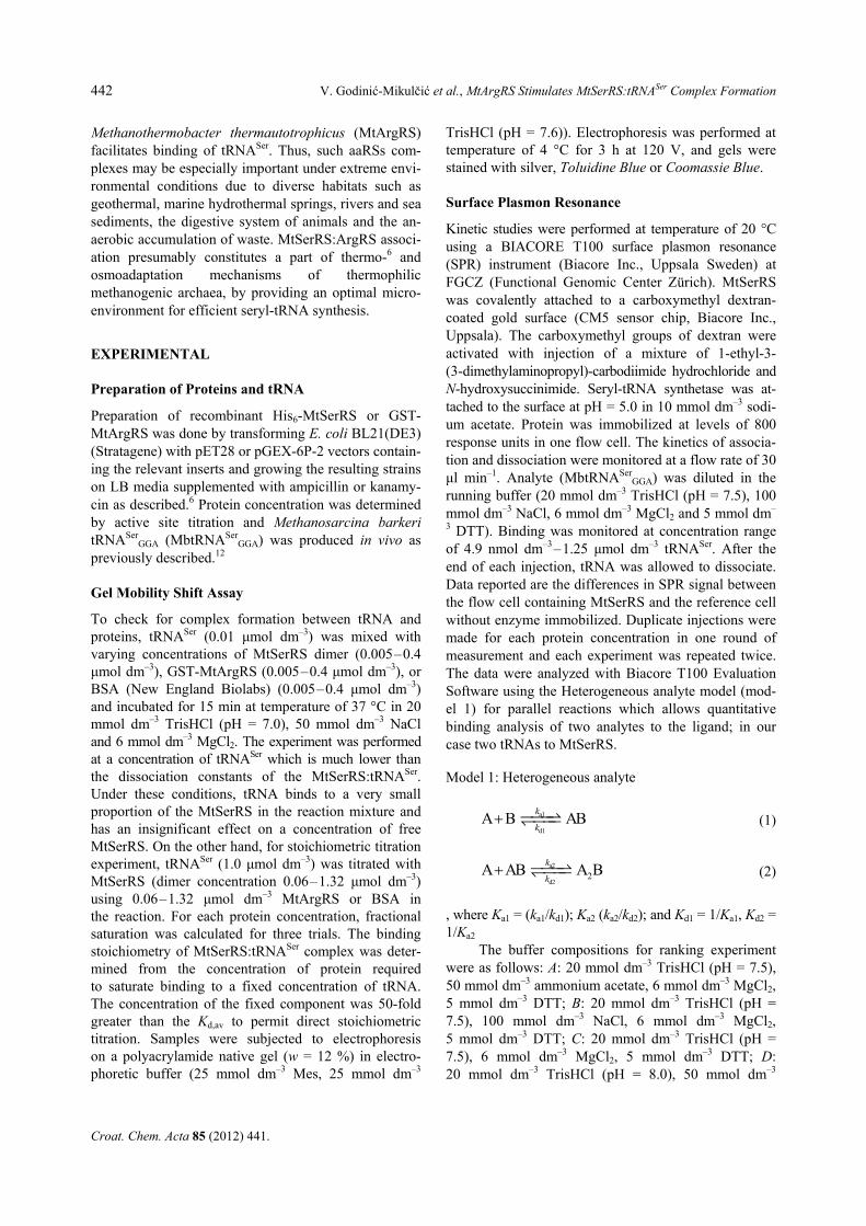
Figure 1. Multiple sequence alignment of N-terminal domains from SerRSs from different organisms. Amino acid sequenceswere aligned using the software ClustalW (http://www.ebi.ac.uk/Tools/msa/clustalw2/). According to the percentage of conserva-tion, 80 % conserved amino acids are shaded as black, 60 % conserved as blue and 40 % conserved as light-blue. Abbreviationsfor names of organisms are the following: Mt (Methanothermobacter thermautotrophicus), mMb (methanogenic-type SerRS fromMethanosarcina barkeri), Mmp (Mathanococcus maripaludis), Mj (Methanocaldococcus jannashii), Ph (Pyrococcus horikoshii),Ta (Thermoplasma acidophilum), Ss (Sulfolobus sulfatarius), Hs (Homo sapiens), Sc (Saccharomyces cerevisiae), Hm(Haloarcula marismortui), Ec (Escherichia coli), Bs (Bacillus sulfolobus), Tt (Thermus thermophilus), Aa (Aquifex aeolicus), Mb(bacterial-type SerRS from Methanosarcina barkeri), Ma (Methanosarcina acetivorans), Mmz (Methanosarcina mazei) and Mst(Methanosarcina stadmanae). Amino acids of metanogenic-type SerRS important for tRNA recogniton are marked in red (a). Kinetic analysis of MbtRNASer binding to immobilized MtSerRS monitored by a surface plasmon resonance. Sensorgrams (red)were obtained for the binding of different concentrations of MbtRNASer (4.9 nmol dm–3–1.25 µmol dm–3) to MtSerRS in 20 mmoldm–3 TrisHCl (pH = 7.5), 100 mmol dm–3 NaCl, 6 mmol dm–3 MgCl2 and 5 mmol dm–3 DTT. Data were fit to the Heterogeneousanalyte model (black). Inset: The experimental curves were evaluated by Steady State Affinity model as well and average dissoci-ation constant Kd,av = 186 ± 19 nmol dm–3 was defined (b).
444 V. Godinić-Mikulčić et al., MtArgRS Stimulates MtSerRS:tRNASer Complex Formation
Croat. Chem. Acta 85 (2012) 441.
of M. barkeri SerRS and probed their involvement in tRNA binding.13 The results obtained by SPR13 revealed that residues Arg76, Lys79 and Arg94 have pronounced effect on SerRS:tRNASer complex formation and the dissociation constants (Kd) in comparison with the wild type SerRS:tRNASer interaction. These amino acids are conserved in the primary structure of MtSerRS (corre-sponding to amino acids Arg77, Lys80 and Arg95) (Figure 1a) and in several other methanogenic-type SerRSs. According to the sequence alignments,11 MtSerRS also possesses other features characteristic for methanogenic-type SerRS enzymes, that is a shorter
motif II, an insertion at the position of 401–417 and a unique insertion of 30 amino acids (at positions 273–305) between motifs I and II, which adopts helix-turn-helix (HTH) structure. Furthermore, tRNASer isoacceptors from M. barkeri and M. thermautotrophicus are strikingly similar: the nucleotide pair G30:C40 is present in all serine tRNAs, all possess characteristic long variable arm, discriminatory base G73, nucleotide pair G1:C72 and one unpaired nucleo-tide at position 48.17 Observed structural resemblance in proteins and tRNAs strengthened the assumption about heterologous recognition between MtSerRS and MbtRNASer. As expected, MtSerRS binds M. barkeri tRNASer with rather high affinity (Figure 1b).
The kinetic parameters for binding MtSerRS and tRNASer were determined using surface plasmon reso-nance (Figure 1b and Table 1) in agreement with the Heterogeneous analyte binding model (model 1). In this model, one tRNA (A) binds to the ligand B (MtSerRS) to form a complex AB. The second tRNA (A) binds to the AB complex and forms a complex A2B. The sensor-gram in this model reflects the sum of the two binding reactions and the equilibrium constant for the second step (Ka2 = 6.853 × 10⁶ mol–1 dm3) is higher than that for the first step (Ka1 = 6.873 × 10⁵ mol–1 dm3) showing the cooperative binding of tRNASer (Table 1). The first step of binding one tRNA which involves formation of a bimolecular complex between tRNASer and the flexibly exposed N-terminal domain of SerRS (Kd1 = 1.455 µmol dm–3, Table 1) changes the affinity for the second tRNA (Kd2 = 0.1459 µmol dm–3, Table 1).
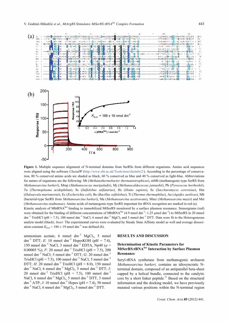
Table 2. Buffer solution composition and ionic strength. We refer to complete solution (A–J ) simply as the buffer. The concen-trations of buffer species (Na+, Cl–, Mg2+, NH4+, CH3COO–) were calculated based on published pKas and the Henderson-Hasselbach equation36
Buffer(a),(b) c Na+ Cl– Mg2+ NH4+ CH3COO– B+ B B– N+ Ic Rmax
F 20 200 217 – – – 17 3 – 17 217 120
H 20 150 175 6 – – 13 7 – 13 181 72
G 20 100 117 – – – 17 3 – 17 117 69
I 20 100 129 6 – – 17 3 – 17 159 66
A 20 – 29 6 50 50 17 3 – 17 83 65
B 20 100 129 6 – – 17 3 – 17 135 65
D 20 – 25 6 50 50 13 7 – 13 81 55
C 20 – 29 6 – – 17 3 – 17 35 35
JH 10 54 62 6 – – – 6 4 6 72 34
EH 10 154 150 – – – – 6 4 6 168 30 (a) All concentrations are expressed in mmol dm–3. (b) B+, positively charged buffer; B–, negatively charged buffer; N+, protonated amine concentration; Ic, ionic strength. All buffers
contained DTT (5 mmol dm–3) except D. Buffer E contained EDTA (ethylenediaminetetraacetic acid) (3 mmol dm–3) that in neutral solution exists mostly as EDTA3– and Np40 (nonionic detergent, φ = 0.00005). Buffer I contained ATP (adenosine tri-phosphate) (3 mmol dm–3). In neutral solution, ATP is ionized and exists mostly as ATP4–, with small proportion of ATP3–. All buffers contained Tris but E and H contained Hepes and this is denoted with subscript H. pH of the buffers was 7.5 except for buffers D and H (pH = 8.0). Additional Na+ and Cl- ions are due to corrections because of pH adjustment. Rmax (in RU) is max-imum binding of MtSerRS:tRNASer in given conditions.
Table 1. Kinetic parameters determined by SPR for MtSerRS:tRNASer interaction in agreement with the Heteroge-neous analyte model. Chi2 = 3.85 and denotes statistical value that describes the accuracy of matching the experimental data with the chosen model of binding. The values 10 are ac-ceptable35
Kinetic parameters
ka1 / mol–1 dm3 s–1 3430 ± 15
kd1 / s–1 0.00499 ± 1 × 10⁻⁵
ka2 / mol–1 dm3 s–1 1.189 × 10⁶ ± 3.3 × 10³
kd2 / s–1 0.1735 ± 5 × 10⁻⁴
Kd1 / µmol dm–3 1.455
Ka1 / mol–1 dm3 6.873 × 10⁵
Kd2 / μmol dm–3 0.1459
Ka2 / mol–1 dm3 6.853 × 10⁶
V. Godinić-Mikulčić et al., MtArgRS Stimulates MtSerRS:tRNASer Complex Formation 445
Croat. Chem. Acta 85 (2012) 441.
The experimental curves were evaluated also in the state of equilibrium by Steady State Affinity model and average dissociation constant Kd,av = 186 ± 19 nmol dm–3 was defined (Figure 1b). The Kd,av value is in good agreement with published data for the enzyme mMbSerRS13 and published Kd values for other synthetase:tRNA complexes involving CysRS,18 GlnRS,19,20 and AspRS.21 Binding Responses Comparison of MtSerRS:tRNASer
Association in Qualitative Analysis by SPR
To shed some light on the nature of binding between MtSerRS and tRNASer, the maximum binding capacity Rmax (in RU) was measured using SPR in the presence of different concentration of salts and buffering compo-nents (Table 2). Analyte (tRNASer) was allowed to flow over immobilized ligand (MtSerRS). In all tested buff-ers the binding of tRNA was successful and reached measurable level of response. We compared the shape of the curves which were very informative without cal-culating individual rate constants in different reactions.
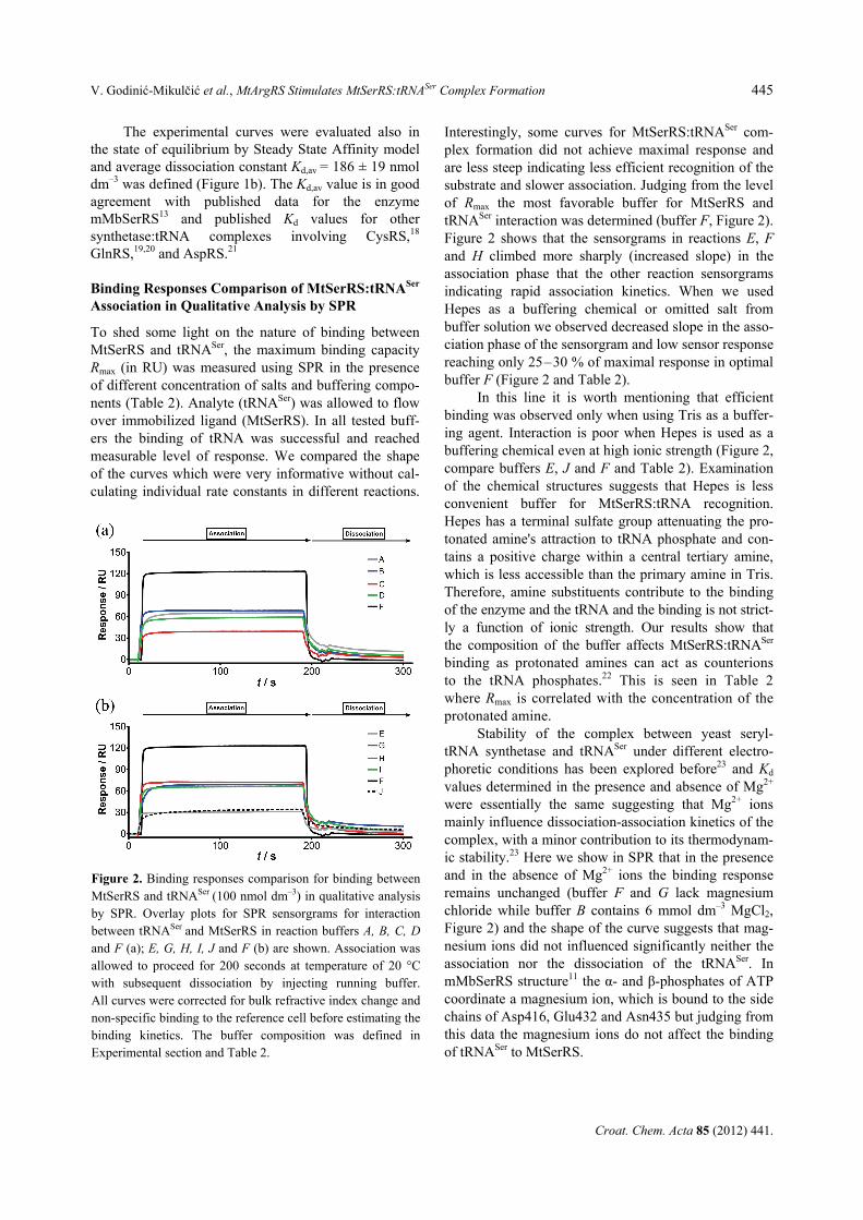
Interestingly, some curves for MtSerRS:tRNASer com-plex formation did not achieve maximal response and are less steep indicating less efficient recognition of the substrate and slower association. Judging from the level of Rmax the most favorable buffer for MtSerRS and tRNASer interaction was determined (buffer F, Figure 2). Figure 2 shows that the sensorgrams in reactions E, F and H climbed more sharply (increased slope) in the association phase that the other reaction sensorgrams indicating rapid association kinetics. When we used Hepes as a buffering chemical or omitted salt from buffer solution we observed decreased slope in the asso-ciation phase of the sensorgram and low sensor response reaching only 25–30 % of maximal response in optimal buffer F (Figure 2 and Table 2).
In this line it is worth mentioning that efficient binding was observed only when using Tris as a buffer-ing agent. Interaction is poor when Hepes is used as a buffering chemical even at high ionic strength (Figure 2, compare buffers E, J and F and Table 2). Examination of the chemical structures suggests that Hepes is less convenient buffer for MtSerRS:tRNA recognition. Hepes has a terminal sulfate group attenuating the pro-tonated amine's attraction to tRNA phosphate and con-tains a positive charge within a central tertiary amine, which is less accessible than the primary amine in Tris. Therefore, amine substituents contribute to the binding of the enzyme and the tRNA and the binding is not strict-ly a function of ionic strength. Our results show that the composition of the buffer affects MtSerRS:tRNASer binding as protonated amines can act as counterions to the tRNA phosphates.22 This is seen in Table 2 where Rmax is correlated with the concentration of the protonated amine.
Stability of the complex between yeast seryl-tRNA synthetase and tRNASer under different electro-phoretic conditions has been explored before23 and Kd values determined in the presence and absence of Mg2+ were essentially the same suggesting that Mg2+ ions mainly influence dissociation-association kinetics of the complex, with a minor contribution to its thermodynam-ic stability.23 Here we show in SPR that in the presence and in the absence of Mg2+ ions the binding response remains unchanged (buffer F and G lack magnesium chloride while buffer B contains 6 mmol dm–3 MgCl2, Figure 2) and the shape of the curve suggests that mag-nesium ions did not influenced significantly neither the association nor the dissociation of the tRNASer. In mMbSerRS structure11 the α- and β-phosphates of ATP coordinate a magnesium ion, which is bound to the side chains of Asp416, Glu432 and Asn435 but judging from this data the magnesium ions do not affect the binding of tRNASer to MtSerRS.
Figure 2. Binding responses comparison for binding betweenMtSerRS and tRNASer (100 nmol dm–3) in qualitative analysisby SPR. Overlay plots for SPR sensorgrams for interactionbetween tRNASer and MtSerRS in reaction buffers A, B, C, Dand F (a); E, G, H, I, J and F (b) are shown. Association wasallowed to proceed for 200 seconds at temperature of 20 °Cwith subsequent dissociation by injecting running buffer.All curves were corrected for bulk refractive index change andnon-specific binding to the reference cell before estimating thebinding kinetics. The buffer composition was defined inExperimental section and Table 2.
446 V. Godinić-Mikulčić et al., MtArgRS Stimulates MtSerRS:tRNASer Complex Formation
Croat. Chem. Acta 85 (2012) 441.
Osmoadaptation of Thermophilic Methanogenic Archaea and Halotolerance of the Enzyme MtSerRS
Archaea had to apply a wide range of strategies to sur-vive in conditions of high salt concentration, e.g. sodi-um chloride. We have previously shown that MtSerRS catalyzed tRNA serylation in vitro suboptimally at low salt concentration.6 Only at salt concentrations above 100 mmol dm–3 the enzyme achieved satisfactory activi-ty. This is in agreement with our current results reveal-ing direct influence of sodium chloride concentration on the interaction between MtSerRS and tRNASer (Figure 2, Table 2). There is a strong correlation between in-creased recognition of the tRNASer and increasing ionic strength (Table 2 and Figure 2). As shown by Rmax val-ues (Table 2), the maximum binding capacity for SerRS:tRNASer interaction was reached when elevating the concentration of sodium ions from 54 mmol dm–3 to 200 mmol dm–3 as well as using TrisHCl as a buffering component, consistent with the increasing number of positive charges of these cations (Table 2). Curve H in Figure 2 shows higher response (Rmax = 72) then binding described with curves C, E and J, and upon addition of more salt to the buffer, the response is increased to a maximal level (curve F, Rmax = 120) (Figure 2), indicat-ing a requirement of substantial quantities of NaCl for the interaction. Curves A, B, D and I show a reduced interaction potential due to decreased concentration of salt (Figure 2). Interestingly, curves C, A, J and G (Figure 2) show less steep curve in the association part of the curve compared to curve F indicating slower association. From ranking experiment (Figure 2) it is evident that c (NaCl) ≥ 100 mmol dm–3 should be present in reaction for optimal recognition between MtSerRS and tRNASer. These findings strengthen the conclusion that association of MtSerRS and tRNASer is actually stabilized by elevated ionic strength. These findings are reminiscent of the aminoacylation assays conducted in the presence of 250 mM KCl for LysRS, LeuRS and ProRS from M. thermautotrophicus.24 In contrast, the standard aminoacylation reactions by bac-terial and eukaryotic SerRSs are usually carried out without salt.25 Thus, salt-dependent association between MtSerRS and tRNASer may provide one of the mecha-nisms of osmoadaptation of methanogenic archaea. Unlike bacteria, most archaea require high concentra-tion of cations (e.g. K+) in the cell for optimal growth
conditions.26 M. thermautotrophicus (strain ΔH) grows very well at the concentrations of sodium chloride up to 0.60 mol dm–3. Moreover, the intracellular concentration of potassium ions27 in M. thermautotrophicus is high as 0.65 to 1.1 mol dm–3. In this respect, the organisms can adapt and evolve proteins that can function at higher salt concentrations. Comparison of the total amino acid content of ribosomal proteins revealed 29 % of acidic ribosomal proteins in M. thermautotrophicus compared to only 7 % in E. coli while halofils contain over 50 % of acidic ribosomal proteins. Moreover, these proteins are less hydrophobic and the effect of salting out with cations is decreased. Therefore, cations can stabilize proper folding of proteins that can function at harsh ionic conditions. Alteration of Kd Value in MtSerRS:tRNASer Associa-tion Upon Addition of MtArgRS
The effect of MtArgRS on the tRNASer binding by MtSerRS was investigated in vitro by gel mobility shift assay. Quantification of free and bound RNA bands in gel mobility shift assay allowed the binding curve for MtSerRS:tRNASer to be analyzed (Figure 3). Saturation binding was displayed graphically, and a sigmoidal curve was obtained as shown in Figure 3a, indicating cooperative binding, and could not be fit to the standard single-site binding model. In SPR, a sensorgram of a heterogeneous analyte binding to immobilized ligand (Figure 1) represents the sum of two separate binding interactions. If second tRNA has a higher binding affini-ty than the first tRNA, the gel-shift data will reflect the binding kinetics of the higher affinity tRNA. If Ka2 is very large compared to Ka1 then the major species pre-sent in solution are either [B] or [A2B] (see Experi-mental) and from gel-shift analysis the apparent dissoci-ation constant Kd,app of 119 nmol dm–3 was obtained for both binding steps occurring at the same time. Interest-ingly, when MtArgRS was added in the reaction, the Kd,app for tRNASer was decreased two-fold to 62.0 nmol dm–3 (Table 3). This finding is in agreement with previ-ous kinetic experiments6 where Km for tRNASer was reduced also two-fold in the presence of MtArgRS. Interaction between MtSerRS and MtArgRS enhanced tRNASer aminoacylation, in agreement with previous reports of improved aminoacylation upon aaRS complex formation in archaea and yeast.4–6,24,28 While the details
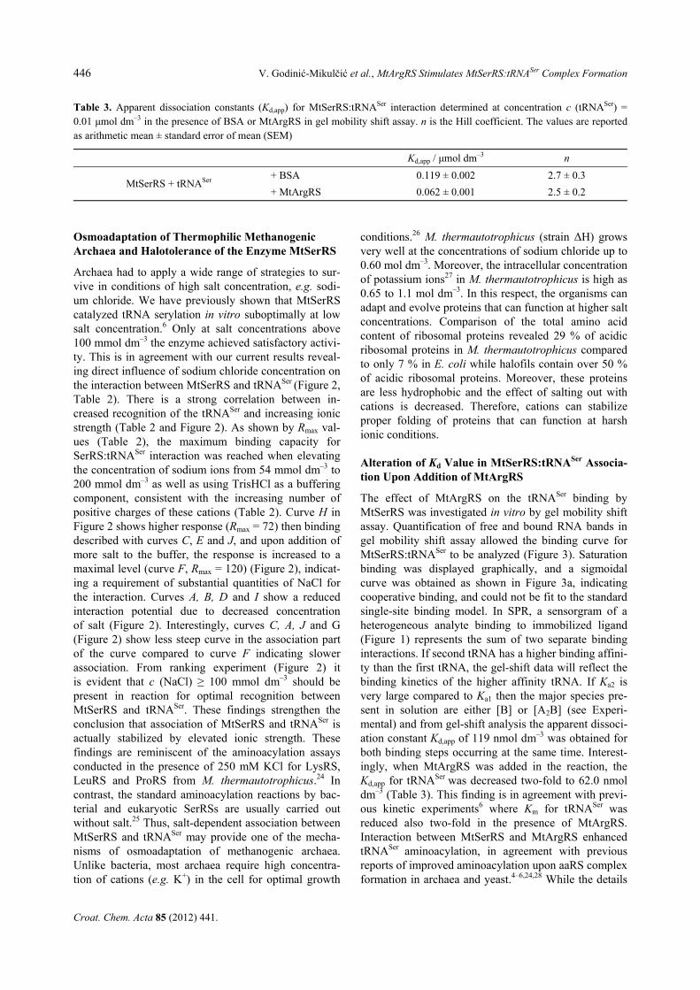
Table 3. Apparent dissociation constants (Kd,app) for MtSerRS:tRNASer interaction determined at concentration c (tRNASer) =0.01 μmol dm–3 in the presence of BSA or MtArgRS in gel mobility shift assay. n is the Hill coefficient. The values are reported as arithmetic mean ± standard error of mean (SEM)
Kd,app / μmol dm–3 n
MtSerRS + tRNASer + BSA 0.119 ± 0.002 2.7 ± 0.3
+ MtArgRS 0.062 ± 0.001 2.5 ± 0.2
V. Godinić-Mikulčić et al., MtArgRS Stimulates MtSerRS:tRNASer Complex Formation 447
Croat. Chem. Acta 85 (2012) 441.
of this interaction remain to be resolved, our data indi-cate that the presence of MtArgRS in this macromolecu-lar assembly could improve the efficiency of the transla-tional machinery by facilitating the recognition of the tRNASer by MtSerRS as shown in this work (Table 3).
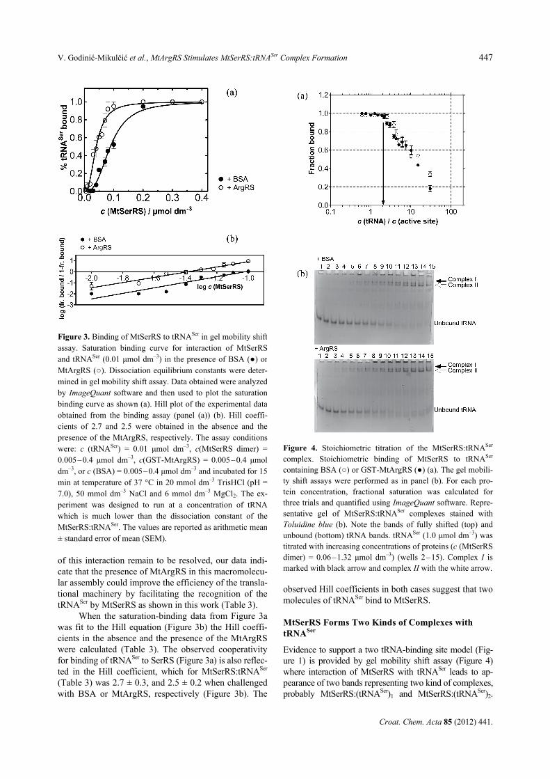
When the saturation-binding data from Figure 3a was fit to the Hill equation (Figure 3b) the Hill coeffi-cients in the absence and the presence of the MtArgRS were calculated (Table 3). The observed cooperativity for binding of tRNASer to SerRS (Figure 3a) is also reflec-ted in the Hill coefficient, which for MtSerRS:tRNASer (Table 3) was 2.7 ± 0.3, and 2.5 ± 0.2 when challenged with BSA or MtArgRS, respectively (Figure 3b). The
observed Hill coefficients in both cases suggest that two molecules of tRNASer bind to MtSerRS. MtSerRS Forms Two Kinds of Complexes with tRNASer
Evidence to support a two tRNA-binding site model (Fig-ure 1) is provided by gel mobility shift assay (Figure 4) where interaction of MtSerRS with tRNASer leads to ap-pearance of two bands representing two kind of complexes, probably MtSerRS:(tRNASer)1 and MtSerRS:(tRNASer)2.
Figure 3. Binding of MtSerRS to tRNASer in gel mobility shiftassay. Saturation binding curve for interaction of MtSerRSand tRNASer (0.01 μmol dm–3) in the presence of BSA (●) orMtArgRS (○). Dissociation equilibrium constants were deter-mined in gel mobility shift assay. Data obtained were analyzedby ImageQuant software and then used to plot the saturationbinding curve as shown (a). Hill plot of the experimental dataobtained from the binding assay (panel (a)) (b). Hill coeffi-cients of 2.7 and 2.5 were obtained in the absence and thepresence of the MtArgRS, respectively. The assay conditionswere: c (tRNASer) = 0.01 μmol dm–3, c(MtSerRS dimer) =0.005–0.4 μmol dm–3, c(GST-MtArgRS) = 0.005–0.4 μmoldm–3, or c (BSA) = 0.005–0.4 μmol dm–3 and incubated for 15min at temperature of 37 °C in 20 mmol dm–3 TrisHCl (pH =7.0), 50 mmol dm–3 NaCl and 6 mmol dm–3 MgCl2. The ex-periment was designed to run at a concentration of tRNAwhich is much lower than the dissociation constant of theMtSerRS:tRNASer. The values are reported as arithmetic mean± standard error of mean (SEM).
Figure 4. Stoichiometric titration of the MtSerRS:tRNASer
complex. Stoichiometric binding of MtSerRS to tRNASer
containing BSA (○) or GST-MtArgRS (●) (a). The gel mobili-ty shift assays were performed as in panel (b). For each pro-tein concentration, fractional saturation was calculated for three trials and quantified using ImageQuant software. Repre-sentative gel of MtSerRS:tRNASer complexes stained with Toluidine blue (b). Note the bands of fully shifted (top) and unbound (bottom) tRNA bands. tRNASer (1.0 μmol dm–3) was titrated with increasing concentrations of proteins (c (MtSerRS dimer) = 0.06–1.32 μmol dm–3) (wells 2–15). Complex I is marked with black arrow and complex II with the white arrow.
448 V. Godinić-Mikulčić et al., MtArgRS Stimulates MtSerRS:tRNASer Complex Formation
Croat. Chem. Acta 85 (2012) 441.
The binding stoichiometry of MtSerRS:tRNASer com-plex was determined from the amount of protein re-quired to saturate binding to a fixed concentration of tRNASer (Figure 4a). The concentration of the fixed component was 50-fold greater than the Kd to permit direct stoichiometric titration. In the presence of BSA, we find that protein dimer fully binds to tRNASer pre-sent in the reaction at concentration of 0.275 μmol dm–3 (protein dimer:tRNA ratio 0.55), while in the presence of GST-MtArgRS, full binding of protein dimer to tRNASer was achieved at concentration of 0.205 μmol dm–3 (protein dimer:tRNA ratio 0.41) in agreement with binding stoichometry of 2 RNA molecules per protein dimer.
Seryl-tRNA synthetases from Thermus thermo-philus and Escherichia coli form two types of noncovalent complexes with tRNASer; SerRS:(tRNASer)1 and SerRS:(tRNASer)2.
29–31 The tRNA is bound across the two subunits of dimeric SerRS, as revealed by the SerRS:(tRNASer)1 crystal structure11,30 and biochemical studies on heterodimers.12,32 SerRS from the yeast Sac-charomyces cerevisiae has also been found to bind one or two tRNASer cooperatively24. Measurements of affini-ty (Figure 3), stoichiometry (Figure 4) and cooperativity (Table 3) suggest that communication among subunits in MtSerRS dimer is required for tRNASer binding. We favor the model of two tRNASer molecules bound per MtSerRS protein dimer in cooperative manner. It can be speculated that binding of one molecule facilitates bind-ing of the other tRNA molecule to MtSerRS as support-ed by gel-shift experiment (Figure 4b). To explore these possibilities in future crystallographic experiments will be an intriguing task.
Overall SerRS structure is very flexible. Docking model of mMbSerRS:tRNA complex12 implies that the N-terminal domain of one protomer in the dimer will bind the variable arm of the tRNASer, whereas the 3'-end of the same tRNA will enter the active site of the other protomer.12 Catalytic and N-terminal domains of SerRS have to act synergistically to provide a high and specific binding affinity for their cognate tRNASer.12 Although tRNA-binding domains in the two SerRS types are non-homologous and evolutionarily unrelated,11 the requirement for a closing movement of the N-terminal domain upon tRNA binding has been observed in the T. Thermophilus SerRS:tRNA co-crystal structure33 and predicted by docking model.11 The initial capture of the tRNA is likely to be facilitated by the flexible dispo-sition of the N-terminal domain, which will then deliver the tRNA to the active site of the enzyme via a hinge-like motion mediated by HTH interactions.12 Unlike some synthetases such as aspartyl-tRNA synthetase, where each monomer interacts with only one tRNA molecule, here each tRNASer binds both monomers of
SerRS (cross-dimer binding)12 and it is tempting to further explore tRNA-induced cooperative effects. Crossubunit interactions with the tRNA have also been identified in ProRS and ThrRS.34 In ThrRS, the binding of tRNA by one monomer, by breaking a salt bridge, presumably modifies the position and flexibility of the ordering loop of the other monomer and consequently affects the binding or release of the substrates in the second active site.34 This type of tRNA-induced inter-subunit communication needs to be further explored in serine system because it could constitute another func-tional link between members of subclass IIa.
CONCLUSION
The kinetic parameters for binding MtSerRS and tRNASer were determined using surface plasmon reso-nance (Figure 1b and Table 1) in agreement with the Heterogeneous analyte binding model. Results show that the affinity of tRNASer for the second binding site is approximately 10-fold stronger compared to affinity for the first binding site (Kd1 = 1.455 µmol dm–3, Kd2 = 0.1459 µmol dm–3 ,Table 1).
The analysis of the binding data from three sepa-rate experiments revealed presence of positive cooperativity in formation of the MtSerRS:tRNASer
binding complexes (Figure 3a) displayed as sigmoidal saturation curve.
There is a correlation between efficiency of bind-ing (Rmax) tRNASer to MtSerRS and elevated ionic strength that was observed only when using Tris rather than Hepes as a buffering chemical. By comparison of the binding responses and shape of sensorgrams we show that magnesium ions do not affect the binding of tRNASer to MtSerRS.
Further evidence to support a two tRNA-binding site model (Figure 1) was provided by gel mobility shift assay (Figure 4) where interaction of MtSerRS with tRNASer leads to appearance of two bands representing two kind of complexes, probably MtSerRS:(tRNASer)1 and MtSerRS:(tRNASer)2.
MtSerRS:tRNASer complex formation was stimu-lated by the addition of MtArgRS, which is an interact-ing partner of MtSerRS. The presence of MtArgRS led to a two-fold decrease in Kd,app for MtSerRS:tRNASer (Table 3), but had no affect on cooperative properties or stoichiometry of the complex (Figure 4).
While the details of this interaction remain to be resolved, our data indicate that the complex formation between MtArgRS and MtSerRS may improve the effi-ciency of the translational machinery, assuring more efficient recognition of the tRNASer by MtSerRS as presented in this work (Table 3).
V. Godinić-Mikulčić et al., MtArgRS Stimulates MtSerRS:tRNASer Complex Formation 449
Croat. Chem. Acta 85 (2012) 441.
Acknowledgements. This work was supported by the grant from the Ministry of Science, Education and Sports of Repub-lic of Croatia (project 119-0982913-1358). Stefan Schauer from the Functional Genomics Center Zürich is acknowledged for assistance in SPR experiments.
REFERENCES
1. S. H. Zaher and R. Green, Cell 136 (2009) 746–762. 2. J. Ling, N. Reynolds, and M. Ibba, Annu. Rev. Microbiol. 63
(2009) 61–78. 3. M. Ibba and D. Söll, Annu. Rev. Biochem. 62 (2000) 617–650. 4. S. G. Park, K. L. Ewalt, and S. Kim, TRENDS in Biochem. Sci.
30 (2005), 569–574. 5. C. D. Hausmann and M. Ibba, FEMS Microbiol. Rev. 32 (2008)
705–721. 6. V. Godinić-Mikulčić, J. Jarić, C. D. Hausmann, M. Ibba, and I.
Weygand-Đurašević, J. Biol. Chem. 286 (2011) 3396–3404. 7. D. Tumbula, U. C. Vothknecht, H. S. Kim, M. Ibba, B. Min, T.
Li, J. Pelaschier, C. Stathopoulos, H. Becker, and D. Söll, Genet-ics 152 (1999) 1269–1276.
8. H. S. Kim, U. C. Vothknecht, R. Hedderich, I. Celic, and D. Söll, J. Bacteriol. 180 (1998) 6446–6449.
9. D. Korencic, I. Ahel, and D. Soll, Food Technol. Biotechnol. 40 (2002) 235–260.
10. S. Bilokapić, N. Ban, and I. Weygand-Đurašević, Croat. Chem. Acta 82 (2009) 493–501.
11. S. Bilokapić, T. Maier, D. Ahel, I. Gruić-Sovulj, D. Söll, I. Wey-gand-Đurašević, and N. Ban, EMBO J. 25 (2006) 2498–2509.
12. S. Bilokapić, N. Ivić, V. Godinić-Mikulčić, I. Piantanida, N. Ban, and I. Weygand-Đurašević, J. Biol. Chem. 284 (2009) 10706–10713.
13. J. Jarić, S. Bilokapić, S. Lesjak, A. Crnković, N. Ban, and I. Weygand-Đurašević, J. Biol. Chem. 284 (2009) 30643–30651.
14. I. Weygand-Đurašević and S. Cusack, Seryl-tRNA Synthetases in: M. Ibba, C. Francklyn, S. Cusack (Eds.), The AminoacyltRNA Synthetases. Landes Biosciences, Georgetown (SAD), 2005, pp. 177–192.
15. V. Biou, A. Yaremchuk, M. Tukalo, and S. Cusack, Science 263 (1994) 1404–1410.
16. V. Godinić, M. Močibob, S. Ročak, M. Ibba, and I. Weygand-Đurašević, FEBS J. 274 (2007) 2788–2799.
17. D. Korenčić, C. Polycarpo, I. Weygand-Đurašević, and D. Söll, J. Biol. Chem. 279 (2004) 48780–48786.
18. C. M. Zhang, J. J. Perona, and Y. M. Hou, Biochemistry 42 (2003), 10931–10937.
19. I. Weygand-Đurašević, E. Schwob, and D. Söll, Proc. Natl. Acad. Sci. U. S. A. 90 (1993) 2010–2014.
20. T. L. Bullock, L. D. Sherlin, and J. J. Perona, Nat. Struct. Biol. 7 (2000) 497–504.
21. M. Frugier, L. Moulinier, and R. Giegé, EMBO J. 19 (2000) 2371–2380.
22. J. R. Wenner and V. A. Bloomfield, Anal. Biochem. 268 (1999) 201–212.
23. I. Gruić-Sovulj, J. Rokov-Plavec, M. Močibob, T. Kamenski, and I. Weygand-Đurašević, Croat. Chem. Acta 77 (2004) 599–604.
24. M. Praetorius-Ibba, C. D. Hausmann, M. Paras, T. E. Rogers, and M. Ibba, J. Biol. Chem. 282 (2007) 3680–3687.
25. C. S. Francklyn, E. A. First, J. J. Perona and Y. M. Hou, Methods 44 (2008) 100-118.
26. R. Ciulla, C. Clougherty, N. Belay, S. Krishnan, C. Zhou, D. Byrd, and M. F. Roberts, J. Bacteriol. 176 (1994) 3177–3187.
27. D. D. Martin, R. A. Ciulla, and M. F. Roberts, Appl. Environ. Microbiol. 65 (1999) 1815–1825.
28. G. Simos, A. Sauer, F. Fasiolo, and E. C. Hurt, Mol. Cell. 1 (1998) 235–242.
29. F. Borel, C. Vincent, R. Leberman, and M. Härtlein, Nucleic Ac-ids Res. 22 (1994) 2963–2969.
30. A. D. Yaremchuk, M. A. Tukalo, I. Krikliviy, N. Malchenko, V. Biou, C. Berthet-Colominas, and S. Cusack, FEBS Lett. 310 (1992) 157–161.
31. S. Price, S. Cusack, F. Borel, C. Berthet-Colominas, and R. Leberman, FEBS Lett. 324 (1993) 167–170.
32. C. Vincent, F. Borel, J. C. Willison, R. Leberman, and M. Härtlein, Nucleic Acids Res. 23 (1995) 1113–1118.
33. S. Cusack, A. Yaremchuk, and M. Tukalo, EMBO J. 15 (1996) 2834–2842.
34. A. Torres-Larios, R. Sankaranarayanan, B. Rees, A.-C. Dock-Bregeon, and Dino Moras, J. Mol. Biol. 331 (2002) 201–211.
35. A. Chenal, P. Nizard, V. Forge, M. Pugniere, M. O. Roy, J. C. Mani, F. Guillain, and D. Gillet, Protein Eng. 15 (2002) 383–391.
36. K. J. Ellis and J. F. Morrison, Methods Enzymol. 182 (1982) 24–38.