56
KUGLER PUBLICATIONS Volume 15 • Issue 1 • 2016 • 1560-2133

KUGLERPUBLICATIONS
Volume 15 • Issue 1 • 2016 • 1560-2133

Submit your article now to the
Asian Journal of OPHTHALMOLOGYa peer-reviewed online open access journal.
There are no publication costs, hidden fees or charges.
Chief editor: Paul Chew
The objectives of Asian Journal of Ophthalmology are as follows:• To provide a platform for the publication of information with a focus on
Ophthalmology in Asia• To disseminate information that will improve the care of patients with all
types of ophthalmological disorders, with a special focus on glaucoma• To increase the understanding of such disorders through reporting of
educational activities• To publish the results of research programmes to expand knowledge about
the causes, prevention, and treatment of ophthalmological disorders• To work closely with Asian and international researchers to achieve these
aims• To provide a forum for young and relatively inexperienced researchers to
present their research results as Original Articles via an international platform• To maintain and promote relationships with any organisation with similar
goals.
For more information, an article template and submission guidelines, see www.asjoo.com

Asian Journal of Ophthalmology is the peer-reviewed journal for The Asian Pacific Glaucoma Society (APGS), The Asia Pacific Ophthalmic Trauma Society (APOTS) and all others with an interest in Ophthalmology.
Advertising inquiriesThe Asian Journal of Ophthalmology offers many sponsorship and advertising opportunities, both online and in print. Please mail us at [email protected] to access the media kit or for queries.
CopyrightAuthors who publish with this journal agree to the following terms:a. Authors retain copyright and grant the journal right of first publica-tion, with the work twelve (12) months after publication simultaneously licensed under a Creative Commons Attribution License that allows others to share the work with an acknowledgement of the work’s author-ship and initial publication in this journal.b. Authors are able to enter into separate, additional contractual arrangements for the non-exclusive distribution of the journal’s published version of the work (e.g., post it to an institutional repository or publish it in a book), with an acknowledgement of its initial publication in this journal.c. Authors are permitted and encouraged to post their work online (e.g., in institutional repositories or on their website) prior to and during the submission process, as it can lead to productive exchanges, as well as earlier and greater citation of published work.
DisclaimersAll articles published, including editorials and letters, represent the opinions of the authors and do not reflect the official policy of Asian Journal of Ophthalmology, the APGS, APOTS, its sponsors, the publisher or the institution with which the author is affiliated, unless this is clearly specified. Although every effort has been made to ensure the technical accuracy of the contents of Asian Journal of Ophthalmology, no responsi-bility for errors or omissions is accepted.Asian Journal of Ophthalmology, APGS, APOTS, and the publisher do not endorse or guarantee, directly or indirectly, the quality or efficacy of any product or service described the advertisements or other material that is commercial in nature in any issue. All advertising is expected to conform to ethical and medical standards. No responsibility is assumed by the APGS, APOTS or the publisher for any injury and/or damage to persons or property as a matter of products liability, negligence or otherwise, or from any use or operation of any methods, products, instructions, or ideas contained in the material herein. Because of rapid advances in the medical Sciences, independent verification of diagnoses and drug dosages should be made.
Chief editor:Paul Chew, [email protected]
Editorial office:Asian Journal of Ophthalmology/ Kugler Publications, P.O. Box 20538, 1001 NM Amsterdam, The Netherlands. [email protected]
Publisher:Kugler Publications, P.O. Box 20538, 1001 NM Amsterdam, The Netherlands. [email protected] www.kuglerpublications.com
Manuscript submissions:Information for authors is available via the website (www.asjoo.com), through which all manuscripts should be submitted. For inquiries please contact us at: [email protected].
Peer-review manager:Kayoko Welsh, [email protected]
Publication frequencyThe Asian Journal of Ophthalmology is published four issues per year (quarterly) electronically. Each issues will consist of approximately 48 pages. A selection of the best papers is published in print twice a year and distributed free of charge at congresses through Kugler Publications or partners.
Open access policyThe Asian Journal of Ophthalmology provides immediate open access to its content after (free) registration, on the principle that making research freely available to the public supports a greater global exchange of knowledge. There are no fees required to publish in the journal.
Focus and scopeAs new technologies and therapeutic interventions are continually being developed, Ophthalmology has become a field of rapid change, particularly in the Asia-Pacific region, where disease patterns and health care delivery differ greatly from those seen in the West. Asian Journal of Ophthalmology was established in 1998 with the aim of disseminating information relevant to Ophthalmology and glaucoma throughout Asia and to interested groups worldwide.
The objectives of Asian Journal of Ophthalmology are as follows:• To provide a platform for the publication of information with a focus on Ophthalmology in Asia.• To disseminate information that will improve the care of patients with all types of ophthalmological disorders, with a special focus on glaucoma.• To increase the understanding of such disorders through reporting of educational activities.• To publish the results of research programmes to expand knowledge about the causes, prevention, and treatment of ophthalmological disorders.• To work closely with Asian and international researchers to achieve these aims.• To provide a forum for young and relatively inexperienced researchers to present their research results as Original Articles via an international platform.• To maintain and promote relationships with any organiza-tion with similar goals.
Although the focus of Asian Journal of Ophthalmology mainly was on glaucoma with close ties to the South-East Asian Glaucoma Interest Group (SEAGIG) in the past, the journal now focuses on the entire spectrum of Ophthal-mology. This resulted in collaboration with the Asia Pacific Ophthalmic Trauma Society (APOTS).
The Asian Journal of Ophthalmology and Kugler Publications have started to collaborate since mid 2012 on the publication of the journal. A new website has been launched (www.asjoo.com), which facilitates all aspects of the peer-review and publication process, from manuscript submission to publication.
BOARD OF EDITORSAUSTRALIAIvan GoldbergPaul HealeyRavi ThomasCHINANing Li Wang HONG KONGJimmy LaiKenneth LiClement ThamINDIAGarudadri Chandra SekharKulin KothariLingam VijayaPrateep VyasJAPANTetsuya YamamotoKOREAMichael S. KookKi Ho ParkMALAYSIARopilah Abdul RahmanTengku Ain Fathlun Bt Tengku KamaldenFang Seng KheongNEW ZEALANDStephen BestTony WellsSINGAPOREMarcus AngTin AungSoon Phaik CheeCaroline CheeSao Bing LeeErlangga MangunkusumoCheryl NgoChelvin C.A. SNGGangadhara SundarSharon TowTHAILANDVilavun PuangsricharernPrin RojanapongpunVisanee TantiseviTAIWANCatherine Liu Da Wen LuTHE PHILIPPINESCecilia AquinoMario AquinoAlejandro ChungPatricia KhuTURKEYPinar Aydin O’DwyerUNITED KINGDOMPaul FosterGus GazzardWinnie NolanMandeep SinghUSARobert Ritch
For further information and manuscript submissions please visit our website: www.asjoo.com.
3Asian Journal of OPHTHALMOLOGY
Table of contentsThe performances of eye drop instillation in glaucoma patients 5Yuvaporn Tangseepha, Anita Manassakorn
Determinants and outcome of periocular dirofilariasis in a cohort of patients with demonstrable live worm from the ocular and adnexal parasitic granulomas 15Padma Balagopal Prabhu, Kuzhupally Vallon Raju
Correlation of refractive error with axial length and corneal topography 25Poonam Kishore, Vinita Singh, Nitin Chaudhary, Surabhi Ruia
Anterior segment optical coherence tomography documentation of Reverse Pupillary Block 34Devendra Maheshwari, Renagappa Ramakrishnan, Neelam Pawar
Posterior segment involvelent in remote lightning strike 40Atul Kumar Singh
Occupational eye hazard—a case of perforating industrial nail injury to the eye 43Lee Elin, Wagle Ajeet Madhav
Chronic Pseudophakic Aqueous Misdirection 47Mona A. Kaleem, Sheldon Oberfeld, Jonathan Eisengart
Asian Pacific Glaucoma Guidelines 3The Asia Pacific Glaucoma Society (APGS) is moving ahead with preparation of the 3rd Edition of our popular Glaucoma Guidelines that are distributed and read widely across the Asia-Pacific Region. The last edition (then known as the SEAGIG Guidelines was published 6 years ago), this version was downloaded thousands of times per year since 2003. The APGG are a very important educational tool for the Asia-Pacific region and are widely used.
This latest edition of the Guidelines will be co-chaired by Profs. Aung Tin (Singapore) and Jonathan Crowston (Melbourne). Currently the Working party is researching and preparing the necessary updates.
Oversight CommitteeTin Aung, Singapore (co-chair)Jonathan Crowston, Australia (co-chair)Ivan Goldberg, AustraliaSimon Bakker, Kugler Publications (publisher)
Working Party MembersHenry Chen, TaiwanRainier Covar, PhilippinesRonnie George, IndiaSeok Hwan Kim, KoreaNaris Kitnarong, ThailandDexter Leung, Hong Kong
Yuanbo Liang, ChinaToru Nakazawa, JapanShamira Perera, SingaporeSushil Vasudevan, MalaysiaAndrew White, AustraliaRenyi Wu, China
SponsorsAsia Pacific Glaucoma Society is very grateful to the below listed sponsors who help make the Asian Pacific Glaucoma Guidelines 3 possible.
Platinum SponsorsALCON SANTEN
Gold SponsorsPFIZER
Silver SponsorsALLERGAN
Bronze SponsorsELLEX, HEIDELBERG
SupportOculus, Zeiss

5Asian Journal of OPHTHALMOLOGY
The performances of eye drop instillation in glaucoma patients
Yuvaporn Tangseepha1, Anita Manassakorn1
1Department of Ophthalmology, Faculty of Medicine, Chulalongkorn University
AbstractAim: To evaluate the performances of the patients’ eye drop instillation and estimate the quantity of eye drop needed per month in glaucoma patients.Design: Cross-sectional, observational and questionnaire studyMethods: 137 glaucoma patients who had visual acuity better than 20/200 and had self-administered eye drops ≥ 6 months were included. All patients were informed to apply artificial tears into their eyes. Performances were directly observed and evaluated according to the following criteria: washing hands before application, applying the drops into lower conjunctival fornix, successful instillation on the first attempt, did not contami-nate the tip of the bottle with eye and adnexa, and occluded their nasolacrimal duct or closing of the eyelids after application. We also interviewed about the same tasks they always do at home. Nonparametric test were used for analyses.Results: Median (IQR) age of the study population was 68 years (18 – 89). Median (IQR) duration since diagnosis of glaucoma was 48 (6 – 576) months. During direct observa-tion, only 1 patient (0.7%) was able to accomplish all 5 criteria whereas 9 patients (6.6%) could not accomplish any of the criteria. Twenty-nine patients (21.2%) successfully instilled a drop in the lower fornix without touching the ocular adnexa. The overall performance under direct observation was significantly lower than the interview score (p<0.001). Younger patients (<60 years old) had higher performance under direct observation (p = 0.006) and knew the correct techniques during interview session better than the older patients (p = 0.014). Fifty-eight patients (42.4%) used more than 1 drop for each attempt. Number of eye drops used reported by the patients was significantly lower than what was directly observed (p<0.001).Discussion: Performance of self-administered eye drop was very poor. Age affected the ability of eye drop application. Standard technique should be emphasized to improve the performance of the glaucoma treatment and prevent contamination.
Key words: Antiglaucoma medications, Compliance, Eye drop, Glaucoma, Intraocular pressure
IntroductionGlaucoma is a chronic degenerative optic neuropathy which usually requires most patients to continue the use of antiglaucoma medications to control the intraoc-ular pressure and prevent the progression of the disease. Patient compliance is the major key factor for successful treatment for glaucoma. Many investigators evaluated the adherence and persistence use of the medication via questionnaires,
Correspondence: Anita Manassakorn, Department of Ophthalmology, Chulalongkorn University and King Chulalongkorn Memorial Hospital, Thai Red Cross Society, Bangkok, Thailand, 10330.E-mail: [email protected]
Original ArticleAsian J Ophthalmol. 2016;15:005-014© Asian Journal of Ophthalmology

The performances of eye drop instillation in glaucoma patients
6 Asian Journal of OPHTHALMOLOGY
electronic monitoring devices and pharmacy claimed data.1-4 Aside from medica-tion compliance, the perfect and aseptic techniques for eye drop applications are also important because ineffective administration will result in unsuccessful treat-ment and increase in the dosage which can cause ocular and systemic side effects, and therefore, non compliance. In addition, majority of our glaucoma patients were elderly and had difficulties administering the eye drops by themselves.
The number of drops used for each attempt needs to be considered. If the patient used more than 1 drop at a time, they will run out of the medication before the next visit. Many patients do not request for refill prior to their next appointment. As a result, the intraocular pressure will become high and the physicians will add more medications or seek surgical intervention to rectify the situation. Since medication is usually used as the initial management nowadays, the purpose of this study was to evaluate the performances of the patients’ eye drop instillation and estimate the quantity of eye drops used per month.
Materials and MethodsThis cross-sectional observational study and questionnaires was conducted at the Department of Ophthalmology, King Chulalongkorn Memorial Hospital, the Thai Red Cross Society, Bangkok, Thailand. We certify that all application institutional and governmental regulations concerning the ethical use of human volunteers were followed during this research. The protocol was reviewed and approved by the IRB of Chulalongkorn University, in concordance with the Declaration of Helsinki. All participants provided written informed consent to participate in the study.
The sample size was calculated using the following formula after a pilot study of 30 patients were completed.
N = Zα2 pq / d2
= 1.962(0.36)(0.64) / 0.082
= 138.3
One hundred and forty participants were diagnosed with glaucoma from outpa-tient clinics and treated with antiglaucoma medications. Patients who had visual acuity better than 20/200 and had self-administered the eye drops for at least 6 months were enrolled. Patients who had any neurological diseases or disability of the musculoskeletal system were excluded. Three patients were also excluded due to unable to perform visual field testing. One hundred and thirty-seven patients were informed to apply artificial tears (2.5-ml bottle) into their eyes under direct observation. We also instructed them to do the same procedures at home. An evalu-ation was performed according to the 5 following criteria: wash their hands before application, apply the drops into the lower conjunctival fornix, successful instilla-tion on the first attempt, did not contaminate the tip of the eye drop bottle with the eye and/or adnexa, and perform nasolacrimal duct occlusion or close the eyelids after application. After that, one of the authors (YT) interviewed the patients about the methods they performed at home using the following questions.
Tangseepha, Manassakorn
7Asian Journal of OPHTHALMOLOGY
• Do you wash your hand before eye drop application?• Do you apply eye drop in lower conjunctival fornix?• Do you think your first drop land on ocular surface?• Do you think the tip of eye drop did not contact with the eye and/or ocular
adnexa?• Do you perform nasolacrimal duct occlusion or close the eyelids after eye
drop application?
The correlations for direct observation and interview scores vs. age group, eye laterality, gender, educational level, duration of glaucoma treatment, visual acuity and disease severity were then analyzed. The disease severity was classified by visual field mean deviation (MD) as mild (MD ≥ −6 dB), moderate (−12 dB ≤ MD <−6 dB), and severe (MD <−12 dB). Our success criteria was defined as using only 1 drop to land on the ocular surface without contamination to the ocular surface. In addition, we assessed the amount of eye drops needed for each attempt and calculated numbered needed per month.
Statistical analysesAll data were analyzed using SPSS software version 17 (SPSS Inc., Chicago, IL, USA). After the normality test was done, we used nonparametric method. Mann-Whitney U test was used for all comparison, except disease severity that was performed by Kruskal-Wallis test.
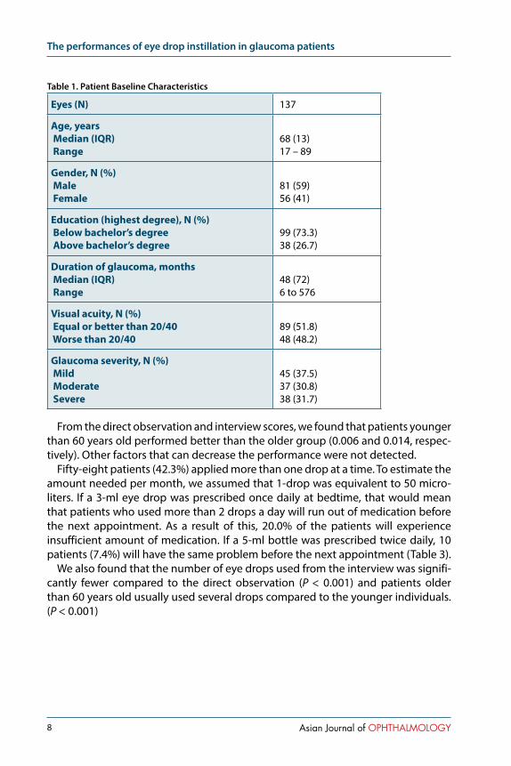
ResultsOne hundred and thirty-seven glaucoma patients were enrolled in the study. The demographic data of the study sample is shown in Table 1. Our study showed that under direct observation, only 1 patient (0.7%) correctly applied the eye drop according to the 5 required criteria mentioned above whereas 9 patients (6.6%) could not accomplish any of the criteria (Fig. 1).
Only 29 patients (21.2%) accomplished the success criteria. According to the interview score, 22 patients (16.1%) got full marks and 3 patients (2.2%) did not know the correct techniques for eye drop application. The results of each applica-tion step and interview are shown in Figure 2.
The interview results showed that most of our patients knew the correct techniques, especially for washing their hands before application because 105 patients (75.6%) reported performing this procedure before applying the medica-tion (Fig. 2). However, only one-third of the patients knew that they had to apply the eye drop into the lower fornix. In addition, more than half of the patients contami-nated the tip of the eye drop bottle by touching it to either the eye or adnexa. Hence, the performances of the eye drop instillation from the interview scores were statistically significantly higher than the observed scores (P < 0.001). The factors associated with the patients’ performance for eye drop application are shown in Table 2.
The performances of eye drop instillation in glaucoma patients
8 Asian Journal of OPHTHALMOLOGY
Table 1. Patient Baseline Characteristics
Eyes (N) 137
Age, years Median (IQR) Range
68 (13)17 – 89
Gender, N (%) Male Female
81 (59)56 (41)
Education (highest degree), N (%) Below bachelor’s degree Above bachelor’s degree
99 (73.3)38 (26.7)
Duration of glaucoma, months Median (IQR) Range
48 (72)6 to 576
Visual acuity, N (%) Equal or better than 20/40 Worse than 20/40
89 (51.8)48 (48.2)
Glaucoma severity, N (%) Mild Moderate Severe
45 (37.5)37 (30.8)38 (31.7)
From the direct observation and interview scores, we found that patients younger than 60 years old performed better than the older group (0.006 and 0.014, respec-tively). Other factors that can decrease the performance were not detected.
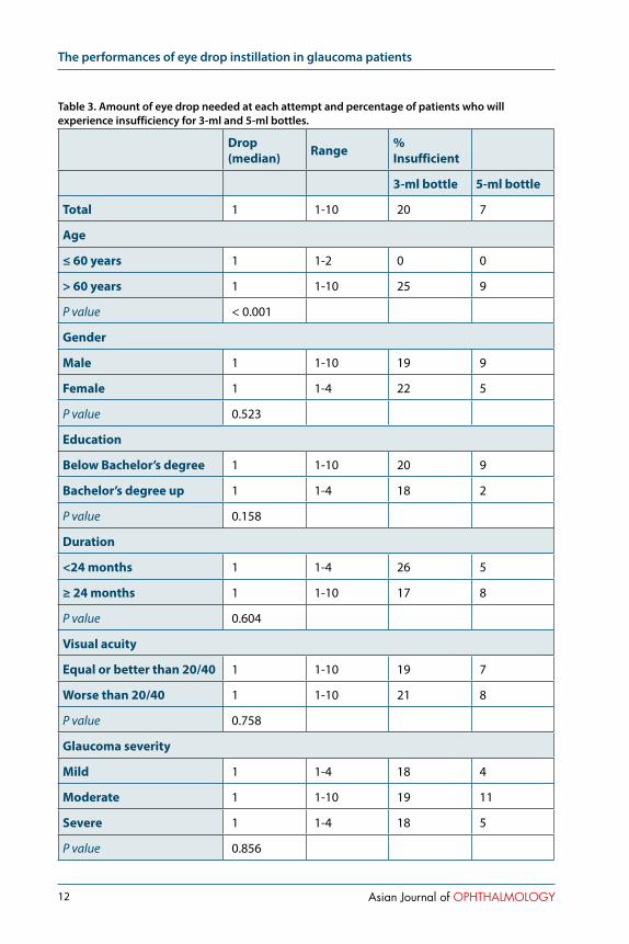
Fifty-eight patients (42.3%) applied more than one drop at a time. To estimate the amount needed per month, we assumed that 1-drop was equivalent to 50 micro-liters. If a 3-ml eye drop was prescribed once daily at bedtime, that would mean that patients who used more than 2 drops a day will run out of medication before the next appointment. As a result of this, 20.0% of the patients will experience insufficient amount of medication. If a 5-ml bottle was prescribed twice daily, 10 patients (7.4%) will have the same problem before the next appointment (Table 3).
We also found that the number of eye drops used from the interview was signifi-cantly fewer compared to the direct observation (P < 0.001) and patients older than 60 years old usually used several drops compared to the younger individuals. (P < 0.001)

Tangseepha, Manassakorn
9Asian Journal of OPHTHALMOLOGY
Table 2. Summary of observation and interview scores
Score Observation Interview
Median (points) Median (points)
Age
≤ 60 years 3 4
> 60 years 2 3
P value 0.006 0.014
Gender
Male 2 3
Female 2 3
P value 0.704 0.574
Education
Below Bachelor’s degree 2 3
Bachelor’s degree up 3 4
P value 0.534 0.123
Duration
< 24 months 2 3
≥ 24 months 2 3
P value 0.690 0.425
Visual acuity
Equal or better than 20/40 2 3
Worse than 20/40 2 3
P value 0.235 0.527
Glaucoma severity
Mild 2 3
Moderate 2 3
Severe 2 3
P value 0.349 0.746

The performances of eye drop instillation in glaucoma patients
10 Asian Journal of OPHTHALMOLOGY
Fig 2. Distribution of observation and interview score
Fig 1. Performance of eye drop instillation according to observational score and interview score
Tangseepha, Manassakorn
11Asian Journal of OPHTHALMOLOGY
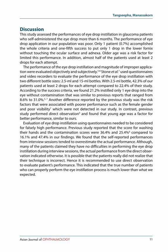
DiscussionThis study assessed the performances of eye drop instillation in glaucoma patients who self-administered the eye drop more than 6 months. The performance of eye drop application in our population was poor. Only 1 patient (0.7%) accomplished the whole criteria and one-fifth success to put only 1 drop in the lower fornix without touching the ocular surface and adnexa. Older age was a risk factor for limited this performance. In addition, almost half of the patients used at least 2 drops for each attempt.
The performance of the eye drop instillation and magnitude of improper applica-tion were evaluated objectively and subjectively.5-8 Stone et al.7 used questionnaires and video recorders to evaluate the performance of the eye drop instillation with two different bottle sizes: 2.5-ml and 15-ml bottles. With 2.5-ml bottle, 42.3% of our patients used at least 2 drops for each attempt compared to 22.4% of their study. According to the success criteria, we found 21.2% instilled only 1 eye drop into the eye without contamination that was similar to previous reports that ranged from 8.6% to 31.0%.5, 7 Another difference reported by the previous study was the risk factors that were associated with poorer performance such as the female gender and poor visibility7 which were not detected in our study. In contrast, previous study performed direct observation9 and found that young age was a factor for better performance, similar to ours.
Evaluation of eye drop instillation using questionnaires needed to be considered for falsely high performance. Previous study reported that the score for washing their hands and the contamination scores were 36.4% and 25.4%6 compared to 16.1% and 47.4% in our findings. We found that the self-reported performances from interview sessions tended to overestimate the actual performance. Although, many of the patients claimed they have no difficulties in performing the eye drop instillation during interview sessions, the actual performance from the direct obser-vation indicated otherwise. It is possible that the patients really did not realize that their technique is incorrect. Hence it is recommended to use direct observation to evaluate patient’s performance. This indicated that the true number of patients who can properly perform the eye instillation process is much lower than what we expected.
The performances of eye drop instillation in glaucoma patients
12 Asian Journal of OPHTHALMOLOGY
Table 3. Amount of eye drop needed at each attempt and percentage of patients who will experience insufficiency for 3-ml and 5-ml bottles.
Drop (median) Range %
Insufficient
3-ml bottle 5-ml bottle
Total 1 1-10 20 7
Age
≤ 60 years 1 1-2 0 0
> 60 years 1 1-10 25 9
P value < 0.001
Gender
Male 1 1-10 19 9
Female 1 1-4 22 5
P value 0.523
Education
Below Bachelor’s degree 1 1-10 20 9
Bachelor’s degree up 1 1-4 18 2
P value 0.158
Duration
<24 months 1 1-4 26 5
≥ 24 months 1 1-10 17 8
P value 0.604
Visual acuity
Equal or better than 20/40 1 1-10 19 7
Worse than 20/40 1 1-10 21 8
P value 0.758
Glaucoma severity
Mild 1 1-4 18 4
Moderate 1 1-10 19 11
Severe 1 1-4 18 5
P value 0.856

Tangseepha, Manassakorn
13Asian Journal of OPHTHALMOLOGY
Aside from the performance of the eye drop instillation, the number of drops used is equally important. In another direct observational study, it was reported that the patients used 1 – 8 drops for each attempt.5 This indicated that the physi-cian should always prescribe extra medication because it is unrealistic to assume that one drop will be accomplished per attempt or instruct the patients to come in for a refill when they run out of medication regardless of their next appointment. Although the results from previous studies varied due to the different settings, we can imply that the performance of eye drop application was poor worldwide. As a result of this, it is even more pertinent that physicians should assess the proper eye instillation process before assuming the dosage and/or efficacy of the medication need to be adjusted or changed.
There were some limitations in our study. We used artificial tear eye drops so the size and shape of the bottles varied which could have affected the applica-tion process. However, we selected bottles composed of the same material to avoid differences in the pressure that is used to apply the medication. Second, the setting during direct observation such as the lighting and having adequate space to move in may not be the same as the patients’ home which could have altered their perfor-mances. Lastly, we did not explore adherence, persistence, handling and storage of the medication in this study. Additional larger study incorporating all of these issues mentioned above is warranted.
In conclusion, our findings were consistent with previous reports that even in experienced patients, proper eye drop instillation was poor. We recommend that training or retraining patients and their relatives is necessary to improve this task. Not only for glaucoma management and prevention of side effects but it will also prevent unnecessary expenses. Physicians need to be aware that extra quantity of the medication should be incorporated into the dosage calculation, especially for the elderly patients.
References1. Okeke CO, Quigley HA, Jampel HD, Ying GS, Plyler RJ, Jiang, Y, et al. Adherence with topical
glaucoma medication monitored electronically the Travatan Dosing Aid study. Ophthalmology. 2009;116(2):191-199.
2. Hermann MM, Bron AM, Creuzot-Garcher CP, Diestelhorst, M. Measurement of Adherence to Brimonidine Therapy for Glaucoma Using Electronic Monitoring. J Glaucoma. 2011;20(8):502-508.
3. Reardon G, Kotak S, Schwartz GF. Objective assessment of compliance and persistence among patients treated for glaucoma and ocular hypertension: a systematic review. Patient Prefer Adher. 2011;5:441-463.
4. Sleath B, Robin AL, Covert D, Byrd JE, Tudor G, Svarstad B. Patient-reported behavior and problems in using glaucoma medications. Ophthalmology. 2006;113(3):431-436.
5. Gupta R, Patil B, Shah BM, Bali SJ, Mishra SK, Dada T. Evaluating eye drop instillation technique in glaucoma patients. J Glaucoma. 2012;21(3):189-192.
6. Tsai T, Robin AL, Smith JP. An Evaluation of How Glaucoma Patients Use Topical Medications: A Pilot Study. Trans Am Ophthalmol Soc. 2007;105:7.
7. Stone JL, Robin AL, Novack GD, Covert DW, Cagle GD. An objective evaluation of eyedrop instillation in patients with glaucoma. Arch Ophthalmol. 2009;127(6):732-736.
The performances of eye drop instillation in glaucoma patients
14 Asian Journal of OPHTHALMOLOGY
8. Hennessy AL, Katz J, Covert D, Protzko C, Robin AL. Videotaped evaluation of eyedrop instillation in glaucoma patients with visual impairment or moderate to severe visual field loss. Ophthalmology. 2010;117(12):2345-2352.
9. Aptel F, Masset H, Burillon C, Robin A, Denis P. The influence of disease severity on quality of eye-drop administration in patients with glaucoma or ocular hypertension. Br J Ophthalmol. 2009;93(5):700-701.
15Asian Journal of OPHTHALMOLOGY
Determinants and outcome of periocular dirofilariasis in a cohort of patients with demonstrable live worm from the ocular and adnexal parasitic granulomas
Padma Balagopal Prabhu, Kuzhupally Vallon RajuDepartment of Ophthalmology, Government Medical college, Kozhikode, Kerala, India
AbstractPurpose: We attempt to describe the unique diagnostic features of dirofilariasis affecting the eye, a rare disease caused by the nematode dirofilaria repens.Case report: The cohort includes 5 adult cases of ocular dirofilariasis. Migratory oedema was present in all but one case. The occurrence of the lesions near the medial canthus in all the cases including subconjunctival mass suggests predictable pattern of migration of the worm. Absence of systemic eosinophila and lack of marked eosinophilic infiltration around the parasitic granuloma in histopathology indicates alternative immune response against the parasite. Persistence of live worm despite antihelminthic drugs can be accounted by the presence of a thick capsule which protects the filaria against adulticidal and larvicidal drugs. Surgical exicision was curative in all cases.Conclusion: Our case series points to the importance of having high index of suspicion and early detection of ocular dirofilariasis as it is amenable to simple and effective treatment.
Key words: Dirofilaria repens, zoonosis, migratory oedema, eosinophilia,
IntroductionDirofilariasis is an emerging zoonosis in India.1 Pulmonary, cardiovascular, perio-cular, intraocular and orbital involvement has been documented both in endemic and nonendemic areas with dirofilariasis.1,2 Scientific information available in the international literature is limited to isolated case reports from different parts of the world.3 This data is insufficient to provide a clear and comprehensive concept regarding the clinical picture, investigative modalities and outcome of treatment in a case of suspected ocular dirofilariasis. Five cases of diagnosed ocular dirofilariasis are reported with an attempt to analyse the diagnostic features and treatment outcome of this rare but evolving entity.
Case detailsThis is a retrospective data analysis of cases diagnosed as periorbital and ocular dirofilariasis confirmed by demonstration of worm (either dead or alive) on excision biopsy during the period of one year. Informed consent was obtained from the
Correspondence: Padma Balagopal Prabhu, Department of OPHthalmology, Government Medical College, Kozhikode, Kerala, India 673008E-mail: [email protected]
Original ArticleAsian J Ophthalmol. 2016;15:015-024© Asian Journal of Ophthalmology
Periocular dirofilariasis in patients with live worm from the ocular and adnexal parasitic granulomas
16 Asian Journal of OPHTHALMOLOGY
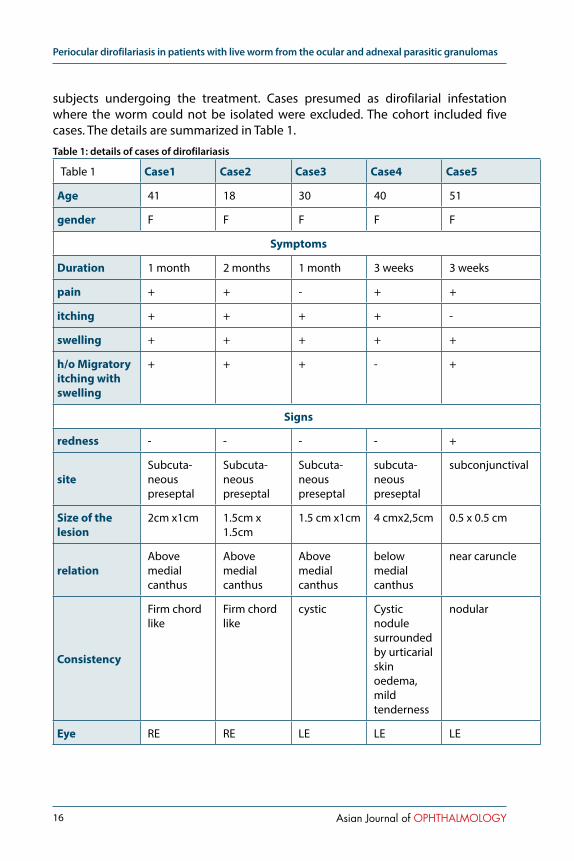
subjects undergoing the treatment. Cases presumed as dirofilarial infestation where the worm could not be isolated were excluded. The cohort included five cases. The details are summarized in Table 1.
Table 1: details of cases of dirofilariasis
Table 1 Case1 Case2 Case3 Case4 Case5
Age 41 18 30 40 51
gender F F F F F
Symptoms
Duration 1 month 2 months 1 month 3 weeks 3 weeks
pain + + - + +
itching + + + + -
swelling + + + + +
h/o Migratory itching with swelling
+ + + - +
Signs
redness - - - - +
siteSubcuta-neouspreseptal
Subcuta-neouspreseptal
Subcuta-neouspreseptal
subcuta-neouspreseptal
subconjunctival
Size of the lesion
2cm x1cm 1.5cm x 1.5cm
1.5 cm x1cm 4 cmx2,5cm 0.5 x 0.5 cm
relationAbove medial canthus
Above medial canthus
Above medial canthus
below medial canthus
near caruncle
Consistency
Firm chord like
Firm chord like
cystic Cystic nodule surrounded by urticarial skin oedema, mild tenderness
nodular
Eye RE RE LE LE LE

Prabhu, Raju
17Asian Journal of OPHTHALMOLOGY
Table 1 Case1 Case2 Case3 Case4 Case5
Eye examination
WNL BCVA 20/20 F=WNL
WNL BCVA 20/20 F=WNL
WNL BCVA 20/20 F=WNL
WNL BCVA 20/20 F=WNL
Subconj nodular tender swelling near caruncle,WNL BCVA 20/20 F=WNL
Investigations
Blood routine WNL WNL WNL WNL WNL
Peripheral smear
WNL WNL WNL WNL WNL
AEC 380 414 670 458 529
Pretreatment
Tab Alben-dazole , tab predniso-lone X 3 weeks
Tab Alben-dazole , tab predniso-lone X 3 weeks
Tab Alben-dazole , tab predniso-lone X 3 weeks
Tab Alben-dazole , tab predniso-lone X 1 week
---------
Excision
capsule Thick illdefined
Thick illdefined
Thick illdefined
Thick illdefined
Thick illdefined
Muscle infiltration
Orbicularis oculi
Orbicularis oculi
Orbicularis oculi
Orbicularis oculi
Medial rectus
pus - - - - +
Status of worm live live live live live
size of the worm
51 x 0.6mm 47 x 0.6mm 60 x 0.5mm 87 x 0.7mm 42 x 0.6 mm
Periocular dirofilariasis in patients with live worm from the ocular and adnexal parasitic granulomas
18 Asian Journal of OPHTHALMOLOGY
Table 1 Case1 Case2 Case3 Case4 Case5
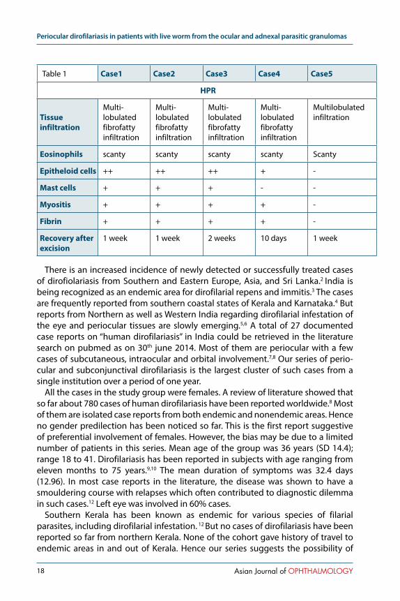
HPR
Tissue infiltration
Multi-lobulated fibrofatty infiltration
Multi-lobulated fibrofatty infiltration
Multi-lobulated fibrofatty infiltration
Multi-lobulated fibrofatty infiltration
Multilobulated infiltration
Eosinophils scanty scanty scanty scanty Scanty
Epitheloid cells ++ ++ ++ + -
Mast cells + + + - -
Myositis + + + + -
Fibrin + + + + -
Recovery after excision
1 week 1 week 2 weeks 10 days 1 week
There is an increased incidence of newly detected or successfully treated cases of dirofiolariasis from Southern and Eastern Europe, Asia, and Sri Lanka.2 India is being recognized as an endemic area for dirofilarial repens and immitis.3 The cases are frequently reported from southern coastal states of Kerala and Karnataka.4 But reports from Northern as well as Western India regarding dirofilarial infestation of the eye and periocular tissues are slowly emerging.5,6 A total of 27 documented case reports on “human dirofilariasis” in India could be retrieved in the literature search on pubmed as on 30th june 2014. Most of them are periocular with a few cases of subcutaneous, intraocular and orbital involvement.7,8 Our series of perio-cular and subconjunctival dirofilariasis is the largest cluster of such cases from a single institution over a period of one year.
All the cases in the study group were females. A review of literature showed that so far about 780 cases of human dirofilariasis have been reported worldwide.8 Most of them are isolated case reports from both endemic and nonendemic areas. Hence no gender predilection has been noticed so far. This is the first report suggestive of preferential involvement of females. However, the bias may be due to a limited number of patients in this series. Mean age of the group was 36 years (SD 14.4); range 18 to 41. Dirofilariasis has been reported in subjects with age ranging from eleven months to 75 years.9,10 The mean duration of symptoms was 32.4 days (12.96). In most case reports in the literature, the disease was shown to have a smouldering course with relapses which often contributed to diagnostic dilemma in such cases.12 Left eye was involved in 60% cases.
Southern Kerala has been known as endemic for various species of filarial parasites, including dirofilarial infestation. 12 But no cases of dirofilariasis have been reported so far from northern Kerala. None of the cohort gave history of travel to endemic areas in and out of Kerala. Hence our series suggests the possibility of
Prabhu, Raju
19Asian Journal of OPHTHALMOLOGY
endemicity in northern Kerala as well.The symptoms at presentation were varied. Pain was present in 4/5 cases.
Erythema was noticed only in one patient. Itching and swelling occurred in all patients. History of migratory oedema was appreciated by 4 subjects. Migratory oedema with itching is suggestive of subcutaneous larva migrans.
Migratory skin edema is frequently reported in dirofilarial infestation involving subcutaneous tissue in other areas of the body also.13,14 The classical description is of creeping erruptions characterized by local swellings with changing locations. However continuous migration as in our series as well as isolated and scattered urticarial reactions have been reported. Similar findings have been noted among canine and feline community infested with Dirofilaria repens and immitis.22 In them, pruritic dermatitis spreading to the adjacent region creating large areas of alopecia has been described.
Presence of itching out of proportion to the pain (and tenderness), associated urticarial reaction in the surrounding dermis, migration of the itchy oedematous areas contiguously to the adjacent site over a period of days and absence of leuko-cytosis and raised ESR led to the suspicion of dirofilarial infestation in the cases.
Dirofilariasis can present as an inflammatory mass or noninflammatory nodules.14,
15 Among inflammatory cases cellulitis like presentation is rarely reported. Itching with or without tender swelling is the usual history. In our series all the cases with pain at the onset had live worms contrary to the observation that inflammation is often associated with dead worm due to arthus like response to the parasitic debris.14 A careful history can give valuable clues regarding the diagnosis in such cases.
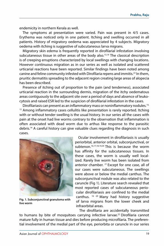
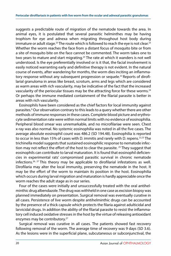
Ocular involvement in dirofilariasis is usually periorbital, anterior orbital, subconjunctival, or subtenon.16,17,18,19,20 This is because the worm has affinity for the subcutaneous tissues. In these cases, the worm is usually well local-ized. Rarely live worm has been isolated from anterior chamber. 21 Except for one case, all of our cases were subcutaneous. The swellings were above or below the medial canthus. The subconjunctival nodule was also related to the caruncle (Fig 1). Literature search revealed that most reported cases of subcutaneous perio-cular dirofilariasis are confined to the medial canthus. 16 -20 Many had history suggestive of larva migrans from the lower cheek and infraorbital areas.
The dirofilaria are accidentally transmitted to humans by bite of mosquitoes carrying infective larvae.22 Dirofilaria cannot mature fully in human tissue and dies before producing microfilaria. The preferen-tial involvement of the medial part of the eye, periorbita or caruncle in our series
Fig. 1. Subconjunctival granuloma with live worm
Periocular dirofilariasis in patients with live worm from the ocular and adnexal parasitic granulomas
20 Asian Journal of OPHTHALMOLOGY
suggests a predictable route of migration of the nematode towards the area. In animal eyes, it is postulated that several parasitic helminthes may be having trophism for eye and adnexa when migrating throughout host body during immature or adult stage.23 The route which is followed to reach the eye is not clear.23
Whether the worm reaches the face from a distant focus of mosquito bite or from a site of mosquito bite on the face cannot be commented. The worm takes one to two years to mature and start migrating.23 The rate at which it wanders is not well understood. Is the eye preferentially involved or is it that, the facial involvement is easily noticed warranting early and definitive therapy is not evident. In the natural course of events, after wandering for months, the worm dies inciting an inflamma-tory response without any subsequent progression or sequele.22 Reports of dirofi-larial granuloma in areas like breast, scrotum, arms and legs which are considered as warm areas with rich vascularity, may be indicative of the fact that the increased vascularity of the periocular tissues may be the attracting force for these worms.24 Or perhaps the immune mediated containment of the filarial parasite is better in areas with rich vascularity.
Eosinophils have been considered as the chief factors for local immunity against parasites.6 Our observation contrary to this leads to a query whether there are other methods of immune responses in these cases. Complete blood picture and erythro-cyte sedimentation rate were within normal limits with no evidence of eosinophilia. Peripheral blood smear was unremarkable, and no microfilariae were seen. Chest x-ray was also normal. No systemic eosinophilia was noted in all the five cases. The average absolute eosinophil count was 486.2 (SD 194.48). Eosinophilia is reported to occur in less than 15% of cases with D. immitis and rarely with D. repens.22,23 The trichinella model suggests that sustained eosinophilic response to nematode infec-tion may not reflect the effort of the host to clear the parasite. 25 They suggest that eosinophils can contribute to larval maturation. It is found that eosinophil deficien-cies in experimental rats’ compromised parasitic survival in chronic nematode infections.26, 27 This theory may be applicable to dirofilarial infestations as well. Dirofilaria may alter the local immunity, preserving the nematode in the host. It may be the effort of the worm to maintain its position in the host. Eosinophilia which occurs during larval migration and maturation is hardly appreciable once the worm reaches the adult stage as in our series.
Four of the cases were initially and unsuccessfully treated with the oral antihel-minthic drug albendazole. The drug was withheld in one case as excision biopsy was planned immediately on presentation. Surgical removal was eventually curative in all cases. Persistence of live worm despite antihelminthic drugs can be accounted by the presence of a thick capsule which protects the filaria against adulticidal and larvicidal drugs. In addition the ability of the filarial parasite to resist the inflamma-tory cell induced oxidative stresses in the host by the virtue of releasing antioxidant enzymes may be contributory.27
Surgical removal was curative in all cases. The patients showed fast recovery following removal of the worm. The average time of recovery was 9 days (SD 3.6). As the lesions were in the superficial plane, subcutaneous or subconjunctival, the
Prabhu, Raju
21Asian Journal of OPHTHALMOLOGY
approach to the mass in each case was simple. However attachment to the under-lying muscle, orbicularis oculi and medial rectus noted among all subjects in our series, has to be kept in mind.
Simple extraction of the worm is the treatment of choice for human dirofila-riasis.28 Unlike D. immitis which requires the use of anti-helminthic agents, use of antifilarial medication for D. repens is not indicated in the literature. In a small number of cases of D. repens, ivermectin and/or diethylcarbamazine has been tried with good results.
The plane of excision was difficult to ascertain due to the absence of a well defined capsule in all the cases. The worms, which were alive in all the subjects, were removed in toto. The surrounding tissue sample was taken for confirmation of the granuloma without disturbing the normal anatomy as far as possible in view of the close relation of the granuloma to the underlying muscle. The cystic cavity containing the worm showed pus only in case 5. However there were no significant signs of inflammation noted postoperatively.
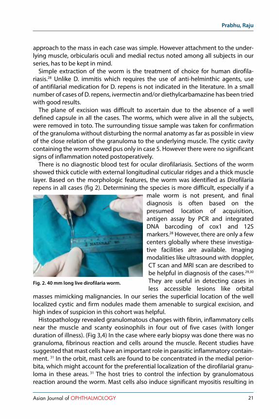
There is no diagnostic blood test for ocular dirofilariasis. Sections of the worm showed thick cuticle with external longitudinal cuticular ridges and a thick muscle layer. Based on the morphologic features, the worm was identified as Dirofilaria repens in all cases (fig 2). Determining the species is more difficult, especially if a
male worm is not present, and final diagnosis is often based on the presumed location of acquisition, antigen assay by PCR and integrated DNA barcoding of cox1 and 12S markers.28 However, there are only a few centers globally where these investiga-tive facilities are available. Imaging modalities like ultrasound with doppler, CT scan and MRI scan are described to be helpful in diagnosis of the cases.29,30 They are useful in detecting cases in less accessible lesions like orbital
masses mimicking malignancies. In our series the superficial location of the well localized cystic and firm nodules made them amenable to surgical excision, and high index of suspicion in this cohort was helpful.
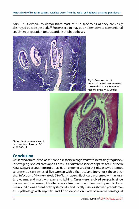
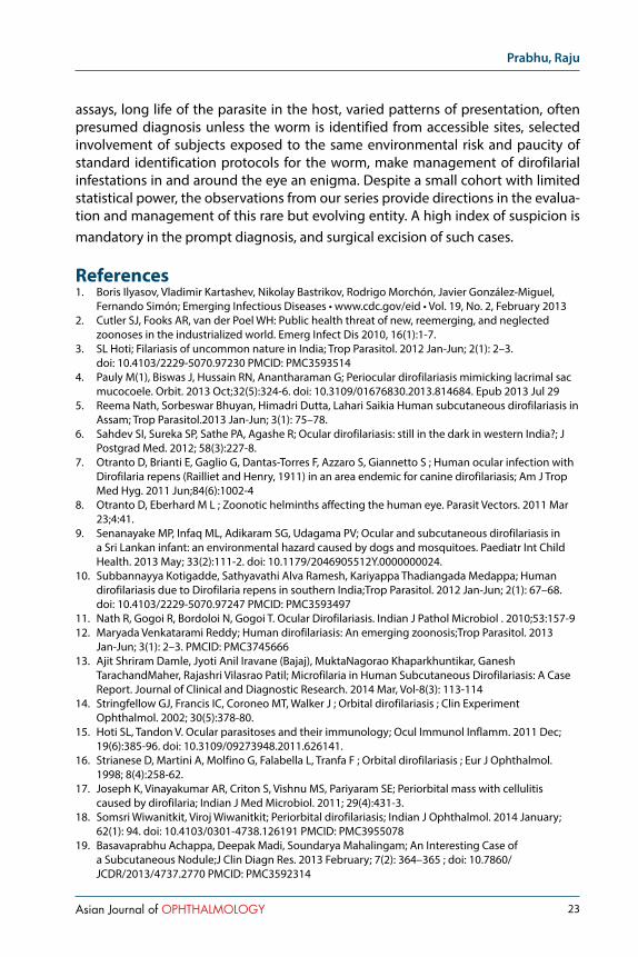
Histopathology revealed granulomatous changes with fibrin, inflammatory cells near the muscle and scanty eosinophils in four out of five cases (with longer duration of illness). (Fig 3,4) In the case where early biopsy was done there was no granuloma, fibrinous reaction and cells around the muscle. Recent studies have suggested that mast cells have an important role in parasitic inflammatory contain-ment. 31 In the orbit, mast cells are found to be concentrated in the medial perior-bita, which might account for the preferential localization of the dirofilarial granu-loma in these areas. 31 The host tries to control the infection by granulomatous reaction around the worm. Mast cells also induce significant myositis resulting in
Fig. 2. 40 mm long live dirofilaria worm.
Periocular dirofilariasis in patients with live worm from the ocular and adnexal parasitic granulomas
22 Asian Journal of OPHTHALMOLOGY
pain.31 It is difficult to demonstrate mast cells in specimens as they are easily destroyed outside the body.32 Frozen section may be an alternative to conventional specimen preparation to substantiate this hypotheses.
ConclusionOcular and orbital dirofilariasis continues to be recognized with increasing frequency, in new geographical areas and as a result of different species of parasites. Northern Kerala, a part of southern India may be an endemic area for this disease. We attempt to present a case series of five women with either ocular adnexal or subconjunc-tival infection of the nematode Dirofilaria repens. Each case presented with migra-tory edema, and most with pain and itching. Cases were resolved surgically, since worms persisted even with albendazole treatment combined with prednisolone. Eosinophilia was absent both systemically and locally. Tissues showed granuloma-tous pathology with myositis and fibrin deposition. Lack of reliable serological
Fig. 3. Cross section of dirofilarial worm in tissue with surrounding granulomatous response H&E X40 300 dpi
Fig. 4. Higher power view of cross section of worm H&E X200 300dpi
Prabhu, Raju
23Asian Journal of OPHTHALMOLOGY
assays, long life of the parasite in the host, varied patterns of presentation, often presumed diagnosis unless the worm is identified from accessible sites, selected involvement of subjects exposed to the same environmental risk and paucity of standard identification protocols for the worm, make management of dirofilarial infestations in and around the eye an enigma. Despite a small cohort with limited statistical power, the observations from our series provide directions in the evalua-tion and management of this rare but evolving entity. A high index of suspicion is mandatory in the prompt diagnosis, and surgical excision of such cases.
References1. Boris Ilyasov, Vladimir Kartashev, Nikolay Bastrikov, Rodrigo Morchón, Javier González-Miguel,
Fernando Simón; Emerging Infectious Diseases • www.cdc.gov/eid • Vol. 19, No. 2, February 20132. Cutler SJ, Fooks AR, van der Poel WH: Public health threat of new, reemerging, and neglected
zoonoses in the industrialized world. Emerg Infect Dis 2010, 16(1):1-7.3. SL Hoti; Filariasis of uncommon nature in India; Trop Parasitol. 2012 Jan-Jun; 2(1): 2–3.
doi: 10.4103/2229-5070.97230 PMCID: PMC35935144. Pauly M(1), Biswas J, Hussain RN, Anantharaman G; Periocular dirofilariasis mimicking lacrimal sac
mucocoele. Orbit. 2013 Oct;32(5):324-6. doi: 10.3109/01676830.2013.814684. Epub 2013 Jul 295. Reema Nath, Sorbeswar Bhuyan, Himadri Dutta, Lahari Saikia Human subcutaneous dirofilariasis in
Assam; Trop Parasitol.2013 Jan-Jun; 3(1): 75–78.6. Sahdev SI, Sureka SP, Sathe PA, Agashe R; Ocular dirofilariasis: still in the dark in western India?; J
Postgrad Med. 2012; 58(3):227-8.7. Otranto D, Brianti E, Gaglio G, Dantas-Torres F, Azzaro S, Giannetto S ; Human ocular infection with
Dirofilaria repens (Railliet and Henry, 1911) in an area endemic for canine dirofilariasis; Am J Trop Med Hyg. 2011 Jun;84(6):1002-4
8. Otranto D, Eberhard M L ; Zoonotic helminths affecting the human eye. Parasit Vectors. 2011 Mar 23;4:41.
9. Senanayake MP, Infaq ML, Adikaram SG, Udagama PV; Ocular and subcutaneous dirofilariasis in a Sri Lankan infant: an environmental hazard caused by dogs and mosquitoes. Paediatr Int Child Health. 2013 May; 33(2):111-2. doi: 10.1179/2046905512Y.0000000024.
10. Subbannayya Kotigadde, Sathyavathi Alva Ramesh, Kariyappa Thadiangada Medappa; Human dirofilariasis due to Dirofilaria repens in southern India;Trop Parasitol. 2012 Jan-Jun; 2(1): 67–68. doi: 10.4103/2229-5070.97247 PMCID: PMC3593497
11. Nath R, Gogoi R, Bordoloi N, Gogoi T. Ocular Dirofilariasis. Indian J Pathol Microbiol . 2010;53:157-912. Maryada Venkatarami Reddy; Human dirofilariasis: An emerging zoonosis;Trop Parasitol. 2013
Jan-Jun; 3(1): 2–3. PMCID: PMC374566613. Ajit Shriram Damle, Jyoti Anil Iravane (Bajaj), MuktaNagorao Khaparkhuntikar, Ganesh
TarachandMaher, Rajashri Vilasrao Patil; Microfilaria in Human Subcutaneous Dirofilariasis: A Case Report. Journal of Clinical and Diagnostic Research. 2014 Mar, Vol-8(3): 113-114
14. Stringfellow GJ, Francis IC, Coroneo MT, Walker J ; Orbital dirofilariasis ; Clin Experiment Ophthalmol. 2002; 30(5):378-80.
15. Hoti SL, Tandon V. Ocular parasitoses and their immunology; Ocul Immunol Inflamm. 2011 Dec; 19(6):385-96. doi: 10.3109/09273948.2011.626141.
16. Strianese D, Martini A, Molfino G, Falabella L, Tranfa F ; Orbital dirofilariasis ; Eur J Ophthalmol. 1998; 8(4):258-62.
17. Joseph K, Vinayakumar AR, Criton S, Vishnu MS, Pariyaram SE; Periorbital mass with cellulitis caused by dirofilaria; Indian J Med Microbiol. 2011; 29(4):431-3.
18. Somsri Wiwanitkit, Viroj Wiwanitkit; Periorbital dirofilariasis; Indian J Ophthalmol. 2014 January; 62(1): 94. doi: 10.4103/0301-4738.126191 PMCID: PMC3955078
19. Basavaprabhu Achappa, Deepak Madi, Soundarya Mahalingam; An Interesting Case of a Subcutaneous Nodule;J Clin Diagn Res. 2013 February; 7(2): 364–365 ; doi: 10.7860/JCDR/2013/4737.2770 PMCID: PMC3592314

Periocular dirofilariasis in patients with live worm from the ocular and adnexal parasitic granulomas
24 Asian Journal of OPHTHALMOLOGY
20. Ilyasov B, Kartashev V, Bastrikov N, Madjugina L, González-Miguel J, Morchón R, Simón F; Thirty cases of human subcutaneous dirofilariasis reported in Rostov-on-Don(Southwestern Russian Federation). Enferm Infecc Microbiol Clin; 2014 Jun 16. pii: S0213-005X(14)00181-5. doi:10.1016/j.eimc.2014.04.002.
21. Viney Gupta, Preeti Sankaran, Mohanraj, Jyotish Chandra Samantaray, Vimla Menon; Bilateral intraocular dirofilariasis ; Indian J Ophthalmol;2014 March; 62(3): 357–358. doi: 10.4103/0301-4738.116252 PMCID: PMC4061683
22. Walter Tarello ; Review Article Clinical Aspects of Dermatitis Associated with Dirofilaria repens in Pets: A Review of 100 Canine and 31 Feline Cases (1990–2010) and a Report of a New Clinic Case Imported from Italy to Dubai Journal of Parasitology Research Volume 2011, Article ID 578385, 7 pages doi:10.1155/2011/578385
23. Simón F, Siles-Lucas M, Morchón R, González-Miguel J, Mellado I, Carretón E, et al; Human and animal dirofilariasis; The emergence of a zoonotic mosaic; Clin Microbiol Rev. 2012;25:471–506. http:// dx.doi.org/10.1128/CMR.00012-12
24. J M Conly, L H Sekla, D E Low; Dirofilariasis presenting as a breast lump;Can Med Assoc J. 1984 June 15; 130(12): 1575–1576. PMCID: PMC1483366
25. Gottstein B, Pozio E, Nöckler K; Epidemiology, diagnosis, treatment, and control of trichinellosis; Clin Microbiol Rev 2009. 22(1):127-145.
26. Bruschi F, Murrell KD;Trichinellosis. In Tropical Infectious Diseases Principals, Pathogens, and Practices. 2 edition. Edited by: Guerrant RL, WalkerDH, Weller PF. Philadelphia: Churchill Livingstone. 2005:1225-1230
27. Kociecki J, Kociecka W; Visual system involvement in selected zoonotic diseases. II Trichinellosis; Klin Oczna 2004. 106(3):371-375.
28. Otranto D, Diniz DG, Dantas-Torres F, Casiraghi M, de Almeida INF, de Almeida LNF, Nascimento dos Santos J, Penha Furtado A, de Almeida Sobrinho AF, Bain O; Human intraocular filariasis caused by Dirofilaria sp., Brazil; Emerg Infect Dis. 2011.
29. M Smitha, VR Rajendran, E Devarajan, PM Anitha; Case report: Orbital dirofilariasis,Indian J Radiol Imaging. 2008 February; 18(1): 60–62. doi: 10.4103/0971-3026.37050PMCID: PMC2766890
30. Thandre N Gopinath, K P Lakshmi, P C Shaji, P C Rajalakshmi; Periorbital dirofilariasis—Clinical and imaging findings: Live worm on ultrasound; Indian J Ophthalmol. 2013 June; 61(6): 298–300. doi: 10.4103/0301-4738.114111 PMCID: PMC3744785
31. McMenamin PG, Morrison SM, McMenamin C; Immunomorphologic studies of mast cell heterogeneity, location, and distribution in the rat conjunctiva ; J allergy clin immunol. 1996;97(6):1375-86.
32. Ribatti D, Crivellato E; Mast cell ontogeny: an historical overview. Immunol Lett 2014 May-Jun; 159(1-2):11-4. doi: 10.1016/j.imlet.2014.02.003. Epub 2014 Feb 14.

25Asian Journal of OPHTHALMOLOGY
Correlation of refractive error with axial length and corneal topography
Poonam Kishore1, Vinita Singh1, Nitin Chaudhary2, Surabhi Ruia1
1 Department of Ophthalmology, King George’s Medical University, Lucknow, India; 2 Swastik Eyecare Centre, Unnao, India
AbstractPurpose: To collect and analyze normative data about corneal topography and axial length in various refractive errors in Indian population.Design: Cross-sectional observational study.Materials and Method: Three hundred eyes (150 patients) of age group 12-35 yrs were arranged in 5 groups according to refractive status; Group 1 (n=44): myopia of Spherical Equivalent (SE) > 6 D; Group 2 (n=67): myopia of SE >0.5 D to 6 D; Group 3 (n=88): nearly emmetropic of SE -0.5 D to +0.5 D; Group 4 (n=59): hypermetropia of SE >0.5 to 6 D; Group 5 (n=42): hypermetropia of SE > 6 D. Axial length(AL), central radius of curvature of cornea (CR), central power of cornea (CK) , Al/CR ratio for each group were documented . Correla-tion with SE and among each other was studied.Results: Mean AL (in mm) of myopic patients (n=111) was 24.23 ± 1.34, emmetropic (n=88) 22.62 ± 0.94 and hypermetropics (n=101) 20.73 ±0.94. Mean CR (in mm) of myopic patients was 7.55 ± 0.35, emmetropics was 7.70 ±0.32, and hypermetropes was 7.99 ±0.35. Mean CK (in D) of myopics was 44.86±2.59, emmetropes was 43.91±1.76, and hyperme-tropes was 42.32±1.89. Mean AL/CR ratio of myopics was 3.22 ± 0.29, emmetropics 2.94 ± 0.07, and hypermetropics 2.60 ± 0.19. AL was negatively correlated with SE(r=-0.91, p<0.0001) and positively with AL/CR(r=0.88, p<0.0001) and CK (r=0.36, p<0.0001). CR was negatively correlated with AL/CR (r=-0.74, p<0.0001) while positively correlated with SE (r=0.62, p<0.0001). CK showed positive correlation with AL/CR (r=0.75, p<0.0001) while negative correlation with SE (r=-0.61, p<0.0001). AL/CR was negatively correlated with SE(r=-0.95, p<0.0001).Conclusion: This study showed a negative correlation between axial length and refractive error and between AL/CR ratio and refractive error with stronger inverse relationship in hypermetropes than myopes. There was a positive correlation of CR with SE with a weaker direct relationship in myopes than hypermetropes.
Keywords: Axial length; central radius of curvature of cornea; corneal power; spherical equivalent.
IntroductionThe refractive state or spherical equivalent(SE) of the eye is determined by refrac-tive components (corneal power, lens power, anterior chamber depth, and axial length) which are interdependent rather than independent variables, and that the eye grows during the early years in life in such a manner that the refractive state tends towards emmetropia.1,2
Correspondence: Poonam Kishore, Department of Ophthalmology, King George’s Medical University, Lucknow, India. 226003.E-mail: [email protected]
Original ArticleAsian J Ophthalmol. 2016;15:025-033© Asian Journal of Ophthalmology
Correlation of refractive error with axial length and corneal topography
26 Asian Journal of OPHTHALMOLOGY
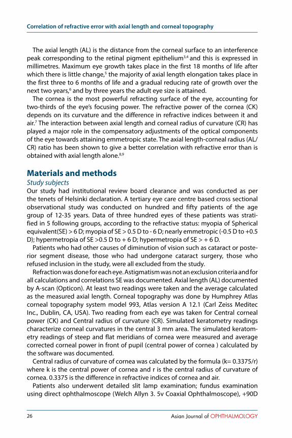
The axial length (AL) is the distance from the corneal surface to an interference peak corresponding to the retinal pigment epithelium3,4 and this is expressed in millimetres. Maximum eye growth takes place in the first 18 months of life after which there is little change,5 the majority of axial length elongation takes place in the first three to 6 months of life and a gradual reducing rate of growth over the next two years,6 and by three years the adult eye size is attained.
The cornea is the most powerful refracting surface of the eye, accounting for two-thirds of the eye’s focusing power. The refractive power of the cornea (CK) depends on its curvature and the difference in refractive indices between it and air.7 The interaction between axial length and corneal radius of curvature (CR) has played a major role in the compensatory adjustments of the optical components of the eye towards attaining emmetropic state. The axial length-corneal radius (AL/CR) ratio has been shown to give a better correlation with refractive error than is obtained with axial length alone.8,9
Materials and methodsStudy subjectsOur study had institutional review board clearance and was conducted as per the tenets of Helsinki declaration. A tertiary eye care centre based cross sectional observational study was conducted on hundred and fifty patients of the age group of 12-35 years. Data of three hundred eyes of these patients was strati-fied in 5 following groups, according to the refractive status: myopia of Spherical equivalent(SE) > 6 D; myopia of SE > 0.5 D to - 6 D; nearly emmetropic (-0.5 D to +0.5 D); hypermetropia of SE >0.5 D to + 6 D; hypermetropia of SE > + 6 D.
Patients who had other causes of diminution of vision such as cataract or poste-rior segment disease, those who had undergone cataract surgery, those who refused inclusion in the study, were all excluded from the study.
Refraction was done for each eye. Astigmatism was not an exclusion criteria and for all calculations and correlations SE was documented. Axial length (AL) documented by A-scan (Opticon). At least two readings were taken and the average calculated as the measured axial length. Corneal topography was done by Humphrey Atlas corneal topography system model 993, Atlas version A 12.1 (Carl Zeiss Meditec Inc., Dublin, CA, USA). Two reading from each eye was taken for Central corneal power (CK) and Central radius of curvature (CR). Simulated keratometry readings characterize corneal curvatures in the central 3 mm area. The simulated keratom-etry readings of steep and flat meridians of cornea were measured and average corrected corneal power in front of pupil (central power of cornea ) calculated by the software was documented.
Central radius of curvature of cornea was calculated by the formula (k= 0.3375/r) where k is the central power of cornea and r is the central radius of curvature of cornea. 0.3375 is the difference in refractive indices of cornea and air.
Patients also underwent detailed slit lamp examination; fundus examination using direct ophthalmoscope (Welch Allyn 3. 5v Coaxial Ophthalmoscope), +90D

Kishore, Singh, Chaudhary, et al
27Asian Journal of OPHTHALMOLOGY
Lens and indirect ophthalmoscope (IO-7 binocular indirect ophthalmoscope, Appaswami).
Statistical AnalysisData were summarized as Mean ± SD and percentage. The age and outcome measures (AL, CR, CK, AL/CR ratio) of five groups were compared by one way analysis of variance (ANOVA). The discrete (categorical) observations of sex of five groups were compared by chi-square (χ2) test. Pearson correlation analysis was used to assess association between the variables. Linear regresion was used to find the strength of associations between two continuous variables. A two-sided (α=2) p<0.05 was considered statistically significant. All analyses were performed on STATISTICA (window version 6.0).
ResultOn comparing the sex proportion (Male/Female), χ2 test revealed no significant difference in proportions of sex between the groups (χ2=6.79; p=0.1473) .The mean age of all groups show no significant difference (F=0.27, p=0.8941).
SE of the five groups was summarized as Group 1: -9.73 ± 5.37 ; Group 2: -2.35 ± 1.17; Group 3: 0.08 ± 0.30; Group 4: 3.20 ± 1.52; Group 5: 8.07 ± 2.0.
AL of the five groups was summarized as: Group 1: 25.25 ± 1.49; Group 2: 23.56 ± 0.62; Group 3: 22.62 ± 0.94; Group 4: 21.28 ± 0.71; Group 5:19.97 ± 0.64. . Linear regression analysis of AL and SE: (Fig. 1) showed decrease in AL with increase in SE from myopia towards hypermetropia.
CR of the five groups was summarized as: Group 1: 7.46 ± 0.44; Group 2: 7.61 ± 0.27; Group 3: 7.70 ± 0.32; Group 4: 7.76 ± 0.27; Group 5: 8.30 ± 0.16 Linear regres-sion analysis between CR and SE: (Fig. 3) demonstrated increase in CR with change in SE from myopia to hypermetropia.
Fig 1. Correlation between axial length (AL) and spherical equivalent (SE).

Correlation of refractive error with axial length and corneal topography
28 Asian Journal of OPHTHALMOLOGY
CK of the five groups was summarized as: Group 1: 44.52 ± 3.61; Group 2: 44.42 ± 1.48; Group 3: 43.91 ± 1.76; Group 4: 43.50 ± 1.53; Group 5: 40.66 ± 0.77. The data exemplified higher value of CK in myopes as compared to that in hypermetropes (Figure not shown).
Al/CR of the five groups was summarized as: Group 1: 3.40 ± 0.37; Group 2: 3.11 ± 0.11; Group 3: 2.94 ± 0.07; Group 4: 2.74 ± 0.10; Group 5: 2.41 ± 0.08. (Fig. 4) illustrates linear regression analysis between AL/CR and SE i.e. as refractive status changes from myopic to hypermetropic side, AL/CR ratio decreases.
Fig 2. Correlation between central radius of curvature of cornea (CR) and spherical equivalent.
Fig 3. Correlation of AL/CR ratio with Spherical equivalent.
Kishore, Singh, Chaudhary, et al
29Asian Journal of OPHTHALMOLOGY
AL showed negative association with CR (r=-0.37, p<0.0001) and SE (r=-0.91, p<0.0001) while positive association with CK (r=0.36, p<0.0001) and AL/CR (r=0.88, p<0.0001). CR showed negative association with CK (r=-0.98, p<0.0001) and AL/CR (r=-0.74, p<0.0001) while positive association with SE (r=0.62, p<0.0001). CK showed positive association with AL/CR (r=0.75, p<0.0001) while negative associa-tion with SE (r=-0.61, p<0.0001). AL/CR ratio showed significantly high and negative association with SE (r=-0.95, p<0.0001). Table not shown.
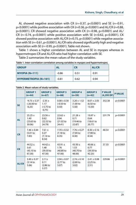
Table 1 shows a higher correlation between AL and SE in myopes whereas in hypermetropes CR and AL/CR ratio had higher correlation with SE.
Table 2 summarizes the mean values of the study variables.
Table 1. Inter-correlation correlation among variables in myopes and hypermetropes.
GROUP AL CR AL/CR
MYOPIA (N=111) -0.86 0.51 0.91
HYPERMETROPIA (N=101) -0.81 0.62 0.94
Table 2. Mean values of study variables.
GROUP 1(n=44)
GROUP 2(n=67)
GROUP 3(n=88)
GROUP 4(n=59)
GROUP 5(n=42)
F VALUE(4,295 DF) P VALUE
SE
-9.73 ± 5.37(-26.00 to -6.25)
-2.35 ±1.17(-5.75 to -0.75)
0.08 ± 0.30(-0.50 to 0.50)
3.20 ± 1.52(1.00 to 5.50)
8.07 ± 2.05(6.50 to 13.50)
352.38 p<0.0001
AL
25.25 ± 1.49(23.63 to 30.06)
23.56 ±0.62(22.32 to 24.79)
22.62 ± 0.94(21.02 to 24.41)
21.28 ± 0.71(19.46 to 22.87)
19.97 ± 0.64(18.61 to 20.77)
231.79 p<0.0001
CR
7.46 ± 0.44(5.01 to 7.83)
7.61 ±0.27(7.14 to 8.54)
7.70 ± 0.32(7.18 to 8.35)
7.76 ± 0.27(7.04 to 8.30)
8.30 ± 0.16(7.98 to 8.54)
48.54 p<0.0001
CK
44.52 ± 3.61(43.10 to 67.30)
44.42 ±1.48(39.50 to 47.30)
43.91 ± 1.76(40.80 to 47.00)
43.50 ± 1.53(40.70 to 48.00)
40.66 ± 0.77(39.50 to 42.25)
37.33 p<0.0001
AL/CR
3.40 ± 0.37(3.14 to 5.06)
3.11±0.11(2.86 to 3.27)
2.94 ± 0.07(2.77 to 3.07)
2.74 ± 0.10(2.60 to 3.02)
2.41 ± 0.08(2.25 to 2.51)
229.86 p<0.0001
Correlation of refractive error with axial length and corneal topography
30 Asian Journal of OPHTHALMOLOGY
DiscussionMean SE of patients included in this study was -0.17 ± 5.64 D. Mean SE in myopic patients was -5.27 ± 5.02 D and in hypermetropic patients was 5.20 ± 3.0 D. Mean SE from group 1 to 5 was -7.55 ± 1.23 D, - 2.35 ± 1.17 D, 0.08 ± 0.30 D, 3.16 ± 1.50 D and 8.08 ± 2.08 D respectively.
Mean AL of patients included in this study was 22.58 ± 1.84 mm. Mean AL of myopic patients was 24.23 ± 1.34 mm and of hypermetropic patients was 20.73 ± 0.94 mm. Mean AL from group 1 to 5 was 24.78 ± 0.84 mm, 23.61 ± 0.69 mm, 22.62 ± 0.94 mm, 21.29 ± 0.71 mm and 19.96 ± 0.64 mm respectively.
Tien Yin Wong et al10 in his study on chinese population in Singapore (2001) found the mean axial length of 23.23± 1.17 mm slightly higher than our study. Elvis Ojaimi et al11 in his study on Australian population (2005) in his study found nearly same mean axial length that was 22.61 ± 0.02 mm (range: 19.64–25.35). Lourdes Llorente et al12 in their study on spanish population (2005) found lower AL 22.62 ± 0.76 mm for hyperopic eyes and higher AL i.e 25.16 ± 1.23 mm for myopic eyes.
Our study revealed a high correlation between AL and SE (r= -0.91, p<0.0001). This correlation was higher than previous studies. Correlation between AL and SE in myopic group (r= -0.86, p<0.0001, slope factor -4.9048) was slightly higher than hypermetropic group (r= -0.81, p<0.0001, slope factor -3.9944).
Dr. Niall C et al13 in his study in 1998 found significant, but lower than our study, relationship (r2 = 0.611, p = 0.0001) between the degree of hyperopia and the measured AL. Stenstrom14 (1948) found the correlation between AL and SE to be -0.76 which was higher than other studies but lower than our study. Touzeau O et al15 in their study on French population (2003) found a significant correlation between AL and SE (r=0.82, p<0.001). Jenny M Ip et al16 in their study on Autralian population (2007) reported correlation of (r = −0.44) between AL and SE in 6 year children and (r = −0.61) in 12-year-old children.
Mean CK of patients included in this study was 43.72 ± 2.39 D. Mean CK of myopic patients was 44.86 ± 2.59 D and of hypermetropic patients was 42.33 ± 1.90 D. Mean CK from group 1 to 5 was 45.52 ± 0.81 D, 44.42 ± 1.48 D, 43.91 ± 1.76 D, 43.49 ± 1.53 D and 40.67 ± 0.78 D.
Sorsby et al17 in their study on British population (1957), in their cross sectional study, reported mean CK of 43.25 D for emmetropic eyes and CK of 44.40 D for myopic eyes, and concluded that corneal power was probably as significant as axial length in production of ametropia upto 4.0 D.
Mean CK in female patients was found higher than male patients in emmetropic and hypermetropic subjects and vice versa in myopic subjects ( female 44.66 ± 1.71 D, 44.62 ± 1.35 D and 42.93 ± 2.06 D, males 45.14 ± 3.51 D, 43.36 ± 1.86 D and 41.95 ± 1.69 D for myopic, emmetropic and hypermetropic subjects respectively).
D Ganguli et al18 (1975) 25 found average corneal power in emmetropic males was 43.57 ± 0.08 D and emmetropic females 44.13 ± 0.12 D. Average corneal power in myopic males was found 43.78 + 0.10 D and in myopic females was 45.29 + 0.11 D. Average corneal power in hypermetropic males was found 43.08 + 0.12 D and in hypermetropic females was 44.06 + 0.13 D. He found that corneal power was more
Kishore, Singh, Chaudhary, et al
31Asian Journal of OPHTHALMOLOGY
in females than males whether the eyes are emmetropic, myopic or hypermetropic, but more marked in female myopes
Our study reveals a high correlation between CK and SE ( r=-0.61, p<0.0001). Tahra Al Mahmoud et al19 in their study on Canadian population found a weaker relationship than our study( r=-0.18, P<0.01).
Mean CR of patients included in this study was 7.74 ± 0.39 mm. Mean CR of myopic patients was 7.55 ± 0.35 mm and of hypermetropic patients was 7.99 ± 0.35 mm. Mean CR from group 1 to 5 was 7.59 ± 0.16 mm, 7.61 ± 0.27 mm, 7.70 ± 0.32 mm, 7.77 ± 0.27 mm and 8.30 ± 0.16 mm.
Tien Yin Wong et al10 (2001) found the mean corneal curvature of 7.65 ± 0.27 mm. Lourdes Llorente et al12 (2005) found the radius of curvature of cornea in myopic eyes (7.86 ± 0.37 mm) to be steeper than in hypermetropic eyes (7.97 ± 0.30 mm). Elvis Ojaimi et al11 (2005) in his study found mean greatest CR was 7.85 ± 0.01 mm and mean least CR was 7.71 ± 0.01 mm.
Our study reveals a high correlation between CR and SE (r=0.62, p<0.0001). Correlation between CR and SE in myopic group (r= 0.51, p<0.0001, slope factor 4.9048) was slightly lower than hypermetropic group (r= 0.62, p<0.0001, slope factor 3.9944). Jenny et al16 (2007) found lower correlation for SE with CR (r ≤ 0.09). Scott and Grosvenor20 in their study on population of America (1993) found a higher correlation between CR and SE (r= +0.96). Dr. Niall C et al13 (1998) found weak but statistically significant relationship (r=0.128, p=0.009) between mean corneal radius measurements and mean spherical refractive errors, with mean corneal radius flattening with increasing hyperopia.
Our study reveals a negative correlation between AL and CR (r=-0.36,p<0.0001). Stenstrom14 (1948) found the correlation between AL and CR to be +0.18, Hirsch et al21 found the correlation to be +0.70. Touzeau O et al15 (2003) found a strong correlation between CR and AL in emmetropic eyes (r=0.63,p<0.001) and a weak but significant correlation in ametropic eyes (r=0.28,p=0.002).
Mean AL/CR ratio of patients included in this study was 2.93 ± 0.34. Mean AL/CR ratio of myopic patients was 3.22 ± 0.29 and of hypermetropic patients was 2.60 ± 0.19. Mean AL/CR ratio from group 1 to 5 was 3.27 ± 0.07, 3.11 ± 0.11, 2.94 ± 0.07, 2.74 ± 0.10 and 2.41 ± 0.08. Elvis Ojaimi et al11 (2005) found distribution of axial length/mean corneal radius ratio was peaked (leptokurtic) with a mean of 2.91. Lourdes Llorente et al12 (2005) found significantly (p<0.0001) higher AL/CR ratio for myopic patients (3.2 ± 0.2) than in hyperopic patients (2.8 ± 0.1).
Our study reveals a high correlation between AL/CR ratio and SE (r=-0.95, p<0.0001). The correlation between AL/CR ratio with refractive status (Myopia r=0.91, p<0.0001, slope factor -4.9048; hypermetropia r=0.94, p<0.0001, slope factor -3.9944). The correlation in hypermetropic patients was slightly higher than myopic patients
Lourdes Llorente et al12 (2005) found a highly significant correlation between AL/CR and refractive error SE (p<.0001, r=-0.93, slope=-0.058) which was almost similar to our study. They found higher correlation for myopes (p<.0001, r=0.87, slope= -0.07) than hyperopes (p<.0001, r=0.7171, slope= -0.04) which was contrary to our study.
Correlation of refractive error with axial length and corneal topography
32 Asian Journal of OPHTHALMOLOGY
An attempt was further made to study the role of AL and CR in various refrac-tive errors with emmetropic patients. The corneal radius was more or less than ± 1 SD different from mean emmetropic eyes in 20.72% in myopes and 53.48 % of hypermetropes. The corresponding figures for axial length variations are 67.57% in myopes and 85.15% in hypermetropes. Thus, indicating a significant role of AL in higher population of patients. Corneal radius had a similar role in 3.60% of myopes and 50.54% of hypermetropes. AL was found to be causative factor in 67.56% of myopes and 85% of hypermetropes.
It is evident from above discussion that axial length plays very important role in causation of refractive errors, while corneal radius plays so in causation of hyperme-tropia. In our best knowledge, no such type of comparison was made in past studies.
The differences across studies may be due to several reasons: different age groups, refractive error ranges, and populations and ethnicities, differences in the statistical power of the studies, and differences across methods of measurement of CR, AL, SE and CK.
ConclusionThis study reveals a highly significant correlation between axial length and spher-ical equivalent, the correlation being slightly higher in myopic group than hyper-metropic group.
A significant correlation between central power of cornea and spherical equiva-lent was found. This study reveals a high correlation between central radius curva-ture of cornea and spherical equivalent. Correlation between central radius curva-ture of cornea and spherical equivalent in myopic group was slightly lower than hypermetropic group.
A significant correlation between AL/CR ratio and spherical equivalent was found. The correlation in hypermetropic patients was slightly higher than myopic patients.
Our study is distinct owing to paucity of studies reported in Indian population on analysis of normative data correlating optical biometry parameters with refractive error. This study corroborates with findings of similar studies carried out in other population.
References1. Mutti DO, Mitchell GL, Jones LA, et al. Axial Growth and Changes in Lenticular and Corneal Power
during Emmetropization in Infants. Invest Ophthalmol Vis Sci. 2005;46(9):074-3080.doi: 10.1167/iovs.04-1040.
2. Pennie FC, Wood ICJ, Olsen C, White S, Charman WN. A longitudinal study of the biometric and refractive changes in full-term infants during the first year of life.Ophthalmology. 1995 ;102(5):827-30. doi:10.1016/S0042-6989(01)00169-9
3. Hitzenberger CK. Optical measurement of the axial eye length by laser Doppler interferometry. Invest Ophthalmol Vis Sci. 1991;32(3);616–624.
4. Schmid GF, Papastergiou GI, Nickla DL et al.Validation of laser Doppler interferometric measurements in vivo of axial eye length and thickness of fundus layers in chicks. Current Eye Research 1996; 15(6), 691–696.

Kishore, Singh, Chaudhary, et al
33Asian Journal of OPHTHALMOLOGY
5. Flitcroft DI, Knight-Nanan D, Bowell R, Lanigan B, & O’Keefe M. Intraocular lenses in children: changes in axial length, corneal curvature, and refraction. Br J Ophthalmol. 1999; 83(3): 265–269. doi:10.1136/bjo.83.3.265.
6. Gordon RA, Donzis PB. Refractive development of the human eye. Arch Ophthalmol.1985;103(6):785–789. doi:10.1001/archopht.1985.01050060045020.
7. Waltman SR and Hart WM. The cornea in Adler’s Physiology of the Eye-Clinical Application( R. A. Moses and W. M. Hart) Eds., pp. 36–59, CV Mosby Coy, St. Louis, Mo, USA, 8th edition, 1987.
8. Iyamu E, Iyamu J, Obiakor CI. The Role of Axial Length-Corneal Radius of Curvature Ratio in Refractive State Categorization in a Nigerian Population. ISRN Ophthalmology. 2011. doi: 10.5402/2011/138941.
9. Grosvenor T, Scott R. Role of the axial length/corneal radius ratio in determining the refractive state of the eye. Optometry and Vision Science. 1994; 71(9): 573–579.
10. Wong T.Y, Foster P.J , Ng T.P, et al. Variations in Ocular Biometry in an Adult Chinese Population in Singapore: The Tanjong Pagar Survey. Invest. Ophthalmol. Vis. Sci. 2001; 42:1 73-80.
11. Ojaimi E , Rose K.A , Morgan I.G, et al . Distribution of Ocular Biometric Parameters and Refraction in a Population-Based Study of Australian Children. Invest. Ophthalmol. Vis. Sci.2005;46(8):2748-2754. doi: 10.1167/iovs.04-1324.
12. Llorente L, Barbero S, Cano D, Dorronsoro C & Marcos S. Axial length, corneal shape and optical aberrations in myopic versus hyperopic eyes. Journal of Vision. 2003;3(12). doi: 10.1167/3.12.27.
13. Strang NC, Schmid KL, Carney LG. Hyperopia is predominantly axial in nature. Current Eye Research. 1998;17(4):380-383. doi:10.1080/02713689808951218.
14. Stenstrom S, “Investigation of the variation and correlation of the optical elements of human eyes,” American Journal of Optometry & Archives of American Academy of Optometry,1949;25:496-504.
15. Touzeau O, Allouch C, Borderie V, et al. Correlation between refraction and ocular biometry. Journal Francais D’ophtalmologie 2003, 26(4):355-363. PMID:12843892.
16. JM Ip, Huynh S.C, Kifley A, et al. Variation of the Contribution from Axial Length and Other Oculometric Parameters to Refraction by Age and Ethnicity. Invest. Ophthalmol. Vis. Sci. 2007;48(10):4846-4853. doi: 10.1167/iovs.07-0101.
17. Sorsby, Arnold, B. Benjamin, et al. “Emmetropia and its aberrations. Medical Research Council Special Report Series no 293.” (1957).
18. D Ganguli, IS Roy, SK Biswas, M Sengupta. Study of corneal power and diameter in simple refractive error. Indian journal of ophthalmology 1975;23(1):6-11.
19. AlMahmoud T, Priest D, Munger R. Correlation between Refractive Error, Corneal Power, and Thickness in a Large Population with a Wide Range of Ametropia. Invest. Ophthalmol. Vis. Sci. 2011; 52(3):1235-1242. doi: 10.1167/iovs.10-5449.
20. Grosvenor T, Scott R, “Role of the axial length/corneal radius ratio in determining the refractive state of the eye,” Optometry and Vision Science, 1994; 71: 573–579.
21. Hirsch, Monroe J and Weymouth F.W. Notes on ametropia; a further analysis of Stenstrom’s data. American journal of optometry and archives of American Academy of Optometry 1947;24(12): 601-608.

34 Asian Journal of OPHTHALMOLOGY
Anterior segment optical coherence tomography documentation of Reverse Pupillary Block
Devendra Maheshwari1, Renagappa Ramakrishnan1, Neelam Pawar1
1Aravind Eye Hospital, Tirunelveli, Tamilnadu, India
AbstractWe report a 10-year-old boy with unusually dense, bilateral central posteriorcapsule pigmentation associated with the characteristic clinical features of pigmentdispersion syndrome, including Krukenberg’s spindle and dense trabecular pigmentation in both eyes. There was no history of trauma , laser or intraocular surgeries . The presence of posterior or backward bowing of iris suggested a reverse pupillary block mechanism of pigment dispersion syndrome. Nd Yag laser peripheral iridotomy was performed in both eyes to relieve reverse pupillary block. Anterior segment optical coherence tomography (AS-OCT) showed reversal of iris concavity after laser iridotomy.
Key words: Pigment dispersion syndrome; Pigmentation; Posterior capsule; Neodymium Yttrium Yag laser peripheral iridotomy. Anterior segment optical coherence tomography.
Pigment dispersion syndrome (PDS) typically develops in young adults and is most commonly diagnosed in the second to fourth decade. This clinical condition is typically seen in young, myopic males. PDS is characterized by the presence of Krukenberg spindles, iris trans-illumination defects, trabecular meshwork pigmen-tation and backward bowing of the iris.1,4
PDS is unusual in a pediatric age, although it has been previously described in the western literature.5-8 To our best knowledge no PDS case has been reported in literature in Indian population .
We describe a case of typical pigment dispersion syndrome in 10-year-old Asian Indian child with elevated Intraocular pressure (IOP).
Case ReportA 10-year-old boy presented with complaint of defective vision of six month duration (Fig. 1).He had no history of trauma , surgery or any systemic illness. There was no history of headache, blurred vision or haloes. He had family history of glaucoma with grandmother being affected with primary open angle glaucoma.He had no history of any spectacle use previously.
On examination, his uncorrected visual acuity was 20/60 in both eyes. The best corrected visual acuity (BCVA) was 20/20 with −1.00 D cylinder ×90 in right eye and −1.50 D cylinder ×90 in left eye. He was not prescribed any spectacles previously. During examination clinical findings of PDS were noted.
Correspondence: Correspondence: Devendra Maheshwari, Medical Officer, Glaucoma Departement, Aravind Eye Hospital,Tirunelveli,Tamilnadu,India.E-mail: [email protected]
Case ReportAsian J Ophthalmol. 2016;15:034-039© Asian Journal of Ophthalmology

Maheshwari, Ramakrishnan, Pawar
35Asian Journal of OPHTHALMOLOGY
The IOP with Goldman Applanation tonometer was 36mm Hg in right eye and 39mmHgin left eye( adjusted according to CCT ). On slit-lamp examination, a pigment deposition on the corneal endothelium in a spindle shaped manner (Krukenberg’s spindle) along with deep anterior chambers with few pigments. Radial, slit like iris transillumination defects were noted in both eyes. These defects were typically located in the periphery of the iris. There was bilateral central poste-rior capsule pigmentation with evident Zentmeyers line and Scheies line (Fig. 2a & 2b).Gonioscopy revealed wide open angle grade 4 (Shaffers Grading) with 4+ pigmentation of the trabecular meshwork with iris concavity in midperiphery in both eyes. Corneal pachymetry was 665 um in right eye and 668 um in left eye. The horizontal corneal diameters were 12 mm and 12.5 mm in right and left eye respectively. Axial length was 24.00mm in right eye and 24.05 in left eye ( Carl Ziess IOL Master).The cup-disc ratio was 0.4 in both eyes with healthy neuroretinal rim and retinal nerve fiber.
Peripheral fundus examination showed lattice degeneration with multiple holes in both eyes for which prophylactic barrage laser was done. Humphrey visual fields (24-2) were normal in both eyes. Anterior segment optical coherence tomography (Visante 1000, Carl Zeiss Meditec Inc, and Dublin, CA, USA) showed a concave iris configuration in both eyes [Figure 2a and 2c].
Fig. 1a. Pigmentation of posterior lens capsule, Pigment deposited on the posterior lens surface, Zentmayer ’s line. (white arrow)
Fig. 1b. Pigment accumulation occurring at the junction of the zonules and posterior capsule Scheie’s line. (white arrow)
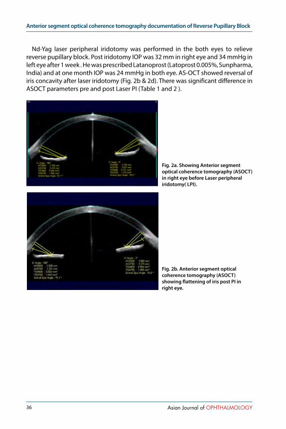
Anterior segment optical coherence tomography documentation of Reverse Pupillary Block
36 Asian Journal of OPHTHALMOLOGY
Nd-Yag laser peripheral iridotomy was performed in the both eyes to relieve reverse pupillary block. Post iridotomy IOP was 32 mm in right eye and 34 mmHg in left eye after 1 week . He was prescribed Latanoprost (Latoprost 0.005%, Sunpharma, India) and at one month IOP was 24 mmHg in both eye. AS-OCT showed reversal of iris concavity after laser iridotomy (Fig. 2b & 2d). There was significant difference in ASOCT parameters pre and post Laser PI (Table 1 and 2 ).
Fig. 2a. Showing Anterior segment optical coherence tomography (ASOCT) in right eye before Laser peripheral iridotomy( LPI).
Fig. 2b. Anterior segment optical coherence tomography (ASOCT) showing flattening of iris post PI in right eye.

Maheshwari, Ramakrishnan, Pawar
37Asian Journal of OPHTHALMOLOGY
Table 1. AS OCT Parameters Pre and Post Laser PI in RE
AS OCT Parameters RE
IC Angle 180Pre Laser PI
IC Angle 180Post Laser PI
P-valueAS OCT Parameters RE
IC Angle0Pre Laser PI
IC Angle0Post Laser PI
P-value
AOD 500 2.705 2.030 0.043* AOD 500 2.745 1.982 0.043*
AOD 750 3.215 2.251 AOD 750 3.031 2.275
TISA500 0.694 0.563 TISA500 0.721 0.594
TISA 750 1.56 1.042 TISA 750 1.374 1.069
SSA 81.7 75.1 SSA 78.5 74.8
*Wilcoxon signed-rank testAOD -Angle opening distance (in mm), TISA(mm2) Trabecular Iris Space Area, SSA –Scleral Spur Angle
Table2. AS OCT Parameters Pre and Post Laser PI in LE
AS OCT Parameters LE
IC Angle 180Pre Laser PI
IC Angle 180Post Laser PI
P-value
IC Angle0Pre Laser PI
IC Angle0Post Laser PI
P-value
AOD 500 2.116 1.534 1.912 1.845
AOD 750 2.408 1.768 0.043* 2.239 2.085 0.043*
TISA500 0.542 0.470 0.594 0.491
TISA 750 1.073 0.882 1.158 0.993
SSA 76.3 70.3 77.1 74.8
*Wilcoxon signed-rank test
DiscussionPigment dispersion syndrome is a typically seen in the younger age group ranging from 20–40 years.1,2 It is quite unusual to find pigment dispersion syndrome in young child. Reports of PDS in the paediatric group is not so common, lack of symptoms despite high IOP in this age group may be reason of this paucity in literature.
The mechanism of the pigment dispersion was elucidated by Campbell, who provided evidence for a rubbing between lens zonules and iris pigment epithelium, with release of pigment granules.4
The backward bowing of the mid-peripheral iris, described by Campbell, is of key importance in the etiology of PDS. The presence of iris back bowing suggests a reverse pupillary block mechanism of pigment dispersion syndrome.4
Karickhoff also described a form of reverse pupillary block, in which he suggested

Anterior segment optical coherence tomography documentation of Reverse Pupillary Block
38 Asian Journal of OPHTHALMOLOGY
that the iris acted as a flap valve against the anterior lens surface. This so-called valve would allow aqueous to flow forward because of a pumping action of the mid-peripheral iris in association with ocular movement. However, the valve effect would prevent the aqueous from flowing backward, thereby increasing anterior chamber pressure and pushing the peripheral iris backward against the lens zonules, causing a further shedding of pigment.8 The similar iris configuration seen in PDS/PG led to Campbell’s and Karickhoff’s theory of reverse pupillary block.
In a series of 407 pigment dispersion syndrome patients, the youngest age described was 14 years old; the youngest with accompanying elevated IOP was 25 years old.4 Lazaro Garcia and associates reported pigment dispersion syndrome with megalocornea in a 12-year-old child.5
Kaiser-Kupfer and associates described three brothers who developed pigmentdispersion syndrome at an early age; Out of three one showed, iris transillumina-
tion defects at the age of 7 years.6
Grassi et al.described PDS, with atypical features in an 8 year old patient.7
Ritch et al.reported an 11-year-old girl with typical features of bilateral PDS and elevated intraocular pressure (IOP) . They also described t in two 12-year-old boys, one with a more severe phenotype with both affected parents and the other having
Fig. 2d. Anterior segment optical coherence tomography (ASOCT) showing flattening of iris post PI in left eye.
Fig. 2c. Showing Anterior segment optical coherence tomography (ASOCT) in left eye before LPI.
Maheshwari, Ramakrishnan, Pawar
39Asian Journal of OPHTHALMOLOGY
a less severe phenotype single affected parent .8 Argon laser peripheral iridotomy was performed in both eyes of one boy and one eye of other.
Preliminary success of treatment by laser peripheral iridotomy (LPI) has been reported by Karickhoff and by Campbell and Schertzer.4,8 LPI is traditionally thought to be of benefit in PDS, because it is supposed to decrease the risk of develop-ment of pigmentary glaucoma. However Scott et al. in a prospective, randomized, controlled trial found there was no benefit of Nd: YAG LPI in preventing progres-sion from PDS with ocular hypertension to pigmentary glaucoma.It is possible that the treatment may be effective in younger patients: those without irreversible trabecular meshwork damage or in those with documented increased iridozonular contact (iris concavity and more posterior iris root insertion).10
However, to the best of our knowledge, 10-year-old boy described in this report is the youngest and first Asian Indian with typical features of bilateral PDS and elevated IOP .Laser peripheral iridotomy relieved reverse pupillary block and additional antiglaucoma medication were needed to lower IOP.
References1. Niyadurupola N, Broadway DC. Pigment dispersion syndrome and pigmentary glaucoma-a major
review. Clin Experiment Ophthalmol 2008;36:868-82.2. Sugar HS. Pigmentary glaucoma: a 25-year review. Am J Ophthalmol1966;62:499-507.3. Scheie HG, Cameron JD. Pigment dispersion syndrome: a clinical study. Br J Ophthalmol
1981;65:264-9.4. Campbell DG. Pigmentary dispersion and glaucoma: a new theory. Arch Ophthalmol
1979;97:1667-72.5. García CL, Feijoó JG, Sardiña JC, Gómez AC, Benítez Del Castillo JM, Benítez JM, et al. Pigment
dispersion syndrome in a patient with megalocornea. Arch Soc Esp Oftalmol 2000;75:347-350.6. Kaiser-Kupfer MI, Kupfer C, McCain L. Asymmetric pigment dispersion syndrome. Trans Am
Ophthalmol Soc 1983;81:310-324.7. Grassi MA, Alward WL, Verdick RL, Langlow EP, Kwon YH. Atypical pigment dispersion syndrome in
a child. Am J Ophthalmol 2004;137:753-6.8. Dorairaj SK, Robin A, Shihadeh W, Greenberg S, Liebmann JM, Ritch R. Phenotypic Variability of
Pigment Dispersion Syndrome in Children .Arch Ophthalmol 2007;125:136-8.9. Karickhoff JR. Pigmentary dispersion syndrome and pigmentary glaucoma: a new mechanism
concept, a new treatment, and a new technique. Ophthalmic Surg 1992;23:269 -77.10. Scott A, Kotecha A, Bunce C, Balidis M, Garway-Heath DF, Miller MH, Wormald R.
YAG laser peripheral iridotomy for the Prevention of Pigment Dispersion Glaucoma. Ophthalmology 2011;118:468-73.

40 Asian Journal of OPHTHALMOLOGY
Posterior segment involvelent in remote lightning strike
Atul Kumar SinghDepartment of Ophthalmology Command Hospital (AF) Bangalore Karnataka, India
IntroductionSome form of ophthalmic injury is seen in the majority of lightening victims. These may be anterior segment involvement, mostly the cornea.1Other lesion on the anterior segment include uveitis, hyphaema, cataract and dislocated lens.2
Posterior segment lesion include vitreous haemorrhage, retinal oedema, retinal haemorrhage, retinal detachment, cystoids macular oedema, chorioretinalrupture, maculopathy, CRVO and CRAO.3 Neuro-ophthalmic lesion include loss of pupillar-yreflex, anisocoria, horner syndrome, multiple cranial nerve palsies and nystagmus.
Case reportA 35 years old patient presented to us with sudden decrease in vision from right eye associated with hearing loss on that side. He gave the history of light falling on the right side of body one day back in the night as he stepped out from his house. As soon as he touched the ground in night around 11 pm he experienced sharp electric current passing through his right side of body for fraction of second and soon after few hours he noticed some visual disturbance. His left side was uninvolved as his left side was covered by tin shade.
On examination his best corrected visual acuity was 6/18 / N/6 RE and 6/9/ N/6 in the LE.Anterior segment exami-nation was within normal limits and intraocular pressure was 15 mm of Hg (both eyes). There was no RAPD. Fundus examination of left eye was within normal limits (Figure 1).Right eye fundus examination shows multiple retinal haemorrhages in all the quadrants with preretinal haemorrhage in the infero temporal area (Figure 2). His systemic examination (Blood pressure, Random Blood sugar) was normal. A
Fig 1. Left eye of patient
Correspondence: Atul Kumar Singh, Department of Ophthalmology Command Hospital (AF) Bangalore Karnataka, 560007, IndiaE-mail: [email protected]
Case ReportAsian J Ophthalmol. 2016;15:040-042© Asian Journal of Ophthalmology

Singh
41Asian Journal of OPHTHALMOLOGY
comprehensive investigations failed to reveal any another underlying etiology (This include CBC CT,BT,INR,Homocysteine level,protein C and S, Antithrombin III and factor V level).In the mean time he was also evaluated by ENT specialist where the diagnosis of moderate conductive hearing loss was made. Patient was subjected to CT scan to find out the cause of hearing loss. CT scan revealed opacification of the mastoid air cells consistent with hemorrhagic effusion with no fracture of bone. After three weeks of follow up the pre retinal haemorrhage was completely resolved and retinal haemorrhage was decreased in number (Figure 3).His visual acuity was improved to 6/12/ N/8 RE. Our patient did not suffer from any skin or eye lid burns.
DiscussionLee at al4 reported four routes by which lightning reaches the victim and causes injuries:
1. Direct strike: When the major current flows directly through the victim and is facilitated by metal object.
2. Splash: Where lightening strikes an object first and then arcs through the path of least resistance.
3. Contact: When lightning strike the object the victim is in contact with such as being while talking electrocuted over the phone or in bathtub by current flowing through wires or pipes
4. Ground current: The bolt strikes the ground and travels along the surface towards the victim
Our patient probably sustained the injury by the fourth mechanism mentioned - ground current passing initially through the right side of the body indicated by loss of hearing in right ear, decreased vision RE due to posterior segment involvement.
Lightning involves a transfer of electric charge. Also lightning contact is instan-taneous taking less time (exposure time usually lasts only 1 to 100 milliseconds) to
Fig 2. Right eye of patient multiple retinal haemorrhages in all the quadrants with pre retinal haemorrhage in the inferotemporal area
Fig 3. resolving haemorrhage
Posterior segment involvelent in remote lightning strike
42 Asian Journal of OPHTHALMOLOGY
cause injury.5 Tissue is destroyed by both heat and electrolysis. The high resistance offered by non-nervous tissue accounts for the thermal effects of electrical injuries, which result in immediate coagulation of the proteins of the cells.6 In the poste-rior segment macula is very sensitive to thermal damage because it contains high melanin granules of its RPE.The electric current damage the RPE also can damage the inner blood retinal barrier resulting in retinal vascular incompetence.7 Krishna et al.7 reported a case of bilateral macular hole with posterior sub capsular cataract.Handa et al.3 states that lack of posterior vitreous detachment and operculum support the diagnosis of lightening maculopathy as opposed to full thickness macular hole. He reported a case of maculpathy which initially presented as a retinal cyst with surrounding macular oedema and later evolved into full thickens macular hole.
Visual prognosis in patients with lightening injury will depend upon extent of involvement of ocular structure extent of macular damage and optic nerve involve-ment. In our case fortunately there is no involvement of macula and optic nerve resulting in very good visual prognosis of the patient.
References1. Edelstein J, Peters W, Cartotto R. Lightning injury: A review and case presentations. Canadian J
Plastic Sur. 1994;2:164–8.2. Norman ME, Albertson D, Younge BR. Ophthalmic manifestations of lightning strike. Surv
Ophthalmol. 2001;46:19–24.3. Handa JT, Jaffe GJ. Lightning maculopathy. Retina. 1994;14:164–724. Lee MS, Gunton KB, Fischer DH, Brucker AJ. Ocular manifestations of remote lightning strike.
Retina. 2002;22:808–10.5. Dimick AR. Harrison’s Principles of Internal Medicine. In: Fauci AS, Braunwald E, Isselbacher KJ,
Wilson JD, Martin JB, Kasper DL, et al., editors. Health Professions Division. 14th ed. New York: McGraw-Hill; 1998. p. 2559.
6. Duke-Elder Sir S, Macfaul PA. In: System of Ophthalmology. Duke-Elder Sir S, editor. Vol XIV. London: Henry Kimpton; 1972. pp. 813–35. Part 2.
7. Krishna A Rao,Lavanya G Rao,Ajay N Kamath and VikramJain Indian J Ophthalmol. 2009 Nov-Dec; 57(6): 470–472.
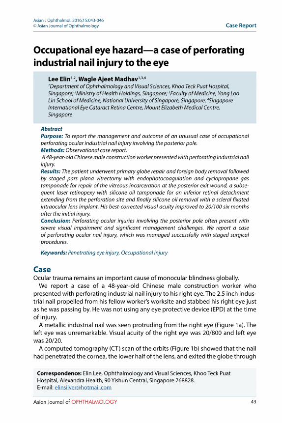
43Asian Journal of OPHTHALMOLOGY
Occupational eye hazard—a case of perforating industrial nail injury to the eye
Lee Elin1,2, Wagle Ajeet Madhav1,3,4
1Department of Ophthalmology and Visual Sciences, Khoo Teck Puat Hospital, Singapore; 2Ministry of Health Holdings, Singapore; 3Faculty of Medicine, Yong Loo Lin School of Medicine, National University of Singapore, Singapore; 4Singapore International Eye Cataract Retina Centre, Mount Elizabeth Medical Centre, Singapore
AbstractPurpose: To report the management and outcome of an unusual case of occupational perforating ocular industrial nail injury involving the posterior pole.Methods: Observational case report. A 48-year-old Chinese male construction worker presented with perforating industrial nail injury.Results: The patient underwent primary globe repair and foreign body removal followed by staged pars plana vitrectomy with endophotocoagulation and cyclopropane gas tamponade for repair of the vitreous incarceration at the posterior exit wound, a subse-quent laser retinopexy with silicone oil tamponade for an inferior retinal detachment extending from the perforation site and finally silicone oil removal with a scleral fixated intraocular lens implant. His best-corrected visual acuity improved to 20/100 six months after the initial injury.Conclusion: Perforating ocular injuries involving the posterior pole often present with severe visual impairment and significant management challenges. We report a case of perforating ocular nail injury, which was managed successfully with staged surgical procedures.
Keywords: Penetrating eye injury, Occupational injury
CaseOcular trauma remains an important cause of monocular blindness globally.
We report a case of a 48-year-old Chinese male construction worker who presented with perforating industrial nail injury to his right eye. The 2.5 inch indus-trial nail propelled from his fellow worker’s worksite and stabbed his right eye just as he was passing by. He was not using any eye protective device (EPD) at the time of injury.
A metallic industrial nail was seen protruding from the right eye (Figure 1a). The left eye was unremarkable. Visual acuity of the right eye was 20/800 and left eye was 20/20.
A computed tomography (CT) scan of the orbits (Figure 1b) showed that the nail had penetrated the cornea, the lower half of the lens, and exited the globe through
Correspondence: Elin Lee, Ophthalmology and Visual Sciences, Khoo Teck Puat Hospital, Alexandra Health, 90 Yishun Central, Singapore 768828.E-mail: [email protected]
Case ReportAsian J Ophthalmol. 2016;15:043-046© Asian Journal of Ophthalmology
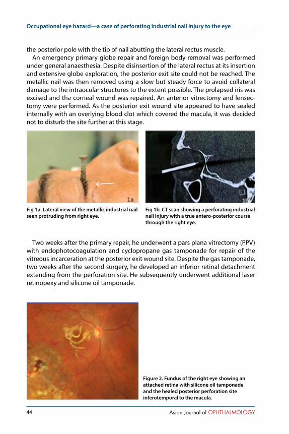
Occupational eye hazard—a case of perforating industrial nail injury to the eye
44 Asian Journal of OPHTHALMOLOGY
the posterior pole with the tip of nail abutting the lateral rectus muscle.An emergency primary globe repair and foreign body removal was performed
under general anaesthesia. Despite disinsertion of the lateral rectus at its insertion and extensive globe exploration, the posterior exit site could not be reached. The metallic nail was then removed using a slow but steady force to avoid collateral damage to the intraocular structures to the extent possible. The prolapsed iris was excised and the corneal wound was repaired. An anterior vitrectomy and lensec-tomy were performed. As the posterior exit wound site appeared to have sealed internally with an overlying blood clot which covered the macula, it was decided not to disturb the site further at this stage.
Two weeks after the primary repair, he underwent a pars plana vitrectomy (PPV) with endophotocoagulation and cyclopropane gas tamponade for repair of the vitreous incarceration at the posterior exit wound site. Despite the gas tamponade, two weeks after the second surgery, he developed an inferior retinal detachment extending from the perforation site. He subsequently underwent additional laser retinopexy and silicone oil tamponade.
Fig 1b. CT scan showing a perforating industrial nail injury with a true antero-posterior course through the right eye.
Fig 1a. Lateral view of the metallic industrial nail seen protruding from right eye.
Figure 2. Fundus of the right eye showing an attached retina with silicone oil tamponade and the healed posterior perforation site inferotemporal to the macula.

Elin, Madhav
45Asian Journal of OPHTHALMOLOGY
The silicone oil was removed four months later and the eye received a scleral fixated intraocular lens. Six months after the initial injury, the best-corrected visual acuity of right eye was 20/100. Fundus examination revealed an attached retina with a healed posterior exit wound inferotemporal to the macula (Figure 2).
DiscussionPerforating ocular injuries often present with significant visual impairment and
have a guarded prognosis. The final visual outcome depends on the site of impact and ocular structure(s) involved. We describe a case of perforating nail injury, which was successfully managed with staged surgical procedures.
The visual prognosis of a perforating injury which involves the posterior pole of the eye is typically very poor. Other factors that prognosticate poor visual outcome are poor initial visual acuity and presence of a relative afferent pupillary defect1. Our patient presented with involvement of the posterior pole and poor initial vision. There was no reverse afferent pupillary defect.
The primary aim of initial surgical management is restoring anatomic integrity of globe and removal of the intraocular foreign body. Because of the true antero-posterior (AP) course of the nail through the eye in our case, the exit wound was not easily approachable externally and hence had to be left unrepaired after removal of the nail.
There are some controversies regarding the initial management of open-globe injuries involving the posterior segment2. One such controversy is early vitrectomy. Vitrectomy has improved the prognosis of perforating injuries, and a meta-analysis showed anatomical success in 69% and a visual acuity of ≥5/200 in 56% of the eyes3. The timing of vitrectomy for perforating injuries can be early (within 2 days), delayed (7-14 days) or late (after 30 days). Many studies advocate early vitrectomy for successful visual outcome especially if there is retinal detachment and concur-rent vitreous haemorrhage, retained intraocular foreign body or traumatic endoph-thalmitis4. However, early vitrectomy often presents technical difficulties due to poor visibility, lack of posterior hyaloidal separation, bleeding and re-opening of the exit wound. As our patient had an unrepaired posterior exit wound, early vitrec-tomy was not attempted due to concern of reopening of the exit wound, the poor visibility and potential guarded prognosis associated due to suspected macular involvement. Multiple staged surgical procedures are often required for recov-ering visual function after the initial wound repair. Our patient underwent multiple staged procedures to restore the ocular anatomy and function.
Preventive measures such as use of EPDs are vital in light of limited surgical abili-ties to manage perforating ocular injuries involving the visual axis. Our patient was not using an EPD when the injury occurred. Woo et al5 have reported that 75.3% of patients with work-related ocular trauma had not used an EPD. Preventive strategies such as overcoming language barriers, providing education to improve compliance of EPDs and increasing availability of EPDs to all workers should be implemented.
In summary, staged surgical procedures can restore anatomy and provide fairly good visual function in eyes with serious ocular perforating nail injuries involving

Occupational eye hazard—a case of perforating industrial nail injury to the eye
46 Asian Journal of OPHTHALMOLOGY
the posterior pole. However, preventive measures continue to be of paramount importance. Implementation and reinforcement of measures of EPD use and creation of awareness of proper use of hazardous tools such as the nail gun can potentially reduce occupation-related perforating ocular injuries.
References:1. Schmidt GW, Broman AT, Hindma HB, Grant MP. Vision Survival after Open Globe Injury Predicted
by Classification and Regression Tree Analysis. Ophthalmology 2008; 115: 202–2092. Mittra RA, Mieler WF. Controversies in the management of open-globe injuries involving the
posterior segment. Surv Ophthalmol 1999; 44: 215–25.3. De Juan E, Sternberg P, Michels RG: Timing of vitrectomy after penetrating ocular injuries.
Ophthalmology 1984; 91: 1072-44. Wani VB, Al-Ajmi M, Thalib L, Azad RV, Abul M, Al-Ghanim M, Sabti K. Vitrectomy for posterior
segment intraocular foreign bodies: visual results and prognostic factors. Retina 2003; 23: 654–60.5. Jyh-Haur Woo, Gangadhara Sundar. Eye Injuries in Singapore – Don’t Risk It. Do More. A
Prospective Study. Ann Acad Med Singapore 2006; 35: 706-18
47Asian Journal of OPHTHALMOLOGY
Chronic Pseudophakic Aqueous MisdirectionMona A. Kaleem1,2, Sheldon Oberfeld1, Jonathan Eisengart1
1Cole Eye Institute, Cleveland Clinic, Cleveland, Ohio, USA; 2Department of Ophthalmology, University of Maryland, Baltimore, Maryland, USA
AbstractA 61 year old female with no prior ocular history developed progressive anterior chamber shallowing following uncomplicated phacoemulsification and intraocular lens implan-tation. This shallowing rapidly accelerated after Nd:YAG laser capsulotomy, and led to a significant myopic shift. Ultrasound biomicroscopy imaging demonstrated anterior displacement of the irido-lenticular diaphragm and anterior rotation of ciliary processes confirming a diagnosis of aqueous misdirection. One year after Nd:YAG anterior hyaloi-dotomy and medical therapy, her anterior chamber deepened and myopic shift resolved. This case demonstrates the successful management of chronic pseudophakic aqueous misdirection without the need for surgical intervention.
Key words: aqueous misdirection, malignant glaucoma, Nd:YAG capsulotomy
IntroductionAqueous misdirection syndrome, also known as malignant glaucoma, is a diagnosis of exclusion characterized by anterior chamber (AC) shallowing and often elevated intraocular pressure (IOP) in the absence of pupillary block or choroidal hemor-rhage. It is an uncommon complication of intraocular surgery, laser therapy, or use of miotic agents. Aqueous misdirection is usually associated with a history of angle closure glaucoma or short axial length. It is most commonly precipitated by glaucoma filtering surgery, with more than 75% of cases occurring after trabeculec-tomy and 3.7-2.8% after a tube shunt procedure.1 There have been a few reports of aqueous misdirection after intra or extra-capsular cataract surgery with an estimated incidence between 0.03%-0.025%, but there is no data from the era of phacoemulsification surgery.1,2 The incidence of misdirection after Nd:YAG capsul-otomy is unknown and only a few case reports exist in the literature.3
Here we report a case of chronic aqueous misdirection presenting with AC shallowing, normal IOP, and a significant myopic shift six months after phacoemul-sification and two weeks after Nd:YAG laser capsulotomy. The features of this case demonstrate the subtle clinical signs and symptoms phacoemulsification surgeons should take into account when evaluating patients in the post-operative period.
Case ReportA 61 year old black female with mature nuclear sclerosis and posterior subcapsular cataracts underwent uncomplicated phacoemulsification cataract extraction and posterior chamber intraocular lens implantation in both eyes. She had no prior
Correspondence: Jonathan Eisengart, Cole Eye Institute, Cleveland Clinic, 2022 E 105th St, Cleveland, OH 44106, USAE-mail: [email protected]
Case ReportAsian J Ophthalmol. 2016;15:047-052© Asian Journal of Ophthalmology
Chronic Pseudophakic Aqueous Misdirection
48 Asian Journal of OPHTHALMOLOGY
ocular history to report; her pre-operative anterior segment exam was normal with the exception of mature cataract. Posterior segment exam was limited by brunescent cataract but a symmetrical red reflex was noted. IOP was 10 mm Hg. Pre-operative refraction was difficult due to density of the cataract, however, was measured as: OD: -4.75 + 3.25 x 10⁵ correcting to hand motion and OS: -1.25 + 3.00 x 081 correcting to count finger at one foot. Axial length as measured by A-scan was OD: 20.95 mm and OS: 20.80 mm. Phacoemulsification and intraocular lens implantation into the capsular bag was performed in the right eye first with no ensuing events to report, followed by the left eye two months later.
An Alcon SN60WF 27.5 D lens was placed in the left eye with a target refraction of -0.91 D. Her post-operative course was uneventful although the surgeon noted the AC of the left eye was slightly shallower than the right eye. IOP ranged from 10-18 mm Hg. At post-op month one her refraction was stable at -1.50 D correcting to 20/30.
She returned for her six month follow up exam complaining of blurry vision. Her refraction measured -2.25 + 0.50 x 085 correcting to 20/40 and she underwent Nd:YAG laser capsulotomy for posterior capsular opacification. Two weeks after laser, her refraction changed to -7.25 + 4.50 x 090 correcting to 20/25. On slit lamp she was noted to have moderate AC shallowing. The lens implant remained securely within the capsular bag in proper orientation with an open posterior capsule. IOP was measured as 17 mm Hg and gonioscopy revealed angle closure with peripheral anterior synchiae in three quadrants, and one quadrant with appositional closure. The fellow eye was wide open to ciliary body band. The UBM image in figure 1 shows angle configuration of the unaffected right eye with AC depth of 3.65 mm. Figures 2 and 3 of the left eye demonstrate an AC depth of 2.42 mm (1.23 mm of shallowing of the left eye AC compared to the right eye), anterior displacement of the IOL-iris diaphragm, and anterior rotation of ciliary processes. There was no iris bombe configuration to suggest pupillary block.
Her posterior segment exam was normal. There was no evidence of choroidal effusion or hemorrhage on a careful peripheral exam, nor was there evidence of a supraciliary effusion on UBM. Because the anterior hyaloid was still visibly intact at the slit lamp, an Nd:YAG anterior hyaloidotomy was performed using 2.3-3.5 mJ. Immediately afterward her AC deepened to 2.92 mm. She was started on atropine 1% and timolol 0.5%.
At one month follow up on topical therapy, her refraction returned to -1.50 correcting to 20/30 and her IOP remained within normal limits at 18 mm Hg. Her AC
Fig 1. UBM image of the right eye on initial presentation. Anterior chamber depth measured as 3.65 mm.

Kaleem, Oberfeld, Eisengart
49Asian Journal of OPHTHALMOLOGY
maintained the post anterior hyaloidotomy configuration which was deeper than prior to treatment, but still shallower than the fellow eye. Atropine and timolol were stopped.
Six months after stopping topical therapy she had a mild recurrence of AC shallowing and anterior displacement of the lens to iris diaphragm with an AC depth of 2.72 mm as documented on UBM imaging, a myopic shift, and IOP of 19 mm Hg. On gonioscopy, she was noted to have angle closure with peripheral anterior synechiae in all four quadrants. Atropine and timolol eye drops were reinstituted for several months. Her AC returned to a normal depth of 3.47 mm Hg (only 0.18 mm shallower than the right eye) with resolution of myopic shift and aqueous misdirection (Figure 4).
DiscussionAqueous misdirection was first described by Dr. von Graefe as an early post-surgical complication in patients with angle closure glaucoma. To date, it has been reported after various procedures including filtering surgery, extra and intra capsular cataract surgery, phakic intraocular lens implantation and corneal transplantation even in the absence of a prior diagnosis of angle closure.1,4 Other risk factors include an
Fig 2 & 3. UBM images of the left eye on initial presentation. Anterior chamber depth measured as 2.42 mm.
Fig 4. UBM image of the left eye one year after following Nd:YAG laser anterior hyaloidotomy on medical therapy. Anterior chamber depth measured as 3.47 mm.

Chronic Pseudophakic Aqueous Misdirection
50 Asian Journal of OPHTHALMOLOGY
axial length of 22.5 mm or less, narrow irido-corneal angle, female sex, and older age.2,5 A handful of reports of misdirection following laser procedures such as laser suture lysis, iridotomy, and capsulotomy also appear in the literature. To our knowledge, this appears to be the fourth published case of misdirection following Nd:YAG capsulotomy.3,6 We believe that a subclinical picture of aqueous misdirec-tion was developing after cataract surgery, and ultimately this was exacerbated by laser treatment.
Although misdirection typically occurs in the acute post-operative period, there have been reports of cases occurring several weeks to as far out as sixteen years after the inciting surgical procedure.7 In this case, we observed progressive AC shallowing and initially unexplained myopic shift occurring six months after uncomplicated phacoemulsification surgery accelerating in the two week post-capsulotomy time frame. Increased myopia has been reported as the first symptom noticed by patients in cases of chronic aqueous misdirection.7 The myopic shift can be explained by anterior displacement of the lens to iris diaphragm secondary to increased posterior pressure.
Classic features of aqueous misdirection include central AC shallowing, normal or elevated IOP, and absence of posterior segment pathology. A flat central and peripheral AC is a defining clinical feature, however, IOP may be quite variable. Normal IOP of ≤ 22 mm Hg has been observed in 21-50% of patients, and IOP is more likely to be normal in the setting of a functional glaucoma surgery such as a trabeculectomy or aqueous tube shunt. Therefore IOP is not a reliable indicator of disease.1
Our patient had several risk factors for misdirection including female gender, short axial length, and recent anterior segment surgery. The patient presented with normal IOPs at each visit despite a shallowed AC and closed angle. The explanation for normal IOPs in this patient is not entirely clear, however, we believe this was possible due to the presence of some, however limited, trabecular outflow.
Since aqueous misdirection is a diagnosis of exclusion, other causes of acute post-operative AC shallowing such as pupillary block, choroidal hemorrhage, or serous effusion must first be excluded.4 We were able to exclude pupillary block by a slit lamp exam that revealed no posterior synechiae and a posterior chamber intraocular lens well placed within the capsular bag. A normal posterior segment exam excluded a serous or hemorrhagic choroidal process.
The pathophysiology of aqueous misdirection is complex and multifactorial. Although the theories regarding how misdirection is initiated are quite diverse, they are all unified by the central concept of an alteration of aqueous flow at the level of the ciliary processes, anterior hyaloid face, zonules, and vitreous body leading to impaired anterior migration of aqueous humor and increased vitreous pressure. It has been demonstrated that the vitreous body becomes less perme-able under conditions of high pressure resulting in decreased transvitreal flow.1
The increased posterior pressure forces anterior movement of the irido-lenticular diaphragm which is observed clinically as AC shallowing and angle closure.3,6,8
The patient underwent Nd:YAG anterior hyaloidotomy with careful attention to

Kaleem, Oberfeld, Eisengart
51Asian Journal of OPHTHALMOLOGY
treat the anterior hyaloid face. She was also medically managed with one topical aqueous suppressant and a cycloplegic agent. The patient’s AC deepened to a normal configuration and myopic shift resolved approximately one year after diagnosis, during which time she underwent laser hyaloidotomy and treat-ment with medical therapy. Disruption of the anterior hyaloid face with laser can sometimes be an effective strategy for treating aqueous misdirection and avoiding more invasive surgery. However, laser treatment and medical management are often only temporizing measures. The definitive treatment is pars plana vitrec-tomy combined with iridectomy, zonulectomy, and hyaloidectomy.9 Only one of the earlier case reports of misdirection following laser capsulotomy was success-fully managed with medical therapy alone; the two other cases required surgical intervention.
We observed a recurrence of clinical signs upon cessation of topical therapy and resolution after restarting, which provided further evidence of the diagnosis of aqueous misdirection in our patient. As of her most recent exam she remains on an aqueous suppressant and a cycloplegic agent. Her AC has deepened to 3.47 mm which is only 0.18 shallower than her non-affected eye and refraction has improved and stabilized at near plano. She defers surgical intervention at this time.
This case is now the fourth published report of aqueous misdirection after laser capsulotomy. Our patient presented with symptoms of myopic shift and AC shallowing in the absence of an acute IOP elevation. The patient was also able to be successfully managed with medical and laser therapy alone. These are subtle features ophthalmologists should be aware of when following patients in the long term post-operative period. UBM was critical in making the appropriate diagnosis and plan of management.
References1. Ramulu PY, Gedde SJ. Aqueous Misdirection. In: Shaarawy TM, Sherwood MB, Hitchings RA,
Crowston JG, eds, Glaucoma: Surgical Management. Vol 2. 1st ed. Saunders, 2009; 211-221.2. Salmon JF. Glaucoma Secondary to Anterior Segment Surgery. In: Albert DM, Miller JW, Azar DT,
Blodi BA, Cohan JE, eds, Albert & Jakobiec’s Principles and Practice of Ophthalmology. 3rd ed. Saunders, 2008; 2671-2674.
3. Alvi NP, Cantor LB. Aqueous Misdirection Syndrome. In: Yanoff M, Duker JS, eds, Ophthalmology. 4th ed. Saunders, 2014; 1092-1093.
4. Dave P, Senthil S, Rao HL, Garudadri CS. Treatment Outcomes in Malignant Glaucoma. Ophthalmology 2013; 120(5):984-990.
5. Wollensak J, Pham DT, Anders N. Ciliolenticular block as a late complication in pseudophakia. Ophthalmologe 1995; 92(3):280-283
6. Pham DT, Wollensak J. Ciliary (Malignant) Glaucoma Following Posterior Chamber Lens Implantation. Ophthalmic Surgery 1987; 18(10):741-744.
7. Reed JE, Thomas JV, Lytle RA, Simmons RJ. Malignant Glaucoma Induced by and Intraocular Lens. Ophthalmic Surgery 1990; 21(3):177-180.
8. Hanish SJ, Lamberg RL, Gordon JM. Malignant Glaucoma Following Cataract Extraction and Intraocular Lens Implant. Ophthalmic Surgery 1982; 13(9):713-714.
9. Halkias A, Magauran DM, Joyce M. Ciliary block (malignant) glaucoma after cataract extraction with lens implant treated with YAG laser capsulotomy and anterior hyaloidotomy. British Journal of Ophthalmology 1992; 76:569-570.
Chronic Pseudophakic Aqueous Misdirection
52 Asian Journal of OPHTHALMOLOGY
10. Tomey KF, Senft SH, Antonios SR, Shammas IV, Shihab ZM, Traverso CE. Aqueous Misdirection of Flat Chamber After Posterior Chamber Implants With and Without Trabeculectomy. Arch Ophthalmology 1987; 105:770-773.
11. Arya SK, Sonika, Kochmar S, Kumar S, Kang M, Sood S. Malignant Glaucoma as a Complication of Nd:YAG Laser Posterior Capsulotomy. Ophthalmic Surgery, Lasers & Imaging 2004; 35(3):248-250.
12. Grazyna M, Joanna S. Malignant glaucoma and Central Retinal Vein Occlusion after Nd:YAG laser posterior capsulotomy. Klinika Oezna 2011; 113:7-9.
13. Mastropasqua L, Ciancaglini M, Carpineto P, Lobefalo L, Gallenga PE. Aqueous misdirection syndrome: A complication of neodymium:YAG posterior capsulotomy. J Cataract and Refractive Surgery 1994; 20:563-565.
14. Quigley HA, Friedman DS, Congdon NG. Possible Mechanisms of Primary Angle-Closure and Malignant Glaucoma 2003; 12(2):167-180.

REGISTRATION NOW OPEN!
#ICATTO2016Sept 29–Oct 1, 2016
Politecnico di Milano, Milan, Italy
The International Congress ofADVANCED TECHNOLOGIES ANDTREATMENTS FOR OPHTHALMOLOGY
Sept 29–Oct 1, 2016
MARK YOUR CALENDAR!
www.icatto.com

Volume 15 • Issue 1 • 2016 • 1560-2133
www.asjoo.comwww.kuglerpublications.com


