1/20/2015 1 Neuroimaging of Headaches in Pregnancy Dara G. Jamieson, M.D. Associate Professor of Clinical Neurology Disclosures: Speaker, Consultant: Boehringer – Ingelheim Consultant: Bayer Pregnancy Changes • Increased: blood volume, cardiac output, stroke volume Pregnancy Changes • Increased: blood volume, cardiac output, stroke volume • Mild increase in coagulopathy • increased: fibrinogen, vWF, FVIII, plasminogen activator inhibitors, platelet aggregation, protein C resistance; • decreased: protein S, ATIII • Venous stasis • IVC, iliac v. compression Headaches & Imaging • Many/most primary headache patients do not need to be imaged. • Avoid CT scans for patients with chronic headache. • Use MRI to image most chronic headache patients. • Unwarranted imaging is increasing. Mufi, et al. J Gen Intern Med 2014 AAN – Quality Standard Sub‐Committee (2008) Significant abnormalities with normal exam: Migraine ‐ 0.2%; TTH ‐ 0%; unspecified ‐ 0‐6.7% (autopsy: AVM 0.8%, aneurysm 2.4%) 1. Avoid testing if there will be no change in management. 2. Avoid testing if the chance of abnormality is not greater than in the general population. 3. Use individual judgment for individual patients. 4. Neuroimaging is usually not warranted with migraine and a normal examination. Prevalence of Incidentalomas: a function of excessive headache imaging Migraine Patients Subcortical white matter lesions: 6 ‐ 40% General Population Developmental venous anomaly: 5 ‐ 10% Cerebral aneurysm: 1 ‐ 5% Cavernous malformation: 0.1 ‐ 0.5% Chiari I malformation: 0.1 ‐ 0.5% Imaging in Pregnancy CT of head (no contrast) Ionizing radiation; less information than MRI; generally avoid CTA of head/neck Contrast dye – proscribed; MRA ‐ alternative MRI/MRA (no contrast) Concerns: EM fields causing teratogenicity; acoustic noise Results: No evidence of fetal effects from EM fields 1.5T in 2/3 rd trimesters: no neonatal hearing loss Suggestions: Maternal diagnosis trumps fetal risk Imaging appears safe even in early pregnancy 1.5T MRI if possible, as late as possible Gad should rarely be used Headache in Pregnancy 1 st trimester • Primary headaches 2 nd /3 rd trimesters • Tumor – increased vascularity, inflammation Post‐partum • Post‐dural puncture headache ‐ immediate • Cerebrovascular disease – first 6 weeks Headache in Pregnancy • Primary headaches – Migraines • Most common cause of headaches in pregnancy, including “thunderclap headache” • Persist/worsen during 1 st trimester with emesis • Improve/resolve during 2 nd &3 rd semesters • New onset migraines may occur pregnancy • Imaging is rarely needed: reassure, monitor
• Avoid CT scans for patients with chronic headache.
• Use MRI to image most chronic headache patients.
• Unwarranted imaging is increasing.
Mufi, et al. J Gen Intern Med 2014
AAN – Quality Standard Sub‐Committee (2008)
Significant abnormalities with normal exam: Migraine ‐ 0.2%; TTH ‐ 0%; unspecified ‐ 0‐6.7%(autopsy: AVM 0.8%, aneurysm 2.4%)
1. Avoid testing if there will be no change in management.
2. Avoid testing if the chance of abnormality is not greater than in the general population.
3. Use individual judgment for individual patients.
4. Neuroimaging is usually not warranted with migraine and a normal examination.
Prevalence of Incidentalomas:a function of excessive headache imaging
Migraine Patients
Subcortical white matter lesions: 6 ‐ 40%
General Population
Developmental venous anomaly: 5 ‐ 10%
Cerebral aneurysm: 1 ‐ 5%
Cavernous malformation: 0.1 ‐ 0.5%
Chiari I malformation: 0.1 ‐ 0.5%
Imaging in PregnancyCT of head (no contrast)
Ionizing radiation; less information than MRI; generally avoid
CTA of head/neck
Contrast dye – proscribed; MRA ‐ alternative
MRI/MRA (no contrast)
Concerns: EM fields causing teratogenicity; acoustic noise
Results: No evidence of fetal effects from EM fields
1.5T in 2/3rd trimesters: no neonatal hearing loss
Suggestions: Maternal diagnosis trumps fetal risk
Imaging appears safe even in early pregnancy
1.5T MRI if possible, as late as possible
Gad should rarely be used
Headache in Pregnancy
1st trimester
• Primary headaches
2nd/3rd trimesters
• Tumor – increased vascularity, inflammation
Post‐partum
• Post‐dural puncture headache ‐ immediate
• Cerebrovascular disease – first 6 weeks
Headache in Pregnancy
• Primary headaches
– Migraines
• Most common cause of headaches in pregnancy, including “thunderclap headache”
• Persist/worsen during 1st trimester with emesis
• Improve/resolve during 2nd & 3rd semesters
• New onset migraines may occur pregnancy
• Imaging is rarely needed: reassure, monitor
1/20/2015
2
Headache in Pregnancy
1st trimester
• Primary headaches
2nd/3rd trimesters
• Tumor – increased vascularity, inflammation
Post‐partum
• Post‐dural puncture headache ‐ immediate
• Cerebrovascular disease – first 6 weeks
Positional headache while pregnant
33 year old woman, 31 weeks pregnant, developed spasm‐like headaches whenever she bent over, lowered her head, sneezed or coughed starting a couple weeks prior to imaging. She denied nausea, vomiting, sensitivity to light or sound. She denied visual changes.
FLAIR
GRE
T1
Positional headache while pregnantPituitary Hemorrhage
• HPI: 34 year old woman noted 3 weeks of headaches, different from her usual migraines. Headaches were holocephalic with photo/phonophobia and nausea/vomiting.
• For 2 weeks she noted L, then R, peripheral visual loss
• 32 weeks pregnant, G4P1012
• PMH: Migraines: (L temporal throbbing)
MRI brain obtained 9‐18‐08
Headache and vision loss in 34 year old pregnant woman
MRI 9‐18‐08
Headache and vision loss in a 34 year old pregnant woman
• General medical examination was normal
• Mental status intact
• On CN testing:
– VA: 20/40 to 20/20 ph OD; 20/200 in nasal field OS
– Color: 10/10 OD, 1/10 OS
– Pupils: APD OS
– VF: superotemporal defect OD, full temporal defect OS
– Rest of CN testing normal
• Motor, sensory, reflex, coordination, gait ‐ normal
Headache and vision loss in a pregnant woman
DWI FLAIR FLAIR
Headache and vision loss in a pregnant woman
Sag T1
Headache and vision loss in a pregnant woman
How would you manage this woman?
A Instruct the neurosurgeon to resect the lesion.
B Instruct the neurosurgeon to get a small biopsy of the lesion.
C Give steroids and follow the lesion.
D Deliver the baby and perform an angiogram.
1/20/2015
3
• CBC: hemoglobin 9.6
• Prolactin 93 (3.3‐26.7 ng/mL)
• TSH, serum cortisol, ACTH,: normal
• LH , FSH low c/w pregnancy
• ESR 62, CRP<0.5
A diagnostic procedure was performed.
Headache and vision loss in a 34 year old pregnant woman Headache and vision loss in a 34 year old pregnant woman
• Underwent an endoscopic transphenoidal biopsy on 8‐2‐10
• The pituitary gland was enlarged with a posterior‐superior hemorrhagic necrotic cyst.
• No evidence of adenoma was noted on the frozen specimens.
• The hypertrophic, normal gland was left intact.
• The dura was repaired; a fat autograft was placed in the pituitary cavity defect.
Classification of hypophysitis• Primary
– Lymphocytic hypophysitis
– Granulomatous hypophysitis
– Xanthomatous hypophysitis
• Secondary
– Systemic Disease• Takayasu’s disease
• Crohn’s disease
• Langerhans cell histiocytosis
• Sarcoidosis
• Inflammatory pseudotumor
– Infective• Bacterial
• Viral
• Fungal
Lymphocytic hypophysitis
• Autoimmune disease of the anterior > posterior pituitary
• Most common in peri‐partum women
• Can occur in non‐pregnant women, men
• Presents with headache, loss of vision, endocrine dysfunction
• Dxs to consider: pituitary adenoma, pituitary apoplexy, meningioma, infectious or inflammatory processes
• Antipituitary antibodies (APAs) not a diagnostic tool for LYH.
MRI features of lymphocytic hypophysitis and pituitary macroadenoma
Hypophysitis
Relatively low
High
Marked
Homogeneous
Common (“dural tail”)
Symmetric
Macroadenoma
Isointense
Usually isointense
Moderate
Focal
Rare
Dumbbell
MRI characteristicSignal on T1Signal on T2
Contrast EnhancementPattern of EnhancementDural Enhancement
Shape
LymphocyticHypophysitis
Pituitary Macroadenoma
Headache and vision loss in a pregnant woman
• Treated with IV Solumedrol with some improvement in visual acuity and field.
• Discharged on hydrocortisone
• Spontaneous L&D at 38 weeks by VBAC.
• Post‐partum pre‐eclampsia (170/80s) treated with antihypertensive medications and IV Mg
• Maintained on steroids, Procardia, labetolol,
Headache and visual loss in pregnant woman(post‐delivery imaging)
Cor T1+C Ax T1+C Sag T1+C
Headache in Pregnancy
1st trimester
• Primary headaches
2nd/3rd trimesters
• Tumor – increased vascularity, inflammation
Peri‐partum/Post‐partum
• Post‐dural puncture headache – immediate
• Cerebrovascular disease – first 6 weeks postpartum
1/20/2015
4
T2
A 34 year old healthy noted a headache after childbirth. The headache was severe when standing, was and relieved on sitting. An MRI was obtained 5 days later.
Post-partum Positional HeadachePost Dural Puncture Headache or
Spontaneous Intracranial Hypotension• Positional headache (standing worse or better)
• MR myelography (MRM) more sensitive than radioisotope cisternography (RIC)
• Improvement with time or epidural patching Am J Neuroradiol 2008;29:853‐56
Am J Neuroradiol. 2008;29:649‐54
PDPH causing CVTA week later she presented again with several days of increasing headache. MRI/MRV showed thrombotic superior sagittal sinus, transverse sinuses, sigmoid sinuses, R IJ vein and superficial cortical veins.
PDPH causing CVT
PPD2 PPD7 15 months later
PDPH causing CVTThe woman continued on warfarin (with intermittent compliance), neurologically intact. Repeat MRV 15 months later showed recanalization of the superior sagittal sinus, distal right transverse sinus and the right internal jugular vein. New lesions noted on MRI, 15 months later, were suspicious for demyelination.
Timing of InfarctionJaigobin and Silver, Stroke & Pregnancy, Stroke 2000. N=50, 711.
0
1
2
3
4
5
6
7
T1 T2 T3 Postpartum
Arterial
Venous
Post‐partum headache and ischemic stroke
36 year old healthy woman had an uneventful first pregnancy with vaginal delivery of a healthy infant. A week later she developed a headache, dizziness, and then progressive left arm, followed by leg, weakness. She had left-sided neglect but visual fields appeared intact.
MRI showed an acute infarct in the territory of the posterior division of the right middle cerebral artery.
Post‐partum ischemic stroke due to arterial dissections
Post-partum arterial dissection Single or multiple vessels Days to < 1 month after vaginal or sectioned delivery Not associated with underlying connective tissue
disorders; can occur in cardiac, renal arteries Reason for post-partum vulnerability unknown
1/20/2015
5
Post‐Partum Headache
• 40 year old woman G2P1
• Normal past medical history; prior pregnancy with post‐partum bleeding
• Conceived with Clomid and artificial insemination
• Pregnancy BPs 120‐130/60‐80
• Delivered normal infant at term by C‐section
• Day 3: d/c’d with BP 127/73 and a headache
Post‐Partum Headache
• Day 8: vaginal bleeding; headache; BP 160/84; admitted to hospital; LE edema
• Day 9: headache; BP 174/81; methergine; d/c’d to home
• Day 17: admitted to hospital with back pain & headache; BP 198/95; trace LE edema & 1+ proteinuria; IV MgS; IV hydralazine (for >180/105)
• Day 18: CT; MRI, MRA, MRV
CT: Post‐partum Day 18
MRI: Post‐partum Day 18FLAIR/DWI MRA/MRV: Post‐partum Day 18 Post‐Partum Headache
• Day 19: headache better; BP 148/78
• Day 20: right arm/hand weakness; BP 172/90‐191/97; MRI, MRV; left leg weakness; steroids; IV nicardipine
• Day 21: weakness bilaterally; dysphasia; BP 115/52 to 133/49; “tremors”
• Day 22: unresponsive; BP 96/48‐143/73; +7.5 lbs in 48 hrs; MRI, MRA, MRV; yawning, “tremors”; IV levetiracetam
MRI: Post‐partum Day 20FLAIR/DWI
MRV: Post‐partum Day 20 MRA: Post‐partum Day 20
1/20/2015
6
MRA: Post‐partum Day 20 Post‐Partum Headache
• Day 19: headache better; BP 148/78
• Day 20: right arm/hand weakness; BP 172/90‐191/97; MRI, MRV; left leg weakness; steroids; IV nicardipine
• Day 21: weakness bilaterally; dysphasia; BP 115/52 to 133/49; “tremors”
• Day 22: unresponsive; BP 96/48‐143/73; +7.5 lbs in 48 hrs; MRI, MRA, MRV; yawning, “tremors”; IV levetiracetam
MRI: Post‐partum Day 22FLAIR/DWI
MRA/MRV: Post‐partum Day 22 Post‐Partum Headache
• Day 23: obtunded; bilateral flexor toes; BP 150/88 ‐109/59
• Day 24: fixed gaze, irregular respirations; reactive pupils; “tremors”; BP 155/90‐142/77;EEG “encephalopathy”; respiratory arrest on way to CT scan; intubated; CT done
• Day 25: brain dead; organ donor
CT; Post‐partum Day 24
Post‐Partum Headache
• Day 23: obtunded; bilateral flexor toes; BP 150/88 ‐109/59
• Day 24: fixed gaze, irregular respirations; reactive pupils; “tremors”; BP 155/90‐142/77;EEG “encephalopathy”; respiratory arrest on way to CT scan; intubated; CT done
– Severe HTN exceeds autoregulatory capacity of brain, failed auto‐regulation, hyperperfusion, endothelial injury, “leaky capillaries,” vasogenic edema
• Theory 2: Vasoconstriction/Hypoperfusion
– Evolving HTN leads to vasoconstriction, decreased perfusion, ischemia, edema
1/20/2015
7
Hypertension in Pregnancy• Complicate 10‐20% of pregnancies with increasing incidence
• ~ 20% of maternal deaths in the US.
• Presents very frequently with headaches prior to diagnosis
• More common in women with migraine
• Categories for Hypertensive Disorders– Chronic Hypertension:
• 5% of pregnancies
• >140/90 x 2 < 20 weeks or > 12 weeks post‐partum
– Gestational Hypertension:
• >140/90 x 2 > 20 weeks without proteinuria
– Preeclampsia/Eclampsia/HELLP
• 5‐8% of pregnancies (10% develop eclampsia with seizures)
• > 20% cases in 6 weeks after delivery (increased stroke risk)
• multiorgan disease, >140/90 x 2 > 20 weeks with proteinuria; severe: > 160/110 x 2, > 5 gm/24 hours, brain/liver/lung/hematologic abnormalities; HELLP
– Preeclampsia superimposed on Chronic Hypertension
Eclampsia ‐ ↑BP, Seizure
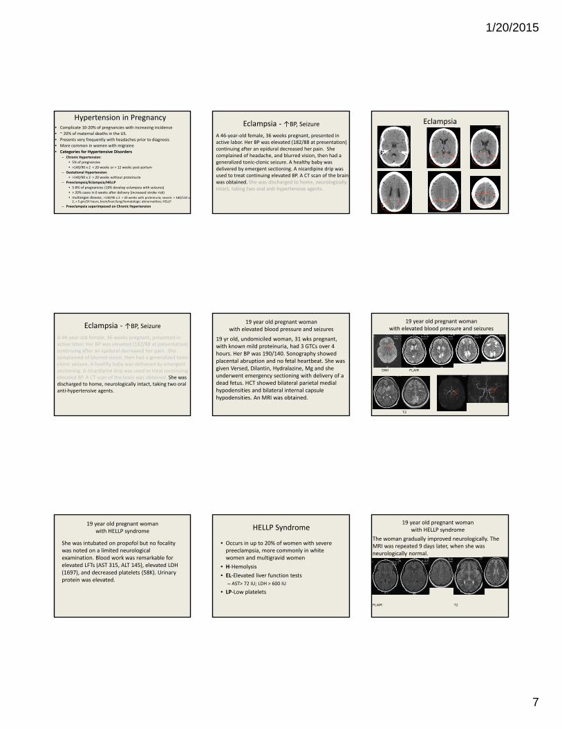
A 46‐year‐old female, 36 weeks pregnant, presented in active labor. Her BP was elevated (182/88 at presentation) continuing after an epidural decreased her pain. She complained of headache, and blurred vision, then had a generalized tonic‐clonic seizure. A healthy baby was delivered by emergent sectioning. A nicardipine drip was used to treat continuing elevated BP. A CT scan of the brain was obtained. She was discharged to home, neurologically intact, taking two oral anti‐hypertensive agents.
Eclampsia
Eclampsia ‐ ↑BP, Seizure
A 46‐year‐old female, 36 weeks pregnant, presented in active labor. Her BP was elevated (182/88 at presentation) continuing after an epidural decreased her pain. She complained of blurred vision, then had a generalized tonic‐clonic seizure. A healthy baby was delivered by emergent sectioning. A nicardipine drip was used to treat continuing elevated BP. A CT scan of the brain was obtained. She was discharged to home, neurologically intact, taking two oral anti‐hypertensive agents.
19 year old pregnant woman with elevated blood pressure and seizures
19 yr old, undomiciled woman, 31 wks pregnant, with known mild proteinuria, had 3 GTCs over 4 hours. Her BP was 190/140. Sonography showed placental abruption and no fetal heartbeat. She was given Versed, Dilantin, Hydralazine, Mg and she underwent emergency sectioning with delivery of a dead fetus. HCT showed bilateral parietal medial hypodensities and bilateral internal capsule hypodensities. An MRI was obtained.
19 year old pregnant woman with elevated blood pressure and seizures
DWI FLAIR
T2
19 year old pregnant woman with HELLP syndrome
She was intubated on propofol but no focality was noted on a limited neurological examination. Blood work was remarkable for elevated LFTs (AST 315, ALT 145), elevated LDH (1697), and decreased platelets (58K). Urinary protein was elevated.
HELLP Syndrome
• Occurs in up to 20% of women with severe preeclampsia, more commonly in white women and multigravid women
• H‐Hemolysis
• EL‐Elevated liver function tests
– AST> 72 IU; LDH > 600 IU
• LP‐Low platelets
19 year old pregnant woman with HELLP syndrome
The woman gradually improved neurologically. The MRI was repeated 9 days later, when she was neurologically normal.
FLAIR T2
1/20/2015
8
Postpartum headache and elevated BPA 42 year old G4P2Ab1 delivered a healthy infant by repeatsectioning at 36 weeks, after SROM. BPs and laboratory testingwere normal. Urine showed trace protein. She was discharged onPPD4 with a BP of 129/78. She was seen in the ED on PPD7 witha 10/10 headache with n/v for a day. Her BPs were consistentlyelevated at 160-180/70-80. Blood work was normal. Urine proteinwas 2+. HCT was read as normal. Pain improved with narcoticsand she was sent home.
Eclampsia & ICHThe woman fell on PPD 8 and was brought to the ED. She wasunresponsive with decorticate posturing. Her BP was 193/112.Blood work was unremarkable. She died on PPD 9. No underlyingbrain lesion was found on autopsy.
Post‐partum HeadacheA 35 year old, healthy, G1P0 woman was admitted at 40 weeks for an elective induction. Her BP on admission was 150/100, staying at 150 ‐160’s/100’s, for 24 hours until vaginal delivery. She complained of a headache and severe abdominal pain after delivery, with persistent BP elevation. At 5 hours after delivery, her headache worsened and left sided weakness developed suddenly. CTs were obtained at onset of weakness and 4 hours later after she was intubated. Liver function tests were markedly elevated from a normal baseline on admission Her platelet count decreased to 40,000 from a normal baseline on admission.
Post‐partum ICH
4 hrslater
L weaknessonset
HELLP & ICH
A 35 year old, healthy, G1P0 woman was admitted at 40 weeks for an elective induction. Her BP on admission was 150/100, staying at 150 ‐160’s/100’s, for 24 hours until vaginal delivery. She complained of a headache and severe abdominal pain after delivery, with persistent BP elevation. At 5 hours after delivery, her headache worsened and left sided weakness developed suddenly. CTs were obtained at onset of weakness and 4 hours later after she was intubated. Liver function tests were markedly elevated from a normal baseline on admission Her platelet count decreased to 40,000 from a normal baseline on admission.
Severe peri‐partum headache in 40 yr old
A 40 yr old woman without significant PMH, G4P1122 had a persistent post partum, 10/10, frontal headache. Her sudden onset headache began prior to delivery and persisted through L&D to post‐partum day #7. Her headache was associated with nausea and dizziness. She had bilateral LE edema since delivery, with proteinuria prior to delivery. Her BP was 136/61. Her neurological examination was normal. Her CBC and metabolic panel were normal. Spinal fluid showed 4100 to 2500 RBCs with xanthochromia. HCT was interpreted as negative.
Severe peri‐partum headache in 40 yr old Post‐Partum Cerebral Angiopathy (RCVS) Post‐Partum Cerebral Angiopathy (RCVS)
1/20/2015
9
Reversible Cerebral Vasoconstriction Syndrome
• Case reports of reversible narrowing of cerebral vessels by Marie Fleming (1987)
• Followed by case series by Call and colleagues (1988) describing characteristic clinical and imaging findings—reversible cerebral segmental vasoconstriction
• Call‐Fleming syndrome
RCVS
Sudden, severe headache, evidence of vasoconstriction in cerebral vessels, and documented resolution of vasoconstriction
Can cause ischemic strokes especially in border‐zone territories
Associated with a variety of clinical states:
Pregnancy (postpartum cerebral angiopathy)
Migraine (migrainous vasospasm)
Drug use (SSRIs, nicotine, cocaine)
Benign angiopathy of the CNS
RCVS
• Clinical:
– Sudden onset severe, “thunderclap” headache
– Nausea
– Vision changes
– Photophobia
– Encephalopathy
– Focal deficits (ischemic, hemorrhagic)
– Generalized seizures (up to 30%)
– 1/3 with moderate‐severe HTN
Post‐partum headache with normal BP
A healthy 43 year old woman delivered her 3nd child vaginally, after an uneventful pregnancy. Her blood pressures were consistently under 140/90 after delivery. She complained of an intermittent mild headache the day after delivery. On the second day after delivery her headache suddenly worsened and her blood pressure increased markedly. She became unresponsive.
Post‐partum ICH – postpartum angiopathy?
No brain lesion was found on autopsy.
Variable Presentations of Postpartum AngiopathyFugate et al. Stroke. 2012; 43: 670‐676
18 patients (mean age, 31 years; range, 15–41)
– Median gestation ‐ 38 weeks.
– 12 (67%) ‐ prior uneventful pregnancy
– Coagulopathy (n=6, 33%) protein S deficiency, antiphospholipid antibody syndrome, essential
– Neurological symptoms (began on median day 5 postpartum) • headache (n=16, 89%),
• focal deficit (n=9, 50%),
• visual disturbance (n=8, 44%),
• encephalopathy (n=6, 33%)
• seizure (n=5, 28%)
Variable Presentations of Postpartum AngiopathyFugate et al. Stroke. 2012; 43: 670‐676
Abnormal brain (MRI/CT) imaging (n=13, 72%)
intracranial hemorrhage (n=7, 39%)
vasogenic edema (n=6, 35%)
infarction (n=6, 35%)
Clinical outcomes
full recovery seen in 9 (50%),
death after a fulminant course in 4 (22%)
residual deficits in 5 (28%)
Headache 3 weeks after delivery
35 year‐old woman had a post‐partum headache for 8 days. Thirty days prior, pre‐eclampsia, HELLP syndrome and intrauterine fetal demise were detected on routine screening of her first pregnancy. The fetus was delivered at 30 weeks. Blood pressures at home after delivery were 110‐130/70‐90, not on medication. She saw her obstetrician for the persistent headache, when her BP was 150/100. An MRI scan was ordered.
Post‐partum headache
SWI
FLAIR
1/20/2015
10
Post‐partum headache Post‐partum headache CVT
1 month after delivery
4 months after delivery
Cerebral Venous Thrombosis
Generally present with headache, less frequently seizures, focal deficitsThrombophilias (Factor V Leiden gene mutation, prothrombin 20210A gene mutation, methylenetetrahydrofolate reductase (MTHFR) 677TT polymorphism)
Exogenous hormones Most common in post‐partum period Treatment with IV heparin to SQ LMWH during pregnancy, to warfarin post‐partum Possible increase in future risk of pregnancy related to prior CVT Avoid further pregnancies for two years. Consider use of anticoagulation (LMWH) with further pregnancies.
Pregnancy, Headache & Stroke
• Headache is an important premonitory symptom for cerebrovascular disease in pregnancy.
• The stroke risk is increasing with older pregnant women and women with migraines.
• The majority of strokes occur in the peri & post‐partum periods.
• ICH & CVT are more common than ischemic stroke.
• Pre‐eclampsia, eclampsia, HELLP, post‐partum cerebral angiopathy may be on a continuum, presenting with headache.
• Monitoring blood pressure after delivery is crucial.
• Hypertensive disorder of pregnancy increases the risk of future ischemic stroke.
Post‐partum sudden headache, then loss of brainstem reflexes
A 23 year old woman, 39 weeks pregnant, underwent emergent sectioning under epidural anesthesia, for fetal bradycardia. At about 10 minutes after the infant was delivered she suddenly complained of a headache, vomited and immediately became unresponsive. She was intubated immediately and a stroke code was called. She had dilated, unreactive pupils; no brainstem reflexes; and no motor response to pain. Her NIHSS was 31. A CT scan, then an MRI scan, were obtained.
Post‐partum sudden onset of unresponsiveness: perimesencephalic air and anesthesia
In about an hour she was awake with a normal neurological examination, denying a headache.