547 J. Exp. Med. The Rockefeller University Press • 0022-1007/98/02/547/14 $2.00 Volume 187, Number 4, February 16, 1998 547–560 http://www.jem.org Association of Phosphorylated Serine/Arginine (SR) Splicing Factors With The U1–Small Ribonucleoprotein (snRNP) Autoantigen Complex Accompanies Apoptotic Cell Death By Paul J. Utz,* Maria Hottelet,* Walther J. van Venrooij, ‡ and Paul Anderson* From the *Department of Medicine, Division of Rheumatology, Immunology, and Allergy, Brigham & Women’s Hospital, Boston, Massachusetts 02115; and the ‡ Department of Biochemistry, University of Nijmegen, 6500 HB Nijmegen,The Netherlands Summary Proteins subject to proteolysis or phosphorylation during apoptosis are commonly precipitated by autoantibodies found in the serum of patients with systemic lupus erythematosus (SLE). We screened a panel of murine monoclonal and human monospecific sera reactive with known au- toantigens for their ability to selectively precipitate phosphoproteins from apoptotic Jurkat T cell lysates. Sera known to recognize the U1–small nuclear ribonucleoprotein (snRNP) com- plex (confirmed by their ability to precipitate U1–snRNA) selectively precipitated a phospho- protein complex (pp54, pp42, pp34, and pp23) from apoptotic lysates. Monoclonal antibodies reactive with U1–snRNP proteins precipitated the same phosphoprotein complex from apop- totic lysates. The phosphorylation and/or recruitment of these proteins to the U1–snRNP complex is induced by multiple apoptotic stimuli (e.g., Fas ligation, gamma irradiation, or UV irradiation), and is blocked by overexpression of bcl-2. The U1–snRNP-associated phosphoprotein complex is immunoprecipitated by monoclonal antibodies reactive with serine/arginine (SR) proteins that comprise a structurally related family of splicing factors. The association of phos- phorylated SR proteins with the U1–snRNP complex in cells undergoing apoptosis suggests a mechanism for regulation of alternative splicing of apoptotic effector molecules. C omponents of ribonucleoproteins (RNPs) 1 such as Ro, La, heterogeneous nuclear (hnRNP), and small nuclear (snRNP) are commonly recognized by autoanti- bodies found in the serum of patients with autoimmune disease (1–4). The mechanisms by which these and other autoantigens escape tolerance are largely unknown. The ob- servation that keratinocytes subjected to ultraviolet radia- tion express autoantigens such as Ro, La, and the U1-70 kD snRNP protein at cell surface blebs suggests that apoptotic cells may play an important role in the production of au- toantibodies (5–7). This is supported by experiments dem- onstrating the development of autoantibodies after immu- nization of mice with apoptotic cells (8). Proteolytic cleavage of at least 13 known protein autoantigens by indi- vidual interleukin-1b converting enzyme (ICE) family pro- teases (now collectively termed cysteine protease with as- partic acid substrate specificity, or “caspases” [9]) during programmed cell death further supports this hypothesis. To date, over half of all caspase targets are autoantigens or are constituents of larger complexes that contain a protein that is cleaved, and include the U1-70 kD snRNP (10), poly A ribose polymerase (PARP; reference 11), DNA-dependent protein kinase (DNA-PK; 12), hnRNP C1 and C2 (13), lamins A, B, and C (14), the nuclear mitotic apparatus pro- tein (NuMA; 15, 16), topoisomerases 1 and 2 (16), the nu- cleolar protein UBF/NOR-90 (16), and a fodrin (17, 18). Although proteolysis could expose novel epitopes re- quired for the production of autoantibodies, only a fraction of the known autoantigens are cleaved during apoptosis. Recently, we reported that phosphoproteins are commonly 1 Abbreviations used in this paper: DNA-PK, DNA-dependent protein ki- nase; HI-FCS, heat-inactivated FCS; hnRNP, heterogeneous nuclear RNP; MCTD, mixed connective tissue disease; NuMA, nuclear mitotic appara- tus protein; PARP, poly A ribose polymerase; PCNA, proliferating cell nuclear antigen; PVDF, polyvinylidene difluoride; RNP, ribonucleopro- tein; snRNP, small nuclear RNP; SLE, systemic lupus erythematosus; Sm, Smith complex; SR, serine/arginine; SRP, signal recognition particle; TIAR, T cell intracellular antigen-related protein.
Transcript
547
J. Exp. Med.
The Rockefeller University Press • 0022-1007/98/02/547/14 $2.00Volume 187, Number 4, February 16, 1998 547–560http://www.jem.org
Association of Phosphorylated Serine/Arginine (SR)Splicing Factors With The U1–Small Ribonucleoprotein (snRNP) Autoantigen Complex Accompanies ApoptoticCell Death
By Paul J. Utz,
*
Maria Hottelet,
*
Walther J. van Venrooij,
‡
and Paul Anderson
*
From the
*
Department of Medicine, Division of Rheumatology, Immunology, and Allergy, Brigham & Women’s Hospital, Boston, Massachusetts 02115; and the
‡
Department of Biochemistry, University of Nijmegen, 6500 HB Nijmegen, The Netherlands
Summary
Proteins subject to proteolysis or phosphorylation during apoptosis are commonly precipitatedby autoantibodies found in the serum of patients with systemic lupus erythematosus (SLE). Wescreened a panel of murine monoclonal and human monospecific sera reactive with known au-toantigens for their ability to selectively precipitate phosphoproteins from apoptotic Jurkat Tcell lysates. Sera known to recognize the U1–small nuclear ribonucleoprotein (snRNP) com-plex (confirmed by their ability to precipitate U1–snRNA) selectively precipitated a phospho-protein complex (pp54, pp42, pp34, and pp23) from apoptotic lysates. Monoclonal antibodiesreactive with U1–snRNP proteins precipitated the same phosphoprotein complex from apop-totic lysates. The phosphorylation and/or recruitment of these proteins to the U1–snRNPcomplex is induced by multiple apoptotic stimuli (e.g., Fas ligation, gamma irradiation, or UVirradiation), and is blocked by overexpression of bcl-2. The U1–snRNP-associated phosphoproteincomplex is immunoprecipitated by monoclonal antibodies reactive with serine/arginine (SR)proteins that comprise a structurally related family of splicing factors. The association of phos-phorylated SR proteins with the U1–snRNP complex in cells undergoing apoptosis suggests amechanism for regulation of alternative splicing of apoptotic effector molecules.
C
omponents of ribonucleoproteins (RNPs)
1
such asRo, La, heterogeneous nuclear (hnRNP), and small
nuclear (snRNP) are commonly recognized by autoanti-bodies found in the serum of patients with autoimmunedisease (1–4). The mechanisms by which these and otherautoantigens escape tolerance are largely unknown. The ob-servation that keratinocytes subjected to ultraviolet radia-tion express autoantigens such as Ro, La, and the U1-70 kDsnRNP protein at cell surface blebs suggests that apoptoticcells may play an important role in the production of au-
toantibodies (5–7). This is supported by experiments dem-onstrating the development of autoantibodies after immu-nization of mice with apoptotic cells (8). Proteolyticcleavage of at least 13 known protein autoantigens by indi-vidual interleukin-1
b
converting enzyme (ICE) family pro-teases (now collectively termed cysteine protease with as-partic acid substrate specificity, or “caspases” [9]) duringprogrammed cell death further supports this hypothesis. Todate, over half of all caspase targets are autoantigens or areconstituents of larger complexes that contain a protein thatis cleaved, and include the U1-70 kD snRNP (10), poly Aribose polymerase (PARP; reference 11), DNA-dependentprotein kinase (DNA-PK; 12), hnRNP C1 and C2 (13),lamins A, B, and C (14), the nuclear mitotic apparatus pro-tein (NuMA; 15, 16), topoisomerases 1 and 2 (16), the nu-cleolar protein UBF/NOR-90 (16), and
a
fodrin (17, 18).Although proteolysis could expose novel epitopes re-
quired for the production of autoantibodies, only a fractionof the known autoantigens are cleaved during apoptosis.Recently, we reported that phosphoproteins are commonly
precipitated from apoptotic cell extracts by autoantibodiesderived from patients with systemic lupus erythematosus(SLE), suggesting that protein modifications accompanyingapoptosis might generally predispose to autoantibody for-mation (19). We previously identified seven phosphopro-teins (termed pp200, pp54, pp46, pp42, pp34, pp23, andpp17) in Jurkat T cells that are specifically precipitated withautoimmune sera in response to apoptotic stimuli (19). Wealso showed that a serine kinase activity is present in immu-noprecipitates prepared from apoptotic Jurkat cell extractsusing sera from patients with SLE and SLE overlap syn-dromes. We proposed that phosphorylation of autoantigensmay be a common sequela of apoptotic cell death, and wepostulated that these phosphoproteins, like other kinasesubstrates, such as c-jun, may be involved in the effectorarm of the cell death pathway.
Well-characterized, monospecific human sera have beenused in several recent studies to identify autoantigens thatare cleaved during apoptosis (12, 16). We have used a simi-lar approach to identify autoantigens that are selectivelyphosphorylated during apoptosis. Although most of the seradid not precipitate phosphoproteins from radiolabeled apop-totic lysates, five sera known to recognize the U1–snRNPcomplex precipitated phosphoproteins migrating with ap-parent molecular masses of 54, 42, 34, and 23 kD by SDS-PAGE. A series of human autoimmune sera directed againstthe U1–snRNP, but not the U2–snRNP, also coprecipi-tated this same phosphoprotein complex. Identical resultswere obtained using anti-U1A human variable domain an-tibody fragments and monoclonal antibodies directed againstindividual components of U–snRNPs. Because the relativemigration of these U1–snRNP-associated phosphoproteinsresembled the serine/arginine (SR) complex of splicing fac-tors, we used antibodies reactive with SR proteins to pre-cipitate phosphoproteins from apoptotic lysates. A mono-clonal antibody specific for a phosphoepitope common toall SR proteins (mAb104) and a monoclonal antibody spe-cific for the phosphorylated form of the SR protein SC35precipitated a similar phosphoprotein complex from theselysates. The identification of SR proteins as potential sub-strates for a serine kinase that is activated during apoptosis hasimportant implications for understanding cell death path-ways, RNA splicing, and the immune response in diseasesthat are characterized by the development of autoantibodies.
Materials and Methods
Cell Culture.
Jurkat cells were grown in 5% CO
2
at 37
8
C us-ing RPMI 1640 (BioWhittaker, Inc., Walkersville, MD) supple-mented with 9% heat-inactivated FCS (HI-FCS; Tissue CultureBiologicals, Tulare, CA), penicillin, and streptomycin (Media-tech, Inc., Herndon, VA). Cells were grown and harvested at mid-log phase. Jurkat T cells engineered to stably overexpress bcl-2(or empty vector), a gift from John Reed (the La Jolla CancerResearch Foundation, La Jolla, CA), were grown in RPMI me-dium as described above supplemented with G418 (GIBCOBRL, Gaithersburg, MD) at a final concentration of 500
m
g/ml.Protein overexpression was confirmed by Western blotting.
Metabolic Labeling.
Labeling was performed as described (19).In brief, Jurkat cells were incubated at a density of 2
3
10
6
cells/ml in labeling medium containing the following: 45% RPMI1640, 45% RPMI 1640 lacking either phosphate (GIBCO BRL)or methionine and cysteine (GIBCO BRL), 2 mM glutamine(Mediatech, Inc.), 5% HI-FCS, and 5% HI-FCS that had beendialyzed to equilibrium against a buffer containing 10 mM Hepesand 140 mM NaCl (Sigma Chemical Co., St. Louis, MO).[
32
P]orthophosphate or [
35
S]methionine and cysteine (Dupont-NEN, Boston, MA) was added at a concentration of 0.1 mCi/ml.Cells were incubated at 37
8
C for 10–16 h to allow the cells toreach steady state before each treatment, unless otherwise indi-cated. For two-dimensional tryptic phosphopeptide mapping ex-periments, cells were labeled for 2 h, followed by a 3-h stimula-tion with anti-Fas antibodies (7C11) in labeling media composedof 90% RPMI 1640 lacking phosphate, 2 mM glutamine, 10% di-alyzed HI-FCS, and 0.15 mCi/ml
32
P-orthophosphate.
Cell Lysis.
Lysis of cells was performed using NP-40 (SigmaChemical Co.) lysis buffer (1% NP-40, 150 mM NaCl, 50 mMTris, pH 7.8, and 1 mM EDTA). NP-40 lysis buffer was supple-mented immediately before use with 1 mM sodium vanadate(Sigma Chemical Co.) and a 100
3
protease inhibitor cocktail pre-pared by dissolving 10 mg chymostatin, 1.5 mg leupeptin, 7 mgpepstatin A, 850 mg phenylmethylsulfonyl fluoride, 500 mg benz-amidine, and 5 mg aprotonin in 50 ml of ethanol by stirring over-night (20). The solution was sterilized by filtration and stored atroom temperature. All chemicals were purchased from SigmaChemical Co. After addition of 1 ml lysis buffer, the lysate wasincubated on ice for 30 min, centrifuged in a refrigerated mi-crofuge (5402; Eppendorf Inc., Hamburg, Germany) at 14,000rpm for 15 min, and then the supernatant was used immediatelyfor each experiment.
UV Irradiation.
Labeled Jurkat cells were plated on 100
3
15-mmpolystyrene petri dishes (Nunc, Thousand Oaks, CA) at a con-centration of 2
3
10
6
cells/ml and irradiated in a Stratalinker2400 (Stratagene, La Jolla, CA) at a distance of 9 cm for 18 s. Af-ter irradiation, cells were incubated at 37
8
C for the indicatedtimes before harvesting.
Gamma Irradiation.
Labeled cells were placed in a 50-ml con-ical tube and irradiated at a dose of 3,300 rad from a Cesium 137source using an irradiator (Gammacell 1000; Nordion Interna-tional, Kanata, Ontario, Canada). After irradiation, cells were placedin culture dishes at 37
8
C and incubated for the indicated timesbefore harvesting.
Cellular Activation.
Labeled Jurkat cells were treated with thefollowing antibodies: anti-Fas antibody 7C11 (provided by Mich-ael Robertson, Indiana University, Bloomington, IN) from hy-bridoma supernatant at a final dilution of 1:500; and anti-CD3antibody (Coulter Immunology, Hialeah, FL) at a concentrationof 5
m
g/ml followed by goat anti–mouse antibody (Jackson Im-munoResearch Laboratories, West Grove, PA) at the same con-centration. Cells were incubated at 37
8
C for the indicated timesbefore harvesting.
Immunoprecipitation and Western Blot Analysis.
Lysates were pre-cleared once with 25
m
l of a 50% solution of protein A–Sepharose(Pharmacia, Uppsala, Sweden) in PBS and 5
m
g rabbit anti–mouse(RAM) IgG (Jackson ImmunoResearch Laboratories) for 1 h, fol-lowed by two preclears with protein A–Sepharose overnight.Mouse monoclonal antibodies (5
m
g) and 5
m
g RAM (IgG orIgM), or 2
m
l patient serum alone were used in precipitation ex-periments. Immunoprecipitation experiments using anti-U1A hu-man antibody fragments were performed as described (21). The fol-lowing human polyclonal antibodies were stored at
2
70
8
C until
549
Utz et al.
used: anti-Ro, anti-La, anti–Smith complex (Sm), anti–Jo-1, anti-nucleolar, anticentromere, anti–Scl-70, anti-DNA, and anti-U1–RNP (Arthritis Foundation/CDC Reference Sera, Atlanta, GA);anti-Th/To, anti–U3-fibrillarin, anti-signal recognition particle(SRP), anti–PL-7, and anti–PL-12 (T. Medsger and N. Fertig,University of Pittsburgh School of Medicine, Pittsburgh, PA); twodifferent anti-U1/U2 monospecific sera (Sera Ya and Ga; refer-ence 22) and anti-SRP (J. Craft, Yale University School of Med-icine, New Haven, CT); anti-NuMA (serum AS), and anti-UBF(serum JO; E. Tan and C. Casiano, The Scripps Institute, La Jolla,CA); anti–RNA polymerase I/III and II (serum KA), anti–poly-merase I/III (serum IM), anti-Th/To, anti–U3-fibrillarin, anti-Ku, and anti–Scl-70 (M. Kuwana, Keio University MedicalSchool, Tokyo, Japan); anti-ribosomal
P
and antihistone/U–RNP (Immunovision Inc., Springdale, AZ); anti–U1-70 kD sn-RNP protein (A. Rosen, the Johns Hopkins University School ofMedicine, Baltimore, MD); anti-sp140, and anti-sp100 (D.Bloch, Massachusetts General Hospital, Boston, MA); seven hu-man sera specific for the U1–snRNP complex (B83, B152, B175,H34, H165, K4, and L41) and a control serum specific for bothU1 and U2–snRNPs (V26) have been reported previously (23,91). Serum from patients with SLE and mixed connective tissuedisease (MCTD) with high titers of antibodies against Sm andRNP components, respectively, were provided by P.H. Schur(Brigham and Women’s Hospital, Boston, MA). Autoimmuneserum capable of precipitating pp54, pp42, pp34, and pp23 (cor-responding to patients 1, 8, 11, and 12) were described previously(19). Serum from a fifth patient (patient 3) also coprecipitatedthese proteins but was unavailable in sufficient quantity to com-plete the studies described below (19). The following mousemonoclonal antisera were stored at
2
70
8
C until used: anti-laminB (E3), and anti–lamin A
1
B (E6; E.A. Nigg, University of Ge-neva, Geneva, Switzerland); anti–lamin B (Calbiochem-Nova-biochem Corp., San Diego, CA); anti–proliferating cell nuclearantigen (PCNA; Zymed Laboratories, Inc., South San Francisco,CA); anti–DNA-PK (D. Weaver, Dana Farber Cancer Institute,Boston, MA); two monoclonal anti-Ku antibodies (C. Zhang,Dana Farber Cancer Institute, Boston, MA); anti-Ki67 (D. Bloch,Massachusetts General Hospital, Boston, MA); anti–U1-70 kD (ref-erence 24; S. Hoch, the Agouron Institute, La Jolla, CA); anti-SmY12 (J. Craft, Yale University School of Medicine, New Haven,CT); anti-U1A/U2B
99
9A9 (25); anti-U2B
99
4G3 (25); anti-Ro602G10 (26); anti-Ro52 and anti-La SW5 (27) have been describedpreviously; mAb104 monoclonal directed against SR proteins(reference 28; R. Reed, Harvard University School of Medicine,Boston, MA); anti-SC35 (Sigma Chemical Co.); and antifibril-larin monoclonal antibodies (72B9.D31 and 17C12.G9) and twomonoclonal antibodies directed against other U3–snRNP com-ponents (7G3.B7 and 6G10.D3; K.M. Pollard, Scripps Institute,La Jolla, CA). Serum from healthy control patients was a gift fromP. Fraser (Brigham and Women’s Hospital, Boston, MA). Immu-noprecipitations were performed after addition of 1% BSA (Inter-gen Company, Purchase, NY) in PBS to a total volume of 500
m
land rotation in a 4
8
C cold room for 2–24 h. Comparison of pre-cipitates showed no difference between incubation times for peri-ods of up to 72 h. Precipitates were harvested by centrifugingfor 15 s at 14,000 rpm in a refrigerated Eppendorf microfuge,washing three times with NP-40 lysis buffer supplemented withprotease inhibitor cocktail, resuspending in SDS loading bufferwith 9% 2-mercaptoethanol, boiling for 5 min, and separating byPAGE as described (29). Proteins were transferred to nitrocellu-lose (Schleicher & Schuell, Inc., Keene, NH) for Western blot-ting or tryptic mapping experiments, or to polyvinylidene difluo-
ride (PVDF; Dupont-NEN, Boston, MA) for phosphoamino acidanalysis, and either exposed for autoradiography or subjected toWestern blot analysis as indicated (30). The anti–bcl-2 mousemonoclonal antibody 4D7 (PharMingen, San Diego, CA) wasused for blotting studies at a dilution of 1:1,000. Nitrocelluloseblots were blocked with 5% Blotto (Bio-Rad Laboratories, Her-cules, CA) in PBS overnight at 4
8
C. Bands were visualized usingRAM conjugated to horseradish peroxidase (Amersham Corp., Ar-lington Heights, IL) at a dilution of 1:7,500 in 5% Blotto in PBS,and developed using ECL chemiluminescence performed accord-ing to the manufacturer’s instructions (Amersham Corp.).
Phosphoamino Acid Analysis.
Immunoprecipitates that had beenelectrophoresed and transferred to PVDF were rinsed thoroughlywith water, exposed for radiography, and then appropriate bandswere excised with a razor blade. The radiolabeled bands werethen subjected to acid hydrolysis as described (31).
RNA Isolation and Identification.
Immunoprecipitates from
32
P-labeled Jurkat cells were prepared as described above. After thethird NP-40 lysis buffer wash, the immunoprecipitate was di-gested in a volume of 300
m
l for 1 h at 37
8
C in a solution con-taining 50
m
g/ml proteinase K (Sigma Chemical Co.), 10 mMTris, pH 7.8, 10 mM EDTA, and 0.5% SDS. The RNA was iso-lated after two extractions with a phenol/chloroform/isoamyl al-cohol (25:24:1) mixture (GIBCO BRL). The RNA was precipi-tated overnight at
2
70
8
C after the addition of 20
m
l 3 M sodiumacetate, 400
m
l ethanol, and 10
m
g transfer RNA (Sigma Chemi-cal Co.) as a carrier. The pellet was obtained after a 15-min cen-trifugation in an Eppendorf centrifuge maintained at 4
8
C. Thepellet was washed once with 70% ethanol, dried in a fume hood,and subjected to PAGE on 6% sequencing gels. A small amountof whole cell lysate was also processed as above and included as aninternal standard on each gel.
Two-Dimensional Phosphopeptide Analysis.
Two-dimensional tryp-tic phosphopeptide mapping was performed as described (32) us-ing trypsin (Worthington Biochemical Corp., Freehold, NJ) at aconcentration of 0.1 mg/ml in 50 mM ammonium bicarbonate.Plates were exposed to film at
2
70
8
C with an intensifying screenfor 2 d.
Results
Autoimmune Sera Precipitate Phosphoproteins from LysatesPrepared from Jurkat T Cells Undergoing Fas-induced Apopto-sis.
Sera reactive with known autoantigens were firsttested for their ability to precipitate the expected protein orcomplex from extracts prepared from
35
S-labeled Jurkatcells, as detected by SDS-PAGE followed by autoradio-graphic exposure. Sera that precipitated ambiguous patternsof proteins were subjected to further analysis by Westernblotting of both whole cell Jurkat extracts and immunopre-cipitates prepared as above, to confirm that the well-char-acterized sera precipitated the expected target antigen. Mostof the sera were derived from patients with autoimmunedisease. In addition, six murine monoclonal antibodies re-active with known autoantigens (Ku, DNA-PK, lamins Aand B, Ki67, and PCNA) were included in this screen.
Jurkat cells metabolically labeled with
32
P-orthophos-phate were cultured for 3 h in the absence or presence of amonoclonal antibody reactive with Fas (anti-7C11), solubi-lized in NP-40 lysis buffer, and immunoprecipitated usingpatient serum or monoclonal antibodies as previously de-
550
Serine/Arginine Splicing Factors in Apoptosis
scribed (19). Immunoprecipitates were separated on a 12%SDS–polyacrylamide gel, transferred to nitrocellulose, andthen subjected to autoradiography. Most sera did not pre-cipitate unique phosphoproteins from apoptotic lysates. Forexample, sera reactive with nuclear proteins implicated inDNA replication, binding, or repair (i.e., Ku, DNA-PK,PCNA, Scl-70, and histone) failed to reproducibly precipi-tate new phosphoproteins from apoptotic Jurkat cell ex-tracts (data not shown). Similarly, proteins present in thenuclear matrix or involved in mitosis (i.e., lamins A and B,centromere A and B, Ki-67, NuMA, sp140, and sp100),the nucleolus (i.e., Th/To, UBF/NOR-90, RNA poly-merase I, II, and III, and U3–snRNPs), or cytoplasmiccomponents of the translational apparatus (i.e., Jo-1, Pl-7,Pl-12, SRP, and ribosomal P) were unmodified in theseexperiments (data not shown). In contrast, several sera spe-cific for U–snRNP
complexes precipitated phosphopro-teins of 54, 42, 34, and 23 kD from apoptotic Jurkat cellextracts (Fig. 1
A
). The phosphorylation of these proteinsdid not result from a nonspecific, general increase in kinaseactivity after Fas engagement, as
32
P-labeled, whole cell ex-tracts prepared from untreated and apoptotic cells wereidentical when analyzed by SDS PAGE (data not shown).Moreover, this pattern was similar to that observed usingfour distinct sera described in our previous report, suggest-ing that these four proteins (termed pp54, pp42, pp34, andpp23) may be previously unrecognized components ofU–snRNP complexes (19). As reported previously (19),the constitutive phosphorylation of La (Fig. 1
A
,
Ro
and
La
) was unaltered in cells undergoing apoptosis.
Autoimmune Sera Coprecipitate U–snRNP Proteins.
Immu-noprecipitates were prepared from
35
S-labeled lysates pre-pared before (
2
) or 3 h after (
1
) Fas ligation (Fig. 1
B
). Al-though immunoprecipitates prepared from apoptotic andnonapoptotic lysates contained
35
S-labeled proteins migrat-ing similarly to the phosphoproteins identified in Fig. 1
A
,we were unable to establish the relationship between the
32
P- and
35
S-labeled proteins. Nevertheless, we did not ob-serve the appearance of new
35
S-labeled proteins corre-sponding to components of the phosphoprotein complex.Moreover, all five sera precipitated U–snRNP components(e.g., the A, B, B
9
, and C proteins, indicated by arrows)from labeled Jurkat cell extracts (Figs. 1
B
and 2, also seebelow). Six other sera from patients with SLE and MCTDpossessing high titers of antibodies against Sm or RNPcomponents (as determined by ELISA assay), also precipi-tated U–snRNP components from
35
S-labeled Jurkat cellextracts, as well as all four phosphoproteins from apoptoticcell extracts prepared from
32
P-labeled Jurkat cells (data notshown). These results demonstrate that autoimmune seracapable of precipitating U–snRNP proteins coprecipitate aphosphoprotein complex containing pp54, pp42, pp34,and pp23 from apoptotic Jurkat cell lysates.
Autoimmune Sera Coprecipitate the U1–snRNA Moleculeand pp54, pp42, pp34, and pp23.
To further establish thatpp54, pp42, pp34, and pp23 are components of U–snRNPcomplexes, we used all five sera shown in Fig. 1
B
, foursera previously reported to precipitate these four phospho-proteins (patients 1, 8, 11, and 12; reference 19), and con-trol sera to identify the RNA molecules present in individ-
Figure 1. Human autoimmune sera specific for U–snRNP complexes precipitate phosphoproteins from apoptotic Jurkat cell lysates. (A) Jurkat cellswere labeled with 32P orthophosphate and lysed either before (2) or 3 h after (1) the addition of anti-Fas 7C11. Proteins were then precipitated usingthe indicated autoimmune serum, separated on a 12% SDS–polyacrylamide gel, transferred to nitrocellulose, and exposed for autoradiography. Individualsera are described in detail in Materials and Methods. U-serum 1, Immunovision antihistone/RNP; U-serum 2, CDC/AF reference serum 4 (anti-U1–RNP);U-serum 3, serum Ga; U-serum 4, serum Ya; U-serum 5, CDC/AF reference serum 5 (anti-Sm). The relative migration of molecular mass markers in kilo-daltons is indicated on the left side of the gel. (B) Immunoprecipitation from 35S-labeled Jurkat cells. Jurkat cells were labeled with 35S-methionine andcysteine and lysed either before (2) or 3 h after (1) the addition of anti-Fas 7C11 before immunoprecipitation using sera derived from the indicated pa-tient. Immunoprecipitates were separated on a 12% SDS–polyacrylamide gel, transferred to nitrocellulose, and then subjected to autoradiographic analy-sis. The relative migration of molecular mass markers in kilodaltons is indicated on the left side of each panel. Bands corresponding to the U–snRNP proteinsA, B, B9, and C are indicated on the right side of the panel.
32P-orthophosphate were solubilized in NP-40 lysisbuffer, RNA was extracted from washed immunoprecipi-tates, separated on a 6% polyacrylamide gel, and subjectedto autoradiography. As shown in Fig. 2, all sera capable ofprecipitating the phosphoprotein complex also precipitatethe U1–snRNA. A similar result was obtained for anotherwell-characterized serum previously shown to recognizethe U1-70 kD protein (Fig. 2, lane 6, U-serum 6, a gift ofA. Rosen; reference 10). We noted that one sample (U-serum2, Fig. 2, lane 10) specifically precipitated the U1– but notthe U2–snRNA molecule, suggesting that pp54, pp42, pp34,and pp23 may be components of the U1–snRNP complex.To confirm this observation, we screened seven additionalhuman sera previously shown to specifically precipitate theU1–snRNP complex (91), and serum V26 (which precipi-tates both the U1– and U2–snRNPs; reference 23) for
their ability to coprecipitate pp54, pp42, pp34, and pp23from 32P-labeled, apoptotic Jurkat cell extracts. All 7 seracoprecipitated the U1–snRNA (Fig. 3 A) and all fourphosphoproteins (Fig. 3 B). Identical results were obtainedusing serum V26 (data not shown). Interestingly, individualsera precipitated phosphoproteins of varying intensity (e.g.,all four phosphoproteins are precipitated by serum B83,Fig. 3 B, lane 2, but mainly pp42 and pp54 are precipitatedby serum H165, Fig. 2, lane 10). This may result from het-erogenous populations of antibodies directed against indi-vidual U1–snRNP components, or by antibodies that rec-ognize the phosphoprotein directly. None of the controlantibodies directed against Ro, La, ribosomal P, or SRP
Figure 2. Coprecipitation of U1–snRNA using selected autoantisera.Jurkat cells were labeled with 32P-orthophosphate and solubilized in NP-40 lysis buffer. After immunoprecipitation with the indicated serum,RNA was extracted and separated on 6% sequencing gels before dryingand autoradiographic exposure. The relative migration of known RNAmoieties is depicted on the right side of the figure. The serum specificityis indicated above each sample. Lanes are numbered at the bottom of thepanel. Lanes 1–4, patients 1, 8, 11, 12 (19); U-serum 1, Immunovision an-tihistone/RNP; U-serum 2, CDC/AF reference serum 4 (anti-U1–RNP);U-serum 3, serum Ga; U-serum 4, serum Ya; U-serum 5, CDC/AF refer-ence serum 5 (anti-Sm); U-serum 6, anti–U1-70 kD serum (gift of A.Rosen).
Figure 3. U1-specific autoantisera coprecipitate the U1–snRNA mole-cule and pp54, pp42, pp34, and pp23 from apoptotic extracts. (A) Jurkatcells were labeled with 32P-orthophosphate and lysed in NP-40 lysisbuffer. After immunoprecipitation with the indicated serum, RNA wasextracted and separated on 6% sequencing gels before drying and autora-diographic exposure. Patient sera specific for the U1–snRNP complexwere used in lanes 1–7. A patient serum (V26, lane 8) capable of precipi-tating both the U1– and U2–snRNPs is shown for comparison. The rela-tive migration of the U1– and U2–snRNAs is depicted on the right sideof the figure. (B) Jurkat cells were labeled with 32P-orthophosphate andlysed either before (2) or 3 h after (1) the addition of anti-Fas (7C11)before immunoprecipitation using sera derived from the indicated patient.Immunoprecipitates were separated on a 12% SDS–polyacrylamide gel,transferred to nitrocellulose, and subjected to autoradiographic analysis.Sera correspond to the seven U1-specific autoantisera shown in Fig. 3 A.The relative migration of molecular size markers in kilodaltons is indi-cated on the left side of the figure. Bands corresponding to pp54, pp42,pp34, and pp23 are shown on the right side of the panel. A high molecu-lar mass complex is indicated with a large arrowhead. Lanes are numberedat the bottom of the figure.
552 Serine/Arginine Splicing Factors in Apoptosis
precipitated either the U–snRNAs or any of the four phos-phoproteins (data not shown). Several sera also precipitatedphosphoproteins between 96 and 200 kD that were nolonger detected after Fas stimulation (e.g., B152, H34, andK4, Fig. 3 B, lanes 3, 7, and 11), again suggesting that thesesera likely recognize heterogeneous epitopes of the U1–snRNP particle. It is unknown if this represents dephosphory-lation, caspase cleavage, or dissociation of these phospho-proteins from the immunoprecipitate after the apoptoticstimulus. These results suggest that the U1–snRNP is a dy-namic particle that is altered by caspases (U1-70 kD pro-tein; reference 10), and potentially by kinases (pp54, pp42,pp34, and pp23; reference 19) and phosphatases (the high
molecular mass protein complex shown in Fig. 3 B) duringapoptosis.
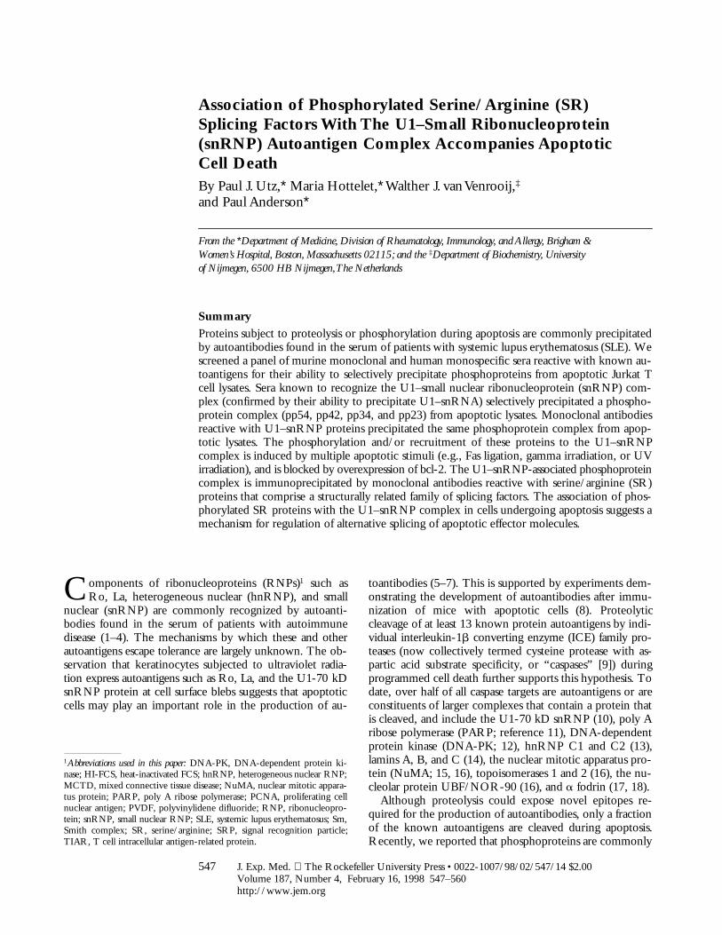
Monoclonal Antibodies Directed Against U1–snRNP Compo-nents Precipitate pp54, pp42, pp34, and pp23 from Apoptotic JurkatCell Lysates. To confirm our results described above us-ing human autoimmune serum, and to determine whetherpp54, pp42, pp34, and pp23 exist in both the U1– andU2–snRNP splicing complexes, we used a series of mono-clonal antibodies directed against proteins common orunique to each U–snRNP complex. Specifically, we testedantibodies directed against the U1-70 kD component ofthe U1–snRNP, the U2B99 protein of the U2–snRNP(4G3), the anti-Sm monoclonal antibody Y12, and a mono-
Figure 4. Monoclonal antibodies directed againstU1–snRNP components precipitate pp54, pp42,pp34, and pp23 from extracts prepared from apop-totic Jurkat cells. (A) Jurkat cells were labeled with32P-orthophosphate and lysed either before (2) or 3 hafter (1) the addition of anti-Fas 7C11. Proteinswere then precipitated using the indicated autoim-mune serum, separated on a 12% SDS–polyacryl-amide gel, transferred to nitrocellulose, and then ex-posed for autoradiography. The relative migration ofmolecular mass markers in kilodaltons is indicated onthe left side of the gel. Bands corresponding to pp90,pp54, pp42, pp34, and pp23 are shown on the rightside of the panel. Lanes are numbered at the bottomof the panel. (B) The identical experiment in 35S-labeledJurkat cells. The relative migration of molecular sizemarkers in kilodaltons is indicated on the left side ofthe gel. Lanes are numbered at the bottom of thefigure. (C) Phosphoamino acid analysis of pp54,pp42, pp34, and pp23. Jurkat cells were labeled with32P-orthophosphate, treated with the anti-Fas mono-clonal antibody 7C11, and solubilized using NP-40
lysis buffer after 3 h. Proteins were then precipitated with the anti-U1A/U2B99 monoclonal antibody 9A9, separated on a 12% SDS–polyacrylamide gel,transferred to PVDF, and exposed for autoradiography. Individual phosphoproteins were localized on the membrane, excised, and then subjected to acidhydrolysis. Phosphoamino acids were separated by two-dimensional electrophoresis in pH 1.9 buffer in the horizontal dimension, followed by pH 3.5buffer in the vertical dimension before autoradiographic analysis. Individual proteins are labeled on the side of each panel. Migration of phosphoaminoacid standards are labeled with circles as follows: phosphoserine (pS), phosphothreonine (pT), phosphotyrosine (pY). (D) Anti-U1A antibody fragmentscoprecipitate pp54, pp42, pp34, and pp23 from apoptotic Jurkat cell lysates. Labeled Jurkat cell extracts were prepared as above. Proteins were precipi-tated using the indicated anti-U1A antibody fragments, separated on a 12% SDS–polyacrylamide gel, transferred to nitrocellulose, and then exposed forautoradiography. The relative migration of molecular mass markers in kilodaltons is indicated on the right side of the gel. Bands corresponding to pp54,pp42, pp34, and pp23 are shown on the left side of the panel.
553 Utz et al.
clonal antibody (9A9) that recognizes an epitope commonto both U1A and U2B99 for their ability to precipitatepp54, pp42, pp34, and pp23 from apoptotic extracts. Jurkatcells metabolically labeled with 32P-orthophosphate werecultured for 3 h in the absence or presence of a monoclonalantibody reactive with Fas (anti-7C11), solubilized in NP-40lysis buffer, and individually immunoprecipitated using eachof these antibodies; control antibodies directed againstother RNA-binding proteins included monoclonal anti-bodies against Ro60, Ro52, La, and the anti-T cell intra-cellular antigen-related protein (TIAR) antibody 6E3 (33).As shown in Fig. 4 A, the Smith antibody Y12, and the9A9 monoclonal antibody specific for the U1A and U2B99proteins (both of which recognize components common toboth U1– and U2–snRNPs) precipitate all four phospho-proteins from apoptotic Jurkat cell lysates (Fig. 4, A and B,lanes 2 and 8). These bands are absent in the lanes corre-sponding to the immunoprecipitation using anti-U2B99(Fig. 4, A and B, lane 6) and anti–U1-70 kD monoclonalantibodies (A and B, lane 4, see Discussion). Interestingly,increased phosphorylation of a 90-kD protein is observedafter Fas stimulation when immunoprecipitates are pre-pared using anti-U2B99 (4G3) antibody (Fig. 4, A and B,lane 6), and on a short exposure of the lanes correspondingto the U1A/U2B99 immunoprecipitate (Fig. 4, A and B,lanes 7 and 8, data not shown), suggesting that a specific phos-phoprotein (henceforth called pp90) is associated with theU2–snRNP during apoptosis. Bands corresponding topp90, pp54, pp42, pp34, and pp23 are absent using mono-clonal antibodies directed against TIAR (6E3, Fig. 4, A andB, lane 10), Ro60 (lane 12), Ro52 (lane 14), La (lane 16),or the putative apoptosis effector TIA-1 (data not shown),another autoantigen that is known to be reversibly phos-phorylated during Fas-mediated apoptosis, but at an earliertime point (19, 34, 35). The same experiment performed incells labeled with [35S]methionine and cysteine (Fig. 4 B)demonstrates no difference between immunoprecipitatesprepared from apoptotic and nonapoptotic cell extracts,consistent with the results shown in Fig. 1 B. Phospho-amino acid analysis of all four proteins precipitated usinganti-U1A/U2B99 (9A9) demonstrates exclusive phosphory-lation of pp54, pp42, pp34, and pp23 on serine residues(Fig. 4 C).
The failure of the U1-70 kD monoclonal antibody toprecipitate pp54, pp42, pp34, and pp23 from apoptotic ly-sates appeared to be inconsistent with the hypothesis thatthese phosphoproteins are specifically associated with theU1–snRNP during apoptosis. To address this apparent par-adox (see Discussion), we used two previously describedhuman variable domain antibody fragments directed againsta different, unique component of the U1–snRNP (the U1Aprotein) and repeated the immunoprecipitation experi-ments (21). Both antibodies (Fig. 4 D, lanes 1–4) coprecip-itate a phosphoprotein complex containing pp54, pp42,pp34, and pp23, but not pp90. A control antibody frag-ment directed against bovine serum albumin precipitatesonly a faint, nonspecific 60-kD protein (Fig. 4 D, lanes 5
and 6). Taken together, these results demonstrate an associ-ation between a phosphoprotein complex (containing pp54,pp42, pp34, and pp23) and the U1–snRNP during apopto-sis and suggests that pp90 may be associated specificallywith the U2–snRNP during apoptosis.
Association of pp54, pp42, pp34, and pp23 with U–snRNPsAccompanies Apoptosis but Not T Cell Receptor Stimulation.Previously, we had demonstrated that, in addition to deathinduced by Fas ligation, phosphorylated autoantigens arealso immunoprecipitated during apoptosis triggered byother stimuli including gamma and UV irradiation, but notby T cell receptor stimulation (19). We repeated this ex-periment using the anti-U1A/U2B99 (9A9) monoclonal an-tibody in immunoprecipitation experiments using 32P-labeledJurkat lysates prepared from cells subjected to apoptoticstimuli or an activation stimulus over a 5-h time course(Fig. 5). This analysis reveals that phosphorylated autoanti-gens are precipitated beginning at the 3-h time point afterFas cross-linking (Fig. 5, lanes 1–4) or UV irradiation (Fig.5, lanes 11–14), and much less intense bands are observed5 h after gamma irradiation (Fig. 5, lanes 5–7), consistentwith our initial observations (19). In contrast, ligation ofthe T cell receptor complex using a monoclonal antibodyreactive with CD3, a stimulus that induces IL-2 productionand enhances proliferation in these cells (data not shown),induced neither precipitation of phosphoproteins (Fig. 5,lanes 8–10) nor DNA fragmentation (data not shown andreference 19) over the course of this experiment.
Bcl-2 Overexpression Blocks Gamma Irradiation–inducedApoptosis and Precipitation of pp54, pp42, pp34, and pp23.Next, we asked whether the precipitation of these phos-
Figure 5. Phosphoprotein components of the U1–snRNP complex areprecipitated after multiple apoptotic stimuli but not an activation stimulus.Jurkat cells were labeled with 32P-orthophosphate, treated with the in-dicated stimulus, and then solubilized using NP-40 lysis buffer at the indi-cated times. Proteins were then precipitated with anti-U1A/U2B99, sepa-rated on a 12% SDS–polyacrylamide gel, transferred to nitrocellulose, andthen exposed for autoradiography. The apoptotic stimulus is indicatedabove each panel. The time in hours after each stimulus is indicated aboveeach lane. The relative migration of molecular mass markers in kilodaltonsis indicated on the left side of the panel. Bands corresponding to pp54,pp42, pp34, and pp23 are shown on the right side of the panel. Lanes arenumbered at the bottom of each panel.
554 Serine/Arginine Splicing Factors in Apoptosis
phorylated U1–snRNP components could be blocked byoverexpression of the bcl-2 protein, which has been shownto efficiently block apoptosis induced by multiple apoptoticstimuli, including gamma irradiation and UV irradiation(19, 36–39). As shown in Fig. 6, Jurkat T cells stably trans-formed with either bcl-2 (lanes 1–4) or empty vector (lanes5–8) were labeled with 32P-orthophosphate and subjectedto gamma irradiation. Cells were solubilized at the indi-cated times and lysates were precipitated using the anti-U1A/U2B99 (9A9) monoclonal antibody. Whereas precip-itation of phosphorylated pp54, pp42, pp34, and pp23 israpidly induced in Jurkat (neo) control cells in response togamma irradiation (Fig. 6, lanes 5–8), the intensity of thebands corresponding to the four phosphoproteins is un-changed in Jurkat (bcl-2) transformants treated with thissame stimulus (Fig. 6, lanes 1–4). Short exposure of the gelin Fig. 6 showed that bcl-2 overexpression also inhibitedthe appearance of pp90 (data not shown). Ectopic expres-sion of bcl-2 effectively inhibited apoptosis in response togamma irradiation, as judged by the induction of DNAfragmentation (data not shown and reference 19). Takentogether, these results demonstrate that the coprecipitationof all five phosphorylated snRNP factors correlates withthe induction of apoptosis and is downstream of the inhibi-tory effects of bcl-2.
Monoclonal Antibodies Specific for SR Splicing Factors Precipi-tate a Phosphoprotein Complex Containing pp54, pp42, pp34,and pp23 from Apoptotic Jurkat Cell Lysates. The identifica-
tion of pp54, pp42, pp34, and pp23 as components of theU1–snRNP complex prompted us to search for knownphosphoprotein components of these splicing particles. Re-view of the literature identified the SR family of splicingfactors as excellent candidates. Five of the most abundantSR proteins are virtually identical in molecular mass to pp54,pp42, pp34, and pp23 (i.e., SRp55, SRp40, SC35, ASF/SF-2, and SRp20) and are phosphorylated on serine resi-dues in vivo and in vitro (40). To test the possibility thatpp54, pp42, pp34, and pp23 might be SR proteins, the ex-periment shown in Fig. 7 was performed. Jurkat cells meta-bolically labeled with 32P-orthophosphate were cultured for3 h in the absence (2) or presence (1) of a monoclonal an-tibody reactive with Fas (anti-7C11), solubilized in NP-40lysis buffer, and then immunoprecipitated using 9A9 (anti-U1A/U2B99), mAb104 (a monoclonal antibody specific forthe phospho-SR domain of these factors; reference 28), oranti-SC35 (a monoclonal antibody specific for the phos-phorylated form of SC35). Labeled proteins were separatedby SDS PAGE and transferred to nitrocellulose. Fig. 7 Ademonstrates that both mAb104 and anti-SC35 coprecipi-tate the same phosphoprotein complex from apoptotic ly-sates. To confirm that the respective proteins precipitatedwith each antibody are identical, bands corresponding topp54, pp42, pp34, and pp23 (from immunoprecipitatesprepared independently using anti-SC35 and 9A9, Fig. 7 A,lanes 2 and 4) were localized by autoradiography, excised,and then subjected to two-dimensional phosphotrypticmapping. As shown for one of the bands (pp34), the phos-photryptic maps are identical (Fig. 7 B). Similar results wereobtained when comparing phosphopeptide maps corre-sponding to pp54, pp42, and pp23 (data not shown).Taken together, these results demonstrate that a phospho-protein complex is selectively associated with the U1–snRNPcomplex during apoptosis, and identify members of the SRfamily of splicing factors as likely components of this phos-phoprotein complex.
Discussion
Autoimmune sera have been used extensively as probesto identify, characterize, and clone proteins and RNA mol-ecules with important cellular functions. Proteins such asLa and the U1-70 kD component of the U1–snRNP com-plex were cloned using serum from SLE patients to screen aphage expression library (41, 42), and antibodies directedagainst Sm proteins and the Th/To antigen have been usedto identify their function in vitro by inhibiting specificsteps in mRNA and nucleolar RNA processing, respec-tively (43, 44). More recently, serum from such patients hasproven useful for the identification of proteins that arecleaved by caspases during apoptosis. We have used a simi-lar strategy to identify proteins that may be phosphorylatedduring stress-induced apoptosis (19). Before our recent re-port (18), only a few such proteins had been identified, andseveral of these proteins have been implicated as critical ef-fectors of cell death. The TIA-1 autoantigen (Utz, P.J., andP. Anderson, unpublished data) is phosphorylated by Fas-
Figure 6. In vivo phosphorylation of U1–snRNP components is inhib-ited in gamma-irradiated Jurkat cells overexpressing bcl-2. Jurkat (bcl-2) trans-formants (lanes 1–4) or Jurkat (neo) control transformants (lanes 5–8) werelabeled with 32P-orthophosphate, subjected to gamma irradiation, solubi-lized in NP-40 lysis buffer, precipitated using anti-U1A/U2B99 antibod-ies, separated on a 12% SDS–polyacrylamide gel, transferred to nitrocellu-lose, and then exposed for autoradiography. The relative migration ofmolecular size markers in kilodaltons is indicated on the left side of thefigure. Bands corresponding to pp54, pp42, pp34, and pp23 are shown onthe right side of the figure. The time, in hours, from initial exposure togamma irradiation is indicated at the top of each lane. Lane numbers ap-pear at the bottom of the figure.
555 Utz et al.
activated serine/threonine kinase (FAST kinase) during ap-optosis (35), and it is postulated that TIA-1 and a related pro-tein, TIAR, differentially regulate RNA metabolism in re-sponse to apoptotic stimuli (Anderson, P., and N. Kedersha,unpublished observations; reference 34). Another kinase(termed JNK) phosphorylates and activates the c-jun tran-scription factor in response to multiple apoptotic stimuli(45–48). Overexpression of an NH2-terminal deletion mu-tant of the c-jun protein acts as a dominant negative sup-pressor of apoptosis. Recent reports, however, suggest thatkinases such as JNK are activated after both lethal and non-lethal stimuli, and thus may be dispensable for execution ofthe apoptotic program under some circumstances (49, 50).
The U–snRNPs are a group of related nuclear particlescontaining a unique, uridine-rich, structural RNA (termedthe U–snRNA) and a core of six or more polypeptides(51). The most abundant of these, the U1–, U2–, U5–, andU4/U6–snRNP complexes are known autoantigens (22,51–54) and play critical roles in the splicing of pre-mRNAmolecules. During splicing, U–snRNPs assemble into amacromolecular structure termed a “spliceosome” whosefunction is to efficiently and precisely process introns frompre-mRNA before export of the mature mRNA from thenucleus. The fidelity of this complex process is facilitatedby other splicing factors that transiently associate with theU–snRNP complexes, particularly the U1– and U2–snRNPs(55, 56). Splicing factors belonging to the SR family arehighly conserved proteins containing one or more RNArecognition motifs (RRMs) at their NH2 termini and a SRrepeat of varying length in their COOH termini (57).Structural analysis of the SR protein ASF/SF2 demon-strates that the SR domains are required for protein phos-phorylation and constitutive RNA splicing but are dispens-able for alternative splicing. Targeted disruption of theRRM domains blocks RNA binding and constitutive
splicing activity (58, 59). At least eight proteins containingSR domains have been identified in humans, including theU1-70 kD protein, SRp75, SRp54, SRp40, ASF/SF2,SC35, U2AF35, and SRp20. Six of these eight proteins(SRp54, SRp40, ASF/SF2, SC35, U2AF35, and SRp20)are similar in size to the proteins described above (pp54,pp42, pp34, and pp23). It has been postulated that SR proteinsenhance splicing by binding to the U1–snRNP during theformation of a commitment complex, thus stabilizing the spli-ceosome assembly (60–62). Individual SR proteins can substi-tute for the U1–snRNP in in vitro splicing assays (63), and SRproteins have been implicated in regulation of both consti-tutive and alternative splicing of several mRNAs (57, 64–69).
Reversible protein phosphorylation is thought to regu-late both constitutive and alternative mRNA splicing. Ex-periments using phosphatase inhibitors, nonhydrolyzable ATPanalogues, or purified phosphatases in in vitro splicing re-actions demonstrates a requirement for reversible proteinphosphorylation for mRNA splicing (70–72), and severalkinases capable of phosphorylating SR proteins have beenidentified. The U1-70 kD snRNP protein is an in vivoand in vitro substrate for an unidentified serine kinase thatcopurifies with the U1–snRNP complex (73). A secondkinase, SR protein kinase-1 (SRPK-1), capable of phos-phorylating multiple different SR proteins has also beenidentified (40, 74–76). Interestingly, this kinase is activeduring mitosis, phosphorylates substrates exclusively on se-rine residues, copurifies with snRNP complexes, and dis-rupts both nuclear speckles and in vitro pre-mRNA splic-ing (40). All five of the known in vitro substrates forSRPK-1 are identical in size to the proteins described inthis report and include SRp55 (pp54), SRp40 (pp40), SC35(pp34), ASF/SF2 (pp34), and SRp20 (pp23; references 19,40). A related kinase, Clk/Sty, has also been shown tophosphorylate SR proteins in vitro (76, 77). Despite these
Figure 7. Monoclonal antibodies specificfor SR proteins precipitate pp54, pp42,pp34, and pp23 from apoptotic Jurkat cellextracts. (A) Jurkat cells were labeled with32P-orthophosphate and lysed either before(2) or 3 h after (1) the addition of anti-Fas7C11. Proteins were then precipitated usingthe indicated monoclonal antibody, sepa-rated on a 12% SDS–polyacrylamide gel,transferred to nitrocellulose, and exposedfor autoradiography. The relative migrationof pp34 is indicated on the left side of thegel. Lanes are numbered at the bottom ofthe panel. The relative migration of molec-ular size markers in kilodaltons is indicatedon the right side of the figure. (B) Two-dimensional phosphotryptic map of pp34.The 34-kD band from the U1A/U2B99 im-munoprecipitate (lane 2) and the anti-SC35immunoprecipitate (lane 4) were excisedand digested with trypsin. Phosphopeptideswere separated electrophoretically at pH 1.9in the first dimension, and by thin-layerchromatography in the second dimensionbefore autoradiographic exposure. Directionof separation is shown by arrows. E, elec-trophoresis; C, chromatography; O, origin.
556 Serine/Arginine Splicing Factors in Apoptosis
intriguing reports, to date there have been no studies di-rectly linking a serine kinase to the phosphorylation ofsplicing factors during stress-induced apoptosis. Experi-ments designed to identify whether SRPK-1, Clk/Sty, theU1-70 kD kinase or a novel serine kinase is responsible forthe apoptosis-specific phosphorylation of SR proteins, andthe role that this modification plays in apoptosis and alter-native mRNA splicing, are in progress.
The evidence suggesting that pp54, pp42, pp34, and pp23are components of the U1–snRNP is compelling. First, all4 autoimmune sera from our initial report (19) and 20 seradescribed herein simultaneously precipitate all 4 phospho-proteins (Figs. 1 and 3 B) together with the U1 RNA (Figs.2 and 3 A), from lysates prepared from Fas-treated Jurkatcells. Second, two different monoclonal antibodies (Y12and 9A9) that recognize core (Sm) components of the U1–snRNP complex also precipitate these same four phospho-proteins from extracts prepared from apoptotic Jurkat cells,whereas monoclonal antibodies directed against six otherRNA-binding proteins do not (Fig. 4). Third, two humanvariable domain antibody fragments directed against over-lapping epitopes of the U1A protein coprecipitate pp54,pp42, pp34, and pp23 from apoptotic Jurkat cell extracts(Fig. 4 D). Finally, the anti-U1A/U2B99 (9A9) monoclonalantibody precipitates all four phosphoproteins from extractsprepared from cells subjected to multiple different apop-totic stimuli but not after engagement of the T cell recep-tor, and the association of these phosphoproteins with theU–snRNPs is blocked in cells engineered to overexpressbcl-2 (Figs. 5 and 6). Thus, all of the experiments describedusing SLE sera from our initial report have been replicatedusing the anti-U1A/U2B99 (9A9) monoclonal antibody (19).
It remains to be determined whether phosphoproteinsare also associated with the U2– and other U–snRNP com-plexes during apoptosis. Two human sera and four mono-clonal antibodies specific for components of the U3–snRNPcomplex failed to coprecipitate pp54, pp42, pp34, or pp23(data not shown). A monoclonal antibody directed againstthe U2B99 protein that uniquely precipitated the U2–snRNA(data not shown) was incapable of precipitating pp54,pp42, pp34, and pp23 in most (.10) experiments. Rarely,faint bands migrating at 54, 42, 34, and 23 kD were ob-served on long exposures (data not shown). While this mayrepresent the direct association of the U2–snRNP and theSR proteins, it is equally plausible that these bands repre-sent the association of SR proteins, U1– and U2–snRNPsin an active spliceosome complex.
The identification of pp54, pp42, pp34, and pp23 as SRproteins is suggested by several observations. The respec-tive SR proteins SRp54, SRp42, SC35, ASF/SF2, andSRp20 have similar migration patterns on SDS PAGE andare phosphorylated exclusively on serine residues (74). SRproteins also interact with components of the spliceosomeand copurify with the U1–snRNA during gel filtration anal-ysis (78, 79) and sucrose gradient centrifugation (our un-published data). All four proteins (pp54, pp42, pp34, andpp23) comigrate with their respective SR counterparts dur-
ing two-dimensional gel electrophoresis, and anti-SC35 iscapable of coprecipitating the U1–snRNA (data not shown).Finally, an identical phosphoprotein complex is precipi-tated by two monoclonal antibodies specific for the phos-phorylated forms of SR proteins (Fig. 7; reference 28, 80).A much more difficult question is whether these proteinsare stable components of the U1–snRNP that are phosphor-ylated de novo after an apoptotic stimulus, or rather are re-cruited to the U1–snRNP complex during apoptosis. SRproteins have few methionine and cysteine residues (twoSR proteins have no methionines other than the initiator),perhaps explaining why bands corresponding to these pro-teins are not consistently observed when immunoprecipi-tates are prepared from 35S-labeled Jurkat cells. Althoughthe de novo phosphorylation model is favored by the iden-tification of at least three kinases capable of phosphorylatingSR domain–containing proteins (40, 73–77), we cannotexclude the possibility that a small fraction of the phosphor-ylated forms of pp54, pp42, pp34, and pp23 (i.e., an amountbelow the level of detection obtained by metabolic labelingwith [35S]methionine and cysteine) are recruited to theU1–snRNP complex during apoptosis. The answer to thisimportant question awaits the development of other re-agents, including anti-SR antibodies that recognize non-phosphorylated SR proteins and epitope-tagged SR pro-teins for use in transfection experiments.
The inability of the U1-70 kD monoclonal antibody tocoprecipitate pp54, pp42, pp34, and pp23 appeared to con-tradict our argument that these phosphoproteins are associ-ated with the U1–snRNP during apoptosis. This promptedus to test monoclonal antibodies specific for other compo-nents of the U1–snRNP for their ability to precipitatethis phosphoprotein complex (Fig. 4 D). We hypothesizethat only a subfraction of the U1–snRNP complexespresent in a cell is associated with SR proteins. In thismodel, the U1A/U2B99 (9A9), anti-Sm (Y12), and anti-U1A monoclonal antibodies recognize this population,while the U1-70 kD monoclonal antibody recognizes a dif-ferent population that is incapable of interacting with SRproteins, perhaps by a steric hindrance. It is also possiblethat caspase-mediated cleavage of U1-70 kD during apop-tosis disrupts the interaction of U1-70 kD with other SRproteins, an event that may explain the observation thatoverexpression of the COOH terminus of U1-70 kD (whichcontains tandem SR domains that are separated aftercaspase cleavage; reference 81) acts as a dominant negativesuppressor of RNA splicing and RNA transport (82). Sev-eral reports identifying a direct interaction between U1-70kD and SR proteins support both possibilities (61, 62).
In addition to transcriptional and translational regulationof apoptosis, our results suggest that a third regulatorymechanism for programmed cell death is at the level ofmessenger RNA splicing. It has been shown that cells ex-pressing the larger splice variant of the bcl-x gene (bcl-xL)are protected against cell death, while cells expressing theshort form lacking the highly conserved BH1 and BH2 in-teraction domains (bcl-xS) have an increased susceptibility
557 Utz et al.
to cell death (36, 83–85). Similar regulation has been de-scribed for the Caenorhabditus elegans ced-4 gene product(86), for the death domain-containing receptor LARD(87), and for caspase 2 (Nedd2/Ich1) in which the proteinproduct of the larger splice variant (Ich1L) is proapoptoticand the shorter variant (Ich1S) is protective (36, 83–85). Ithas been shown that reversible phosphorylation of SR pro-teins (e.g., ASF/SF2) can alter their ability to select alterna-tive mRNA splice sites (65, 88, 89). It is tempting to spec-ulate that SR protein phosphorylation may regulate levelsof prosurvival factors such as bcl-xL and Ich1S, or of proap-optotic factors such as bcl-xS and Ich1L, thus altering thesusceptibility of a particular cell to an apoptotic trigger.Although this is unlikely to be an important mechanism af-ter engagement of a dedicated death receptor such as Fas orthe TNF receptor, both of which rapidly activate irrevers-ible caspase cascades, alternative splicing of bcl-x, caspase 2,and other unidentified mRNAs may be a critical check-point when cells are subjected to slowly lethal or sublethalstimuli.
Autoantibodies reactive with core components of theU1–snRNP (anti-Sm) are specifically found in patients withSLE. The observation that snRNP particles reside in plasmamembrane blebs formed at the surface of cells undergoingapoptosis suggests that antigens presented in this mannermight bypass normal mechanisms of tolerance. In additionto its subcellular localization, the U1–snRNP complex un-dergoes profound structural alterations in cells undergoingapoptosis. These structural alterations could produce novelpeptide epitopes to which T cells have not been renderedtolerant. This may be particularly important for the pro-duction of autoantibodies reactive with the U1–snRNP
complex, which is subject to the phenomenon of “epitopespreading” whereby an immune response to one compo-nent of the particle promotes the formation of antibodiesreactive with other components of the particle (90). Wepropose that a T cell response directed against modifiedcomponents of the U1–snRNP complex (e.g., caspase-cleaved U1-70 kD and/or phosphorylated SR-derivedpeptides) may promote the formation of antibodies reactivewith other components of the complex. By driving thematuration of potentially self reactive B cells specific forcomponents of the U1–snRNP particle, T cells recogniz-ing these neoepitopes could be essential for autoantibodyproduction. It is currently unknown whether human au-toantisera directly recognize SR proteins. With the identi-fication of monoclonal antibodies capable of recognizingpp54, pp42, pp34, and pp23 in apoptotic Jurkat cells, itshould now be possible to address these and several otherimportant questions. Are SR proteins stable components ofthe U1–snRNP complex that are phosphorylated duringapoptosis, or are they recruited to the complex during celldeath? What is the kinase responsible for SR protein phos-phorylation, and what is its role in programmed cell deathpathways and RNA splicing? Answers to these importantquestions and the identification of other posttranslationalmodifications of autoantigens during apoptosis are certainto yield valuable clues to the pathogenic mechanisms un-derlying autoimmune diseases such as SLE, scleroderma,MCTD, and Sjögren’s Syndrome. Further studies may iden-tify components of this putative kinase pathway as novelmolecular targets for pharmacologic therapy of autoim-mune disease.
The authors thank J. Craft and members of the laboratories of P. Anderson, R. Reed, and M. Streuli for in-sights and helpful comments; Q. Medley and A. Da Silva for assisting with two-dimensional peptide map-ping; V. Shifrin and Q. Medley for critical review of the manuscript; the Brigham & Women’s HospitalClinical Immunology Laboratory; P.H. Schur, P. Fraser, J. Craft, M. Kuwana, C. Casiano, E. Tan, E.A.Nigg, C. Zhang, D. Weaver, T. Medsger, N. Fertig, D. Bloch, A. Rosen, S. Hoch, N. Kedersha, R. Reed,K.M. Pollard, and M. Robertson for providing monoclonal and polyclonal antibodies used in this study; R.de Wildt and R.M.A. Hoet for providing the anti-U1A human variable chain antibody fragments; and J.Reed for the gift of the bcl-2– and neo-overexpressing Jurkat cells.
This work was supported in part by the Arthritis Foundation (P.J. Utz and P. Anderson); the National Insti-tutes of Health grants AI33600 and CA67929 (P. Anderson); and the Peabody Foundation (P. Anderson). P.Anderson is a Scholar of the Leukemia Society of America. The work of W.J. van Venrooij was supported inpart by the Netherlands Foundation for Chemical Research (SON) with financial aid from the NetherlandsOrganization for Scientific Research (NWO) and the Netherlands Technology Foundation (STW).
Address correspondence to Paul J. Utz, Department of Medicine, Division of Rheumatology, Immunology,and Allergy, Brigham & Women’s Hospital, Smith Bldg., Rm. 608, 75 Francis St., Boston, MA 02115.Phone: 617-525-1216; Fax: 617-525-1310; E-mail: [email protected]
Received for publication 30 September 1997 and in revised form 21 November 1997.
558 Serine/Arginine Splicing Factors in Apoptosis
References1. Astaldi-Ricotti, G., M. Bestagno, A. Cerino, C. Negri, R.
Caporali, F. Cobianchi, M. Longhi, and C. Montecucco.1989. Antibodies to hnRNP core protein A1 in connectivetissue diseases. J. Cell. Biochem. 40:43–47.
2. Van Venrooij, W., and P. Sillekens. 1989. Small-nuclearRNA-associated proteins: autoantigens in connective tissuediseases. Clin. Exp. Rheum. 7:635–639.
3. von Muhlen, C.A., and E.M. Tan. 1995. Autoantibodies inthe diagnosis of systemic rheumatic diseases. Semin. ArthritisRheum. 24:323–358.
4. Montecucco, M., R. Caporali, C. Negri, F. deGennaro, A.Cerino, M. Bestagno, F. Cobianchi, and G. Astaldi-Ricotti.1990. Antibodies from patients with rheumatoid arthritis andsystemic lupus erythematosus recognize different epitopes ofa single heterogeneous nuclear RNP core protein. ArthritisRheum. 33:180–186.
5. LeFeber, W.P., D.A. Norris, S.R. Ryan, J.C. Huff, L.A. Lee,M. Kubo, S.T. Boyce, B.L. Kotzin, and W.L. Weston. 1984.Ultraviolet light induces binding of antibodies to selected nu-clear antigens on cultured keratinocytes. J. Clin. Invest. 74:1545–1551.
6. Golan, T.D., K.B. Elkon, A.E. Gharavi, and J.G. Krueger.1992. Enhanced membrane binding of autoantibodies to cul-tured keratinocytes of systemic lupus erythematosus patientsafter ultraviolet B/ultraviolet A irradiation. J. Clin. Invest. 90:1067–1076.
7. Casciola-Rosen, L.A., G. Anhalt, and A. Rosen. 1994. Au-toantigens targeted in systemic lupus erythematosus are clus-tered in two populations of surface blebs on cultured kerati-nocytes. J. Exp. Med. 179:1317–1330.
8. Mevorach, D., J. Zhou, and K. Elkon. 1996. Immunizationof mice with apoptotic cells induces low levels of autoanti-bodies. Arthritis Rheum. 39:S143. (Abstr.)
9. Alnemri, E., D. Livingston, D. Nicholson, G. Salveson, N.Thornberry, W. Wong, and J. Yuan. 1996. Human ICE/CED-3 protease nomenclature. Cell. 87:171.
10. Casciola-Rosen, L.A., D.K. Miller, G.J. Anhalt, and A.Rosen. 1994. Specific cleavage of the 70 kDa protein com-ponent of the U1 small nuclear riboprotein is a characteristicbiochemical feature of apoptotic cell death. J. Biol. Chem.269:30757–30760.
11. Lazebnik, Y., S. Kaufmann, S. Desnoyers, G. Poirier, and W.Earnshaw. 1994. Cleavage of poly (ADP-ribose) polymeraseby a proteinase with properties like ICE. Nature. 371:346–347.
12. Casciola-Rosen, L.A., G.J. Anhalt, and A. Rosen. 1995.DNA-dependent protein kinase is one of a subset of autoanti-gens specifically cleaved early during apoptosis. J. Exp. Med.182:1625–1634.
13. Waterhouse, N., S. Kumar, Q. Song, P. Strike, L. Sparrow,G. Dreyfuss, E.S. Alnemari, G. Litwack, M. Lavin, and D.Watters. 1996. Heterogeneous ribonucleoproteins C1 and C2,components of the spliceosome, are specific targets of inter-leukin 1b-converting enzyme-like proteases in apoptosis. J.Biol. Chem. 271:29335–29341.
14. Lazebnik, Y., A. Takahashi, R. Moir, R. Goldman, G. Poir-ier, S. Kaufmann, and W. Earnshaw. 1995. Studies of thelamin proteinase reveal multiple parallel biochemical path-ways during apoptotic execution. Proc. Natl. Acad. Sci. USA.92:9042–9046.
15. Weaver, V., C. Carson, P. Walker, N. Chaly, B. Lach, Y.Raymond, D. Brown, and M. Sikorska. 1996. Degradationof nuclear matrix and DNA cleavage in apoptotic thymo-
cytes. J. Cell. Sci. 109:45–56.16. Casiano, C.A., S.J. Martin, D.R. Green, and E.M. Tan.
1996. Selective cleavage of nuclear autoantigens during CD95(Fas/Apo-1)-mediated T cell apoptosis. J. Exp. Med. 184:765–770.
17. Marin, S., G. O’Brien, W. Nishioka, A. McGahon, A. Mah-boubi, T. Saido, and D. Green. 1995. Proteolysis of fodrin(non-erythroid spectrin) during apoptosis. J. Biol. Chem. 270:6425–6428.
18. Haneji, N., T. Nakamura, K. Takio, K. Yanagi, H. Higashi-yama, I. Saito, S. Noji, H. Sugino, and Y. Hayashi. 1997.Identification of alpha-fodrin as a candidate autoantigen inprimary Sjogren’s syndrome. Science. 276:604–607.
19. Utz, P.J., M. Hottelet, P. Schur, and P. Anderson. 1997. Pro-teins phosphorylated during stress-induced apoptosis arecommon targets for autoantibody production in patients withsystemic lupus erythematosus. J. Exp. Med. 185:843–854.
20. Karwan, R., J.L. Bennett, and D.A. Clayton. 1991. NuclearRNase MRP processes RNA at multiple discrete sites: inter-action with an upstream G box is required for subsequentdownstream cleavages. Genes Dev. 5:1264–1276.
21. de Wildt, R., R. Finnern, W. Ouwehand, A. Griffiths, W.van Venrooij, and R. Hoet. 1996. Characterization of humanvariable domain antibody fragments against the U1 RNA-associated A protein, selected from a synthetic and a patient-derived combinatorial V gene library. Eur. J. Immunol. 26:629–639.
22. Craft, J., T. Mimori, T. Olsen, and J. Hardin. 1988. The U2small nuclear ribonucleoprotein particle as an autoantigen. J.Clin. Invest. 81:1716–1724.
23. Sillekens, P., R. Beijer, W. Habets, and W. van Venrooij.1989. Molecular cloning of the cDNA for the human U2 sn-RNA-specific U1A9 protein. Nucleic Acids. Res. 17:1893–1906.
24. Billings, P., R. Allen, F. Jensen, and S. Hoch. 1982. Anti-RNP monoclonal antibodies derived from a mouse strain withlupus-like autoimmunity. J. Immunol. 128:1176–1180.
25. Habets, W., M. Hoet, B. de Jong, A. van der Kemp, and W.van Venrooij. 1989. Mapping of B cell epitopes on small nu-clear ribonucleoproteins that react with human autoantibod-ies as well as with experimentally-induced mouse monoclonalantibodies. J. Immunol. 143:2560–2566.
26. Veldhoven, C.I.A., G. Pruijn, J. Meilof, J. Thijssen, A. vander Kemp, W. van Venrooij, and R. Smeenk. 1995. Charac-terization of murine monoclonal antibodies against 60 kDaRo/SS-A and La/SS-B autoantigens. Clin. Exp. Immunol.101:45–54.
27. Pruijn, G., J. Thijssen, P. Smith, D. Williams, and W. vanVenrooij. 1995. Anti-La monoclonal antibodies recognizingepitopes within the RNA-binding domain of the La proteinshow different capacities to immunoprecipitate RNA-associ-ated La. Eur. J. Biochem. 232:611–619.
28. Roth, M., C. Murphy, and J. Gall. 1990. A monoclonal anti-body that recognizes a phosphorylated epitope stains lamp-brush chromosome loops and small granules in the amphibiangerminal vesicle. J. Cell Biol. 111:2217–2223.
29. Laemmli, E. 1970. Cleavage of structural proteins during theassembly of the head of bacteriophage T4. Nature. 227:680–685.
30. Harlow, E., and D. Lane. 1988. Immunoblotting. In Anti-bodies: A Laboratory Manual. Cold Spring Harbor Labora-tory Publications, Cold Spring Harbor, NY. 474–510.
559 Utz et al.
31. Coligan, J., A. Kruisbeek, D. Margulies, E. Shevach, and W.Strober. 1994. Two dimensional phosphopeptide mapping.In Current Protocols in Immunology. John Wiley & Sons,Inc. 3:11.2.1–11.2.8.
32. Medley, Q., N. Kedersha, S. O’Brien, Q. Tian, S. Schloss-man, M. Streuli, and P. Anderson. 1996. Characterization ofGMP-17, a granule membrane protein that moves to theplasma membrane of natural killer cells following target rec-ognition. Proc. Natl. Acad. Sci. USA. 93:685–689.
33. Taupin, J., Q. Tian, N. Kedersha, M. Robertson, and P.Anderson. 1995. The RNA-binding protein TIAR is trans-located from the nucleus to the cytoplasm during Fas-medi-ated apoptotic cell death. Proc. Natl. Acad. Sci. USA. 92:1629–1633.
34. Tian, Q., M. Streuli, H. Saito, S. Schlossman, and P. Ander-son. 1991. A polyadenylate binding protein localized to thegranules of cytolytic lymphocytes induces DNA fragmenta-tion in target cells. Cell. 67:629–639.
35. Tian, Q., J. Taupin, S. Elledge, M. Robertson, and P. Ander-son. 1995. Fas-activated serine/threonine kinase (FAST) phos-phorylates TIA-1 during Fas-mediated apoptosis. J. Exp. Med.182:865–874.
36. Boise, L., A. Gottschallk, J. Quintans, and C. Thompson.1995. Bcl-2 and Bcl-2-related proteins in apoptosis regula-tion. Curr. Top. Microbiol. Immunol. 200:107–121.
37. Itoh, N., Y. Tsujimoto, and S. Nagata. 1993. Effect of bcl-2on Fas antigen-mediated cell death. J. Immunol. 151:621–627.
38. Reed, J. 1994. Bcl-2 and the regulation of programmed celldeath. J. Cell. Biol. 124:1–6.
39. Sentman, C., J. Shutter, D. Hockenberry, O. Kanagawa, andS. Korsmeyer. 1991. Bcl-2 inhibits multiple forms of apopto-sis but not negative selection in thymocytes. Cell. 67:879–888.
40. Gui, J.-F., W. Lane, and X.-D. Fu. 1994. A serine kinaseregulates intracellular localization of splicing factors in the cellcycle. Nature. 369:678–682.
41. Chambers, J., and J. Keene. 1985. Isolation and analysis ofcDNA clones expressing human lupus La antigen. Proc. Natl.Acad. Sci. USA. 82:2115–2119.
42. Query, C., R. Bentley, and J. Keene. 1989. A commonRNA recognition motif identified within a defined U1 RNAbinding domain of the 70K small nuclear ribonuclearproteincomponent. Cell. 57:89–101.
43. Padgett, R., S. Mount, J. Steitz, and P. Sharp. 1983. Splicingof messenger RNA precursors is inhibited by antisera to smallnuclear ribonucleoprotein. Cell. 35:101–107.
44. Gold, H.A., J.N. Topper, D. Clayton, and J. Craft. 1989.The RNA processing enzyme RNase MRP is identical tothe Th RNP and related to RNase P. Science. 245:1377–1380.
45. Chen, Y.-R., C.F. Meyer, and T.-H. Tan. 1996. Persistentactivation of c-Jun N-terminal kinase 1 (JNK1) in gamma ra-diation-induced apoptosis. J. Biol. Chem. 271:631–634.
46. Derijard, B., M. Hibi, I.-H. Wu, T. Barrett, S. Bing, T.Deng, M. Karin, and R. Davis. 1994. JNK1: a protein kinasestimulated by UV light and Ha-Ras that binds and phospho-rylates the c-Jun activation domain. Cell. 76:1025–1037.
47. Kyriakis, J., P. Banerjee, E. Nikolakaki, T. Dai, E. Rubie, M.Ahmad, J. Avruch, and J. Woodgett. 1994. The stress-acti-vated protein kinase subfamily of c-jun kinases. Nature. 369:156–160.
48. Xia, Z., M. Dickens, J. Raingeaud, R.J. Davis, and M.E.Greenberg. 1995. Opposing effects of ERK and JNK-p38MAP kinases on apoptosis. Science. 270:1326–1331.
49. Liu, Z.-G., H. Hsu, D. Goeddel, and M. Karin. 1996. Dis-section of TNF receptor 1 effector functions: JNK activationis not linked to apoptosis while NF-kB activation preventscell death. Cell. 87:565–576.
50. Lenczowski, J., L. Dominguez, A. Eder, L. King, C. Zachar-chuk, and J. Ashwell. 1997. Lack of a role for jun kinase andAP-1 in Fas-induced apoptosis. Mol. Cell. Biol. 17:170–181.
51. Craft, J. 1992. Antibodies to snRNPs in systemic lupuserythematosus. Rheum. Dis. Clin. North Amer. 18:311–335.
52. Lerner, M., and J. Steitz. 1979. Antibodies to small nuclearRNAs complexed with proteins are produced by patientswith systemic lupus erythematosus. Proc. Natl. Acad. Sci. USA.76:5495–5499.
53. Okano, Y., and T. Medsger. 1991. Newly identified U4/U6snRNP-binding proteins by serum autoantibodies from a pa-tient with systemic sclerosis. J. Immunol. 146:535–542.
54. Okano, Y., and T. Medsger. 1991. Novel serum autoanti-bodies reactive with U5 small nuclear ribonucleoprotein par-ticle (snRNP) specific proteins obtained from a patient withsystemic sclerosis-polymyositis overlap. Arthritis Rheum. 34:S103. (Abstr.)
55. Horowitz, D., and A. Krainer. 1994. Mechanisms for select-ing 59 splice sites in mammalian pre-mRNA splicing. TrendsGenet. 100:100–106.
56. Hodges, P., and J. Beggs. 1994. U2 fulfills a commitment.Curr. Biol. 4:264–267.
57. Screaton, G., J. Caceres, A. Mayeda, M. Bell, M. Plebanski,D. Jackson, J. Bell, and A. Krainer. 1995. Identification andcharacterization of three members of the human SR family ofpre-mRNA splicing factors. EMBO (Eur. Mol. Biol. Organ.)J. 14:4336–4349.
58. Caceres, J., and A. Krainer. 1993. Functional analysis of pre-mRNA splicing factor ASF/SF2 structural domains. EMBO(Eur. Mol. Biol. Organ.) J. 12:4715–4726.
59. Zuo, P., and J. Manley. 1993. Functional domains of the hu-man splicing factor ASF/SF2. EMBO (Eur. Mol. Biol. Organ.)J. 12:4727–4737.
60. Fu, X.-D. 1993. Specific commitment of pre-mRNAs tosplicing by single SR proteins. Nature. 365:82–85.
61. Kohtz, J., S. Jamison, C. Will, P. Zuo, R. Luhrmann, M.Garcia-Blanco, and J. Manley. 1994. Protein-protein interac-tions and 59-splice-site recognition in mammalian mRNAprecursors. Nature. 368:119–124.
62. Wu, J., and T. Maniatis. 1993. Specific interactions betweenproteins implicated in splice site selection and regulated alter-native splicing. Cell. 75:1061–1070.
63. Crispino, J., B. Blencowe, and P. Sharp. 1994. Complemen-tation by SR proteins of pre-mRNA splicing reactions de-pleted of U1 snRNP. Science. 265:1866–1869.
64. Caceres, J., F. Stamm, D. Helfman, and A. Krainer. 1994.Regulation of alternative splicing by overexpression of antag-onistic splicing factors. Science. 265:1706–1709.
65. Ge, H., and J. Manley. 1991. Primary structure of the humansplicing factor ASF reveals similarities with Drosophila regula-tors. Cell. 66:373–382.
66. Krainer, A., A. Mayeda, D. Kozak, and G. Binns. 1991.Functional expression of cloned human splicing factor SF2:homology to RNA binding proteins, U1-70K, and Drosophilasplicing regulators. Cell. 66:383–394.
67. Fu, X.-D., A. Mayeda, T. Maniatis, and A. Krainer. 1992.General splicing factors SF2 and SC35 have equivalent activi-ties in vitro, and both affect alternative 59 and 39 splice site se-lection. Proc. Natl. Acad. Sci. USA. 89:11224–11228.
560 Serine/Arginine Splicing Factors in Apoptosis
68. Mayeda, A., and A. Krainer. 1992. Regulation of alternativepre-mRNA splicing by hnRNPA1 and splicing factor SF2.Cell. 68:365–375.
69. Zahler, A., K. Neugebauer, W. Lane, and M. Roth. 1993.Distinct functions of SR proteins in alternative pre-mRNAsplicing. Science. 260:219–222.
70. Mermoud, J., P. Cohen, and A. Lamond. 1992. Ser/Thr-spe-cific phosphatases are required for both catalytic steps of pre-mRNA splicing. Nucleic Acids Res. 20:5263–5269.
71. Mermoud, J., P. Cohen, and A. Lamond. 1994. Regulationof mammalian spliceosome assembly by a protein phosphor-ylation mechanism. EMBO (Eur. Mol. Biol. Organ.) J. 13:5679–5688.
72. Tazi, J., M.-C. Daugeron, G. Cathala, C. Brunel, and P.Jeanteur. 1992. Adenosine phosphorothioates (ATPaS andATPgS) differentially affect the two steps of mammalian pre-mRNA splicing. J. Biol. Chem. 267:4322–4326.
73. Woppmann, A., C. Will, U. Kornstadt, P. Zuo, J. Manley,and R. Luhrmann. 1993. Identification of an snRNP-associ-ated kinase activity that phosphorylates arginine/serine richdomains typical of splicing factors. Nucleic Acids Res. 21:2815–2822.
74. Gui, J.-F., and X.-D. Fu. 1994. A serine kinase regulates in-tracellular localization of splicing factors in the cell cycle. Na-ture. 369:678–682.
75. Gui, J.-F., H. Tronchere, S. Chandler, and X.-D. Fu. 1994.Purification and characterization of a serine kinase specific forthe serine- and arginine-rich pre-mRNA splicing factors.Proc. Natl. Acad. Sci. USA. 91:10824–10828.
76. Colwill, K., L. Feng, J. Yeakley, G. Gish, J. Caceres, T. Paw-son, and X.-D. Fu. 1996. SRPK1 and Clk/Sty protein ki-nases show distinct substrate specificities for serine/arginine-rich splicing factors. J. Biol. Chem. 271:24569–24575.
77. Melcher, M., and J. Thorner. 1996. Identification and char-acterization of the CLK1 gene product, a novel CaM kinase-like protein kinase from the yeast Saccaromyces cerevisiae. J.Biol. Chem. 271:29958–29968.
78. Staknis, D., and R. Reed. 1994. SR proteins promote thefirst specific recognition of pre-mRNA and are present to-gether with the U1 small nuclear ribonucleoprotein particlein a general splicing enhancer complex. Mol. Cell Biol. 14:7670–7682.
79. Staknis, D., and R. Reed. 1995. Members of a family of pro-teins (the RD family) detected by a U1 70K monoclonal an-
tibody are present in spliceosomal complexes. Nucleic AcidsRes. 23:4081–4086.
80. Zahler, A., W. Lane, J. Stolk, and M. Roth. 1992. SR pro-teins: a conserved family of pre-mRNA splicing factors.Genes Dev. 6:837–847.
81. Casciola-Rosen, L., D. Nicholson, T. Chong, K. Rowan, N.Thornberry, D. Miller, and A. Rosen. 1997. Apopain/CPP32 cleaves proteins that are essential for cellular repair: afundamental principle of apoptotic death. J. Exp. Med. 183:1957–1964.
82. Romac, J.-J., and J. Keene. 1995. Overexpression of the argi-nine-rich carboxy-terminal region of U1 snRNP 70K inhib-its both splicing and nucleocytoplasmic transport of mRNA.Genes Dev. 9:1400–1410.
83. Fang, W., J. Rivard, D. Mueller, and T. Behrens. 1994.Cloning and molecular characterization of mouse bcl-x in Band T lymphocytes. J. Immunol. 153:4388–4398.
84. Boise, L., M. Gonzalez-Garcia, C. Postema, L. Ding, T.Lindsten, L. Turka, X. Mao, G. Nunez, and C. Thompson.1993. Bcl-x, a bcl-2-related gene that functions as a dominantregulator of apoptotic cell death. Cell. 74:597–608.
85. Yang, E., and S. Korsmeyer. 1996. Molecular thanatopsis: adiscourse on the BCL2 family and cell death. Blood. 88:386–401.
86. Shaham, S., and H. Horvitz. 1996. An alternatively spliced C.elegans ced-4 RNA encodes a novel cell death inhibitor.Cell. 86:201–208.
87. Screaton, G., X.-N. Xu, A. Olsen, A. Cowper, R. Tan, A.McMichael, and J. Bell. 1997. LARD: A new lymphoid-spe-cific death domain containing receptor regulated by alterna-tive pre-mRNA splicing. Proc. Natl. Acad. Sci. USA. 94:4615–4619.
88. Krainer, A., G. Conway, and D. Kozak. 1990. Purificationand characterization of pre-mRNA splicing factor SF2 fromHeLa cells. Genes Dev. 4:1158–1171.
89. Kuo, H., F. Nasim, and P. Grabowski. 1991. Control of al-ternative splicing by the differential binding of U1 small nu-cleoprotein particle. Science. 251:1045–1050.
90. Craft, J., and S. Fatenejad. 1997. Self antigens and epitopespreading in systemic autoimmunity. Arthritis Rheum. 40:1374–1382.
91. Hoet, R.M., P. de Weerd, J. Klein Gunnewiek, I. Koorn-neef, and W.J. van Venrooij. 1992. Epitope regions on U1small nuclear RNA recognized by anti-U1RNA-specific au-toantibodies. J. Clin. Invest. 90:1753–1762.