ELSEVIER Biochimica et Biophysica Acta 1292 (1996) 215-222
BB Biochi~ic~a et Biophysica A~ta
Molecular characteristics of dimethylnitrosamine induced fibrotic liver collagen
Joseph George, Gowri Chandrakasan * Department of Biochemistry, Central Leather Research Institute, A@ar, Madras 600 020, India
Received 10 July 1995; accepted 7 September 1995
Abstract
The molecular characteristics of purified pepsin solubilized collagen from rat liver was studied in control and dimethylnitrosamine administered animals. The ~- and /3-chains of purified pepsin solubilized liver collagen were separated by subjecting the denatured collagen to SDS-polyacrylamide gel electrophoresis. The c~ 1 (II1) chains were resolved from the c~ l(I) chains by interrupted electrophore- sis with delayed reduction of the disulfide bonds of type III collagen. The aldehyde content of the purified pepsin solubilized collagen was estimated in control and experimental samples in order to assess the extent of collagen cross-links. Fibril formation curves were studied with purified pepsin solubilized collagen to see the rate of formation of cross-links within the fibrillar mesh. The results of the unreduced electrophoretic studies revealed a significant increase in the /3-subunit of type I collagen with a remarkable decrease of a/ /3 ratio in DMN treated animals. Reduction with /3-mercaptoethanol indicated the presence of type III collagen in the electrophoretic field with a proportionate increase on the 21st day. A significant increase in the aldehyde content and an increased rate of fibril formation were noticed in DMN induced fibrotic liver collagen. The data of the present investigation revealed that the DMN induced fibrotic fiver collagen is more cross-linked than normal liver collagen and the deposition of type III collagen is more prominent than type I collagen in early fibrosis.
The biochemical properties of the connective tissue depend essentially upon the structural and the three-dimen- sional mesh work of the collagen bundles embedded in a hydrated ground substance. During biosynthesis, collagen is modified by a series of post-translational modifications which are necessary to carry the nascent polypeptide chains to the extracellular matrix and to ensure proper fibril formation [1]. Disorders affecting any of these steps have profound effects on the connective tissue structure. A variety of human conditions, normal and pathological, involve the ability of tissues to repair and regenerate the collagenous framework. Some of these conditions are char- acterized by excessive formation and deposition of colla- gen in various tissues and are generally classified under the term 'fibrosis' [2].
To ensure the formation of functional extracellular ma- trix, it is necessary to control the production of collagen in vivo. In addition, the mechanism responsible for the con- trol of this normally stable protein must be capable of adjusting the requirements of altered synthesis and degra- dation, as in periods of rapid growth or during the tissue remodelling that follows injury. Dimethylnitrosamine ad- ministration is found to cause excessive deposition of extracellular matrix proteins, especially of collagen in the rat liver [3-5]. This animal model appears appropriate for the study of the early events associated with the develop- ment of hepatic fibrosis [6]. Quantitation of various colla- gen types in normal and cirrhotic human liver has been carried out by Rojkind [7]. A detailed study on the role of intracellular enzymes in collagen biosynthesis in rat liver during hepatic injury induced by dimethylnitrosamine has been reported [8,9]. However no data is available on the influence of dirnethylnitrosamine on the biochemical prop- erties of connective tissue collagens during hepatic fibro- sis. Similarly, no work has been carried out on the effect of dimethylnitrosamine on the cross-linking of liver colla-
216 J. George. G. Chandrakasan /Biochimica et Biophysica Acta 1292 (1996) 215-222
gen. Therefore, a systematic investigation has been under- taken to study the molecular characteristics of purified liver collagen in experimentally induced hepatic fibrosis.
2. Materials and methods
2.1. Chemicals
Dimethylnitrosamine, phenylmethylsulfonyl fluoride (PMSF), N-ethylmaleimide (NEM), pepstatin, pepsin, L- hydroxyproline, Chloramine-T, acrylamide, N,N'-methyl- ene-bis-acrylamide, sodium dodecyl sulfate, ammonium persulfate, N,N,N' ,N'- te tramethyl-ethylenediamine (TEMED), /3-mercaptoethanol and Coomassie brilliant blue R-250 were purchased from Sigma (St. Louis, MO, USA). Formaldehyde and methyl cellosolve were procured from Fluka AG, Switzerland and p-dimethylaminobenzaldehyde from E. Merck, Darmstadt, West Germany. Ethylene- diaminetetraacetic acid (EDTA) and 3-methyl-2-benzo- thiazolinone hydrazone (MBTH) were obtained from Loba Chemie, Bombay, India. All other chemicals used were of analytical grade.
2.2. Animals
Adult male albino rats of Wistar strain at the age group of three months and weighing between 180-200 g were used for the induction of liver injury. The animals were bred and maintained under 12 h light/12 b dark cycles in the air-conditioned animal house with commercial rat feed (Hindustan Lever, Bombay, India) and water available ad libitum. They were housed in polypropylene cages with a wire mesh top and a hygienic bed of husk.
2.3. blduction of hepatic fibrosis
Hepatic fibrosis was induced by intraperitoneal injec- tions of dimethylnitrosamine (DMN) in doses of 1 /xl (diluted to 1:100 with 0.15 M sterile NaC1)/100 g body weight. The injections were given on the first three consec- utive days of each week over a period of 21 days. Control animals also received an equal volume of 0.15 M NaCI without DMN. The animals were injected without anaes- thesia. Treated animals were sacrificed on days 7, 14 and 21 of the experiment. Some of the control animals were sacrificed at the beginning of the experiment and some together with the treated animals on days 7, 14 and 21. All the animals were anaesthetized with diethyl ether before sacrifice. The livers were rapidly removed, rinsed in cold saline and stored at -70°C until analyzed.
2.4. Assessment of the degree of hepatic fibrosis
The degree of hepatic fibrosis was evaluated both histopathologically and biochemically. The histopathologi-
cal examination of DMN treated liver tissue showed Mal- lory's hyaline within cytoplasm, mitosis, apoptosis, bridg- ing necrosis and centrilobular fibrosis. The biochemical evaluation revealed a 4-fold increase in the total collagen content in the liver by the 21 st day of DMN treatment.
2.5. Extraction of liver collagen
The liver collagen was extracted and fractionated ac- cording to the method described by Rojkind et al. [10] with certain modifications. About 50 g liver tissue was homoge- nized in a Polytron homogenizer (Kinematica GmbH, CH- 6010 Kriens, Switzerland) in 10 volumes of double dis- tilled water containing 20 mM EDTA for inhibiting metal- loproteinases, 1 mM phenylmethylsulfonyl fluoride for inhibiting serine proteinases, 2 mM N-ethylmaleimide for inhibiting sulfhydryl proteinases and 1 /xg/ml pepstatin for inhibiting carboxyl proteinases [11]. The entire opera- tion was carried out at 4°C in a cold room in order to minimize bacterial contamination, enhance the solubility of native collagens and ensure the retention of native confor- mation on the part of the solubilized collagen. The homog- enized suspension was stirred for 24 h using a magnetic stirrer and centrifuged at 15 000 X g for 30 min at 4°C in a Hitachi, model himac SCR 20B refrigerated centrifuge (Hitachi, Tokyo, Japan). The supernatant containing blood-borne contaminants was discarded and the residue was collected.
2.5.1. Neutral salt soluble collagen The above residue was homogenized and resuspended
in 10 volumes of 1 M NaCi with 0.05 M Tris (pH 7.5) containing all the proteinase inhibitors. It was stirred for 24 hours and centrifuged at 18 000 × g for 30 min in a refrigerated centrifuge. The supernatant was collected and the remaining residue was further extracted with 1 M NaC1 containing 0.05 M Tris with inhibitors under identical conditions. It was centrifuged and the supernatant obtained was mixed with the first one.
2.5.2. Acid soluble collagen The residue obtained after neutral salt soluble collagen
extraction was homogenized and dissolved in 10 volumes of 0.5 M acetic acid with proteinase inhibitors. It was stirred for 24 h, centrifuged at 18000 X g for 30 min and the supernatant was collected. The extraction was repeated and the supernatant obtained was combined with the above acid extract.
2.5.3. Pepsin solubilized collagen The above residue was resuspended in 10 volumes of
0.5 M acetic acid and mixed with 500 mg pepsin per 10 g solid (5%). It was digested for 24 h at 4°C by stirring gently using a magnetic stirrer. The digested suspension was centrifuged at 18 000 × g for 1 h and the supematant was collected. The digestion was repeated with further
J. George, G. Chandrakasan / Biochimica et Biophysica Acta 1292 (1996)215-222 217
addition of pepsin (5%) and the supematant was combined with the above pepsin extract.
2.5.4. Insoluble collagen The residue left behind after pepsin digestion was dis-
solved in 10 volumes of 0.5 M acetic acid and analyzed for collagen by hydroxyproline assay. The amount obtained was treated as insoluble collagen.
2.6. Purification of pepsin solubilized liver collagen
The pepsin solubilized liver collagen was purified ac- cording to the method described by Rojkind et al. [11] with modifications. To the collagen extract, crystalline NaC1 was added slowly with constant stirring to a maximum of 3 M. The precipitation of collagen was completed by stirring for 2 hours in the cold. The precipitated collagen was allowed to settle down without stirring overnight at 4°C. It was centrifuged at 27 000 × g for 1 h in a Hitachi ultracentrifuge, model himac SCP 70G and the residue was redissolved in 0.5 M acetic acid and reprecipitated by adding 3 M NaC1. The precipitated collagen was again separated by centrifuging at 27 000 × g for 1 h. It was redissolved in 0.5 M acetic acid and dialyzed against 0.02 M disodium hydrogen phosphate until the precipitation was complete. The dissolution of collagen in 0.5 M acetic acid and precipitation by dialysis against 0.02 M disodium hydrogen phosphate was repeated for another two times. Finally the precipitated collagen was dissolved in a small volume of 0.5 M acetic acid, dialyzed exhaustively against 0.15 M acetic acid and lyophilized.
2.7. Estimation of total collagen in liver extracts
The total collagen in the liver extracts was determined by the estimation of hydroxyproline, a characteristic imino acid present in the collagen. For the determination of hydroxyproline, all the samples were hydrolyzed in 6 N HC1 in sealed tubes at l l0°C for 16 h. The hydrolyzed samples were evaporated to dryness in a boiling water bath to remove acid and the residue was redissolved in distilled water and made up to a known volume. It was treated with activated charcoal to remove the color and filtered through Whatman filter paper. The clear filtrate was used for the determination of hydroxyproline according to the method of Woessner [12]. The collagen content in the liver and collagen fractions were calculated by multiplying the hy- droxyproline content by the factor 7.46 as postulated by Neuman and Logan [13] and the results were expressed as m g / g liver tissue wet weight.
2.8. Characterization of ¢e and E-chains of collagen by sodium dodecyl sulfate polyacrylamide gel electrophoresis (SDS-PAGE)
The c~- and /3-chains of purified pepsin solubilized liver collagen were separated by subjecting the denatured colla-
gen to SDS-polyacrylamide gel electrophoresis according to the method of Chan and Cole [14]. The reagents were prepared as described by Blackshear [15]. The purified collagen molecule was dissolved (1 m g / m l ) in sample buffer (pH 6.8) containing 2% SDS and denatured by heating at 50°C for 30 min. Polyacrylamide slab gel of 180 mm length, 150 mm width and 1.5 mm thickness was used. Collagen chains were separated on vertical slab gels consisted of 3.5% stacking gel and 5% resolving gel. The c~l(III) chains were resolved from the a l(I) chains by interrupted electrophoresis with delayed reduction of the disulfide bonds of type III collagen using 5% /3-mercapto-
Fig. 1. Unreduced SDS-polyacrylamide gel electrophoretic pattern of purified pepsin solubilized liver collagen, The purified collagen chains were separated on vertical slab gel consisting of 3.5% stacking gel and 5% resolving gel. The collagen molecule was dissolved (1 mg/ml) in sample buffer (0.76 g Tris~ 2 g SDS, 10 ml glycerol and water to 100 ml, pH 6.8) and denatured by heating at 50°C for 30 rain. About 20/xl of this preparation was loaded into the well. Electrophoresis was carried out at a constant current of 150 V at 20°C until the tracking dye (Bromophenol blue) reached the bottom. Staining was performed with 0.25% Coomassie brilliant blue R-250. Purified rat tail tendon collagen was used as standard, lane A, control; lane B, day 7: lane C, day 14; lane D, day 21; lane E, standard type I collagen from rat tail tendon.
218 J. George. G. Chandrakasan / Biochimica et Biophysica Acta 1292 (1996) 215-222
Table 1 Relative distribution of total liver collagen fractions in DMN induced hepatic fibrosis in rats
Values are mean __. standard error (n = 5). * P < 0.01 and * * P < 0.001 (by ANOVA).
J. George, G. Chandrakasan / Biochimica et Biophysica Acta 1292 (1996)215-222 219
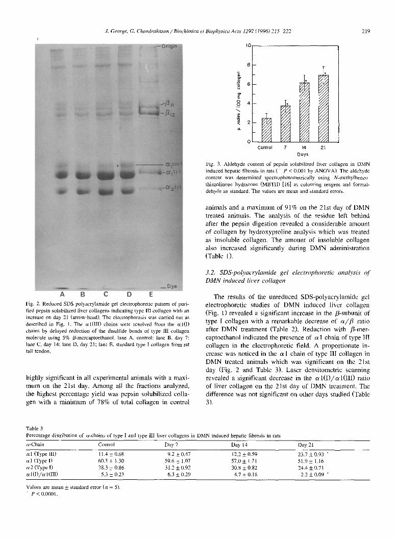
A B C D E Fig. 2. Reduced SDS-polyacrylamide gel electrophoretic pattern of puri- fied pepsin solubilized liver collagens indicating type III collagen with an increase on day 21 (arrow-head). The electrophoresis was carried out as described in Fig. 1. The al(III) chains were resolved from the al(I) chains by delayed reduction of the disulfide bonds of type III collagen molecule using 5% /3-mercaptoethanol. lane A, control; lane B, day 7; lane C, day 14; lane D, day 21: lane E, standard type I collagen from rat tail tendon.
h ighly s ignif icant in all exper imenta l animals wi th a maxi -
m u m on the 21st day. A m o n g all the fract ions analyzed,
the h ighes t percentage yield was pepsin solubi l ized col la-
gen with a m i n i m u m of 78% of total co l l agen in control
I0
o 6
o ° 4
=3.
Control 7 14 Days
Y/A I I I I Y/A . / / . ~
//'~d
"//A / / / . 4
21
Fig. 3. Aldehyde content of pepsin solubilized liver collagen in DMN induced hepatic fibrosis in rats (* P < 0.001 by ANOVA). The aldehyde content was determined spectrophotometrically using N-methylbenzo- thiazolinone hydrazone (MBTH) [16] as colouring reagent and f(~rmal- dehyde as standard. The values are mean and standard errors.
animals and a m a x i m u m of 91% on the 21st day o f D M N
treated animals. The analysis of the res idue lef t beh ind
after the peps in digest ion revea led a cons iderable amount
o f co l lagen by hydroxypro l ine analysis which was treated
as insoluble col lagen. The amount of insoluble co l lagen
also increased s ignif icant ly during D M N adminis t ra t ion
(Table 1).
3.2. SDS-polyacrylamide gel electrophoretic analysis o f DMN induced liver collagen
The results o f the unreduced S D S - p o l y a c r y l a m i d e gel
e lec t rophore t ic studies of D M N induced l iver co l lagen
(Fig. 1) r evea led a s ignif icant increase in the /3-subunit of
type I co l l agen with a remarkable decrease o f a/13 ratio
after D M N t rea tment (Table 2). Reduc t ion with /3-mer-
captoethanol indica ted the presence of a 1 chain of type III
co l lagen in the e lec t rophore t ic field. A propor t ionate in-
crease was no t iced in the a 1 chain o f type II! co l l agen in
D M N treated animals which was s ignif icant on the 21st
day (Fig. 2 and Table 3). Laser dens i tomet r ic scanning
revea led a s ignif icant decrease in the a l ( I ) / a l ( I I I ) ratio
o f l iver co l lagen on the 21st day of D M N treatment. The
d i f ference was not s ignif icant on other days s tudied (Table
3).
Table 3 Percentage distribution of a-chains of type I and type IIl liver collagens in DMN induced hepatic fibrosis in rats
Values are mean _+ standard error (n = 5). * P < 0.0001.
220 J. George. G. Chandrakasan / Biochimica et Biophysica Acta 1292 (1996) 215-222
,oo1 :6
60 , Cor,,rol
40~- / t , I f ° ~ ' ' 4
0 20 40 60
Time ( min )
Fig. 4. Normalized fibril formation curves of pepsin solubilized liver collagen presented as percentage final turbidity versus time during vary- ing periods of DMN treatment in rats. The collagen solution was prepared by dissolving purified pepsin solubilized liver collagen in ice cold 0.5 M acetic acid at a concentration of I mg/ml with gentle stirring. Two ml of this solution was mixed with 2 ml of 30 mM phosphate buffer (pH 7.3) containing 0.15 M NaCI. The cuvettes, one with blank were transferred to the water jacketed sample compartment of a double beam spectrophotom- eter maintained at 37°C. The increase in opacity was continuously monitored at 313 nm in order to obtain the fibril formation curve.
3.3. Aldehyde contents
The aldehyde content of the pepsin solubilized liver collagen was significantly increased on all days of DMN treatment (Fig. 3). The maximum increase was noticed on the 21st day of DMN administration which was about 3 times. The pattern of elevation coincided with the increase in total collagen. This was confirmed by the correlation coefficient studies which revealed a significant positive correlation (r = 0.983) between the two.
3.4. Fibri l f o rmat ion curves
The fibril formation curves of pepsin solubilized liver collagen of control rats and rats treated with DMN on different days were presented in Fig. 4. It was observed that under identical conditions, the fibril formation of DMN treated collagen started rapidly in comparison with control. This was very prominent in the case of the 21st day of DMN treated collagen (Fig. 4). A significant shift was observed in the fibril formation curves of the 14th and 21st days of DMN treated collagen due to quick and early formation of the gel. But regarding collagen on the 7th day, no such significant difference was observed in the curve, even though the fibril formation started much early. In all the cases, the fibril formation was completed at around 45 min with a maximum absorption on the 21 st day of DMN treated collagen. An increase was noticed in the rate of fibril formation on the 14th and 21st days of DMN treated liver collagen.
4. Discussion
It is now well established that hepatic fibrosis is accom- panied by deterioration of liver functions and accumulation of collagens in the liver [18-21]. An increase of total collagen content upto 10-fold in humans [22] and upto 8-fold in rats [23] were reported in various types of hepatic fibrosis. The approximate 4-fold increase of total liver collagen observed in the present study coincides with the previous investigations on DMN induced hepatic fibrosis [5,24].
Quantitative data regarding the relative distribution of total liver collagen fractions in DMN induced hepatic fibrosis is not available. An increase in type I and type III collagens were reported in human alcoholic cirrhosis [25,26]. The significant decrease noticed in the level of neutral salt soluble collagen on all days of DMN treatment with a maximum decrease on the 21st day clearly indicates the rapid and extensive formation cross-links in DMN induced liver collagen. This is further supported by the observation of increased aldehyde content and a rapid rate of fibril formation in the present investigation. It was reported that the increase in collagen content in hepatic fibrosis is associated with a higher proportion of insoluble collagen [27,28]. Kucharz [29] observed a reduction of collagen solubility during the induction of fibrosis of the liver which also substantiates the present findings. The absence of a significant increase in the case of acid soluble collagen and an increase of total and pepsin solubilized collagen except on the 21st day of DMN treatment indi- cates the rapid formation of cross-links which limits the solubility of collagen in dilute acid. The percentage in- crease noticed in the amount of pepsin solubilized collagen was analogous with an earlier report [7].
It was reported that the peptides derived by cyanogen bromide cleavage of type I collagen of normal and cir- rhotic livers have the same electrophoretic mobility on polyacrylamide gels [25]. In the present investigation, both reduced and unreduced SDS-polyacrylamide gel elec- trophoretic studies provided evidence that the subunit com- position of the a- and /3-chains of type I collagen have the same electrophoretic characteristics in control and DMN treated animals. The ratio of the p r o a l ( I ) mRNA to the p r o a 2 ( I ) mRNAs was about 2:1 in the normal rat liver and remained unchanged during DMN treatment [5]. In the present study, laser densitometric scanning of polyacryl- amide gels revealed almost 2:1 ratio for c~ l(I) and c~2(I) chains of control and all the three groups of DMN treated animals (Table 3).
It was observed that fibrosis in the human liver is due to an increase in both type I and type III collagens and that the increase in type III was proportionately larger [25]. A relatively greater accumulation of type III collagen than that of type I collagen was reported in rats with carbon tetrachloride induced hepatic fibrosis [30]. Besides accu- mulation of other collagen types, an excess deposition of
J. George, G. Chandrakasan / Biochimica et Biophysica Acre 1292 (1996) 215-222 221
type III collagen has been noticed in fibrotic human livers also [31]. In the present study, a proportionate increase in type III collagen was noticed in DMN treated animals which was significant on the 21st day (Table 3). This caused a significant decrease in the a l ( I ) / a l ( I I I ) colla- gen ratio on the 21st day of DMN treatment. In this connection, it is interesting to note that slot-blot hybridiza- tion studies by using specific cDNA probes revealed that the procollagen mRNA of a l(I) collagen chain increased 354% while the procollagen mRNA of a l(III) collagen chain increased upto 486% on the 21st day of DMN induced hepatic fibrosis [5,24]. At the same time the procollagen mRNA of a l(IV) collagen chain increased upto 655% [5] and upto 866% [24] on the 21st day of DMN treatment. The marked increase in the concentration of the mRNA for type IV collagen suggests that enhanced production of basement membrane collagen may be an early event in the development of hepatic fibrosis [5].
The native preparation of type III collagen molecules are trimers joined by disulfide bonded y-component which can be reduced to a chains. Under reducing conditions, these bonds are cleaved and the released a l(III) monomers migrate slightly slowly than a l(I) chains [32]. In the present investigation, delayed reduction of disulfide link- ages with /3-mercaptoethanol permitted well separation of a l(III) collagen chain from a 1(I) chain (Fig. 2).
One important post-synthetic alteration of collagen molecule is the formation of cross-links between peptide chains [33]. The cross-linkage is achieved in many ways. Siegel [34] observed that any increase in collagen synthesis leading to increase in newly formed collagen is associated with an increase in aldehyde content. It was also indicated that an increase in aldehyde content shows a greater potential for cross-link formation [35].
An increase in the level of insoluble collagen content, consistent with an enhanced activity of lysyl oxidase was reported in experimentally induced hepatic fibrosis in the rat [36,37]. An elevated level of serum lysyl oxidase activity was also observed in patients with various liver diseases [38]. Lysyl oxidase is an extracellular copper containing enzyme which oxidatively deaminates the e- amino group of certain lysine and hydroxylysine residues of collagen yielding reactive aldehydes which form stable intra and intermolecular covalent cross-links by further reaction [39,40]. The significant increase observed in the aldehyde content of pepsin solubilized liver collagen ob- tained from DMN treated animals clearly indicates the higher degree of cross-linkage. In addition, the significant decrease in the neutral salt soluble collagen and the in- crease in the pepsin solubilized collagen levels noticed in this study further substantiate the above observation.
In the present investigation, it was also observed that the rate of fibril formation was more rapid in DMN induced liver collagen when compared to control. This is substantiated by the observation of reduction of collagen solubility [29] and the increase in the level of insoluble
collagen [36,37] during the induction of hepatic fibrosis in rats. Fibril formation is a critical step in the cross-linking and self-assembly of collagen which purely depends upon the parent molecule. The results of the present study and the previous observations clearly indicate that the fibrotic liver collagen is more cross-linked than control liver colla- gen.
References
[l] Prockop, D.J., Berg, R.A., Kivirikko, K.I. and Uitto, J. (1976) Intracellular steps in the biosynthesis of collagen, in Biochemistry of Collagen (Ramachandran, G.N. and Reddi, A.H., eds.), pp. 163-273, Plenum Press, New York.
turini, C. and Orlandi, F. (1987) J. Hepatol. 5, 174-181. [4] Jenkins, S.A., Grandison, A., Baxter, J.N., Day, D.W., Taylor, I. and
Shields, R. (1985) J. Hepatol. 1,489-499. [5] Ala-Kokko, L., Pihlajaniemi, T., Myers, J.C., Kivirikko, K.I. and
Savolainen. E.R. (1987) Biochem. J. 244, 75-79. [6] Jezequel, A.M., Mancini, R., Rinaldesi, M.L., Ballardini, G., Fallani,
M., Bianchi, F. and Orlandi, F. (1989) J. Hepatol. 8, 42-52. 17] Rojkind, M., Giambrone, M.A. and Biempica, L. (1979) Gastro-
enterology 76, 710-719. [8] Risteli, J. and Kivirikko, K.I. (1976) Biochem. J. 158, 361-367. [9] Risteli, J., Tuderman, L. and Kivirikko, K.I. (1976) Biochem. J. 158,
369-376. [10] Rojkind, M., Giambrone, M.A. and Biempica, L. (1979) Gastro-
enterology 76, 710-719. [111 Rojkind, M. and Martinez-Palomo, A. (1976) Proc. Natl. Acad. Sci.
USA 73, 539-543. [12] Woessner, J.F., Jr. (1961) Arch. Biochem. Biophys. 93, 440-447. [13] Neuman, R.E. and Logan, M.A. (1950) J. Biol. Chem. 186, 549-556. 1141 Chart, D. and Cole, W.G. (1984) Anal. Biochem. 139, 322-328. [15] Blackshear, P.J. (1984) Methods Enzymol. 104, 237-255. [16] Paz, M.A., Blumenfeld, O.O., Rojkind, M,, Henson, E., Furfine, C.
and Gallop, P.M. (1965) Arch. Biochem. Biophys. 109, 548-559. [17] Gelman, R.A., Williams, B.R. and Piez, K.A. (1979) J. Biol. Chem.
254, 180-186. [18] Rojkind, M. and Mourelle, M. (1988) The liver as a bioecological
system: Modifications during regeneration and repair, in Collagen (Nimni, M.E., ed.), Vol. II, pp. 137-159, CRC Press, Boca Raton, FL.
[19] Sherlock, S. (1989) Diseases of the Liver and Biliary System. 8th Ed., pp. 410-448, Blackwell, Oxford.
[20] Hall, P. De La M., Plummer, J.L., Ilsley, A.H. and Cousins, M.J. (1991) Hepatology 13, 815-819.
[21] Ariosto, F., Riggio, O., Cantafora, A., Colucci, S., Gaudio, E., Mechelli, C., Merli, M., Seri, S. and Capocaccia. L. (1989) Eur. Surg. Res. 21,280-286.
[22] Perier, C., Gautier, M., Baril, A., Bayle, J.J., Patouillard, G. and Frey, J. (1984) Clin. Physiol. Biochem. 2, 279-286.
[23] Rojkind, M., Rojikind, M.H. and Cordero-Hernandez, J. (1983) Collagen Rel. Res. 3, 335-347.
[24] Savolainen, E.R., Brocks, D., Ala-Kokko, L. and Kivirikko, K.I. (1988) Biochem. J. 249, 753-757.
[25] Rojkind, M. and Martinez-Palomo, A. (1976) Proc. Natl. Acad. Sci. USA 73, 539-543.
[26] Seyer, J.M., Hutcheson, E,T. and Kang, A.H. (1977) J. Clin. Invest. 59, 241-248.
[27] Bazin, S., Le Louis, M. and Delaunay, A. (1976) Agents Actions 6, 272-276.
[28] Frey, J. and Bayle, J.J. (1978) Lyon Med. 239, 87-93 (in French).
222 J. George, G. Chandrakasan / Biochimica et Biophysica Acta 1292 (1996) 215-222
[29] Kucharz, E.J. (1987) Postepy Hig. Med. Dosw. 41, 302-330. [30] Kucharz, E.J. (1987) Connect. Tissue Res. 6, 143-151. [31] Rauterberg, J., Schlief, E. and Pott, G. (1982) The Collagen of the
normal and fibrotic liver. Characterization of collagen derived pep- tides solubilized by cyanogen bromide cleavage and by low tempera- ture pepsin treatment, in Proceedings of Connective Tissue of the Normal and Fibrotic Human Liver (Popper, H., Gerlach, U. and Kuhn, K., eds.), pp. 36-37, Georg Thieme, Stuttgart.
[32] Sykes, B., Puddle, B., Francis, M. and Smith, R. (1976) Biochem. Biophys. Res. Commun. 72, 1472-1480.
[33] Gallop, P.M., Blumenfeld, O.O. and Seifter, S. (1972) Annu. Rev. Biochem. 41, 617-672.
[34] Siegel, R.C. (1976) J. Biol. Chem. 251, 5786-5792. [35] Tanzer, M.L. (1976) Cross-linking, in Biochemistry of Collagen
(Ramachandran, G.N. and Reddi, A.H., eds.), pp. 137-162, Plenum Press, New York.
[36] Siegel, R.C., Chen, K.H., Greenspan, J.S. and Aguiar, J.M. (1978) Proc. Natl. Acad. Sci. USA 75, 2945-2949.
[37] Shiota, G., Murawaki, Y. and Hirayama, C. (1987) Res. Commun. Chem. Pathol. Pharmacol. 58, 115-127.
[38] Sakamoto, M., Murawaki, Y. and Hirayama, C. (1985) Serum lysyl oxidase activity in patients with various liver diseases. Comparison with serum monoamine oxidase and procollagen III peptide, in Pathobiology of Hepatic Fibrosis (Hirayama, C. and Kivirikko, K.I., eds.), pp. 209-216, Excerpta Medica, Amsterdam.
[39] Rucker, R.B. and Murray, J. (1978) Am. J. Clin. Natr. 31, 1221- 1236.


![Biochi~ic~a et Biophysica A~ta - COnnecting REpositoriesc a and o has been purified from A. vinelandii membranes [12]. This complex was found to be able to oxidize ascor- bate-N, N,](https://static.documents.pub/doc/80x56/60cf08ca3bd7762e217d18c5/biochiica-et-biophysica-ata-connecting-repositories-c-a-and-o-has-been-purified.jpg)


