23
Bio 178 Lecture 6 Cell Structure Sui Huang and Donald E. Ingber, http://w3.mit.edu/i-m/image2big.htm
| Date post: | 20-Dec-2015 |
| Category: |
Documents |
| View: | 213 times |
| Download: | 0 times |

Bio 178 Lecture 6Cell Structure
Sui Huang and Donald E. Ingber, http://w3.mit.edu/i-m/image2big.htm
Reading
• Chapter 5
Quiz Material
• Questions on P 104
• Chapter 5 Quiz on Text Website (www.mhhe.com/raven7)
Outline
• Characteristics of cells
• Microscopy
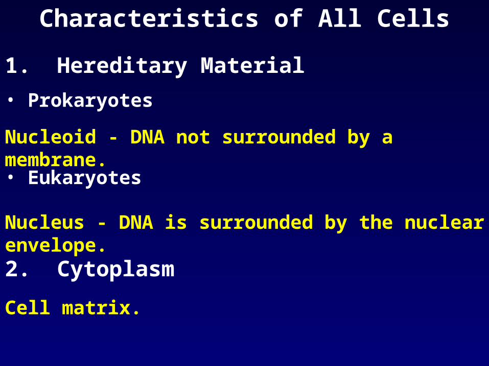
Characteristics of All Cells
1. Hereditary Material
• Prokaryotes
Nucleoid - DNA not surrounded by a membrane.
• Eukaryotes
Nucleus - DNA is surrounded by the nuclear envelope.
2. Cytoplasm
Cell matrix.

Characteristics of All Cells
3. Phosopholipid Bilayer boundary
Plasma membrane is 5-10 nm thick and contains embedded proteins.
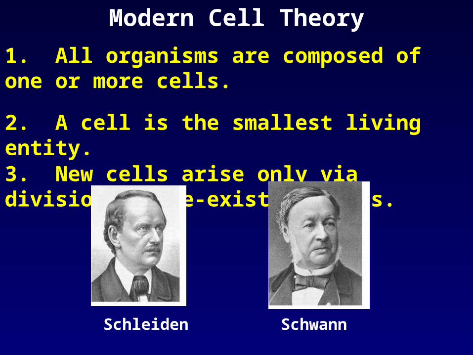
Modern Cell Theory
1. All organisms are composed of one or more cells.
2. A cell is the smallest living entity.
3. New cells arise only via division of pre-existing cells.
Schleiden Schwann
Cell SizeMost eukaryotic cells are 5-20 µm
Reasons• Surface to Volume Ratio
1. Cells need a large a S:V to allow transport of nutrients, gases, & waste across the plasma membrane.
2. Much easier to achieve if an organism is made of numerous small cells rather than few large cells.
• One Command Center per Cell
The nucleus can more easily provide for the cell if the cell is small.
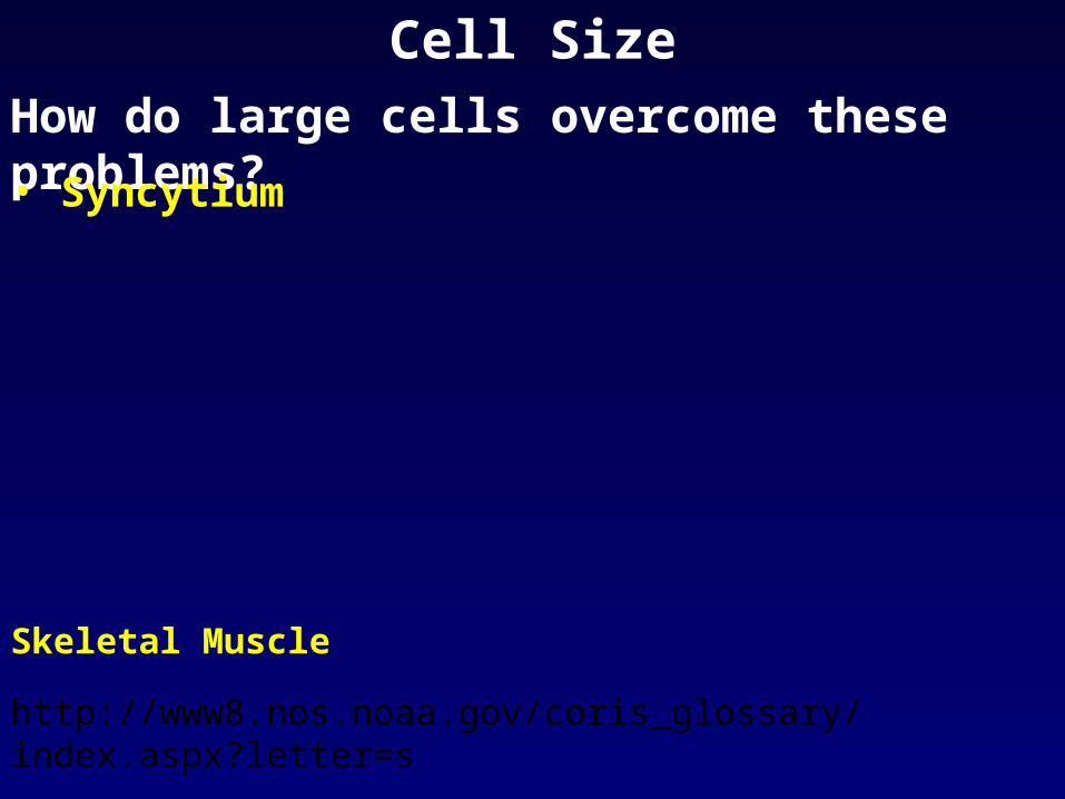
Cell Size
How do large cells overcome these problems?
• Syncytium
Skeletal Muscle
http://www8.nos.noaa.gov/coris_glossary/index.aspx?letter=s

Cell SizeHow do large cells overcome these problems?• Syncytium (cntd.)
Soybeet syncytium induced by nematode parasite Heterodera glycines.
http://www.apsnet.org/education/IllustratedGlossary/PhotosS-V/syncytium.htm

Cell Size
How do large cells overcome these problems?
• Cell Shape
http://www.nature.com/news/2004/040531/images/nerve_180.jpg
http://web.sfn.org/content/Publications/BrainBackgrounders/communication.htm

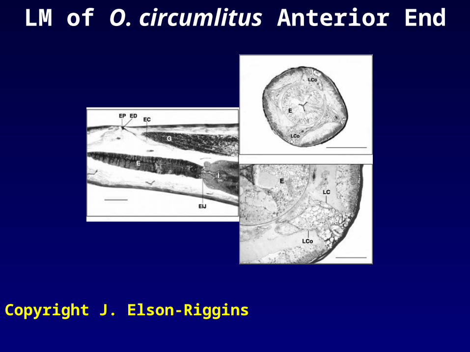
Cell Size
How do large cells overcome these problems?
• Large Nuclei
ES gland of Otostrongylus circumlitus (copyright J. Elson-Riggins)

Microscopy
Resolution
The minimum distance between 2 points at which they can be distinguished as separate objects.
Human Eye:
~ 100 µm
Light Microscope:
0.2 µm (in theory)
Electron Microscope:
0.1 nm

MicroscopyResolution (R)R = 0.61 = 1/2 angular width cone of light rays collected by objective lens
n sin n = refractive index of medium separating object from objective and condenser lenses = wavelength
William H. Heidcamp, http://homepages.gac.edu/~cellab/chpts/chpt1/figure1-3.html
What limits the resolution of the light microscope?
LM of O. circumlitus Anterior End
Copyright J. Elson-Riggins

Electron MicroscopeTransmission Electron Microscope (TEM)Electrons are transmitted through the specimen (thin section).
http://euch3i.chem.emory.edu/~nmr/apk/instrumentation.html http://www.barrettresearch.ca/
teaching/nanotechnology/nano02.htm
(light) = 0.53 µm (electrons) = 0.004 nm

TEM (O. circumlitus ES Gland)
Copyright J. Elson-Riggins

Scanning Electron Microscope (SEM)
Electrons are reflected off the surface of the specimen - gives 3D images.
Ant head
http://www.ucmp.berkeley.edu/esem/gallery.html

Staining Specimens for Microscopy
Staining increases contrast (cells are 70% water little to impede passage of light rays/electrons).
O. Circumlitus ES gland stained with PAS and aniline blue-black.
Copyright J. Elson-Riggins
Specific stains are used to visualize structures.

Staining Specimens for Microscopy
Immunocytochemistry
Antibodies are labeled with fluorescent molecules (or other substances) and used to stain specific structures.
NANCY KEDERSHA / IMMUNOGEN / SCIENCE PHOTO, LIBRARY
http://www.sciencephoto.com/images/imagePopUpDetails.html?id=771320363
Squamous carcinoma cells. Blue = Nuclei,
Red = Cytoplasm,
Green = Plasma membrane


Characteristics of Principle Cell Types
2 structurally different types of cells:
PROKARYOTE EUKARYOTE
True Nucleus?
Membrane bound
organelles?
Kingdoms


