Bioengineering Bacterial Derived Immunomodulants: a Novel IBD Therapeutic Approach. Julie Champion, PhD Department of Chemical Engineering Georgia Institute of Technology Atlanta GA, USA. Andrew S. Neish, MD Department of Pathology Emory University School of Medicine Atlanta GA, USA. - PowerPoint PPT Presentation
Bioengineering Bacterial Derived Immunomodulants: a Novel IBD Therapeutic Approach Andrew S. Neish, MD Department of Pathology Emory University School of Medicine Atlanta GA, USA Julie Champion, PhD Department of Chemical Engineering Georgia Institute of Technology Atlanta GA, USA
Transcript
Bioengineering Bacterial Derived Immunomodulants: a Novel IBD
Therapeutic Approach
Andrew S. Neish, MD
Department of Pathology
Emory University School of Medicine
Atlanta GA, USA
Julie Champion, PhD
Department of Chemical Engineering
Georgia Institute of Technology
Atlanta GA, USA
Disclosures
• Nothing to disclose
Acute intestinal inflammation
• Intraepithelial and luminal accumulation of neutrophils
• Enterocyte apoptosis/injury• Attendant loss of epithelial barrier
integrity• Results from activation of
signaling pathways with upregulation of proinflammatory effector molecules
MKK
JNK NF-B
TAK
TNF-R
TNF MAMPs
IKK
TLRs
INNATE IMMUNITYAPOPTOSIS
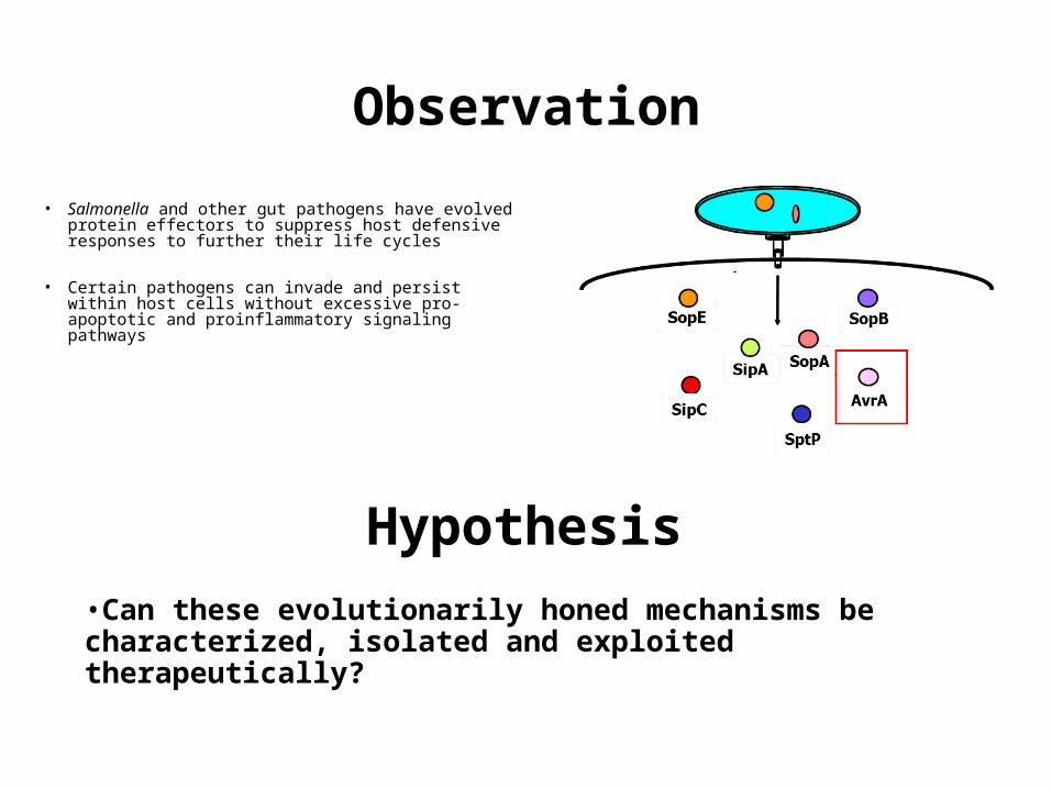
• Salmonella and other gut pathogens have evolved protein effectors to suppress host defensive responses to further their life cycles
• Certain pathogens can invade and persist within host cells without excessive pro-apoptotic and proinflammatory signaling pathways
Observation
Hypothesis
•Can these evolutionarily honed mechanisms be characterized, isolated and exploited therapeutically?
Background
• Salmonella AvrA is a member of an class of bacterial effectors (acetyltransferases) involved in host-pathogen interactions
• AvrA allows innate immune suppression without cell death, consistent with its role facilitating the lifestyle of an intracellular pathogen
• AvrA expression in a living animal is non toxic but suppresses inflammatory and apoptotic responses
MKK
JNK NF-B
TAK
TNF-R
TNF MAMPs
IKK
TLRs
INNATE IMMUNITYAPOPTOSIS
AvrA
Goal :Use chemical engineering/nanotechnology systems to study exploit the anti-inflammatory mechanisms of the Salmonella effector protein AvrA on intestinal inflammation