ORIGINAL RESEARCH ARTICLE published: 27 May 2013 doi: 10.3389/fcimb.2013.00018 Biofilm-derived Legionella pneumophila evades the innate immune response in macrophages Arwa Abu Khweek 1 , Natalia S. Fernández Dávila 1 , Kyle Caution 1 , Anwari Akhter 1 , Basant A. Abdulrahman 1,2 , Mia Tazi 1 , Hoda Hassan 1 , Laura A. Novotny 3 , Lauren O. Bakaletz 1,3 and Amal O. Amer 1 * 1 Department of Microbial Infection and Immunity, Center for Microbial Interface Biology, College of Medicine, The Ohio State University, Columbus, OH, USA 2 Department of Biochemistry and Molecular Biology, Faculty of Pharmacy, Helwan University, Cairo, Egypt 3 Center for Microbial Pathogenesis, Nationwide Children’s Hospital, Columbus, OH, USA Edited by: Yongqun Oliver "He", University of Michigan School of Medicine, USA Reviewed by: Sunny Shin, University of Pennsylvania, USA Cyril Guyard, Public Health Ontario, Canada *Correspondence: Amal O. Amer, Department of Microbial Infection and Immunity, Center for Microbial Interface Biology, Dorothy Heart and Lung Research Institute, Ohio State University, Biological Research Tower, 460 W 12th Ave., Room 706, Columbus, OH 43210, USA. e-mail: [email protected]Legionella pneumophila, the causative agent of Legionnaire’s disease, replicates in human alveolar macrophages to establish infection. There is no human-to-human transmission and the main source of infection is L. pneumophila biofilms established in air conditioners, water fountains, and hospital equipments. The biofilm structure provides protection to the organism from disinfectants and antibacterial agents. L. pneumophila infection in humans is characterized by a subtle initial immune response, giving time for the organism to establish infection before the patient succumbs to pneumonia. Planktonic L. pneumophila elicits a strong immune response in murine, but not in human macrophages enabling control of the infection. Interactions between planktonic L. pneumophila and murine or human macrophages have been studied for years, yet the interface between biofilm-derived L. pneumophila and macrophages has not been explored. Here, we demonstrate that biofilm-derived L. pneumophila replicates significantly more in murine macrophages than planktonic bacteria. In contrast to planktonic L. pneumophila, biofilm-derived L. pneumophila lacks flagellin expression, do not activate caspase-1 or -7 and trigger less cell death. In addition, while planktonic L. pneumophila is promptly delivered to lysosomes for degradation, most biofilm-derived bacteria were enclosed in a vacuole that did not fuse with lysosomes in murine macrophages. This study advances our understanding of the innate immune response to biofilm-derived L. pneumophila and closely reproduces the natural mode of infection in human. Keywords: biofilm, inflammasome, flagellin, caspase-1, Legionella pneumophila, innate immunity INTRODUCTION Legionella pneumophila (L. pneumophila) is a Gram nega- tive facultative bacterium with fastidious growth requirements. Although Legionella exists as free-living planktonic forms in the environment, they are more commonly found as intracellular par- asites of protozoans such as Acanthamoeba spp., Hartmannella spp., and Tetrahymena spp. (Atlas, 1999; Brown and Barker, 1999) and as inhabitants of mixed-community biofilms (Rogers et al., 1994; Lau and Ashbolt, 2009). Replication of L. pneumophila within amoeba is utilized as a survival strategy to overcome the low-nutrient environment and increases the resistance to disin- fectant (Lau and Ashbolt, 2009). This opportunistic pathogen most often thrives in bacterial communities encased in extra- cellular polymeric matrix known as biofilm (Costerton et al., 1978; Donlan et al., 2005). Biofilms have been recognized as one of the most important factors of survival and proliferation of L. pneumophila in warm, humid environments like showers, air conditioners, and spa baths (Fraser et al., 1979; Fliermans et al., 1981; Sethi and Brandis, 1983; Spitalny et al., 1984; Abu Kwaik et al., 1993; Lettinga et al., 2002). These communities have been identified as a causative source of infection in susceptible hosts who inhale aerosols of contaminated water containing L. pneu- mophila. In the human lung environment, L. pneumophila repli- cates exponentially within alveolar macrophages prior to lysing the host cell and invading other macrophages causing a type of walking pneumonia called Legionnaire’s disease or Legionellosis (Horwitz and Silverstein, 1980; Harb and Abu Kwaik, 2000). Legionellosis has two clinically distinct forms: Legionnaires’ dis- ease, a severe type of infection, which includes pneumonia and Pontiac fever, a milder self-limiting illness (Lau and Ashbolt, 2009). Approximately 20,000 cases of Legionnaire’s disease are reported yearly in the US with no person-to-person transmission (Marston et al., 1997). Thus, using biofilm-derived L. pneu- mophila to study the innate immune response to infection reca- pitulates natural mode of infection in human. The murine innate immune response to planktonic L. pneu- mophila has been studied extensively. Nlrc4 and Naip5 detect flagellin monomers in the host cytosol in a process that is depen- dent upon a functional bacterial type IV secretion system. The sensing of contaminating molecules of flagellin promotes the for- mation of a multi-protein complex called the inflammasome. Within the inflammasome, caspase-7 is activated downstream Frontiers in Cellular and Infection Microbiology www.frontiersin.org May 2013 | Volume 3 | Article 18 | 1 CELLULAR AND INFECTION MICROBIOLOG Y
Transcript
ORIGINAL RESEARCH ARTICLEpublished: 27 May 2013
doi: 10.3389/fcimb.2013.00018
Biofilm-derived Legionella pneumophila evades the innateimmune response in macrophagesArwa Abu Khweek1, Natalia S. Fernández Dávila1, Kyle Caution1, Anwari Akhter1,Basant A. Abdulrahman1,2, Mia Tazi1, Hoda Hassan1, Laura A. Novotny3, Lauren O. Bakaletz1,3 and
Amal O. Amer1*
1 Department of Microbial Infection and Immunity, Center for Microbial Interface Biology, College of Medicine, The Ohio State University, Columbus, OH, USA2 Department of Biochemistry and Molecular Biology, Faculty of Pharmacy, Helwan University, Cairo, Egypt3 Center for Microbial Pathogenesis, Nationwide Children’s Hospital, Columbus, OH, USA
Edited by:
Yongqun Oliver "He", University ofMichigan School of Medicine, USA
Reviewed by:
Sunny Shin, University ofPennsylvania, USACyril Guyard, Public Health Ontario,Canada
*Correspondence:
Amal O. Amer, Department ofMicrobial Infection and Immunity,Center for Microbial InterfaceBiology, Dorothy Heart and LungResearch Institute, Ohio StateUniversity, Biological ResearchTower, 460 W 12th Ave., Room 706,Columbus, OH 43210, USA.e-mail: [email protected]
Legionella pneumophila, the causative agent of Legionnaire’s disease, replicates inhuman alveolar macrophages to establish infection. There is no human-to-humantransmission and the main source of infection is L. pneumophila biofilms established inair conditioners, water fountains, and hospital equipments. The biofilm structure providesprotection to the organism from disinfectants and antibacterial agents. L. pneumophilainfection in humans is characterized by a subtle initial immune response, giving timefor the organism to establish infection before the patient succumbs to pneumonia.Planktonic L. pneumophila elicits a strong immune response in murine, but not inhuman macrophages enabling control of the infection. Interactions between planktonicL. pneumophila and murine or human macrophages have been studied for years,yet the interface between biofilm-derived L. pneumophila and macrophages has notbeen explored. Here, we demonstrate that biofilm-derived L. pneumophila replicatessignificantly more in murine macrophages than planktonic bacteria. In contrast toplanktonic L. pneumophila, biofilm-derived L. pneumophila lacks flagellin expression, donot activate caspase-1 or -7 and trigger less cell death. In addition, while planktonicL. pneumophila is promptly delivered to lysosomes for degradation, most biofilm-derivedbacteria were enclosed in a vacuole that did not fuse with lysosomes in murinemacrophages. This study advances our understanding of the innate immune responseto biofilm-derived L. pneumophila and closely reproduces the natural mode of infection inhuman.
INTRODUCTIONLegionella pneumophila (L. pneumophila) is a Gram nega-tive facultative bacterium with fastidious growth requirements.Although Legionella exists as free-living planktonic forms in theenvironment, they are more commonly found as intracellular par-asites of protozoans such as Acanthamoeba spp., Hartmannellaspp., and Tetrahymena spp. (Atlas, 1999; Brown and Barker, 1999)and as inhabitants of mixed-community biofilms (Rogers et al.,1994; Lau and Ashbolt, 2009). Replication of L. pneumophilawithin amoeba is utilized as a survival strategy to overcome thelow-nutrient environment and increases the resistance to disin-fectant (Lau and Ashbolt, 2009). This opportunistic pathogenmost often thrives in bacterial communities encased in extra-cellular polymeric matrix known as biofilm (Costerton et al.,1978; Donlan et al., 2005). Biofilms have been recognized as oneof the most important factors of survival and proliferation ofL. pneumophila in warm, humid environments like showers, airconditioners, and spa baths (Fraser et al., 1979; Fliermans et al.,1981; Sethi and Brandis, 1983; Spitalny et al., 1984; Abu Kwaiket al., 1993; Lettinga et al., 2002). These communities have beenidentified as a causative source of infection in susceptible hosts
who inhale aerosols of contaminated water containing L. pneu-mophila. In the human lung environment, L. pneumophila repli-cates exponentially within alveolar macrophages prior to lysingthe host cell and invading other macrophages causing a type ofwalking pneumonia called Legionnaire’s disease or Legionellosis(Horwitz and Silverstein, 1980; Harb and Abu Kwaik, 2000).Legionellosis has two clinically distinct forms: Legionnaires’ dis-ease, a severe type of infection, which includes pneumonia andPontiac fever, a milder self-limiting illness (Lau and Ashbolt,2009). Approximately 20,000 cases of Legionnaire’s disease arereported yearly in the US with no person-to-person transmission(Marston et al., 1997). Thus, using biofilm-derived L. pneu-mophila to study the innate immune response to infection reca-pitulates natural mode of infection in human.
The murine innate immune response to planktonic L. pneu-mophila has been studied extensively. Nlrc4 and Naip5 detectflagellin monomers in the host cytosol in a process that is depen-dent upon a functional bacterial type IV secretion system. Thesensing of contaminating molecules of flagellin promotes the for-mation of a multi-protein complex called the inflammasome.Within the inflammasome, caspase-7 is activated downstream
Frontiers in Cellular and Infection Microbiology www.frontiersin.org May 2013 | Volume 3 | Article 18 | 1
Abu Khweek et al. Murine macrophages response to L. pneumophila biofilm
of caspase-1 which results in bacterial restriction via fusion ofL. pneumophila-containing vacuoles with lysosomes (Coers et al.,2000; Akhter et al., 2009; Amer, 2010). Conversely, human mono-cytes do not activate this response upon L. pneumophila infectionand phagosomes containing L. pneumophila evade fusion with thelysosome allowing bacterial replication (Roy, 2002; Isberg et al.,2009).
Here we demonstrate that biofilm-derived L. pneumophilareplicates significantly more than planktonic L. pneumophilain murine macrophages due to diminished flagellin expres-sion. Biofilm-derived L. pneumophila does not activate caspase-1or caspase-7, evades fusion with lysosomes, and promotes lesscell death. Taken together, our study characterizes the innateimmune response to biofilm-derived L. pneumophila in murineand human macrophages.
METHODSBACTERIAL STRAINSL. pneumophila strain JR32 a wild-type (WT) strain andflaA mutant is deficient in flagellin, were kindly provided byDr. Howard Shuman, University of Chicago. The dotA mutant,a JR32-derived strain defective in the Dot/Icm Type IV secre-tion system was kindly provided by Dr. Craig Roy, Yale Schoolof Medicine. L. pneumophila expressing green fluorescent protein(GFP) was used for microscopy.
L. pneumophila GROWTHL. pneumophila strains were grown on buffered charcoal yeastextract (BCYE) plates at 37◦C. Three days later, the bacteria wereresuspended in 5 ml of L. pneumophila medium (BYE) with addi-tives (ferric nitrate, L-cysteine, thymidine) and vortexed 100× athigh speed. For biofilm formation, a bacterial suspension with anoptical density (OD) at 600 nm of 3.5 was diluted to 1:2500 insupplemented broth and 200 μl of this suspension was inoculatedinto each well of an 8-well chamber slide (Thermo Scientific LabTek chambered coverglass with cover #155411 and/or #177402).Slides were incubated at 37◦C, 5% CO2 incubator with humid-ified atmosphere without shaking. Biofilms were fed by deliveryof fresh medium to one side of the chamber slide well every24 h for 6 days using 100 μl of L. pneumophila medium. The ODvalues were 3.4–3.6 and 3.8–4 for the JR32 and dotA mutant,respectively. L. pneumophila was grown for planktonic cultureas previously described (Amer et al., 2006; Akhter et al., 2009,2012).
MACROPHAGE INFECTIONC57BL/6 mice were purchased from Jackson laboratory. Bonemarrow derived macrophages (BMDMs) were prepared fromthe femurs of 6 to 8-week-old mice as previously described(Akhter et al., 2009, 2012). Isolation and preparation of thehuman monocyte-derived macrophages (hMDMs) from periph-eral blood was carried out as previously described (Santic et al.,2005; Al-Khodor et al., 2008). Planktonic infections were usedfrom post-exponential cultures as previously described (Ameret al., 2006; Akhter et al., 2009). Infection from biofilm derivedL. pneumophila was carried as follows. Briefly, on day seven, themedia was aspirated and transferred to a 50 mL tube. Biofilms
were scraped from the chamber slide wells. Chambers werewashed 2× with 200 μl of fresh LP medium with the previouslymentioned additives and drained into the 50 ml tube. The col-lected biofilms were vortexed 100× and the OD at 600 nm of thecollected suspension was used to calculate the desired multiplicityof infection. Equivalent inocula of planktonic bacteria were usedfor infection (MOIs).
L. pneumophila is an intracellular pathogen that replicates onlywithin eukaryotic cells since the culture media do not containrequired nutrients such as iron and cysteine.
CONFOCAL LASER SCANNING MICROSCOPE VISUALIZATIONOn day seven, biofilms were washed gently with 200 μl of ster-ile saline (0.9% sodium chloride) (Hospira 0409-4888-10), andstained using the Live/Dead BacLight Bacterial Viability Kit(Invitrogen #7007) for 15 min at room temperature protectedfrom the light. Wells were washed 2× with sterile saline and200 μl of 10% formalin was added for 24 h to fix the biofilm andstored at room temperature protected from the light with beforeit was visualized using inverted confocal Zeiss LSM 510 METAmicroscope with a 63× water objective. Z-stacks were capturedevery 1 μm.
ENZYME-LINKED IMMUNOSORBENT ASSAY (ELISA)L. pneumophila JR32 and dotA strains from post-exponentialplanktonic and biofilm cultures were used to infect murinemacrophages for 24 h. Supernatants were collected and cen-trifuged at 1200 rpm for 10 min and stored at −80◦C as previouslydescribed (Abdulrahman et al., 2011). The plates were coated withprimary antibody for IL-1β and ELISA were performed accord-ing to the manufacture specifications (R&D) (Abdulrahman et al.,2011).
WESTERN BLOTMacrophage lysates were prepared following infection with eitherplanktonic or biofilm JR32 or dotA mutant and immunoblot-ted with caspase-1, caspase-7, or β-actin antibodies (caspase-1,1:3000; caspase-7, 1:300). Blots were washed and the corre-sponding secondary antibody was added. For flagellin detectionby western blot, one OD of bacterial culture was pelleted andresuspended in SDS-containing sample buffer from planktonicor biofilm grown bacteria. Eighteen μl were loaded on 12%SDS-PAGE gel. The blot was probed with flagellin antibody(1:100) kindly provided by Dr. Howard Shuman, University ofChicago, followed by the secondary antibody, donkey anti-rabbit(1:5000). Blots were developed after adding ECL Western BlottingDetection Reagent (GE Healthcare Amersham).
MACROPHAGE CYTOTOXICITY ASSAYPercentage of macrophage necrosis was determined by measuringthe release of host cell cytoplasmic lactate dehydrogenase (LDH)using the cytotoxicity detection kit (Roche Applied Science) tothe specification of the manufacturer. BMDMs were infectedwith JR32 or the dotA mutant from either planktonic or biofilmculture for 4 or 24 h at an MOI of 0.5. Supernatants were col-lected and LDH release was calculated as previously described(Abdulrahman et al., 2011).
Frontiers in Cellular and Infection Microbiology www.frontiersin.org May 2013 | Volume 3 | Article 18 | 2
Abu Khweek et al. Murine macrophages response to L. pneumophila biofilm
L. pneumophila COLOCALIZATION WITH LYSOTRACKERL. pneumophila JR32 from post-exponential planktonic or biofilmcultures were used to infect macrophages plated in 24-well platescontaining sterilized coverslips. Lysotracker red (1:500) was addedbefore fixation and 4′, 6-diamidino-2-phenylindole (DAPI) wasadded after fixation. Coverslips were mounted on slides andviewed using the Olympus Flow View FV10i CLSM. Three hun-dred bacteria were counted from 2 coverslips for each condition.
CONTACT-DEPENDENT HEMOLYSISSheep RBCs (sRBCs) were diluted in RPMI, and washed 3× bycentrifugation for 10 min at 2000× g until the supernatant didnot show any signs of hemolysis; the cells were counted using ahemo-cytometer chamber. Reactions were set up in a final vol-ume of 1 ml with a final concentration of 1 × 10⁷ sRBCs/ml. ThesRBCs were incubated with the planktonic or biofilm bacteriaat an MOI of 20 and RBC lysis was determined as previouslydescribed (Kirby et al., 1998; Alli et al., 2000).
SCANNING ELECTRON MICROSCOPY (SEM)L. pneumophila strains were grown on 12-well plate coverslips forseven days. On the seventh day, the medium was aspirated and thecoverslips were washed with 1× DPBS. Coverslips were fixed with2.5% gluteraldehyde in 0.1 M phosphate buffer pH 7.4, processedand viewed by SEM.
STATISTICAL ANALYSISExperiments were performed 2–3 independent times each in trip-licate or quadruplicate and yielded similar results. Comparisonsof groups for statistical significance were performed usingStudent’s tow tailed t-test. P-values ≤ was considered significant.
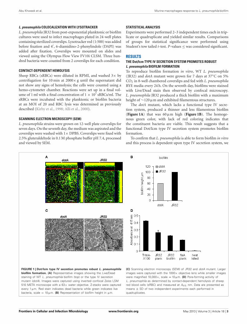
RESULTSTHE Dot/Icm TYPE IV SECRETION SYSTEM PROMOTES ROBUSTL. pneumophila BIOFILM FORMATIONTo reproduce biofilm formation in vitro, WT L. pneumophila(JR32) and dotA mutant were grown for 7 days at 37◦C on 5%CO2 in 8-well chambered coverslips and fed with L. pneumophilaBYE media every 24 h. On the seventh day, biofilms were stainedwith Live/Dead stain then observed by confocal microscopy.L. pneumophila JR32 produced a thick biofilm with a maximumheight of ∼120 μm and exhibited filamentous structures.
The dotA mutant, which lacks a functional type IV secre-tion system, produced a thinner and less filamentous biofilm(Figure 1A) that was 60 μm high (Figure 1B). The homoge-nous green color, with lack of red coloring indicates thatthe constituent bacteria are viable. This result suggests that afunctional Dot/Icm type IV secretion system promotes biofilmformation.
To confirm that L. pneumophila is able to form biofilm in vitroand this process is dependent upon type IV secretion system, we
FIGURE 1 | Dot/Icm type IV secretion promotes robust L. pneumophila
biofilm formation. (A) Representative images showing the Live/Deadstaining of WT L. pneumophila biofilm (top) or the type IV secretionmutant (dotA). Images were captured using inverted confocal Zeiss LSM510 META microscope with a 63× water objective. Z-stacks were capturedevery 1 μm. Red stain indicates dead bacteria while green indicates livebacteria, scale = 10 μm. (B) Representation of biofilm height in μm.
(C) Scanning electron microscopy (SEM) of JR32 and dotA mutant. Largerimages were captured with the 1000× objective lens while smaller imageswere magnified 10,000×, scale = 10 μm. (D) Pore-forming activity ofL. pneumophila as determined by contact-dependent hemolysis of sheepred blood cells (sRBC) and measured at A415 nm. Data are presented asmeans ± SD of two independent experiments each performed inquadruplicates.
Frontiers in Cellular and Infection Microbiology www.frontiersin.org May 2013 | Volume 3 | Article 18 | 3
Abu Khweek et al. Murine macrophages response to L. pneumophila biofilm
examined biofilm formation using scanning electron microscopy(SEM). Our data showed that JR32 strain formed a robust biofilmwith characteristic towers whereas the dotA mutant failed to doso (Figure 1C). These data confirm that robust biofilm formationrequires type IV secretion system.
The pore forming activity of L. pneumophila has been shown tocontribute to macrophage cytotoxicity and requires a functionaltype IV secretion system (Kirby et al., 1998; Alli et al., 2000).To examine whether biofilm-derived L. pneumophila exhibitpore-forming activity, contact-dependent hemolysis of sheep redblood cells (RBCs) was performed (Kirby et al., 1998). Triton-X100 and heat-killed bacteria were used as positive and nega-tive controls, respectively. The flaA mutant lacking flagellin andexpresses a functional type IV secretion system was also exam-ined (Figure 1D). Biofilm-derived and planktonic L. pneumophilaand flaA mutant were capable of lysing the RBCs, suggestingthat biofilm-derived L. pneumophila exhibit a functional type IVsecretion system.
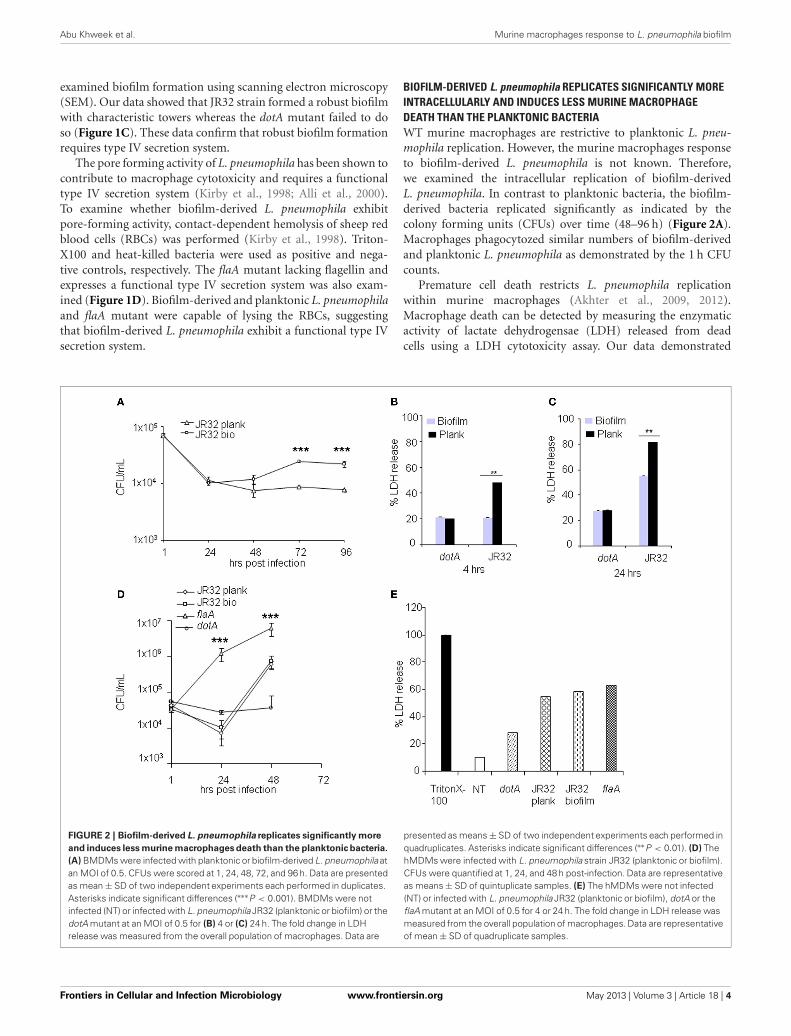
BIOFILM-DERIVED L. pneumophila REPLICATES SIGNIFICANTLY MOREINTRACELLULARLY AND INDUCES LESS MURINE MACROPHAGEDEATH THAN THE PLANKTONIC BACTERIAWT murine macrophages are restrictive to planktonic L. pneu-mophila replication. However, the murine macrophages responseto biofilm-derived L. pneumophila is not known. Therefore,we examined the intracellular replication of biofilm-derivedL. pneumophila. In contrast to planktonic bacteria, the biofilm-derived bacteria replicated significantly as indicated by thecolony forming units (CFUs) over time (48–96 h) (Figure 2A).Macrophages phagocytozed similar numbers of biofilm-derivedand planktonic L. pneumophila as demonstrated by the 1 h CFUcounts.
Premature cell death restricts L. pneumophila replicationwithin murine macrophages (Akhter et al., 2009, 2012).Macrophage death can be detected by measuring the enzymaticactivity of lactate dehydrogensae (LDH) released from deadcells using a LDH cytotoxicity assay. Our data demonstrated
FIGURE 2 | Biofilm-derived L. pneumophila replicates significantly more
and induces less murine macrophages death than the planktonic bacteria.
(A) BMDMs were infected with planktonic or biofilm-derived L. pneumophila atan MOI of 0.5. CFUs were scored at 1, 24, 48, 72, and 96 h. Data are presentedas mean ± SD of two independent experiments each performed in duplicates.Asterisks indicate significant differences (∗∗∗P < 0.001). BMDMs were notinfected (NT) or infected with L. pneumophila JR32 (planktonic or biofilm) or thedotA mutant at an MOI of 0.5 for (B) 4 or (C) 24 h. The fold change in LDHrelease was measured from the overall population of macrophages. Data are
presented as means ± SD of two independent experiments each performed inquadruplicates. Asterisks indicate significant differences (∗∗P < 0.01). (D) ThehMDMs were infected with L. pneumophila strain JR32 (planktonic or biofilm).CFUs were quantified at 1, 24, and 48 h post-infection. Data are representativeas means ± SD of quintuplicate samples. (E) The hMDMs were not infected(NT) or infected with L. pneumophila JR32 (planktonic or biofilm), dotA or theflaA mutant at an MOI of 0.5 for 4 or 24 h. The fold change in LDH release wasmeasured from the overall population of macrophages. Data are representativeof mean ± SD of quadruplicate samples.
Frontiers in Cellular and Infection Microbiology www.frontiersin.org May 2013 | Volume 3 | Article 18 | 4
Abu Khweek et al. Murine macrophages response to L. pneumophila biofilm
that macrophages infected with biofilm-derived L. pneumophilaproduced less LDH at 4 and 24 h post-infection when comparedto those infected with planktonic bacteria (Figures 2B,C).Biofilm-derived and planktonic dotA mutants led to the sameextent of macrophage death. These data suggest that biofilm-derived L. pneumophila induced less cell death in murinemacrophages than did planktonic-derived L. pneumophila.
HUMAN MACROPHAGES ARE PERMISSIVE TO BIOFILM-DERIVEDL. pneumophila AS THEY ARE TO PLANKTONIC CULTURESIn contrast to murine macrophages, human monocytes-derived macrophages (hMDMs) are permissive to planktonicL. pneumophila at least in part due to diminished caspase-1and -7 activation (Horwitz, 1983; Abdelaziz et al., 2011a,b).Replication within macrophages is essential for establishingLegionnaire’s pneumonia. Thus, we evaluated the intracellu-lar growth of biofilm-derived L. pneumophila in hMDMs. Thebiofilm-derived L. pneumophila replicated similar to plank-tonic L. pneumophila in hMDMs (Figure 2D). As expected thedotA mutant did not replicate whereas L. pneumophila mutantlacking flagellin replicated the most (Figure 2D). This differ-ence was not due to differential uptake since phagocytosisof all tested strains was similar as shown by the 1 h CFUs(Figure 2D).
Furthermore, we tested macrophage death by measuring per-centage of LDH released after 24 h of infection. Planktonic andbiofilm-derived L. pneumophila caused similar amount of LDHrelease from hMDMs while dotA mutant caused less cell death(Figure 2E). Human macrophages infected with the flaA mutantalso released comparable amounts of LDH (Figure 2E). These
data suggest that biofilm-derived L. pneumophila behave similarlyto planktonic L. pneumophila in hMDMs.
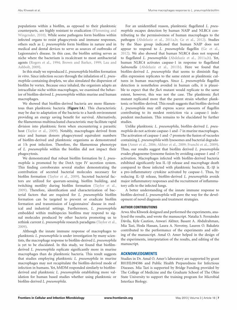
BIOFILM-DERIVED L. pneumophila AVOIDS CASPASE-1 AND -7ACTIVATION IN MURINE MACROPHAGES DUE TO LACK OFFLAGELLIN EXPRESSIONUpon detection of bacterial flagellin by Nlrc4 and Naip5,WT murine macrophages restrict planktonic L. pneumophilareplication via caspase-1 and -7 activation. Caspase-7 pro-motes the fusion of the L. pneumophila-containing vacuolewith the lysosome and bacterial degradation whereas caspase-1 contributes to pyroptosis and IL-1β release (Akhter et al.,2009, 2012). Because biofilm-derived L. pneumophila replicatemore efficiently in murine macrophages and exhibited less celldeath when compared to planktonic, we tested whether mousemacrophages activated caspase-1 in response to biofilm-derivedbacteria. The dotA and flaA mutants were used as negativecontrols since they both avoid caspase-1 activation. In con-trast to planktonic bacteria, biofilm-derived L. pneumophila didnot promote caspase-1 activation as denoted by the detec-tion of the cleaved active band by western blot (Figure 3A).These data indicate that murine macrophages respond tobiofilm-derived L. pneumophila differentially than to planktonicL. pneumophila.
IL-1β maturation is promoted by active caspase-1 in WTmacrophages infected with planktonic L. pneumophila. Therefore,we tested IL-1β release in culture supernatants from murinemacrophages infected with planktonic, biofilm-derived L. pneu-mophila and the dotA mutant. Our data demonstrate that murinemacrophages infected with biofilm-derived bacteria released
FIGURE 3 | Biofilm-derived L. pneumophila did not promote caspase-1
or 7 activation in murine macrophages and showed significantly less
IL-1β release due to lack of flagellin expression. (A) Pro and activecaspase-1 were detected in cell extracts using caspase-1 antibody. WTBMDMs were either not treated (NT) or infected with L. pneumophila JR32(biofilm or planktonic), the dotA or the flaA mutant for 2 h. (B) The amount of
IL-1β was determined in supernatants of WT infected with JR32 (biofilm orplanktonic) or the dotA mutant after 24 h. Data are presented as means ± SDof one experiment performed in quadruplicate. Asterisks indicate significantdifferences (∗∗∗P < 0.001). (C) Activation of caspase-7 was detected in cellextracts using caspase-7 antibody. (D) Western blot analysis of planktonicand biofilm-derived L. pneumophila with flagellin antibody.
Frontiers in Cellular and Infection Microbiology www.frontiersin.org May 2013 | Volume 3 | Article 18 | 5
Abu Khweek et al. Murine macrophages response to L. pneumophila biofilm
30% less IL-1β compared to that released after planktonicL. pneumophila infection (Figure 3B). This result indicates thatthe inflammatory response to biofilm-derived L. pneumophilain murine macrophages is less than that elicited in response toplanktonic bacteria.
During infection of planktonic L. pneumophila, murinemacrophages activate caspase-7 via the inflammasome com-plex contributing to bacterial restriction (Akhter et al., 2009,2012), but this response has not been characterized in biofilm-derived L. pneumophila. Therefore, we tested caspase-7 activa-tion of murine macrophages in response to biofilm-derived andplanktonic L. pneumophila. In contrast to planktonic bacteria,biofilm-derived L. pneumophila did not promote caspase-7 cleav-age (Figure 3C), indicating that biofilm-derived bacteria do notelicit caspase-7 activation, thereby allowing them to evade arestrictive mechanism employed by murine macrophages.
Flagellin mediates restriction of L. pneumophila in murinemacrophages and the flaA mutant has been shown to replicatesignificantly more than the parent strain (Amer et al., 2006).Since biofilm-derived bacteria replicated significantly in murinemacrophages and failed to activate caspase-1 or 7, we hypoth-esized that biofilm-derived bacteria down regulates flagellinexpression. Western blot analysis of bacterial lysates using specificflagellin antibodies demonstrated that biofilm-derived bacteriadiminished flagellin expression compared to planktonic bacte-ria (Figure 3D). Collectively, these results indicate that biofilm
L. pneumophila do not activate the inflammasome because of lackof flagellin expression compared to planktonic L. pneumophila.
PHAGOSOMES CONTAINING BIOFILM-DERIVED L. pneumophila EVADEFUSION WITH THE LYSOSOMESIn murine macrophages, L. pneumophila replication is restrictedby caspase-1 and -7 activation that result in phagosome-lysosome fusion, promoting bacterial degradation (Akhter et al.,2009, 2012). We examined the colocalization of planktonic andbiofilm-derived bacteria with lysosomes at 1 h post-infection.Approximately 55% of biofilm-derived L. pneumophila residedin lysosomes (Figures 4A,B). Yet, 72% of planktonic bacte-ria resided in lysosomes. These results suggests that signifi-cantly more biofilm-derived L. pneumophila evade lysosomaldegradation in macrophages allowing the bacteria to survivewithin the host and replicate as indicated by increased CFUs in(Figure 4A).
DISCUSSIONLegionnaire’s disease is a severe pneumonia that infects theelderly and the immune compromised. There is no humanto human transmission whereas infection occurs by inhalationof contaminated droplets from biofilms lining air condition-ers and fresh water fountains. A biofilm is a highly-organized,multicellular community affixed to an inert or biological sur-face and is the preferred lifestyle of most bacteria. Bacterial
FIGURE 4 | Vacuoles harboring biofilm-derived L. pneumophila bacteria
significantly evade fusion with lysosomes. (A) Representative images ofWT BMDMs infected for 1h with JR32 planktonic or biofilm. Nuclei are stainedblue with DAPI and L. pneumophila stained green with L. pneumophila-specificantibody. Lyso-tracker red was used to stain acidified lysosomes. White arrows
indicate L. pneumophila colocalization with lysotracker. (B) Percentcolocalization of L. pneumophila with lysotracker. Images were captured withthe 60× objective and magnified 3×, scale bar = 10 μm. Data are presented asmeans ± SD of three independent experiments each performed in triplicates.Asterisks indicate significant difference (∗∗P < 0.01).
Frontiers in Cellular and Infection Microbiology www.frontiersin.org May 2013 | Volume 3 | Article 18 | 6
Abu Khweek et al. Murine macrophages response to L. pneumophila biofilm
populations within a biofilm, as opposed to their planktoniccounterparts, are highly resistant to eradication (Flemming andWingender, 2010). While some pathogens form biofilms withininfected organs to resist phagocytosis and immune responses,others such as L. pneumophila form biofilms in nature and inmedical and dental devices to serve as sources of outbreaks ofLegionnaire’s disease. In this case, the biofilm setting serves asniche where the bacterium is recalcitrant to most antibacterialagents (Rogers et al., 1994; Brown and Barker, 1999; Lau andAshbolt, 2009).
In this study we reproduced L. pneumophila biofilm formationin vitro. Since infection occurs through the inhalation of L. pneu-mophila-containing droplets, we also simulated the dispersion ofbiofilm by vortex. Because once inhaled, the organism adapts anintracellular niche within macrophages, we examined the behav-ior of biofilm-derived L. pneumophila within murine and humanmacrophages.
We showed that biofilm-derived bacteria are more filamen-tous than planktonic bacteria (Figure 1A). This characteristicmay be due to adaptation of the bacteria to a harsh environmentproviding an energy saving benefit for survival. Alternatively,the filamentous multinucleated characteristic may facilitate rapiddivision into planktonic form when encountering a parasitichost (Taylor et al., 2009). Notably, macrophages derived frommice and human donors phagocytozed equivalent numbersof biofilm-derived and planktonic organisms as demonstratedat 1 h post infection. Therefore, the filamentous phenotypeof L. pneumophila within the biofilm did not impact theirphagocytosis.
We demonstrated that robust biofilm formation by L. pneu-mophila is promoted by the DotA type IV secretion system.This finding corroborates several studies demonstrating thecontribution of secreted bacterial molecules necessary forbiofilm formation (Taylor et al., 2009). Secreted bacterial fac-tors are utilized for quorum-sensing, biofilm building, andtwitching motility during biofilm formation (Taylor et al.,2009). Therefore, identification and characterization of bac-terial factors that are required for L. pneumophila biofilmformation can be targeted to prevent or eradicate biofilmformation and transmission of Legionnaires’ disease in med-ical and industrial settings. Furthermore, L. pneumophilaembedded within multispecies biofilms may respond to sig-nal molecules produced by other bacteria promoting us torethink current L. pneumophila research paradigms (Taylor et al.,2009).
Although the innate immune response of macrophages toplanktonic L. pneumophila is under investigation by many scien-tists, the macrophage response to biofilm-derived L. pneumophilais yet to be elucidated. In this study, we found that biofilm-derived L. pneumophila replicate significantly more in murinemacrophages than do planktonic bacteria. This result suggeststhat studies employing planktonic L. pneumophila in murinemacrophages may not recapitulate the biofilm-derived mode ofinfection in humans. Yet, hMDM responded similarly to biofilm-derived and planktonic L. pneumophila establishing more val-idation for human based studies whether using planktonic orbiofilm-derived L. pneumophila.
For an unidentified reason, planktonic flagellated L. pneu-mophila escapes detection by human NAIP and NLRC4 con-tributing to the permissiveness of human macrophages to thepathogen (Abdelaziz et al., 2011a,b; Ge et al., 2012). Studiesby the Shao group indicated that human NAIP does notappear to respond to L. pneumophila flagellin (Ge et al.,2012). We also showed that human NLRC4 does not respondto flagellated L. pneumophila (Abdelaziz et al., 2011a,b). Yet,human NLRC4 activates caspase-1 in response to flagellatedSalmonella (Abdelaziz et al., 2011b). Here we found thatbiofilm-derived L. pneumophila that seems to diminish flag-ellin expression replicates to the same extent as planktonic cul-tures in human macrophages. Since L. pneumophila flagellindetection is nonetheless avoided in human cells, it is plausi-ble to expect that the flaA mutant would replicate to the sameextent, however, this was not the case. The planktonic flaAmutant replicated more that the parent strain whether plank-tonic or biofilm-derived. This result suggests that biofilm-derivedL. pneumophila may still express scarce amounts of flagellincontributing to its modest restriction via a caspase-1 inde-pendent mechanism. This remains to be elucidated by furtherstudies.
Unlike planktonic L. pneumophila, biofilm-derived L. pneu-mophila do not activate caspase-1 and -7 in murine macrophages.The activation of caspase-1 and -7 promote the fusion of vacuolescontaining L. pneumophila with lysosomes and bacterial degrada-tion (Amer et al., 2006; Akhter et al., 2009; Franchi et al., 2009).Thus, our results suggest that biofilm derived L. pneumophilaevades phagosome-lysosome fusion by avoiding caspase-1 and -7activation. Macrophages infected with biofilm-derived bacteriaexhibited significantly less IL-1β release and macrophage deathcompared to those infected with planktonic bacteria. IL-1β isa pro-inflammatory cytokine activated by caspase-1. Thus, byreducing IL-1β release, biofilm-derived L. pneumophila avoidsmajor inflammatory responses and the recruitment of inflamma-tory cells to the infected lungs.
A better understanding of the innate immune response tobiofilm-derived L. pneumophila will pave the way for the devel-opment of novel diagnosis and treatment strategies.
AUTHOR CONTRIBUTIONSArwa Abu Khweek designed and performed the experiments, ana-lyzed the results, and wrote the manuscript. Natalia S. FernándezDávila, Kyle Caution, Anwari Akhter, Basant A. Abdulrahman,Mia Tazi, Hoda Hassan, Laura A. Novotny, Lauren O. Bakaletzcontributed to the performance of the experiments and edit-ing of the manuscript. Amal O. Amer helped in the design ofthe experiments, interpretation of the results, and editing of themanuscript.
ACKNOWLEDGMENTSStudies in Dr. Amal O. Amer’s laboratory are supported by grantR01HL094586 and Public Health Preparedness for InfectiousDiseases. Mia Tazi is supported by Bridge Funding provided byThe College of Medicine and the Graduate School of The OhioState University to support the training program for MicrobialInterface Biology.
Frontiers in Cellular and Infection Microbiology www.frontiersin.org May 2013 | Volume 3 | Article 18 | 7
Abu Khweek et al. Murine macrophages response to L. pneumophila biofilm
REFERENCESAbdelaziz, D. H., Gavrilin, M. A.,
Akhter, A., Caution, K., Kotrange,S., Khweek, A. A., et al. (2011a).Apoptosis-associated speck-likeprotein (ASC) controls Legionellapneumophila infection in humanmonocytes. J. Biol. Chem. 286,3203–3208.
Abdelaziz, D. H., Gavrilin, M. A.,Akhter, A., Caution, K., Kotrange,S., Khweek, A. A., et al. (2011b).Asc-dependent and indepen-dent mechanisms contribute torestriction of Legionella pneu-mophila infection in murinemacrophages. Front. Microbiol. 2:18.doi: 10.3389/fmicb.2011.00018
Abdulrahman, B. A., Khweek, A.A., Akhter, A., Caution, K.,Kotrange, S., Abdelaziz, D. H.,et al. (2011). Autophagy stimu-lation by rapamycin suppresseslung inflammation and infectionby Burkholderia cenocepacia in amodel of cystic fibrosis. Autophagy7, 1359–1370.
Abu Kwaik, Y., Eisenstein, B. I.,and Engleberg, N. C. (1993).Phenotypic modulation byLegionella pneumophila uponinfection of macrophages. Infect.Immun. 61, 1320–1329.
Akhter, A., Caution, K., Abu Khweek,A., Tazi, M., Abdulrahman, B. A.,Abdelaziz, D. H., et al. (2012).Caspase-11 promotes the fusion ofphagosomes harboring pathogenicbacteria with lysosomes by mod-ulating actin polymerization.Immunity 37, 35–47.
Akhter, A., Gavrilin, M. A., Frantz, L.,Washington, S., Ditty, C., Limoli, D.,et al. (2009). Caspase-7 activationby the Nlrc4/Ipaf inflammasomerestricts Legionella pneumophilainfection. PLoS Pathog. 5:e1000361.doi: 10.1371/journal.ppat.1000361
Al-Khodor, S., Price, C. T.,Habyarimana, F., Kalia, A., andAbu Kwaik, Y. (2008). A Dot/Icm-translocated ankyrin protein ofLegionella pneumophila is requiredfor intracellular proliferationwithin human macrophages andprotozoa. Mol. Microbiol. 70,908–923.
Alli, O. A., Gao, L. Y., Pedersen,L. L., Zink, S., Radulic, M.,Doric, M., et al. (2000). Temporalpore formation-mediated egressfrom macrophages and alveo-lar epithelial cells by Legionellapneumophila. Infect. Immun. 68,6431–6440.
Amer, A. O. (2010). Modulation ofcaspases and their non-apoptoticfunctions by Legionella pneu-mophila. Cell. Microbiol. 12,140–147.
Amer, A., Franchi, L., Kanneganti, T.D., Body-Malapel, M., Ozoren, N.,Brady, G., et al. (2006). Regulationof Legionella phagosome matura-tion and infection through flagellinand host Ipaf. J. Biol. Chem. 281,35217–35223.
Atlas, R. M. (1999). Legionella: fromenvironmental habitats to diseasepathology, detection and control.Environ. Microbiol. 1, 283–293.
Brown, M. R., and Barker, J. (1999).Unexplored reservoirs of pathogenicbacteria: protozoa and biofilms.Trends Microbiol. 7, 46–50.
Coers, J., Kagan, J. C., Matthews, M.,Nagai, H., Zuckman, D. M., andRoy, C. R. (2000). Identification ofIcm protein complexes that play dis-tinct roles in the biogenesis of anorganelle permissive for Legionellapneumophila intracellular growth.Mol. Microbiol. 38, 719–736.
Costerton, J. W., Geesey, G. G., andCheng, K. J. (1978). How bacteriastick. Sci. Am. 238, 86–95.
Donlan, R. M., Forster, T., Murga, R.,Brown, E., Lucas, C., Carpenter,J., et al. (2005). Legionella pneu-mophila associated with the proto-zoan Hartmannella vermiformis ina model multi-species biofilm hasreduced susceptibility to disinfec-tants. Biofouling 21, 1–7.
Flemming, H. C., and Wingender, J.(2010). The biofilm matrix. Nat.Rev. Microbiol. 8, 623–633.
Fliermans, C. B., Cherry, W. B.,Orrison, L. H., Smith, S. J., Tison,D. L., and Pope, D. H. (1981).Ecological distribution of Legionellapneumophila. Appl. Environ.Microbiol. 41, 9–16.
Franchi, L., Eigenbrod, T., Munoz-Planillo, R., and Nunez, G. (2009).The inflammasome: a caspase-1-activation platform that regulatesimmune responses and diseasepathogenesis. Nat. Immunol. 10,241–247.
Fraser, D. W., Deubner, D. C., Hill,D. L., and Gilliam, D. K. (1979).Nonpneumonic, short-incubation-period Legionellosis (Pontiac fever)in men who cleaned a steam turbinecondenser. Science 205, 690–691.
Ge, J., Gong, Y. N., Xu, Y., and Shao,F. (2012). Preventing bacterial DNArelease and absent in melanoma2 inflammasome activation by a
Harb, O. S., and Abu Kwaik, Y. (2000).Essential role for the Legionellapneumophila rep helicase homo-logue in intracellular infection ofmammalian cells. Infect. Immun. 68,6970–6978.
Horwitz, M. A. (1983). TheLegionnaires’ disease bacterium(Legionella pneumophila) inhibitsphagosome-lysosome fusion inhuman monocytes. J. Exp. Med.158, 2108–2126.
Horwitz, M. A., and Silverstein, S.C. (1980). Legionnaires’ diseasebacterium (Legionella pneumophila)multiples intracellularly in humanmonocytes. J. Clin. Invest. 66,441–450.
Isberg, R. R., O’Connor, T. J., andHeidtman, M. (2009). TheLegionella pneumophila replica-tion vacuole: making a cosy nicheinside host cells. Nat. Rev. Microbiol.7, 13–24.
Kirby, J. E., Vogel, J. P., Andrews,H. L., and Isberg, R. R. (1998).Evidence for pore-forming abil-ity by Legionella pneumophila. Mol.Microbiol. 27, 323–336.
Lau, H. Y., and Ashbolt, N. J. (2009).The role of biofilms and protozoain Legionella pathogenesis: impli-cations for drinking water. J. Appl.Microbiol. 107, 368–378.
Lettinga, K. D., Verbon, A., Weverling,G. J., Schellekens, J. F., Den Boer,J. W., Yzerman, E. P., et al. (2002).Legionnaires’ disease at a Dutchflower show: prognostic factors andimpact of therapy. Emerg. Infect. Dis.8, 1448–1454.
Marston, B. J., Plouffe, J. F., File, T.M. Jr., Hackman, B. A., Salstrom,S. J., Lipman, H. B., et al. (1997).Incidence of community-acquiredpneumonia requiring hospitaliza-tion. Results of a population-basedactive surveillance Study inOhio. The Community-BasedPneumonia Incidence StudyGroup. Arch. Intern. Med. 157,1709–1718.
Rogers, J., Dowsett, A. B., Dennis,P. J., Lee, J. V., and Keevil, C.W. (1994). Influence of tempera-ture and plumbing material selec-tion on biofilm formation andgrowth of Legionella pneumophilain a model potable water sys-tem containing complex microbialflora. Appl. Environ. Microbiol. 60,1585–1592.
Roy, C. R. (2002). The Dot/lcmtransporter of Legionella pneu-mophila: a bacterial conductorof vesicle trafficking that orches-trates the establishment of areplicative organelle in eukaryotichosts. Int. J. Med. Microbiol. 291,463–467.
Santic, M., Molmeret, M., and AbuKwaik, Y. (2005). Maturation of theLegionella pneumophila-containingphagosome into a phagolysosomewithin gamma interferon-activatedmacrophages. Infect. Immun. 73,3166–3171.
Sethi, K. K., and Brandis, H. (1983).Direct demonstration and isola-tion of Legionella pneumophila(serogroup 1) from bathroom waterspecimens in a hotel. Zentralbl.Bakteriol. Mikrobiol. Hyg. B 177,402–405.
Spitalny, K. C., Vogt, R. L., Orciari,L. A., Witherell, L. E., Etkind, P.,and Novick, L. F. (1984). Pontiacfever associated with a whirlpoolspa. Am. J. Epidemiol. 120,809–817.
Taylor, M., Ross, K., and Bentham,R. (2009). Legionella, protozoa,and biofilms: interactions withincomplex microbial systems. Microb.Ecol. 58, 538–547.
Conflict of Interest Statement: Theauthors declare that the researchwas conducted in the absence of anycommercial or financial relationshipsthat could be construed as a potentialconflict of interest.