33
Biology 161 The Eye and The Ear [email protected]

External Eye Stuctures

External Eye Structures

External Eye Structures
Palpebral Fissure The slit between the eyelids
Tarsus The edge of the palpebral fissure where sebaceous glands sit
Conjunctiva Is a transparent mucous membrane The two layers line the eyelids and folds back over the
anterior surface of the eyeball (does not cover the cornea) The function of the conjunctiva is to produce mucus so the
eye does not dry out.

Internal Eye Structures

Cornea Transparent Covering Extension of the sclera Forms a window for light to
enter the eye Cornea is avascular as to
not hinder transparencySclera Hard covering The white of the eye Protects and shapes the
eyeball Provides structure for
muscle attachment
Cornea
Sclera
Internal Eye Structures

Choroid A highly vascular dark
membrane Brown pigment helps
absorb light and keep it from scattering and reflecting in the eye
Blood vessels provide nutrients to all eye layers
Retina the innermost layer of the
eye Contains the
photoreceptors (rods and cones)
Choroid
Retina
Internal Eye Structures

Iris
Colored portion of the eye.
Made of two smooth muscle layers
The muscle fibers allow it to act as a reflexively activated diaphragm to vary pupil size.
Pupil
The round central opening.
It allows light to enter the eye.
Iris Pupil
Internal Eye Structures
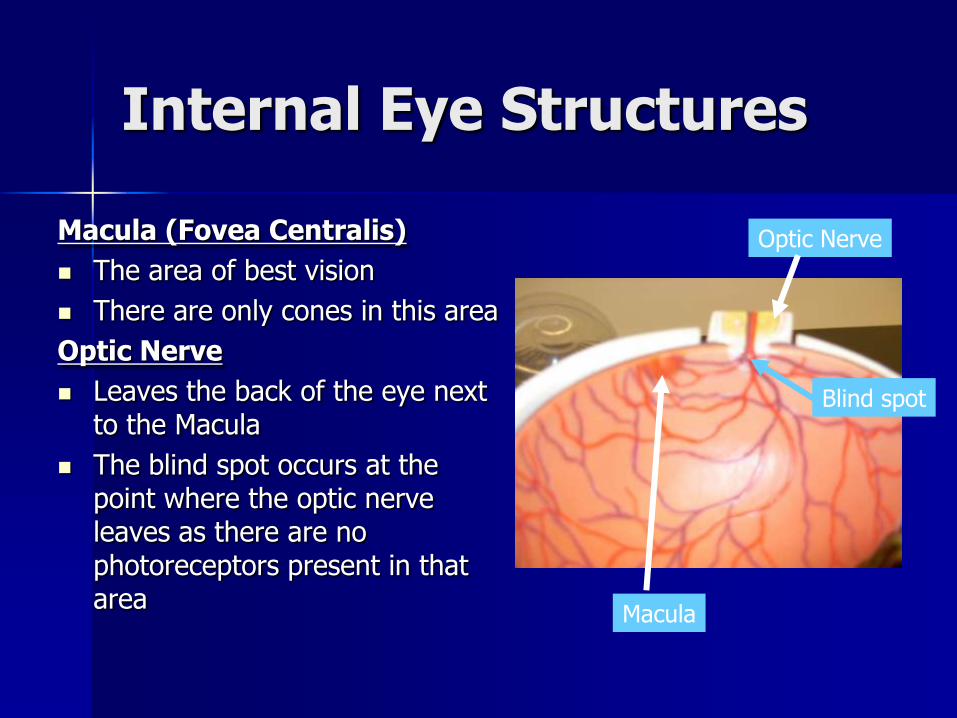
Macula (Fovea Centralis)
The area of best vision
There are only cones in this area
Optic Nerve
Leaves the back of the eye next to the Macula
The blind spot occurs at the point where the optic nerve leaves as there are no photoreceptors present in that area
Macula
Optic Nerve
Blind spot
Internal Eye Structures

Anterior Chamber Filled with aqueous humor
(a clear fluid similar to blood plasma)
Posterior Chamber Filled with vitreous humor
(a clear gel-like substance) The vitreous humor 1. Transmits light2. Supports the retina3. Contributes to intraocular
pressure
Anterior Chamber
Posterior Chamber
Internal Eye Structures

Lens
Is a biconvex, transparent, flexible structure the can change shape to allow precise focusing.
The lens is avascular
New lens fibers are added continuously through life so as you age the lens becomes denser, more convex and less elastic which impairs its ability to focus light properly. Lens
Internal Eye Structures

Canal of Schlemm
Drains the aqueous humor into the venous blood to maintain a constant pressure in the anterior chamber.
Internal Eye Structures

Eye Disorders Glaucoma
Cause – with age the aqueous humor production exceeds aqueous humor re-absorption. This puts pressure on the blood vessels and causes some areas to die. Peripheral vision is lost first, nicknamed “the silent thief of sight”
Prevention – Have pressure checked regularly after 50 years of age.

Eye Disorders – Macular Degeneration (leading cause of blindness in
North America)
Cause – “dry” – small yellow deposits lead to a thinning of the macula
“wet” - blood vessels may bleed and leak fluid causing the macula to bulge
Effect – leads to loss of central eyesight
Treatment – there is no cure after the disease has progressed

Eye Disorders - Cataracts
Cause – the lens of the eye becomes opaque. Risk increases with diabetes and increased UV exposure.
Treatment – the capsule which incases the lens is opened. The lens is fragmented and removed. A new lens is implanted and the capsule is closed.
Prevention – wear UV protective lens.


Eye Disorders -Astigmatism
Cause – eye is more oblong (football shape) than round.
- Ridges on the cornea cause uneven visual perception.
Treatment – Corrective lens

Eye Disorders - Stye
Cause – infection of the sebaceous gland on the eyelid ridge.
Treatment – incise and drain
Stye

Eye Disorders -Conjunctivitis
Cause –inflammation of the conjunctiva, often due to a bacterial infection.
Treatment –antibiotic drops

Vision Tests – Stereoscopic Vision (Binocular Vision)
Stereoscopic vision requires input from both eyes.
1. Look at the Reindeer with 3-D glasses
2. Close one eye or the other
3. The numbers on the grid appear to project away from the page toward you.
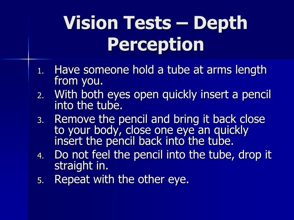
Vision Tests – Depth Perception
1. Have someone hold a tube at arms length from you.
2. With both eyes open quickly insert a pencil into the tube.
3. Remove the pencil and bring it back close to your body, close one eye an quickly insert the pencil back into the tube.
4. Do not feel the pencil into the tube, drop it straight in.
5. Repeat with the other eye.

Vision Tests – Illusion
Closer seems larger
Angles make a difference

Vision Tests – Negative After Image
1. Look at the green paper for 40 seconds.
2. Look at the white area.
3. Since the cones for seeing green have been exposed and used the negative after image is seen.
4. Look at the square patterns. In between the squares a gray area is seen as the rods are exposed to the light.

Color Blindness
There are 3 cone types which absorb varying wavelengths: blue, red and green.
Intermediate colors of the visible light spectrum are due to overlapping input from more then one cone type.

Vision Tests – Color Blindness (Color Vision
Tests)Holmgren Test
1. Match the dyed wool to the wool in the folder.
Ichikawa Test
1. Check plates and find the numbers that are present.

Vision Tests –Astigmatism
1. View chart with one eyen then the other focusing on the center of the chart.
2. If some of the lines are lighter or blurred then there is some astigmatism

Vision Tests – Accomodation and Near Point
1. Have a person hold a pencil at arms length.
2. Hold a ruler to the eye gradually bring the pencil towards the eye.
3. Note the distance where the pencil tip becomes unclear.
4. With age the length at which it becomes unclear will increase

Vision Tests – Snellen Eye Chart for Visual AcuityTest one eye at a time.
1. Standing 20 feet away, read the last line that is clear to you.
2. 20 / 100 indicates that you can read from 20 feet what is normally seen from 100 feet.
3. 20 / 15 indicates that you can see from 20 feet what is normally seen from 15.
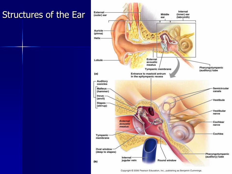
Structures of the Ear

Structures of the Ear
Pinna
Shell shaped projection surrounding the external ear opening.
Directs sound waves into the external ear canal
Ossicles
Are the three smallest bones in the body.
They transmit the vibratory motion of the eardrum which in turn sets the fluid in the inner ear in motion.
Pinna
Ossicles

Structures of the Ear
Semicircular Canals
Are circular canals in the inner ear
These canals are involved in the equilibrium and balance
Auditory Nerve
Cochlear branch extends from the back of the cochlea and transmits sensory information to the brain
Semicircular Canals
Auditory Nerve

Structures of the Ear
Cochlea
Looks like a snail
Houses the receptor organ for hearing
Eustachian Tube
Is a tube which runs downward to link the middle ear cavity to the nasopharynx
Normally the tube is flattened and closed but swallowing or yawning opens it briefly to equalize the pressure in the middle ear cavity
Cochlea
Eustachian Tube

Structures of the Ear
Tympanic Membrane
Ear drum
Sound waves make the eardrum vibrate which in turn transfers the mechanical energy of the sound waves to the ossicles
External Auditory Canal
Extends from the pinna to the tympanic membrane Tympanic Membrane
External AuditoryCanal


