CMC-3 and Chem-3
Biophysical Chemistry
Biomolecular Interactions
Professor Alan CooperRm. B4-20 (Joseph Black Building)
© Alan Cooper - October 2004 Lecture # 1
Aims: To discuss the theoretical and experimental basis ofintermolecular forces in complex, condensed systems andhow this applies to the stabilization of biomolecular structuresand interactions.
Recommended reading/Chemistry Library reference/reserve shelf:
Physical Biochemistry: principles and applications, David Sheehan, WileyBiophysical Chemistry, C R Cantor and P R Schimmel, W H Freeman & Co.Physical Biochemistry, D Freifelder, W H Freeman & Co.Enzyme Structure and Mechanism, A Fersht, W H Freeman & Co.Protein Structure - a Practical Approach, T E Creighton, IRL Press.Introduction to Protein Structure, C Branden and J Tooze, Garland Publishing
+ new for 2004...Biophysical Chemistry, A. Cooper (RSC Tutorial Chemistry Text)
© Alan Cooper - October 2004 CMC-3/Chem-3 BiomolecularInteractions
+ website material (to come)
© Alan Cooper - October 2004 CMC-3/Chem-3 BiomolecularInteractions
What is special (if anything) about biological molecules ?
• they are big (usually)
• they are polymers (usually) with specific sequence
• they have specific structure which determines function
• these structures are determined (mainly) bynon-covalent forces
• they exist and perform in water
• they are the products of evolution
© Alan Cooper - October 2004 CMC-3/Chem-3 BiomolecularInteractions
H2N C C
R1
O
C C
R2
NH
O
C C
R3
OH
OH
H
Hn
φ ψ
Proteins are polymers made up of specific sequences of L-aminoacids linked together by covalent peptide (amide) bonds (Fig.1).Amino acids are chosen from a basic set of 20 building blocksdiffering in sidechain (Fig.2), with occasional special-purposeside chains made to order (e.g. hydroxyproline).
Figure 1: Polypeptide structure showing rotatable φ/ψ angles.The planar peptide (amide) bonds are shown in bold, and areusually trans.
Proteins & Polypeptides
© Alan Cooper - October 2004 CMC-3/Chem-3 BiomolecularInteractions
The 20 naturally-occurring amino acid side chains (residues) withstandard 3-letter and single-letter abbreviations.
© Alan Cooper - October 2004 CMC-3/Chem-3 BiomolecularInteractions
Primary structure: the sequence of amino acids in the polypeptidechain. This is unique to each protein, and is determined (primarily)by the genetic information encoded in the DNA of the relevantgene.
KVFERCELAR TLKRLGMDGY RGISLANWMC LAKWESGYNTRATNYNAGDR STDYGIFQIN SRYWCNDGKT PGAVNACHCSASALLQDNIA DAVACAKRVV RDPQGIRAWV AWRNRCQNRDVRQYVQGCGV
Primary structure of a 130-residue protein (human lysozyme)shown using the single-letter amino acid codes
© Alan Cooper - October 2004 CMC-3/Chem-3 BiomolecularInteractions
Secondary structure: regular, repeating structures such as α-helix,β-sheets, etc.
Tertiary structure: thethree-dimensionalarrangement of secondarystructure elements thatdefines the overallconformation of the(globular) protein.
© Alan Cooper - October 2004 CMC-3/Chem-3 BiomolecularInteractions
Phosphoglycerate kinaseLysozyme (withbound inhibitor)
Examples of protein tertiary structure...
© Alan Cooper - October 2004 CMC-3/Chem-3 BiomolecularInteractions
Quaternary structure: in multi-subunit proteins, the three-dimensional arrangement of the subunits.
© Alan Cooper - October 2004 CMC-3/Chem-3 BiomolecularInteractions
Polynucleotides
The genetic information which encodes protein sequences is found inDNA (deoxyribonucleic acid), and the transcription and translationprocess involves RNA (ribonucleic acid). Both are polynucleotidesconsisting of long sequences of nucleic acids made up of a phospho-ribose backbone, with a choice of four different purine or pyrimidineside-chains or “bases” attached.
© Alan Cooper - October 2004 CMC-3/Chem-3 BiomolecularInteractions
N
NN
N
NH2
O
HO
HHHH
POO-
HO
O
NH
N
NO
NH2N
O
H
HHHHO
POO-
O O
H
HHHH
N
NO
OPOO-
O O
H
HHHH
HN
N
O
O
OPOO-
O-
NH2
Adenine (A)
Guanine (G)
Cytosine (C)
Thymine (T)
DNA structureillustrating thedeoxyribose-phosphatebackbone, to whichmay be attachedpurine (A, G) orpyrimidine (C, T)bases
© Alan Cooper - October 2004 CMC-3/Chem-3 BiomolecularInteractions
N
NN
N
NH2
O
OHO
HHHH
POO-
HO
O
NH
N
NO
NH2N
O
OH
HHHHO
POO-
O O
OH
HHHH
N
NO
OPOO-
O
NH2
O
OH
HHHH
HN
N
O
O
OPOO-
O-
Adenine (A)
Guanine (G)
Cytosine (C)
Uracil (U)
RNA structureillustrating the sugar(ribose) -phosphatebackbone, to whichmay be attachedpurine (A, G) orpyrimidine (C, U)bases
© Alan Cooper - October 2004 CMC-3/Chem-3 BiomolecularInteractions
DNA double-helix withcomplementary basepairing…
purine-pyrimidine
A-T (A-U in RNA)
G-C
…genetic coding
© Alan Cooper - October 2004 CMC-3/Chem-3 BiomolecularInteractions
Polysaccharides
Complex polysaccharides such as starch, glycogen,cellulose, and so forth, play an important part inbiochemistry both as energy stores and structuralcomponents. Many proteins are glycosylated(“glycoproteins”), with oligosaccharide chains (oftenbranched) attached to specific amino acid residues, usuallyat the protein surface. The carbohydrate portion ofglycoproteins is often involved in antigenicity, cellreceptor and other molecular recognition processes.
© Alan Cooper - October 2004 CMC-3/Chem-3 BiomolecularInteractions
Fats, Lipids and Detergents
Fats and lipids are common terms for those bits ofbiological organisms that are insoluble in water but can beextracted with organic solvents such as trichloromethane(chloroform), ethers, etc. They generally consist of a polarhead group attached to non-polar tails of unbranchedhydrocarbons. This amphiphilic nature – hydrophilic head,hydrophobic tail – gives this class of molecule importantproperties that are exploited both by biology itself, and bybiophysical chemists in studying such systems.
Broadly speaking, the number of hydrocarbon tails governsthe behaviour in water.
© Alan Cooper - October 2004 CMC-3/Chem-3 BiomolecularInteractions
Detergents generally contain a polar head group attached to a single non-polar tail (or equivalent). This allows them to form micelles in water:roughly globular assemblies of a number of molecules clustered together,with their head groups exposed to water, while their non-polar tails areburied inside the cluster and away from direct contact with thesurrounding water.
© Alan Cooper - October 2004 CMC-3/Chem-3 BiomolecularInteractions
Lipids have two tails. This makes it difficult to pack the hydrocarbonchains effectively into a globular micelle structure, but they can formlipid bilayers instead. Here the molecules form into two-dimensionalarrays or sheets, in which two layers of lipids bury their tails inside,leaving the hydrophilic heads exposed either side to the water. Theselipid bilayers provide the basic structures of cell membranes.
© Alan Cooper - October 2004 CMC-3/Chem-3 BiomolecularInteractions
Neutral fats or triglycerides commonly have three tails. This makesit difficult to form a compromise between the hydrophilic head andthe bulky hydrophobic tails, so these substances tend to be veryinsoluble and just form an amorphous mass in water. This is whatwe commonly see as “fat”.
© Alan Cooper - October 2004 CMC-3/Chem-3 BiomolecularInteractions
Covalent versus Non-Covalent interactions...
Covalent
Definition: a covalent bond is an interaction between adjacentatoms involving sharing of an electron pair
Properties: Strong (∆Hof ≈ - 400 kJ mol-1 ; >> RT)
Difficult to break (except with enzymes, etc.)
Stiff (don’t stretch or bend, but may rotate)
Short range
Responsible for: Primary structure of biopolymers...
DNA sequence
RNA sequence
Protein sequence...
© Alan Cooper - October 2004 CMC-3/Chem-3 BiomolecularInteractions
Covalent versus Non-Covalent interactions...
Non-Covalent
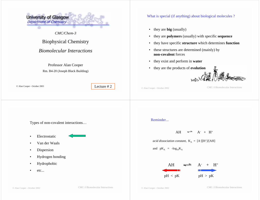
Definition: a non-covalent interaction is any interaction(attractive or repulsive) not involving sharing of anelectron pair.
Properties: Weak (≤ 50 kJ mol-1 ; ≈ RT)Easily disrupted by thermal motionMobile, flexible, non-specificLong range
Responsible for: Macromolecular conformationsDNA/RNA helices; protein secondary/tertiary/quaternary structures; active site binding, subunitassociation… (liquid/solid properties)
CMC/Chem-3
Biophysical Chemistry
Biomolecular Interactions
Professor Alan CooperRm. B4-20 (Joseph Black Building)
© Alan Cooper - October 2003 Lecture # 2 © Alan Cooper - October 2002 CMC-3 Biomolecular Interactions
What is special (if anything) about biological molecules ?
• they are big (usually)
• they are polymers (usually) with specific sequence
• they have specific structure which determines function
• these structures are determined (mainly) bynon-covalent forces
• they exist and perform in water
• they are the products of evolution
© Alan Cooper - October 2002 CMC-3 Biomolecular Interactions
Types of non-covalent interactions…
• Electrostatic
• Van der Waals
• Dispersion
• Hydrogen bonding
• Hydrophobic
• etc...
© Alan Cooper - October 2002 CMC-3 Biomolecular Interactions
AH A- + H+
acid dissociation constant, KA = [A-][H+]/[AH]
and pKA = -log10KA
Reminder...
���������
AH A- + H+
��������������
��������
pH < pK pH > pK
© Alan Cooper - October 2002 CMC-3 Biomolecular Interactions
Group pH < pKA Typical pKA pH > pKA
C-terminus -COOH 3 -COO-
Glu, Asp -COOH 4 -COO-
His -Im-H+ 6 -ImN-terminus -NH3
+ 8 -NH2Cys -SH 8 -S-
Lys -NH3+ 11 -NH2
Tyr -φ-OH 11 -φ-O-
Arg -C(NH2)2+ 12.5 -C(NH)(NH2)
Phosphoglycerol, g =CH2(OH)CH(OH)CH2-O
g-P(OH)O2- 5.6 g-PO3 2-
Typical pKA and charge state for protein amino acid residues andother groups in water
© Alan Cooper - October 2002 CMC-3 Biomolecular Interactions
Electrostatic Interactions
(A) Point Charges: “Coulomb Interaction”
Potential energy between two point charges, q1 and q2 :-
Vqq = q1q2/4πε0εrr
ε0 = vacuum permittivity = 8.85 x 10⁻¹² C2 J-1 m-1
εr = relative permittivity or “dielectric constant” r = distance between charges (in metres)q1 , q2 are the charges (in Coulomb)
(electronic charge: 1 e = 1.6 x 10⁻¹⁹ C )
r
q2q1
© Alan Cooper - October 2002 CMC-3 Biomolecular Interactions
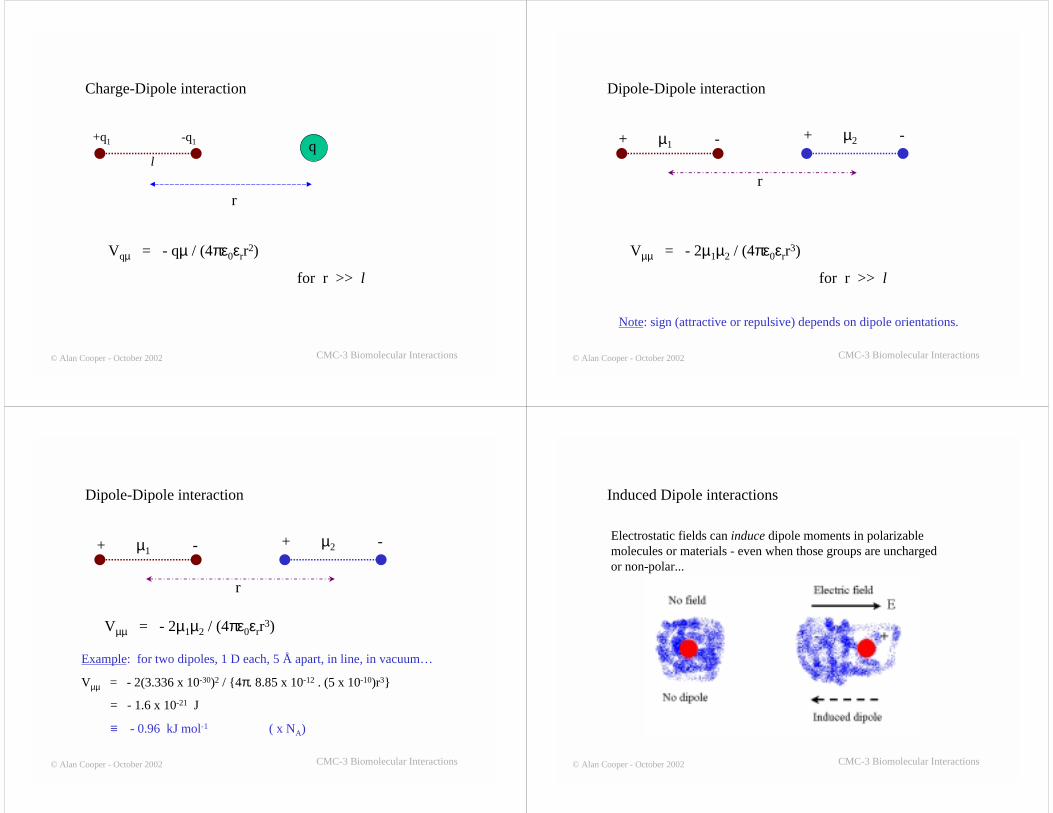
The dielectric constant or relative permittivity (εr) of a substance is a measure of itspolarizability in an electric field. For water at room temperature, εr ≈ 80 (comparedto 1 for a vacuum). This very high value arises because the dioplar water moleculestend to re-orient and align parallel to the electric field. This has the effect of partiallycancelling the electric field, and results in a weakening of electrostatic interactionsbetween charged groups.
-
δ- δ+
+E
© Alan Cooper - October 2002 CMC-3 Biomolecular Interactions
Example: What is the electrostatic potential energy between Na+ andCl- ions, 5 Å apart, in vacuum ?
qNa+ = - qCl- = 1.6 x 10⁻¹⁹ C ; εr = 1 for vacuum
r = 5 Å = 0.5 nm = 5 x 10⁻¹⁰ m
Vqq = q1q2/4πε0εrr = -(1.6 x 10⁻¹⁹)2/ (4π. 8.85 x 10⁻¹². 5 x 10⁻¹⁰) = - 4.6 x 10⁻¹⁹ J note minus sign, attractive interaction
× NA ≡ -276 kJ mol-1
Now add water… εr = 80
Vqq = - 3.5 kJ mol-1 comparable to thermal energy
kT (per molecule) ≡ RT (per mole) ≈ 2.5 kJ mol-1
© Alan Cooper - October 2002 CMC-3 Biomolecular Interactions
Ionic screening...
- +++
Dissolved ions tend to cluster around fixed charges and partialcancel (“screen”) electrostatic interactions.
Debye-Hückel limiting law for low ionic strengths…
V ≈ Vqq . exp(-r/rD)
© Alan Cooper - October 2002 CMC-3 Biomolecular Interactions
Debye-Hückel limiting law for low ionic strengths…
V ≈ Vqq . exp(-r/rD)
rD = “Debye length” = (ε0εrRT / 2ρF2I)1/2
ρ = density of water (103 kg m-3)
F = 1 Faraday (96,500 C)
I = “ionic strength” = ½Σzi mi ≡ [salt] for 1:1 electrolytes
For 1:1 electrolytes, rD = 3.04/[salt]1/2 Å
© Alan Cooper - October 2002 CMC-3 Biomolecular Interactions
Typical physiological ionic strength, I = 0.14
rD = (ε0εrRT / 2ρF2I)1/2 = 8.2 x 10⁻¹⁰ m = 8.2 Å
e.g. for previous calculation… (ions 5 Å apart)
V = -3.5 x exp(-5.0/8.2) = - 1.9 kJ mol-1
© Alan Cooper - October 2002 CMC-3 Biomolecular Interactions
Electrostatic Interactions
(B) Dipole Interactions
+q -q
l
Dipole moment:
µ = ql
Units: 1 Debye = 1 D = 3.336 x 10⁻³⁰ C m
e.g. ±e , separated by 0.1 nm… µ = 4.8 D
Typical dipole moment ≈ 1 D for small molecules/groups
Interaction energies of dipoles with other charges may becalculated by summation of individual point charge interactions,or by formulas in special cases...
© Alan Cooper - October 2002 CMC-3 Biomolecular Interactions
Charge-Dipole interaction
+q1 -q1
l
r
q
Vqµ = - qµ / (4πε0εrr2)
for r >> l
© Alan Cooper - October 2002 CMC-3 Biomolecular Interactions
Dipole-Dipole interaction
+ µ1 - + µ2 -
r
Vµµ = - 2µ1µ2 / (4πε0εrr3)
for r >> l
Note: sign (attractive or repulsive) depends on dipole orientations.
© Alan Cooper - October 2002 CMC-3 Biomolecular Interactions
Dipole-Dipole interaction
+ µ1 - + µ2 -
r
Vµµ = - 2µ1µ2 / (4πε0εrr3)
Example: for two dipoles, 1 D each, 5 Å apart, in line, in vacuum…
Vµµ = - 2(3.336 x 10⁻³⁰)2 / {4π. 8.85 x 10⁻¹² . (5 x 10⁻¹⁰)r3}
= - 1.6 x 10⁻²¹ J
≡ - 0.96 kJ mol-1 ( x NA)
© Alan Cooper - October 2002 CMC-3 Biomolecular Interactions
Induced Dipole interactions
Electrostatic fields can induce dipole moments in polarizablemolecules or materials - even when those groups are unchargedor non-polar...
© Alan Cooper - October 2002 CMC-3 Biomolecular Interactions
Example: Dipole - Induced Dipole interaction...
non-polar
r
VµI = - C1/r6 - always attractive
where C1 = µ2 α / (4πε0)2 -- (corrected Nov 2004)
and α = polarizability of the non-polar group
© Alan Cooper - October 2002 CMC-3 Biomolecular Interactions
3 Å
VµI ≈ - 0.8 kJ mol-1
Typical value…
… for a 1D dipole, 3 Å from a benzene molecule
CMC/Chem-3
Biophysical Chemistry
Biomolecular Interactions
Professor Alan CooperRm. B4-20 (Joseph Black Building)
© Alan Cooper - October 2003 Lecture # 3 © Alan Cooper - October 2002 CMC-3 Biomolecular Interactions
Types of non-covalent interactions…
• Electrostatic
• Van der Waals
• Dispersion
• Hydrogen bonding
• Hydrophobic
• etc...
Previous lecture:-
© Alan Cooper - October 2002 CMC-3 Biomolecular Interactions
Types of non-covalent interactions…
• Electrostatic
• Van der Waals
• Dispersion
• Hydrogen bonding
• Hydrophobic
• etc...
}© Alan Cooper - October 2002 CMC-3 Biomolecular Interactions
Reminder: Dipole - Induced Dipole interaction...
non-polar
r
VµI = - C1/r6 - always attractive
where C1 = µ2 α / 4πε0
and α = polarizability of the non-polar group
© Alan Cooper - October 2002 CMC-3 Biomolecular Interactions
(London) Dispersion Forces
Fritz London (1900-1954)
Transient dipole - Induced dipoleinteraction
All atoms or molecules will tend toattract each other due to a quantummechanical effect...
Heisenberg Uncertainty:-→ transient fluctuations in electron density→ transient dipoles (even in totally non-polar groups)→ transient induced dipoles in adjacent molecules→ attractive “London dispersion forces”
© Alan Cooper - October 2002 CMC-3 Biomolecular Interactions
(London) Dispersion Forces
VDispersion = - C2/r6 - always attractive
r
© Alan Cooper - October 2002 CMC-3 Biomolecular Interactions
VDispersion = - C2/r6
where C2 =
α1, α2 = polarizabilities of the interacting groups
3 α1α2 . I1I22 (4πε0)2 (I1 + I2)
I1, I2 = ionization energies of interacting groups
© Alan Cooper - October 2002 CMC-3 Biomolecular Interactions
VDispersion = - C2/r6
where C2 =
α1, α2 = polarizabilities of the interacting groups
3 α1α2 . I1I22 (4πε0)2 (I1 + I2)
I1, I2 = ionization energies of interacting groups
at high (optical) frequencies - since quantumfluctuations very rapid… hence “dispersion”
© Alan Cooper - October 2002 CMC-3 Biomolecular Interactions
VDispersion = - C2/r6
Typical value…
For 2 methane molecules, 3 Å apart
VDispersion ≈ - 5 kJ mol-1
© Alan Cooper - October 2002 CMC-3 Biomolecular Interactions
Distance dependence of various interactions...
charge-charge Vqq ∝ r-1
charge-dipole Vqµ ∝ r-2
dipole-dipole V µ µ ∝ r-3
dispersion & VDisp ∝ r-6
dipole-induced dipole
long range
short range
© Alan Cooper - October 2002 CMC-3 Biomolecular Interactions
Short-range repulsions...
Orbital overlap/Pauli exclusion at short range
→ very strong repulsion
→ “hard sphere” approximation
→ Van der Waals radii
“Van der Waals interaction” is the term (loosely)used to designate all these attractive/repulsive non-covalent interactions
© Alan Cooper - October 2002 CMC-3 Biomolecular Interactions
Van der Waals/Lennard-Jones potential
V(r) = (A/r)12 - (B/r)6
repulsive attractive
© Alan Cooper - October 2002 CMC-3 Biomolecular Interactions
The Hydrogen Bond
Hydrogen “bonds” are extreme examples of dipole-dipole interactions…
-A-H --- B-
where A and B are electronegative atoms such as F, N,O, and (to a lesser extent) Cl and S.
δ+ δ-δ-
© Alan Cooper - October 2002 CMC-3 Biomolecular Interactions
The Hydrogen Bond
Examples:
-OH --- O- alcohols, water
-NH --- O=C- amides, protein secondary structure
-NH --- N- ammonia (liqu.), DNA/RNA base pairing
H-bonds may be inter- or intra-molecular
© Alan Cooper - October 2002 CMC-3 Biomolecular Interactions
Evidence for Hydrogen Bonds
• High melting points
• High boiling points
• High ∆H of melting/boiling/sublimation…
© Alan Cooper - October 2002 CMC-3 Biomolecular Interactions
Melting pointK
Boiling pointK
∆∆∆∆H(vap)kJ mol-1
H-bond ?
H2O 273 373 40.3 YH2S 190 212 18.8 N
HF 181 292 30.1 YHCl 161 189 16.2 N
NH3 195 240 23.3 YCH4 89 112 9.2 N
© Alan Cooper - October 2002 CMC-3 Biomolecular Interactions
Evidence for Hydrogen Bonds
• High melting points
• High boiling points
• High ∆H of melting/boiling/sublimation…
• Gas phase dimers & higher aggregates
CHO
HOHC
O
OH
H O
HH O
H
water vapour methanoic (formic) acid
© Alan Cooper - October 2002 CMC-3 Biomolecular Interactions
Evidence for Hydrogen Bonds
• High melting points
• High boiling points
• High ∆H of melting/boiling/sublimation…
• Gas phase dimers & higher aggregates
• Anomalous vibrational spectra
IR band shifts and broadening (e.g -OH)
© Alan Cooper - October 2002 CMC-3 Biomolecular Interactions
Evidence for Hydrogen Bonds
• High melting points
• High boiling points
• High ∆H of melting/boiling/sublimation…
• Gas phase dimers & higher aggregates
• Anomalous vibrational spectra
• Crystal structures...
- atom-atom contacts closer than expected for Van derWaals radii, but longer than covalent bond lengths
© Alan Cooper - October 2002 CMC-3 Biomolecular Interactions
How “strong” is a Hydrogen Bond ?
Measurements from gas-phase monomer-dimer equilibrium…
A-H + B AH---B
K = [AH---B]/[AH][B]
∆Go = -RT.lnK = ∆Ho - T. ∆So
���������
Example: formic (methanoic) aciddimers…
K = 260 atm-1 at 300K
CHO
HOHC
O
OH
From experiment: ∆Go = - 13.9 kJ mol-1
∆Ho = - 59.0 kJ mol-1
∆So = - 150 J K-1 mol-1
} 2 H-bondsper dimer
© Alan Cooper - October 2002 CMC-3 Biomolecular Interactions
For water dimers, in the gas phase...
H O
HH O
H
∆Ho ≈ - 20 kJ mol-1
© Alan Cooper - October 2002 CMC-3 Biomolecular Interactions
Water
• High m.p.
• High b.p.
• High heat of vaporization & sublimation - but low heat of melting
• Liquid more dense than solid - ice floats
• 4 oC maximum density of liquid
• High surface tension
• High heat capacity of the liquid
• …etc
Anomalous properties (related to polarity/H-bonding):
© Alan Cooper - October 2002 CMC-3 Biomolecular Interactions
0 5 10 15 20 25 30
0.996
0.998
1.000
Den
sity
/ g
ml-1
Temperature oC
Density ofliquid water,showing 4 oCmaximum
© Alan Cooper - October 2002 CMC-3 Biomolecular Interactions
CMC/Chem-3
Biophysical Chemistry
Biomolecular Interactions
Professor Alan CooperRm. B4-20 (Joseph Black Building)
© Alan Cooper - October 2003 Lecture # 4 © Alan Cooper - October 2002 CMC-3 Biomolecular Interactions
Types of non-covalent interactions…
• Electrostatic
• Van der Waals
• Dispersion
• Hydrogen bonding
• Hydrophobic
• etc...
}
© Alan Cooper - October 2002 CMC-3 Biomolecular Interactions
Effects of Water on Biomolecular Interactions
(1) Electrostatic interactions
High dielectric constant of water (ε ≈ 80)
Reduces interaction potentials coming fromstatic charges
Note: This has much less an effect on high-frequency dispersion forces,since the water dipoles do not have sufficient time to re-orient duringthe lifetime of the quantum fluctuations
-
δ- δ+
+E
© Alan Cooper - October 2002 CMC-3 Biomolecular Interactions
Effects of Water on Biomolecular Interactions
(2) Hydrogen bonding thermodynamics
Hydrogen bonds in vacuum are relatively strong…
H O
HH O
H
Biomolecular secondary & tertiary structures contain lots of H-bonds...
CHO
HOHC
O
OH
H O
H
H O
H+ ∆Ho ≈ + 20 kJ mol-1
∆Ho ≈ + 59 kJ mol-1HCO
OH HCO
OH
+
© Alan Cooper - October 2002 CMC-3 Biomolecular Interactions
Lysozyme structure showing H-bonds
CMC-3 Biomolecular Interactions
protein - DNAcomplex
α-helical secondarystructure in proteins
© Alan Cooper - October 2002
A paradox…?
Polar, hydrogen-bonding groups tend to be very soluble in water
Water molecules can form strong H-bonds with other polargroups (solvation/hydration)…
… so how come H-bonds in proteins and DNA (etc.) ???
-NH + -C=O
-NH---O=C-
e.g. H-bond formation during protein folding ?
H O
HH O
H
H O
HHO
H
+
In water ?
Nochangein totalH-bondcount ?
© Alan Cooper - October 2002 CMC-3 Biomolecular Interactions
Thermodynamics of hydrogen bonding interactions inwater
Experiments involving model compounds that mimicprocesses in larger biomolecules
Ref: Klotz & Franzen (1962) J.Am.Chem.Soc. 84, 3461.
Klotz & Farnham (1968) Biochemistry 7, 3879.
Eberhardt & Raines (1994) J.Am.Chem.Soc. 116, 2149.
Cooper (2000) Biophys. Chem. 85, 25.
There is a natural tendency for allthings (even atoms & molecules) toroll downhill - to fall to lower energy.
∆H tends to want to be negative
This is opposed (at the molecularlevel) by the equally naturaltendency for thermal/Brownianmotion (otherwise known as“entropy”) to make things go theother way…
…and this effect gets bigger as thetemperature increases.
T.∆S tends to want to be positive
A bluffer’s guide to Thermodynamic Equilibrium…
© Alan Cooper - October 2002
Thermodynamic Equilibrium, expressed in terms of the Gibbs Free Energy change,reflects just the balance between these opposing tendencies…
∆G = ∆H - T.∆S
Equilibrium is reached when these two forces just balance (∆G = 0).
The standard free energy change, ∆G°, is just another way of expressing the equilibriumconstant, or affinity (K) for any process, on a logarithmic scale…
∆G° = -RT.lnK
© Alan Cooper - October 2002
CH3
C O
NH
CH3
CH3
C O
NH
CH3
�����������
�����+
CH3
C O
NH
CH3
CH3
H N
OC
CH3
Association (dimerization) of N-methylacetamide (NMA) insolution:-
(Ref: Klotz & Franzen (1962) J.Amer.Chem.Soc. 84, 3461-3466.)
K = [NMA2]/[NMA]2
∆G° = -RT.ln(K) = ∆H° - T.∆S°
d(ln.K)/d(1/T) = - ∆H°/R (van’t Hoff equation)
Low conc. 6800 cm-1 ---> High conc. 6450 cm-1IR:-(NH
stretch)
CH3
C O
NH
CH3
CH3
C O
NH
CH3
�������
�����+
CH3
C O
NH
CH3
CH3
H N
OC
CH3
Association (dimerization) of N-methylacetamide (NMA) insolution:-
(Ref: Klotz & Franzen (1962) J.Amer.Chem.Soc. 84, 3461-3466.)
Solvent K ∆G° ∆H° ∆S°/M-1 /kJ mol-1 /kJ mol-1 /J K-1 mol-1
CCl4 4.7 - 3.8 -17.6 - 46
Dioxane 0.52 + 1.6 - 3.3 - 16.5
Water 0.005 + 13.1 0 - 44
Results(for 25 °C)
Conclusion: Peptide (amide) H-bonding is thermodynamicallyfavourable in non-polar solvents, but not in water.
Better model ?
N-H (aq) + C=O (aq) NH---OC (non-aq)1
Thermodynamic cycle:-
N-H (non-aq) + C=O (non-aq) NH---OC (non-aq)
N-H (aq) + C=O (aq)1
2
3
1
Thermodynamic cycle:-
N-H (non-aq) + C=O (non-aq) NH---OC (non-aq)
N-H (aq) + C=O (aq)1
2
3
3 2= +
fromNMA in
CCl4
from partitioning ofNMA between aq and
non-aq solvents
© Alan Cooper - October 2002 CMC-3 Biomolecular Interactions
Thermodynamics of transfer: partitioningexperiments...
solventlayer
H2O
CCl4
At equilibrium:-
D =
= 1.1 x 10⁻³ (from expt.)
[NMA]CCl4
[NMA]H2O
∆G°(3) = -RT.ln(D) = + 16.9 kJ mol-1
1
Thermodynamic cycle:-
N-H (non-aq) + C=O (non-aq) NH---OC (non-aq)
N-H (aq) + C=O (aq)1
2
3
3 2= +
from partitioning ofNMA between aq and
non-aq solvents
fromNMA in
CCl4
1
Thermodynamic cycle:-
N-H (non-aq) + C=O (non-aq) NH---OC (non-aq)
N-H (aq) + C=O (aq)1
2
3
16.9 -3.8= +∆Go = + 13.1 kJ mol-1
fromNMA in
CCl4
from partitioning ofNMA between aq and
non-aq solvents
Conclusion: Even taking into account the transfer to non-aqueousenvironment, peptide-peptide H-bonding is still appearsthermodynamically unfavourable in the presence of water.
?? Maybe the model compounds are wrong (or inappropriate models)
?? Maybe there are other interactions more important
From model compound experiments:-
N-H (aq) + C=O (aq) NH---OC (non-aq)
∆Go = + 13.1 kJ mol-1
∆Ho
∆Soboth positive (data not shown)
release of bound water ?
CMC/Chem-3
Biophysical Chemistry
Biomolecular Interactions
Professor Alan CooperRm. B4-20 (Joseph Black Building)
© Alan Cooper - October 2003 Lecture # 5
Conclusion: Even taking into account the transfer to non-aqueousenvironment, peptide-peptide H-bonding is still appearsthermodynamically unfavourable in the presence of water.
?? Maybe the model compounds are wrong (or inappropriate models)
?? Maybe there are other interactions more important
From model compound experiments:-
N-H (aq) + C=O (aq) NH---OC (non-aq)
∆Go = + 13.1 kJ mol-1
Previous lecture...
© Alan Cooper - October 2002 CMC-3 Biomolecular Interactions
Types of non-covalent interactions…
• Electrostatic
• Van der Waals
• Dispersion
• Hydrogen bonding
• Hydrophobic
• etc...
Effects of Water on Biomolecular Interactions
(1) Hydrophobic Interactions
References: Cantor & Schimell I pp. 279-288
Tanford (1980) “The Hydrophobic Effect”
Kauzmann (1959) Adv. Protein Chem. 14, 1.
Oil and water don’tmix.. Why not ?
© Alan Cooper - October 2002 CMC-3 Biomolecular Interactions
Oil and water don’t mix.. Why not ?
Non-polar molecules & groups are insoluble in water…
Water does not wet non-polar surfaces (Teflon®, Gortex®, etc.)
Water appears to repel non-polar molecules (and vice versa)
© Alan Cooper - October 2002 CMC-3 Biomolecular Interactions
(London) Dispersion Forces
VDispersion = - C2/r6 - always attractive
r
© Alan Cooper - October 2002 CMC-3 Biomolecular Interactions
Reminder: Dipole - Induced Dipole interaction...
non-polar
r
VµI = - C1/r6 - always attractive
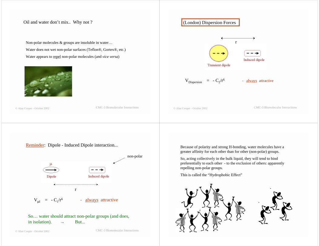
So… water should attract non-polar groups (and does,in isolation). → But...
Because of polarity and strong H-bonding, water molecules have agreater affinity for each other than for other (non-polar) groups.
So, acting collectively in the bulk liquid, they will tend to bindpreferentially to each other - to the exclusion of others: apparentlyrepelling non-polar groups.
This is called the “Hydrophobic Effect”
How can we measure this ?
Since protein folding, ligand binding, etc., (usually) involvesburial of groups in a non-polar environment..
What is the difference in free energy (∆Go) and otherthermodynamic parameters (∆Ho, ∆So , etc.) between groups inan aqueous and non-polar environment ?
© Alan Cooper - October 2002 CMC-3 Biomolecular Interactions
Partitioning of model compounds between solvents
Reminder from Chem-2X (Molecular Thermodynamics)
-- read the “Hitch-hiker’s Guide”
Chem-2X Molecular Thermodynamics © Alan Cooper - October 2002
What is thesituation at
equilibrium ?
Molecules (A say) will tend to move from regions (phases) of highchemical potential to lower µ (low probability to high), until anequilibrium situation is reached in which the chemical potential of A is thesame in all phases...
What is thesituation at
equilibrium ?
The chemical potential of a particular molecule is the same in all phases...
i.e. at equilibrium µA(in phase 1) = µA(in phase 2)µB(in phase 1) = µB(in phase 2)
.. ..... and so on.
(Important note: This does not mean that µA = µB , etc...)
Chemical potentials vary with concentration, etc…
In general: µi = µoi + RT.ln ai
where µi is the actual chemical potential of species i
µoi is the chemical potential of i under standard conditions
ai = γi[i] = activity of i
and the term RT.ln ai takes account of the effects of concentration andintermolecular forces.
Rules for specific circumstances →
(a) For solutions:-
Chemical potential of component A in solution =
µA = µoA + RT.ln γA[A]
≈ µoA + RT.ln [A]
if interaction effects can be ignored (i.e. if γA ≈ 1). This is often the case indilute solutions.
µoA = chemical potential of A when [A] = 1 M
So the “standard state” for solutions is a concentration of 1 M.
Reminder: “activity” a = γA[A] = concentration, corrected for intermolecularinteractions
(b) For liquid mixtures:-
Composition of liquid mixtures is usually expressed in mole fractions (xi)
µi = µoi + RT.ln γixi
Standard state is the pure liquid, for which xi = 1 and γi =1
(c) For gases:-
Composition of gas mixtures is usually expressed in terms of the partialpressures (Pi , in atm), and we can usually ignore activity corrections sincethe molecules are too far apart to interact (ideal gas assumption, γ = 1).
µi = µoi + RT.ln Pi
Standard state is when Pi = 1 atm.
Chem-2X Molecular Thermodynamics © Alan Cooper - October 2002
Liquid-Liquid Equilibrium
Imagine two immiscibleliquids (e.g. water &cyclohexane) in whichtest compound A isdissolved
What is the situation at equilibrium ? What is the concentration of i ineach phase (layer) ?
At equilibrium, for any component A:
µA(liquid 1) = µA(liquid 2)
← liquid 1
← liquid 2
µA(liquid 1) = µA(liquid 2)
For A in liquid 1: µA(liquid 1) =µoA(liquid 1) + RT.ln aA(liquid 1)
For A in liquid 2: µA(liquid 2) = µoA(liquid 2) + RT.ln aA(liquid 2)
So, overall:
µoA(liquid 1) + RT.ln aA(liquid 1) = µo
A(liquid 2) + RT.ln aA(liquid 2)
Re-arrange:ln [aA(liquid 2)/ aA(liquid 1)] = -{µo
A(liquid 2) - µoA(liquid 1)}/RT
= -∆G°transfer(1 → 2)/RT
where ∆G°transfer(1 → 2) = standard free energy of transfer = µo
A(liquid 2) - µoA(liquid 1)
© Alan Cooper - October 2002 CMC-3 Biomolecular Interactions
∆G°transfer(1 → 2) = -RT.ln [aA(liquid 2)/ aA(liquid 1)]
For dilute solutions: aA ≅ [A]
∆G°transfer(1 → 2) ≅ -RT.ln D
where D = [A]2 / [A]1
= “distribution” or “partition” coefficient
© Alan Cooper - October 2002 CMC-3 Biomolecular Interactions
Thermodynamics of transfer: partitioningexperiments...
solventlayer
non-polar
H2O
At equilibrium:-
D =[A]non-polar
[A]H2O
∆G°transfer(H2O → non-polar) = -RT.ln(D)
where [A] = concentration or activity(more accurate) of model compound,A, in appropriate layer
∆G°transfer(1 → 2) = -RT.ln {aA(liquid 2)/ aA(liquid 1)}
Thermodynamics from Solubility
Special case:
If the test molecule, A, is the liquid itself (e.g. liquid 2) -partially soluble in water (liquid 1)
aA(liquid 2) = activity of pure liquid 2 (pure A)
= 1 (by definition)
∆G°transfer(1 → 2) = -RT.ln {1/ aA(liquid 1)} ≅ -RT.ln{1/[A]1} = RT.ln [A]1
[A]1 = solubility of A in solvent 1 (water)∴ measure solubility to give ∆G°transfer
temperature dependence → ∆H°transfer and ∆S°transfer
© Alan Cooper - October 2002 CMC-3 Biomolecular Interactions
Example: Partitioning of fatty acids between heptane and water (pH 7)
Fatty acid N carbons D ∆∆∆∆Go (water →→→→ heptane)/ kJ mol-1
Octanoic 8 0.012 11.0Decanoic 10 0.21 3.9
Lauric 12 3.0 -2.7Myristic 14 54 -9.9
JACS 80, 3817 (1958)
© Alan Cooper - October 2002 CMC-3 Biomolecular Interactions
8 9 10 11 12 13 14
-10
-5
0
5
10
15
∆∆Go = -3.5 kJ mol-1 per -CH2-
∆G
o (wat
er --
> he
ptan
e) /
kJ m
ol-1
N carbons
Example: Partitioning of fatty acids between heptane and water (pH 7)
- “linear free energy” / “group additivity” effects
CMC/Chem-3
Biophysical Chemistry
Biomolecular Interactions
Professor Alan CooperRm. B4-20 (Joseph Black Building)
© Alan Cooper - October 2003 Lecture # 6 © Alan Cooper - October 2002 CMC-3 Biomolecular Interactions
Types of non-covalent interactions…
• Electrostatic
• Van der Waals
• Dispersion
• Hydrogen bonding
• Hydrophobic
• etc...
© Alan Cooper - October 2002 CMC-3 Biomolecular Interactions
This lecture…
(a) Effect of temperature on hydrophobic interactions
(b) Biomolecular interactions in real systems
→ The “Protein Folding Problem”
Effect of temperature onhydrophobic interactions
Anomaloussolubility of non-polar groups in water
∆G°transfer(1 → 2) = -RT.ln {aA(liquid 2)/ aA(liquid 1)}
Thermodynamics from Solubility
Special case:
If the test molecule, A, is the liquid itself (e.g. liquid 2) -partially soluble in water (liquid 1)
aA(liquid 2) = activity of pure liquid 2 (pure A)
= 1 (by definition)
∆G°transfer(1 → 2) = -RT.ln {1/ aA(liquid 1)} ≅ -RT.ln{1/[A]1} = RT.ln [A]1
[A]1 = solubility of A in solvent 1 (water)∴ measure solubility to give ∆G°transfer
temperature dependence → ∆H°transfer and ∆S°transfer© Alan Cooper - October 2002 CMC-3 Biomolecular Interactions
0 10 20 30 40 50
-7.92
-7.90
-7.88
-7.86
-7.84
-7.82
-7.80
-7.78
-7.76
-7.74
T oC
Solubility of benzene in waterBiophysical ChemistryGlasgow
Date: 31/10/2002File: Benzene-sol2.opjOrigin v. 5.046
- ln(
solu
bilit
y)
T oC
0 10 20 30 40 50-25
-20
-15
-10
-5
0
5
10
15
20
25
T.∆S(benzene --> water)
∆Go(benzene --> water)
∆H(benzene --> water)
Thermodynamics of benzene-water solubility
© Alan Cooper - October 2002 CMC-3 Biomolecular Interactions
T oC
0 10 20 30 40 50-25
-20
-15
-10
-5
0
5
10
15
20
25
T.∆S(benzene --> water)
∆Go(benzene --> water)
∆H(benzene --> water)
Thermodynamics of benzene-water solubility
∆G°transfer(benzene → water)is positive (unfavourable)
∆S°transfer(benzene → water) isnegative (unfavourable), andvaries with temperature
∆H°transfer(benzene → water)is exothermic at lowtemperatures, endothermic athigh temperatures
© Alan Cooper - October 2002 CMC-3 Biomolecular Interactions
Thermodynamics of hydrophobic interactions…
Conclusions from model compound transfer experiments:
∆H°transfer and ∆S°transfer depend on temperature
For breaking (pulling apart) hydrophobic interactions, ∆G° is positive,and both ∆H° and ∆S° increase with temperature
Heat capacity change
∆Cp°transfer = ∂(∆H°transfer )/∂T = T.∂(∆S°transfer )/∂T
is positive
© Alan Cooper - October 2002 CMC-3 Biomolecular Interactions
Heat capacity
Both enthalpy and entropy are fundamentally related to the heat capacity (or specific heat)of an object.
∆H(T) = ∆H(Tref) + Tref
T
∫ ∆Cp .dT
and ∆S(T) = ∆S(Tref) + Tref
T
∫ (∆Cp /T).dT
where ∆Cp is the heat capacity change at constant pressure, which is related to thetemperature dependence of both entropy and enthalpy:
∆Cp = ∂∆H/∂T = T. ∂∆S /∂T
Heat capacity is the quantity which tells us how much heat energy (H) we need to add to asystem in order to increase its temperature. It is also related to entropy (S) because, if w ishigh, there are lots of different ways in which the added energy might be distributed withoutraising the temperature, and the heat capacity is consequently also high.
Interpretation in terms of water clustering...
- formation of H-bonded water clusters around non-polargroups can explain (in part) the thermodynamics
© Alan Cooper - October 2002 CMC-3 Biomolecular Interactions
This lecture…
(a) Effect of temperature on hydrophobic interactions
(b) Biomolecular interactions in real systems
→ The “Protein Folding Problem”
© Alan Cooper - October 2002 CMC-3 Biomolecular Interactions
So what’s the problem… ?
Biopolymers fold spontaneouslyto form specific structures …synthetic polymers don’t
The structure is determined by the sequence→ Anfinsen “thermodynamic hypothesis”
© Alan Cooper - October 2002 CMC-3 Biomolecular Interactions
Christian B. Anfinsen
Nobel Prize in Chemistry 1972
"for his work on ribonuclease, especiallyconcerning the connection between the aminoacid sequence and the biologically activeconformation"
© Alan Cooper - October 2002 CMC-3 Biomolecular Interactions
H2N C C
R1
O
C C
R2
NH
O
C C
R3
OH
OH
H
Hn
φ ψ
Proteins are polymers made up of specific sequences of L-aminoacids linked together by covalent peptide (amide) bonds (Fig.1).Amino acids are chosen from a basic set of 20 building blocksdiffering in sidechain (Fig.2), with occasional special-purposeside chains made to order (e.g. hydroxyproline).
Figure 1: Polypeptide structure showing rotatable φ/ψ angles.The planar peptide (amide) bonds are shown in bold, and areusually trans.
© Alan Cooper - October 2002 CMC-3 Biomolecular Interactions
Primary structure: the sequence of amino acids in the polypeptidechain. This is unique to each protein, and is determined (primarily)by the genetic information encoded in the DNA of the relevantgene.
KVFERCELAR TLKRLGMDGY RGISLANWMC LAKWESGYNTRATNYNAGDR STDYGIFQIN SRYWCNDGKT PGAVNACHCSASALLQDNIA DAVACAKRVV RDPQGIRAWV AWRNRCQNRDVRQYVQGCGV
Primary structure of a 130-residue protein (human lysozyme)shown using the single-letter amino acid codes
© Alan Cooper - October 2002 CMC-3 Biomolecular Interactions
Secondary structure: regular, repeating structures such as α-helix,β-sheets, etc.
Tertiary structure: thethree-dimensionalarrangement of secondarystructure elements thatdefines the overallconformation of the(globular) protein.
© Alan Cooper - October 2002 CMC-3 Biomolecular Interactions
Phosphoglycerate kinaseLysozyme (withbound inhibitor)
Examples of protein tertiary structure...Problem 1: How does a protein “find” the correct conformer ?
(Levinthal paradox)
- stereochemical constraints limit the number of Φ-Ψ anglesavailable, but there are still many different possibilities
© Alan Cooper - October 2002 CMC-3 Biomolecular Interactions
The Levinthal “paradox”:C. Levinthal, J. Chim. Phys. 65 (1968) 44; D.B. Wetlaufer, Proc. Natl. Acad. Sci. USA 70 (1973) 691.
Each φ or ψ angle in a peptide might have roughly three possible values,giving 3 × 3 = 9 possible conformers for each peptide (not counting sidechain conformers).
How many possible conformers for a protein (polypeptide) of n residues ?
For n peptide units, no. of possible conformers = 9n
Even for a small protein (n = 100), this is an enormous number of possibleconformers…
9100 = 2.6 x 1095
… only one of which is “correct”
© Alan Cooper - October 2002 CMC-3 Biomolecular Interactions
Problem 1: How does a protein “find” the correct conformer ?
(Levinthal paradox)
How long might it take to explore all possible conformations ?
Assuming (optimistically) that peptide conformations can switchon the femtosecond time scale (10-15 sec) -
Time taken = no. of conformers x time per conformer
= 2.6 x 1095 x 10-15 sec (for n = 100)
= 2.6 x 1080 sec
≈ 1073 years (1 year ≈ 3 x 10⁷ sec)
→ kinetic pathways for folding
- just like every other physical/chemical process...
- the “folding funnel”
© Alan Cooper - October 2002 CMC-3 Biomolecular Interactions
Problem 2: Why does a protein fold ?
- what are the thermodynamic driving forces ?
Random coil conformations…
• much more probable than specific folded structure
• higher entropy
• intrinsically lower free energy
What is ∆S for folding ?
Chem-2X Molecular Thermodynamics © Alan Cooper - October 2002
The absolute entropy of any object is given by:
S = k.ln(w)per object (molecule)
or S = R.ln(w)per mole of objects
What is Entropy ?
Ludwig Boltzmann (1844-1906)
For a polypeptide in the “random coil” state (in which all Φ,Ψ angles arepossible)…
w ≈ 9n
Sconf(random coil) = R.ln(w)
≈ nR.ln(9) ≈ 18n J K-1 mol-1
≈ 1800 J K-1 mol-1 for n = 100-residue protein
For “native” conformation…
w ≈ 1
Sconf(native) = R.ln(1) = 0
∆Sconf(random coil → native) ≈ -18n J K-1 mol-1
∆Goconf(random coil → native) = -T. ∆Sconf
≈ +540 kJ mol-1 for 100-residue protein
- thermodynamically unfavourable free energy change
- this must be overcome by more favourable interactions from other sources
How can we measure these forces in real proteins ?
0 1 2 3 4 5 6
0
50
100
CD
Fluorescence
% c
hang
e
[GuHCl] / mol dm -330 40 50 60 70
Tm
Fluo
resc
ence
inte
nsity
Temperature / oC
- most proteins are marginally stable, and can be unfoldedby relatively small perturbations
- transitions can be followed indirectly through changes inspectroscopic and other properties
© Alan Cooper - October 2002 CMC-3 Biomolecular Interactions
For a 2-state unfolding transition:N U ; K = [U]/[N]
���� ��
∆G°unf = ∆H°unf - T.∆S°unf
= -RT.lnK
K = (F - F0)/(Finf - F)
20 25 30 35 40 45 50 55 60
50
55
60
65
70
75
53.8
71.9
62.5
F
T oC
Finf = 75
F0 = 50
t F K ∆G° ∆H° ∆S°
/ C / kJ mol-1 /kJ mol-1 K-1
35 53.8 0.18 +4.4 255 0.81
40 62.5 1 0 300 0.96
45 71.9 7.1 -5.2 345 1.10
Numericalexample...
CMC/Chem-3
Biophysical Chemistry
Biomolecular Interactions
Professor Alan CooperRm. B4-20 (Joseph Black Building)
© Alan Cooper - October 2003 Lectures # 7 & 8
20 25 30 35 40 45 50 55 60
50
55
60
65
70
75
53.8
71.9
62.5
F
T oC
Finf = 75
F0 = 50
t F K ∆G° ∆H° ∆S°
/ C / kJ mol-1 /kJ mol-1 K-1
35 53.8 0.18 +4.4 255 0.81
40 62.5 1 0 300 0.96
45 71.9 7.1 -5.2 345 1.10
Numericalexample...
© Alan Cooper - October 2002 CMC-3 Biomolecular Interactions
Lectures # 7 & 8
• Microcalorimeter methods
• Protein unfolding thermodynamics
• Ligand binding
• Other experimental methods
© Alan Cooper - October 2002 CMC-3 Biomolecular Interactions
Calorimetry began with Joseph Black in Glasgow250 years ago...
“He waited with impatience for the winter” inGlasgow so that he could do experiments on thefreezing and melting of water and water/alcoholmixtures.
This led to the concept of latent heat and the earlieststudies of the heat capacity of hydrogen-bondednetworks.
CalorimetryCalorimetry
Simpson, A.D.C. (1982) “Joseph Black 1728-1799: a commemorativesymposium” (Royal Scottish Museum, Edinburgh, 1982)
Differential scanning calorimeter
Sample volume: 0.5 ml Temperature range: 10 - 120 oC
Sample concentration: 0.2 - 2 mg/ml© Alan Cooper - October 2002 CMC-3 Biomolecular Interactions
Worked Problem:
Q: What might be the temperature difference between sample and identicalbuffer reference solutions for a sample comprising 1 mg cm-3 of a protein ofRMM 50000 undergoing a thermal transition with ∆H = 80 kJ mol-1 ?
A: 1 mg ≡ 1 x 10⁻³ / 50,000 = 2 x 10⁻⁸ moles of protein
x ∆H ≡ 1.6 x 10⁻³ J heat energy uptake per mg of protein.
The specific heat capacity of water (assume identical for buffer and proteinsolution) = 4.2 J K-1 mol-1
Assuming that all this heat energy is absorbed by the 1 cm3 sample∆T1 = 1.6 x 10⁻³ / 4.2 = 3.8 x 10⁻⁴ °C
(In practice, thermal transitions in biomolecules do not occur all at once,but take place over a finite temperature range. This means that temperaturechanges observed by DSC are usually very much smaller than this.)
Typical data for the heatcapacity increment (∆Cp)observed upon thermalunfolding of a globularprotein in aqueoussolution
A.Cooper (1999). Thermodynamicsof protein folding and stability.“Protein: A ComprehensiveTreatise”, Volume 2, pp. 217-270.(Editor: Geoffrey Allen. JAI PressInc., Stamford CT, 1999)
Protein (un)folding...
0 20 40 60 80 100
1
2
3
4
5
6
∆ Cp
Tm
T / oC
Cp
/ J
K -1
g-1
From: Cooper, Lakey, Johnson & Nollman (2001) - Biophysical Chemistry 93, 215-230.
Typical DSC data for theunfolding of a small globularprotein (lysozyme) in solution atvarious pH values. The insertshows the variation in mid-pointunfolding temperature (Tm) as afunction of pH. The increase inarea under each endotherm withhigher Tm, and the higher heatcapacity baselines after theunfolding transitions, are bothindications of the significantpositive ∆Cp commonlyassociated with such processes.
20 40 60 80
0
20
40
60
80pH 3.8
pH 2.5
pH 1.2
Temperature / o C
C P /
kJ
K-1 m
ol-1
40
60
80
1 2 3 4 5
T m /
o C
pH
© Alan Cooper - October 2002 CMC-3 Biomolecular Interactions
-40
-20
0
2 0
0 2 0 4 0 6 0
∆G u n f
0 2 0 4 0 6 0
0
2 0 0
4 0 0
6 0 0
∆H u n f
T .∆S u n f
T (oC )
kJ m
ol -1
Typical protein unfolding thermodynamics...Typical protein unfolding thermodynamics...
∆H and T.∆S compensate togive small changes in ∆G
Large variations in ∆H and ∆S
Positive ∆Cp
Traditionally characteristic ofexposure of hydrophobic groups(Kauzmann)
-40
-20
0
20
0 20 40 60
∆Gunf
0 20 40 60
0
200
400
600
∆Hunf
T.∆Sunf
T (oC)
kJ m
ol -1
1. Folded proteins are only marginally stable
∆Gunf << RT per residue
∴ needs cooperativity for stability
2. ∆Gunf = ∆Hunf - T.∆Sunf
= small difference between large numbers
∴ very difficult to predict theoretically
3. Mid-point for thermal unfolding…
∆Gunf = 0 ; Tm = ∆Hunf /∆Sunf
4. Extrapolation suggests “cold denaturation”
- unfolding at low temperatures (as well as high)
-40
-20
0
20
0 20 40 60
∆Gunf
0 20 40 60
0
200
400
600
∆Hunf
T.∆Sunf
T (oC)
kJ m
ol -1
What does this suggest aboutthe forces that stabilise thefolded protein ?
- compare to model compounddata for hydrophobicinteractions...
© Alan Cooper - October 2002 CMC-3 Biomolecular Interactions
T oC
0 10 20 30 40 50-25
-20
-15
-10
-5
0
5
10
15
20
25
T.∆S(benzene --> water)
∆Go(benzene --> water)
∆H(benzene --> water)
Thermodynamics of benzene-water solubility
∆G°transfer(benzene → water)is positive (unfavourable)
∆S°transfer(benzene → water) isnegative (unfavourable), andvaries with temperature
∆H°transfer(benzene → water)is exothermic at lowtemperatures, endothermic athigh temperatures
-40
-20
0
20
0 20 40 60
∆Gunf
0 20 40 60
0
200
400
600
∆Hunf
T.∆Sunf
T (oC)
kJ m
ol -1
T oC
0 10 20 30 40 50-25
-20
-15
-10
-5
0
5
10
15
20
25
T.∆S(benzene --> water)
∆Go(benzene --> water)
∆H(benzene --> water)
Protein unfolding Transfer of non-polargroups to water
- suggests that hydrophobic interactions are (mainly) responsible forprotein folding stability
-40
-20
0
20
0 20 40 60
∆Gunf
0 20 40 60
0
200
400
600
∆Hunf
T.∆Sunf
T (oC)
kJ m
ol -1
What does this suggest aboutthe forces that stabilise thefolded protein ?
- compare to model compound datafor hydrophobic interactions...
- suggests that hydrophobicinteractions are (mainly) responsiblefor protein folding stability
Textbook version
Is there an alternative ?
0 20 40 60 80 100
1
2
3
4
5
6
Typical data for the heat capacity increment (∆Cp )observed upon thermal unfolding of a globular
protein in aqueous solution
T / oC
C p /
J K -1
g-1
Cooper (2000) Biophys. Chem. 85, 25-39.
Positive ∆Cp forprotein unfolding isnormally attributed toexposure ofhydrophobic groups.
However...
-40 -20 0 20 40
2
4W ater
Cp
/ J K
-1 g
-1
∆ t / oC
2
4 Ammonia
1
2 Ethanol
1
2 CH3COOH
1
2
-40 -20 0 20 40SOLID LIQUID
Examples of absolute heat capacities for pure compoundsas a function of temperature, plotted with respect to
the normal melting point (∆t = T - Tm).
Hydrocarbons
Cooper (2000) Biophys. Chem. 85, 25-39.
However...
Most, if not all, order-disorder transitions inhydrogen-bondednetworks also takeplace with a positive∆Cp
The melting ofH-bondedlattices
Cooper (2000) Biophys. Chem. 85, 25-39.
Positive ∆Cp is normal for any order-disorder transitionespecially in hydrogen-bonded lattices ...
-40 -20 0 20 40
2
4Water
Cp
/ J K
-1 g
-1
∆ t / oC
2
4 Ammonia
1
2 Ethanol
1
2 CH3COOH
1
2
Hydrocarbons
0 20 40 60 80 100
1
2
3
4
5
Protein
t / oC
Cp
/ J K
-1 g
-1
Low T
High T
Folded Unfolded
Low T
High T
Folded Unfolded
© Alan Cooper - October 2002 CMC-3 Biomolecular Interactions
Ligand binding +
P + L PL
Association constant: KAss = [PL]/[P][L]
Dissociation constant: KDiss = [P][L]/[PL] = 1/ KAss
Note: KDiss = [L]50% (when [P] = [PL] , 50% of protein bound)
How can we measure this ?
����� ���
Ligand binding
0 4 8 12
Lig
and
fluo
resc
ence
inte
nsity
[Protein] / µ mol dm -3
+
Indirect methods using spectroscopic changes (e.g. fluorescence)
Ligand binding
0 4 8 12
[Protein] / µ mol dm -3
+
Indirect methods using spectroscopic changes (e.g. fluorescence)
Fraction bound
= [PL]/([P] + [PL])
=
Finf
F0
F - F0
F - F0
Finf - F0
→ K , ∆G° , etc.
Isothermal titrationmicrocalorimeter
Microcal VP-ITC
Sample volume: 1-2 ml ; [protein] ~ 5-50 µM
Direct method...
Typical ITC data for binding of atrisaccharide inhibitor (tri-N-acetyl-glucosamine; tri-NAG) to hen egg whitelysozyme, in 0.1M acetate buffer, pH 5.Each exothermic heat pulse (upper panel)corresponds to injection of 10 µl of tri-NAG (0.45mM) into the protein solution(36µM). Integrated heat data (lower panel)constitute a differential binding curve thatmay be fit to a standard single-site bindingmodel to give, in this instance, thestoichiometry of binding, N = 0.99,binding affinity, Kass = 3.9 x 10⁵ M-1
(Kdiss = 2.6 µM) , and enthalpy of binding,∆H = -51.7 kJ mol-1 .
-6
-4
-2
0
0 20 40 60 80 100 120
Time (min)
µW
0.0 0.5 1.0 1.5 2.0 2.5 3.0
-50
-40
-30
-20
-10
0
Molar Ratio
kJ/m
ole o
f inj
ecta
nt
From: Cooper, Lakey, Johnson & Nollman (2001) - Biophysical Chemistry 93, 215-230.
+
Binding Equilibrium
Suppose we know the equilibrium constant for binding of a ligand to a protein. How do weknow how much is bound under particular conditions? Typically we might know the totalprotein and total ligand concentrations, but how much is bound?
For protein-ligand binding (or anything equivalent):
P + L PL
Dissociation constant: K = [P][L]/[PL] ……………{1}
Total ligand concentration: CL = [L] + [PL] ……………{2}
Total protein concentration: CP = [P] + [PL]
= K[PL]/[L] + [PL] using {1}
= K[PL]/(CL – [PL]) + [PL] using {2}
Rearrange to give the quadratic equation for [PL]:-
[PL]2 - (CP + CL + K)[PL] + CPCL = 0
Binding Equilibrium
For which the two solutions are:
[PL] = [(CP + CL + K) ± {(CP + CL + K)2 - 4CPCL}½ ]/2
By inspection, the minus sign is the physically correct solution, giving the exact expression forprotein-ligand complex formation, [PL], as a function of the total protein and ligandconcentrations:
[PL] = [(CP + CL + K) - {(CP + CL + K)2 - 4CPCL}½ ]/2
If there are n binding sites per mole of protein, then CP = n.C0 , where C0 is the estimated proteinconcentration, giving:
[PL] = [(n.C0 + CL + K) - {(n.C0 + CL + K)2 - 4nC0CL}½ ]/2
The fraction (φ) of sites occupied at any ligand concentration is given by:
φ(CL) = [PL]/n.C0
= [(n.C0 + CL + K) - {(n.C0 + CL + K)2 - 4nC0CL}½ ]/2nC0
Equilibrium dialysis: ligand binding
semi-permeablemembrane: onlyligands may pass
[L][L] , [P]and [PL]
Equilibrium dialysis: ligand binding
At equilibrium, measure…
(a) Total concn of L on RHS: CL(right) = [L]
(b) Total concn of L on LHS: CL(left) = [L] + [PL]
(c) Total concn of P on LHS: CP(left) = [P] + [PL]
→ [PL] = (b) - (a) ; [P] = (c) - [PL]
K = [PL]/[P][L] ….. directly from concentration measurements © Alan Cooper - October 2002 CMC-3 Biomolecular Interactions
Lectures # 7 & 8
• Microcalorimeter methods
• Protein unfolding thermodynamics
• Ligand binding
• Other experimental methods
UV/visible absorbance spectroscopy
II0
Absorbance A = -log10(I/I0) = -log(T)
= εcl
(Beer-Lambert law)
Excited state
Ground state
Ene
rgy
Interatomic distance
I0
IBeam
Splitter
S
R
Analysis
& displayDetectorMonochromatorLight
Source
UV/visible absorbance spectroscopy
II0
Absorbance A = -log10(I/I0) = -log(T)
= εcl
(Beer-Lambert law)
0
1000
2000
3000
4000
5000
6000
240 260 280 300 320
Trp
Mol
ar e
xtin
ctio
n ε
/
(mol
dm
-3 )-1
cm
-1
240 260 280 300 320
0
100
200
300Phe
240 260 280 300 320
0
1000
2000
Tyr
W avelength λ / nm
240 260 280 300 320
0
1000
2000
3000
4000
5000
6000
Phe
Tyr
Trp
UV/visible absorbance spectroscopy
Near-UV absorbance spectra for aromatic amino acid side chains.When superimposed on the same scale (bottom right panel), the dominantcontribution from tryptophan and tyrosine residues becomes more obvious.
© Alan Cooper - October 2002 CMC-3 Biomolecular Interactions
E
Circular Dichroism (“CD”)
Differential absorption of left- and right-circularly polarized lightby chiral molecules...
Circular dichroism is defined as thenormalized difference in molarextinction:
∆ε = (εR - εL)/ (εR + εL)
where εR and εL refer to the molarabsorbance at a particular wavelength forright- or left-circularly polarized light,respectively.
© Alan Cooper - October 2002 CMC-3 Biomolecular Interactions
Fluorescence spectroscopy
hνexc
hνemE
nerg
y
Interatomic distance
Energy level diagramillustrating electronicexcitation followed byfluorescence emission.After initial (vertical)excitation from the groundstate, the system rapidlyrelaxes to the energyminimum of the excitedstate.
Emission
Monochromator
S
Analysis
& display
Detector
Excitation
Monochromator
Light
Source
Fluorescence spectroscopy
200 250 300 350 400 450 500
aqueous buffer
EmissionExcitation
Fluo
resc
ence
inte
nsity
(ar
bitr
ary
units
)
Wavelength λ / nm
Typical fluorescence excitation and emission spectra for a globular protein in aqueousbuffer at room temperature. The excitation wavelength, λexc, is 290 nm (arrow).The excitation spectrum baseline measured with buffer in the absence of protein isshown offset for clarity.
Raman spectroscopy
- vibrational spectroscopy using visible light
hν0
Monochromator
S
Analysis & display
ω = ν 0 - νDetector
Laser
Raman (inelastic) scattering
Raman spectroscopy
- vibrational spectroscopy using visible light
hν0
Monochromator
S
Analysis & display
ω = ν 0 - νDetector
Laser
anti-Stokes Stokes
0
Rayleigh scatter
ν 0
Scat
tere
d in
tens
ity
Relative Frequency (ν 0 - ν )
Hydrodynamic methods: Analytical Ultracentrifuge
Analytical ultracentrifugerotor
1. Sedimentation equilibrium
Boltzmann (barometric law)exponential concentration gradientat equilibrium.
c(r1)/c(r2) = exp{-m'(r12-r2
2)ω2/2RT}
where m' is the buoyant mass of themolecule.
Hydrodynamic methods: Analytical Ultracentrifuge
Analytical ultracentrifugerotor
1. Sedimentation rate
Gives information about size andshape of macromolecules insolution.
frictional drag
F = fv
centrifugal force
mrw2
velocity v
Hydrodynamic methods: Viscometry
The rate of flow under gravity of a liquid through a capillary tube depends on a number of factorsincluding the viscosity (η) and density (ρ) of the liquid as well as the size and shape of the tube. For astandard capillary viscometer (see Figure) the time taken (t) for a set volume of liquid to flow betweenpoints A and B is proportional to η/ρ so that, after appropriate calibration with known liquids, theviscosity of any sample can be determined from its flow time, t.
The viscosity of asolution ofmacromoleculesdepends on theirsize and shape
Hydrodynamic methods: Dynamic Light Scattering
Scat
tere
d in
tens
ity
Time
Analysis of the shape and frequency of this flickering pattern gives the “autocorrelationtime” (τ) which is related to the diffusion constant (D) of the molecules. This informationcan be used to determine the relative molecular masses and heterogeneities ofmacromolecular samples.
The diffusional or Brownian motion of molecules in aliquid or gas gives rise to fluctuations in density orconcentration that can be observed by optical methods.
This is the basis of the technique known as “dynamic lightscattering” (DLS). A laser beam is passed through the solutionof macromolecules, and the time dependence of the lightscattered from a small volume within the sample is recorded.