Biosynthesis and Defensive Function of Nd-Acetylornithine, aJasmonate-Induced Arabidopsis Metabolite C W
Adewale M. Adio,a Clare L. Casteel,a Martin De Vos,a Jae Hak Kim,a,1 Vijay Joshi,a,2 Baohua Li,b Caroline Juery,a
Josquin Daron,a Daniel J. Kliebenstein,b and Georg Jandera,3
a Boyce Thompson Institute for Plant Research, Ithaca, New York 14853b Department of Plant Sciences, University of California, Davis, California 95616
Since research on plant interactions with herbivores and pathogens is often constrained by the analysis of already known
compounds, there is a need to identify new defense-related plant metabolites. The uncommon nonprotein amino acid
Nd-acetylornithine was discovered in a targeted search for Arabidopsis thalianametabolites that are strongly induced by the
phytohormone methyl jasmonate (MeJA). Stable isotope labeling experiments show that, after MeJA elicitation, Arg, Pro,
and Glu are converted to Orn, which is acetylated by NATA1 to produce Nd-acetylornithine. MeJA-induced Nd-acetylorni-
thine accumulation occurs in all tested Arabidopsis accessions, other Arabidopsis species, Capsella rubella, and Boechera
stricta, but not in less closely related Brassicaceae. Both insect feeding and Pseudomonas syringae infection increase
NATA1 expression and Nd-acetylornithine accumulation. NATA1 transient expression in Nicotiana tabacum and the addition
of Nd-acetylornithine to an artificial diet both decrease Myzus persicae (green peach aphid) reproduction, suggesting a
direct toxic or deterrent effect. However, since broad metabolic changes that are induced by MeJA in wild-type Arabidopsis
are attenuated in a nata1 mutant strain, there may also be indirect effects on herbivores and pathogens. In the case of
P. syringae, growth on a nata1 mutant is reduced compared with wild-type Arabidopsis, but growth in vitro is unaffected by
Nd-acetylornithine addition.
INTRODUCTION
Plants exhibit a wide array of defenses against herbivores and
pathogens, ranging from the production of toxins and feeding
deterrents (Agrawal, 1998; Kessler and Baldwin, 2002) to com-
pensatory growth changes that allow tolerance of high damage
levels (Agrawal, 2000; Stowe et al., 2000; Tiffin, 2000). Many,
perhaps most, of the likely several hundred thousand secondary
metabolites found in the plant kingdom contribute to defense
against herbivores and pathogens (Dixon, 2001; Bino et al.,
2004). Broadly distributed classes of secondary metabolites,
which can be found as components of both constitutive and
inducible plant defense responses, include phenolics, terpenes,
alkaloids, and nonprotein amino acids (Howe and Jander, 2008).
In addition to the standard 20 proteinogenic amino acids,
hundreds of other amino acids have been found inmany different
plant species (Fowden, 2001; Bell, 2003). Some of these, for
example, Orn and homoserine, are essential in primary metab-
olism, whereas others are secondary metabolites that contribute
to plant defense against herbivores and pathogens. For instance,
canavanine, which is found in the seeds of many legumes, is
toxic to herbivores because it competes with Arg in enzymatic
reactions (Rosenthal, 2001). Similarly, azetidine-2-carboxylate,
which is particularly abundant in Convallaria majalis (lily of the
valley), can disrupt protein structures by binding to tRNA in place
of Pro during protein synthesis (Peterson and Fowden, 1963;
Norris and Fowden, 1972).
Another function for nonprotein amino acids may be to store
nitrogen in a form that makes it metabolically unavailable to
herbivores. About half of the 20 protein amino acids are essential
dietary constituents because they cannot be synthesized by
most animals. Degradation of Arg to Orn by tomato (Solanum
lycopersicum) arginase significantly reduces Manduca sexta
(tobacco hornworm) weight gain (Chen et al., 2005), showing
that availability of a single essential amino acid can be growth-
limiting for insects. Similarly, conversion of an essential amino
acid into an uncommon nonprotein amino acid that herbivores
cannot use would reduce the nutritive value of plant tissue.
The nonprotein amino acid Nd-acetylornithine (Figure 1A) has
been reported in Corydalis ochotensis (Manske, 1937), Asple-
nium nidus (Virtanen and Linko, 1955), Bistorta bistortoides
(Lipson et al., 1996), some grasses (Fowden, 1958), and several
legumes (Brown and Fowden, 1966; Zacharius, 1970; Kite and
Ireland, 2002; Marona et al., 2003). Abundance of Nd-acetylorni-
thine in legume seeds (Brown and Fowden, 1966; Zacharius,
1970; Kite and Ireland, 2002) and B. bistortoides rhizomes (38%
of free amino acids; Lipson et al., 1996) suggests a role in
nitrogen storage. However, given that Nd-acetylornithine has
1Current address: Monsanto Company, St. Louis, MO 63167.2 Current address: Dow Agrosciences, Woodburn, OR 97071.3 Address correspondence to [email protected] author responsible for distribution of materials integral to thefindings presented in this article in accordance with the policy describedin the Instructions for Authors (www.plantcell.org) is: Georg Jander([email protected]).CSome figures in this article are displayed in color online but in blackand white in the print edition.WOnline version contains Web-only data.www.plantcell.org/cgi/doi/10.1105/tpc.111.088989
The Plant Cell, Vol. 23: 3303–3318, September 2011, www.plantcell.org ã 2011 American Society of Plant Biologists. All rights reserved.
been found sporadically in ferns, monocots, and dicots (in the
Aspleniaceae, Fabaceae, Papaveraceae, Poaceae, and Poly-
gonaceae), there may be additional functions associated with
this plant metabolite. Like most nonprotein amino acids found in
plants, Nd-acetylornithine has not been studied in a genetically
tractable model species and almost nothing is known about the
biosynthetic pathway(s).
Research with Arabidopsis thaliana has identified diverse
classes of secondary metabolites (D’Auria and Gershenzon,
2005), and, in particular, the defense-related glucosinolates
have been studied extensively (Halkier and Gershenzon, 2006).
However, very little is known about defensive nonprotein amino
acids in Arabidopsis, and the majority of the estimated 5000
small molecules in a typical leaf remain unidentified (Bino et al.,
2004). The plant hormone jasmonic acid (JA) and itsmore volatile
form, methyl jasmonate (MeJA), induce numerous defense re-
sponses, including production of secondary metabolites, in
Arabidopsis and other plants (Howe and Jander, 2008). There-
fore, in an effort to identify and study previously unknown plant
chemical defenses, we initiated a search for the MeJA-induced
production of nonprotein amino acids in Arabidopsis. These
experiments resulted in the identification of Nd-acetylornithine,
an Orn acetyltransferase that synthesizes this compound and
likely defensive functions.
RESULTS
Induced Production of Nd-Acetylornithine in Arabidopsis
Assays of phloem exudates from MeJA-induced Arabidopsis
accession Columbia-0 (Col-0) showed accumulation of an un-
known amino acid, which was determined to be Nd-acetylorni-
thine (Figure 1A) by mass spectrometry (MS) and NMR (see
Supplemental Figure 1 online).Nd-acetylornithine, which was not
commercially available, was synthesized and shown to have the
same properties as the purified Arabidopsis compound (see
Supplemental Figures 1 and 2 online). The absolute configuration
of Arabidopsis Nd-acetylornithine was determined to be L by
comparing the polarity of the purified Arabidopsis metabolite
to synthesized L-Nd-acetylornithine and a racemic D-L mixture.
Nd-acetylornithine, which has not been previously reported in
Arabidopsis or other Brassicaceae, is distinct from Na-acetyl-
ornithine (Figure 1A; see Supplemental Figure 2 online), an
intermediate in plant Arg and Pro metabolism that is found in
most, perhaps all, plants (Verslues and Sharma, 2010).
After treatment of with MeJA,Nd-acetylornithine was detected
in Arabidopsis leaves, stems, flowers, and roots (Figure 1B).
Accumulation in rosette leaves peaked after 4 d and returned to
undetectable levels by day 10 (Figure 1C), suggesting that there
Figure 1. Regulation of Nd-Acetylornithine Production in Arabidopsis.
(A) Structures of L-Nd-acetylornithine and L-Na-acetylornithine.
(B) Accumulation of Nd-acetylornithine in plant tissues 4 d after MeJA treatment. Mean 6 SE of n = 3. ND, not detected.
(C) Time course of Nd-acetylornithine accumulation in rosette leaves after MeJA treatment. Mean 6 SE of n = 3.
(D) Nd-acetylornithine accumulation 4 d after spraying with 450 mM MeJA, SA, ACC, or 100 mM ABA. Mean 6 SE of n = 3.
(E) Inhibition of Nd-acetylornithine biosynthesis by SA. SA was added 24 h before, concomitant with MeJA, or 24 h later. Nd-acetylornithine in rosette
leaves was measured 4 d after the treatment. Mean 6 SE of n = 5.
(F) Nd-acetylornithine in Arabidopsis defense signaling mutants, 4 d after MeJA treatment. Mean6 SE of n = 4 or 5. *P < 0.05, t test relative to unelicited
control plants or day 0 time point (C).
3304 The Plant Cell
is also an Nd-acetylornithine catabolic pathway in Arabidopsis.
Other defense-related plant hormones were tested to determine
whether they induceNd-acetylornithine accumulation.Nd-acetyl-
ornithine abundance in leaves increased with increasing con-
centrations of exogenously added abscisic acid (ABA) (Figure
1D; see Supplemental Figure 3 online). By contrast, there was no
detectable Nd-acetylornithine after treatment with salicylic acid
(SA) or the ethylene precursor 1-aminocyclopropane-1-carbox-
ylate (ACC). SA inhibited the MeJA-induced production of
Nd-acetylornithine (Figure 1E), albeit only if it was applied 24 h
before the MeJA. This is consistent with several prior studies
showing antagonistic effects in plant defense induction and
reduced effectiveness of MeJA elicitation once SA-mediated
defenses are turned on (Thaler et al., 2002; Spoel et al., 2003;Mur
et al., 2006). The nonexpresser of pr genes1 (npr1), salicylic acid
induction deficient1 (sid1), and phytoalexin deficient4 (pad4)
mutations, which result in defects in SA-related defense signal-
ing, had no significant effect on Nd-acetylornithine induction by
MeJA (Figure 1F). Furthermore, these mutants did not have any
detectable Nd-acetylornithine in the absence of MeJA elicitation.
By contrast, there was no MeJA-induced Nd-acetylornithine
accumulation in the coronatine insensitive1-1 (coi1-1) mutant
(Figure 1F), indicating that induction requires a functional COI1
receptor, similar to most other JA-mediated responses in Arabi-
dopsis (Chini et al., 2007; Thines et al., 2007).
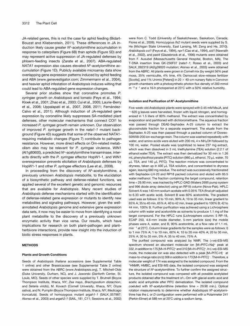
Biosynthesis ofNd-Acetylornithine from Other Amino Acids
The structure of Nd-acetylornithine suggested that it could be
synthesized from Arg, Glu, or Pro, with Orn as an intermediate
(Figure 2A; other pathways are also possible). If so, addition of
[U-13C]Arg, [U-13C]Pro, or [U-13C]Glu to Arabidopsis leaves
should result in the synthesis of [13C]Nd-acetylornithine with a
mass of +5 atomic mass units (see Supplemental Figure 4
online). As predicted, gas chromatography (GC)-MS analysis of
Nd-acetylornithine in MeJA-treated plants after the addition of13C-labeled precursor amino acids showed significant incor-
poration from Arg, Pro, and Glu (Figure 2B). Although the
labeling results showed a direct conversion of the three amino
acids into Nd-acetylornithine, the multiple pathways leading to
Orn formation (Figure 2A) suggested that knockout of any one
amino acid catabolic enzyme would not significantly reduce
Nd-acetylornithine accumulation. Consistent with this hypoth-
esis, knockout mutations in either of the two Arabidopsis
arginases (ARGININE AMINOHYDROLYASE1 [ARGAH1] and
ARGAH2; Brownfield et al., 2008) did not decrease Nd-acetyl-
ornithine abundance (Figure 2C). In fact, for as yet unknown
reasons, the argah2 mutation increased MeJA-induced Nd-
acetylornithine levels.
Orn Acetyltransferase Mutant Isolation
and Characterization
Given the incorporation of five carbon atoms from Arg, Pro, and
Glu into Nd-acetylornithine, we hypothesized that there is an
Orn Nd-acetyltransferase in Arabidopsis that converts Orn into
Nd-acetylornithine (Figure 2A). Analysis of publications report-
ing MeJA-regulated Arabidopsis gene expression showed el-
evated transcription of At2g39030, a member of the GNAT
(Gcn5-relatedN-acetyltransferase) family of acetyltransferases
(Vetting et al., 2005; Yan et al., 2007). Publicly available DNA
microarray data also showed that At2g39030 is induced by JA,
Figure 2. Biosynthesis of Nd-Acetylornithine from Other Amino Acids.
(A) Possible pathways leading from Arg, Pro, and Glu to Nd-acetylornithine. 1, Arginase (At2g39020 and At4g08900); 2, Pro dehydrogenase (At3g30775
and At5g38710); 3, Orn d-aminotransferase (At5g46180); 4, pyrroline-5-carboxylate synthase (At2g39800 and At3g55610); 5, Orn Nd-acetyltransferase
(At2g39030).
(B) Incorporation of [U-13C]Arg, [U-13C]Pro, and [U-13C]Glu into Nd-acetylornithine in MeJA-treated Arabidopsis leaves, expressed as the percentage of
labeled [13C5]Nd-acetylornithine and unlabeled [12C]Nd-acetylornithine in rosette leaves. Mean 6 SE of n = 3.
(C) Nd-acetylornithine accumulation in arginase mutants. Mean 6 SE of n = 5. Letters indicate significant differences, P < 0.05, analysis of variance,
followed by Tukey’s HSD test.
Nd-Acetylornithine, a Defense Metabolite 3305
ABA, mechanical wounding, and a variety of biotic stresses,
including Myzus persicae (green peach aphid) and Pseudomo-
nas syringae (www.genevestigator.com; Zimmermann et al.,
2004).
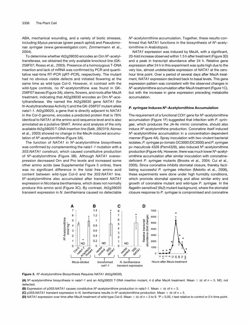
To determine whether At2g39030 encodes an Orn Nd-acetyl-
transferase, we obtained the only available knockout line (GK-
256F07; Rosso et al., 2003). Presence of a homozygous T-DNA
insertion and lack of mRNA was confirmed by PCR and quanti-
tative real-time RT-PCR (qRT-PCR), respectively. The mutant
had no obvious visible defects and initiated flowering at the
same time as wild-type Col-0. However, in contrast with the
wild-type controls, no Nd-acetylornithine was found in GK-
256F07 leaves (Figure 3A), stems, flowers, and roots afterMeJA
treatment, indicating that At2g39030 encodes an Orn Nd-ace-
tyltransferase. We named the At2g39030 gene NATA1 (for
N-Acetyltransferase Activity1) and theGK-256F07mutant allele
nata1-1. At2g39020, a gene that is directly adjacent to NATA1
in the Col-0 genome, encodes a predicted protein that is 78%
identical to NATA1 at the amino acid sequence level and is also
annotated as a putative GNAT. Amino acid analysis of the only
available At2g39020 T-DNA insertion line (Salk_092319; Alonso
et al., 2003) showed no change in the MeJA-induced accumu-
lation of Nd-acetylornithine (Figure 3A).
The function of NATA1 in Nd-acetylornithine biosynthesis
was confirmed by complementing the nata1-1 mutation with a
35S:NATA1 construct, which caused constitutive production
of Nd-acetylornithine (Figure 3B). Although NATA1 overex-
pression decreased Orn and Pro levels and increased some
other amino acids (see Supplemental Figure 5 online), there
was no significant difference in the total free amino acid
content between wild-type Col-0 and the 35S:NATA1 line.
Nd-acetylornithine also accumulated after transient NATA1
expression in Nicotiana benthamiana, which does not normally
produce this amino acid (Figure 3C). By contrast, At2g39020
transient expression in N. benthamiana caused no detectable
Nd-acetylornithine accumulation. Together, these results con-
firmed that NATA1 functions in the biosynthesis of Nd-acety-
lornithine in Arabidopsis.
NATA1 expression was induced by MeJA, with a significant,
20-fold increase observedwithin 1.5 h after treatment (Figure 3D)
and a peak in transcript abundance after 24 h. Relative gene
expression after 24 h in this experiment was quite high due to the
very low, almost undetectable expression of NATA1 at the zero-
hour time point. Over a period of several days after MeJA treat-
ment, NATA1 expression declined back to basal levels. This gene
expression pattern was consistent with the observed changes in
Nd-acetylornithine accumulation afterMeJA treatment (Figure 1C),
but with the increase in gene expression preceding metabolite
accumulation.
P. syringae Induces Nd-Acetylornithine Accumulation
The requirement of a functional COI1 gene for Nd-acetylornithine
accumulation (Figure 1F) suggested that infection with P. syrin-
gae, which produces the JA-Ile mimic coronatine, should also
induce Nd-acetylornithine production. Coronatine itself induced
Nd-acetylornithine accumulation in a concentration-dependent
manner (Figure 4A). Spray inoculation with two virulent bacterial
isolates,P. syringaepv tomatoDC3000 (DC3000) andP. syringae
pv maculicola 4326 (Psm4326), also induced Nd-acetylornithine
production (Figure 4A). However, therewasmuch lowerNd-acetyl-
ornithine accumulation after similar inoculation with coronatine-
deficient P. syringae mutants (Brooks et al., 2004; Cui et al.,
2005). Since coronatine inhibits stomatal closure, thereby facil-
itating successful P. syringae infection (Melotto et al., 2006),
these experiments were done under high humidity conditions,
which promote stomatal opening and allow similar entry and
growth of coronatine mutant and wild-type P. syringae. In the
flagellin-sensitive2 (fls2) mutant background, where the stomatal
closure response to P. syringae is compromised and coronatine
Figure 3. Nd-Acetylornithine Biosynthesis Requires NATA1 (At2g39030).
(A) Nd-acetylornithine biosynthesis in nata1-1 and an At2g39020 T-DNA insertion mutant, 4 d after MeJA treatment. Mean 6 SE of n = 5. ND, not
detected.
(B) Expression of p35S:NATA1 causes constitutive Nd-acetylornithine production in nata1-1. Mean 6 SE of n = 3.
(C) p35S:NATA1 transient expression in N. benthamiana results in Nd-acetylornithine production. Mean 6 SE of n = 5.
(D) NATA1 expression over time after MeJA treatment of wild-type Col-0. Mean 6 SE of n = 3 to 9. *P < 0.05, t test relative to control or 0 h time point.
3306 The Plant Cell
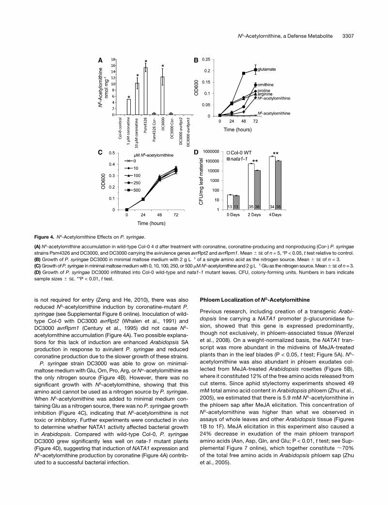
is not required for entry (Zeng and He, 2010), there was also
reduced Nd-acetylornithine induction by coronatine-mutant P.
syringae (see Supplemental Figure 6 online). Inoculation of wild-
type Col-0 with DC3000 avrRpt2 (Whalen et al., 1991) and
DC3000 avrRpm1 (Century et al., 1995) did not cause Nd-
acetylornithine accumulation (Figure 4A). Two possible explana-
tions for this lack of induction are enhanced Arabidopsis SA
production in response to avirulent P. syringae and reduced
coronatine production due to the slower growth of these strains.
P. syringae strain DC3000 was able to grow on minimal-
maltosemediumwith Glu, Orn, Pro, Arg, orNa-acetylornithine as
the only nitrogen source (Figure 4B). However, there was no
significant growth with Nd-acetylornithine, showing that this
amino acid cannot be used as a nitrogen source by P. syringae.
When Nd-acetylornithine was added to minimal medium con-
taining Glu as a nitrogen source, there was no P. syringae growth
inhibition (Figure 4C), indicating that Nd-acetylornithine is not
toxic or inhibitory. Further experiments were conducted in vivo
to determine whether NATA1 activity affected bacterial growth
in Arabidopsis. Compared with wild-type Col-0, P. syringae
DC3000 grew significantly less well on nata-1 mutant plants
(Figure 4D), suggesting that induction of NATA1 expression and
Nd-acetylornithine production by coronatine (Figure 4A) contrib-
uted to a successful bacterial infection.
Phloem Localization of Nd-Acetylornithine
Previous research, including creation of a transgenic Arabi-
dopsis line carrying a NATA1 promoter b-glucuronidase fu-
sion, showed that this gene is expressed predominantly,
though not exclusively, in phloem-associated tissue (Wenzel
et al., 2008). On a weight-normalized basis, the NATA1 tran-
script was more abundant in the midveins of MeJA-treated
plants than in the leaf blades (P < 0.05, t test; Figure 5A). Nd-
acetylornithine was also abundant in phloem exudates col-
lected from MeJA-treated Arabidopsis rosettes (Figure 5B),
where it constituted 12% of the free amino acids released from
cut stems. Since aphid stylectomy experiments showed 49
mM total amino acid content in Arabidopsis phloem (Zhu et al.,
2005), we estimated that there is 5.9 mM Nd-acetylornithine in
the phloem sap after MeJA elicitation. This concentration of
Nd-acetylornithine was higher than what we observed in
assays of whole leaves and other Arabidopsis tissue (Figures
1B to 1F). MeJA elicitation in this experiment also caused a
24% decrease in exudation of the main phloem transport
amino acids (Asn, Asp, Gln, and Glu; P < 0.01, t test; see Sup-
plemental Figure 7 online), which together constitute ;70%
of the total free amino acids in Arabidopsis phloem sap (Zhu
et al., 2005).
Figure 4. Nd-Acetylornithine Effects on P. syringae.
(A) Nd-acetylornithine accumulation in wild-type Col-0 4 d after treatment with coronatine, coronatine-producing and nonproducing (Cor-) P. syringae
strains Psm4326 and DC3000, and DC3000 carrying the avirulence genes avrRpt2 and avrRpm1. Mean6 SE of n = 5, *P < 0.05, t test relative to control.
(B) Growth of P. syringae DC3000 in minimal maltose medium with 2 g L�1 of a single amino acid as the nitrogen source. Mean 6 SE of n = 3.
(C)Growth ofP. syringae inminimalmaltosemediumwith 0, 10, 100, 250, or 500mMNd-acetylornithine and 2 g L�1 Glu as the nitrogen source.Mean6 SE of n = 3.
(D) Growth of P. syringae DC3000 infiltrated into Col-0 wild-type and nata1-1 mutant leaves. CFU, colony-forming units. Numbers in bars indicate
sample sizes 6 SE. **P < 0.01, t test.
Nd-Acetylornithine, a Defense Metabolite 3307
Nd-Acetylornithine Effects on Insect Herbivory
Given the abundance of Nd-acetylornithine in Arabidopsis
phloem exudates (Figure 5B), we predicted that this amino acid
would also be ingested by phloem-feedingM. persicae. This was
confirmed through amino acid analysis of whole aphids, which
contained Nd-acetylornithine after short-term (24 h) feeding from
MeJA-induced Col-0 but not after feeding from uninduced plants
(Figure 5B). Longer infestation with M. persicae induced both
NATA1 gene expression (Figure 5C) and Nd-acetylornithine ac-
cumulation (Figure 5D).
To determine whether there was a direct toxic or deterrent
effect onM. persicae, we addedNd-acetylornithine to an artificial
diet that also contained the 20protein amino acids. This caused a
significant reduction in M. persicae progeny production (Figure
5E) at Nd-acetylornithine concentrations that are comparable to
those that we have observed in phloem exudates. Similarly,
exogenous addition of Nd-acetylornithine to nata1-1 leaves via
their petioles (Figure 5F) significantly reduced aphid reproduc-
tion. However, in this detached-leaf experiment, Nd-acetylorni-
thine addition may also affect aphid reproduction through the
reduced abundance of three amino acids (Gly, Arg, and Thr) in
the treated leaves relative to controls (see Supplemental Figure 8
online). As a percentage of total amino acid content, Nd-acetyl-
ornithine was 5.9-fold more abundant in the honeydew of M.
persicae than in the artificial diet from which they were feeding
(0.5 mM Nd-acetylornithine = 0.29% of total amino acids in the
artificial diet; 1.7% 6 0.3% Nd-acetylornithine in the honeydew;
mean 6 SE of n = 3; P < 0.05, t test). This indicated that Nd-
acetylornithine was taken up and/or metabolized less efficiently
than the 20protein amino acids the aphid diet. Consistent with this
observation, addition of Orn or Gln, but not Nd-acetylornithine, as
Figure 5. NATA1 Expression and Nd-Acetylornithine Affect Arabidopsis–M. persicae Interactions.
(A) Expression of NATA1 in dissected leaf blades and midveins (inset), with and without 24 h MeJA elicitation. Mean 6 SE of n = 4 or 5.
(B) Nd-acetylornithine in petiole exudates and aphids feeding for 1 d from plants treated 4 d earlier with MeJA. Mean 6 SE of n = 3.
(C) NATA1 expression with 25 aphids feeding on one leaf. Fold induction, with 0 h set to 1. Mean 6 SE of n = 3 or 4.
(D) Nd-acetylornithine accumulation in Col-0 wild type after 4 d of M. persicae feeding. Mean 6 SE of n = 3.
(E) Aphid reproduction on artificial diet containing 0 (control), 0.5, 5, or 10 mM Nd-acetylornithine. Mean 6 SE of n = 8.
(F)M. persicae reproduction on detached nata1-1 leaves with petioles in a tube containing Nd-acetylornithine (see inset). Number of aphid progeny after
4 d. Mean 6 SE of n = 22.
(G) M. persicae reproduction wild type and nata1-1 after MeJA treatment. Mean 6 SE of n = 19.
(H) M. persicae reproduction on N. tabacum transiently expressing p35S:NATA1. Number of aphid progeny after 7 d. Mean 6 SE of n = 25.
(I) M. persicae reproduction on Arabidopsis wild-type Col-0 and nata1 mutant transformed with p35S:NATA1. Number of aphid progeny after 7 d.
Mean 6 SE of n = 25 to 27. ND, not detected. *P < 0.05, t test relative to control samples.
3308 The Plant Cell
the only nitrogen source in artificial diet increased aphid progeny
production relative to a Suc-only control diet (see Supplemental
Figure 9 online).
Prior work showed that MeJA treatment significantly decreases
M. persicae reproduction on Arabidopsis (Ellis et al., 2002). This
effectwas confirmedwith ourM.persicae strain on flower stalks of
wild-type Col-0 (Figure 5G). However, on nata1-1 mutant plants,
there was no significant reduction inM. persicae reproduction due
to MeJA treatment, which suggested that NATA1 plays a role in
defense against aphids. Transient expression of NATA1 from the
35S promoter in tobacco (Nicotiana tabacum) caused Nd-acetyl-
ornithine accumulation and decreased M. persicae progeny pro-
duction relative to vector-only controls (Figure 5H). However,
stable transgenic expression of the same construct in wild-type
Col-0 or nata1-1 mutant Arabidopsis did not have a measureable
effect (Figure 5I). SinceNd-acetylornithine inhibition ofM. persicae
reproduction was concentration dependent (Figures 5E and 5F),
this difference may have resulted from lower accumulation of
Nd-acetylornithine in p35S:NATA1Arabidopsis than in transiently
expressing p35S:NATA1N. tabacum orMeJA-elicitedArabidop-
sis (Figure 3).
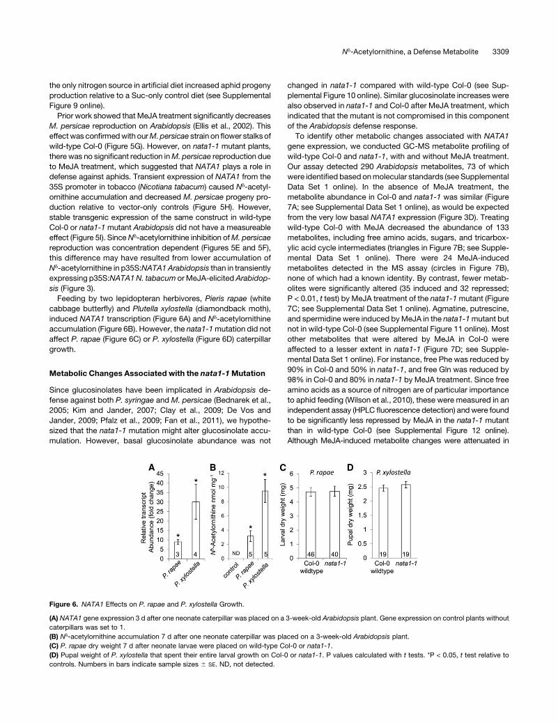
Feeding by two lepidopteran herbivores, Pieris rapae (white
cabbage butterfly) and Plutella xylostella (diamondback moth),
induced NATA1 transcription (Figure 6A) and Nd-acetylornithine
accumulation (Figure 6B). However, the nata1-1mutation did not
affect P. rapae (Figure 6C) or P. xylostella (Figure 6D) caterpillar
growth.
Metabolic Changes Associated with the nata1-1Mutation
Since glucosinolates have been implicated in Arabidopsis de-
fense against both P. syringae and M. persicae (Bednarek et al.,
2005; Kim and Jander, 2007; Clay et al., 2009; De Vos and
Jander, 2009; Pfalz et al., 2009; Fan et al., 2011), we hypothe-
sized that the nata1-1 mutation might alter glucosinolate accu-
mulation. However, basal glucosinolate abundance was not
changed in nata1-1 compared with wild-type Col-0 (see Sup-
plemental Figure 10 online). Similar glucosinolate increases were
also observed in nata1-1 and Col-0 after MeJA treatment, which
indicated that the mutant is not compromised in this component
of the Arabidopsis defense response.
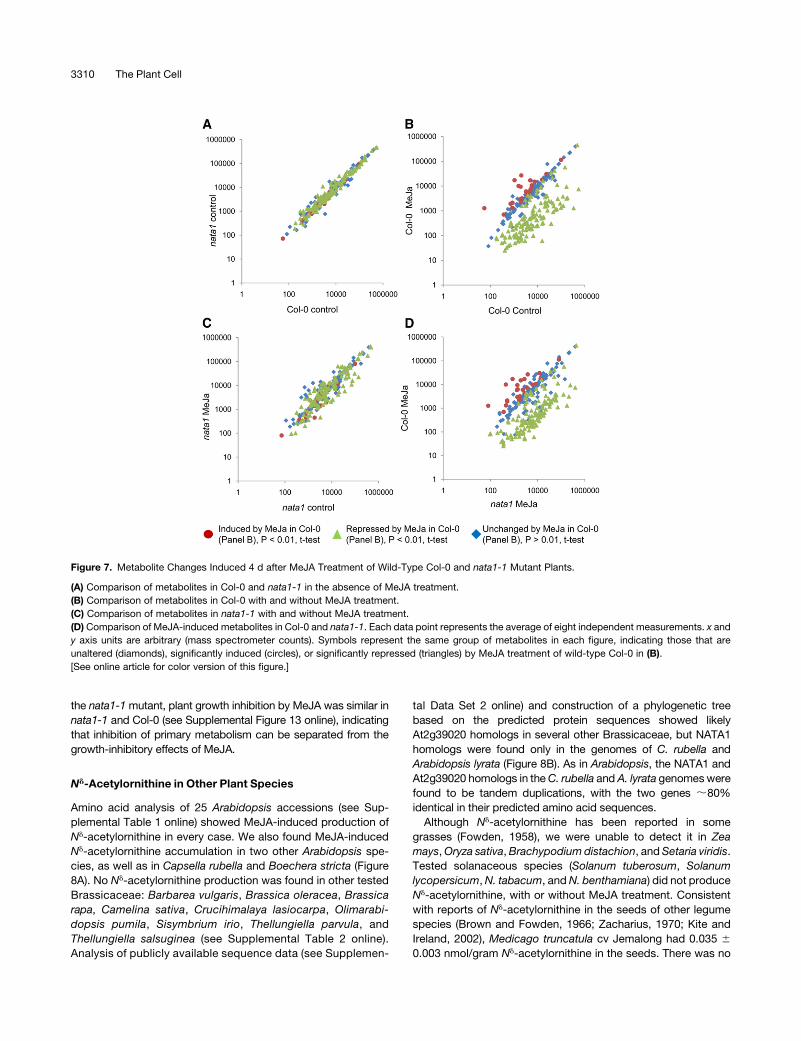
To identify other metabolic changes associated with NATA1
gene expression, we conducted GC-MS metabolite profiling of
wild-type Col-0 and nata1-1, with and without MeJA treatment.
Our assay detected 290 Arabidopsis metabolites, 73 of which
were identified based onmolecular standards (seeSupplemental
Data Set 1 online). In the absence of MeJA treatment, the
metabolite abundance in Col-0 and nata1-1 was similar (Figure
7A; see Supplemental Data Set 1 online), as would be expected
from the very low basal NATA1 expression (Figure 3D). Treating
wild-type Col-0 with MeJA decreased the abundance of 133
metabolites, including free amino acids, sugars, and tricarbox-
ylic acid cycle intermediates (triangles in Figure 7B; see Supple-
mental Data Set 1 online). There were 24 MeJA-induced
metabolites detected in the MS assay (circles in Figure 7B),
none of which had a known identity. By contrast, fewer metab-
olites were significantly altered (35 induced and 32 repressed;
P < 0.01, t test) by MeJA treatment of the nata1-1mutant (Figure
7C; see Supplemental Data Set 1 online). Agmatine, putrescine,
and spermidine were induced byMeJA in the nata1-1mutant but
not in wild-type Col-0 (see Supplemental Figure 11 online). Most
other metabolites that were altered by MeJA in Col-0 were
affected to a lesser extent in nata1-1 (Figure 7D; see Supple-
mental Data Set 1 online). For instance, free Phe was reduced by
90% in Col-0 and 50% in nata1-1, and free Gln was reduced by
98% in Col-0 and 80% in nata1-1 by MeJA treatment. Since free
amino acids as a source of nitrogen are of particular importance
to aphid feeding (Wilson et al., 2010), these were measured in an
independent assay (HPLC fluorescence detection) andwere found
to be significantly less repressed by MeJA in the nata1-1 mutant
than in wild-type Col-0 (see Supplemental Figure 12 online).
Although MeJA-induced metabolite changes were attenuated in
Figure 6. NATA1 Effects on P. rapae and P. xylostella Growth.
(A) NATA1 gene expression 3 d after one neonate caterpillar was placed on a 3-week-old Arabidopsis plant. Gene expression on control plants without
caterpillars was set to 1.
(B) Nd-acetylornithine accumulation 7 d after one neonate caterpillar was placed on a 3-week-old Arabidopsis plant.
(C) P. rapae dry weight 7 d after neonate larvae were placed on wild-type Col-0 or nata1-1.
(D) Pupal weight of P. xylostella that spent their entire larval growth on Col-0 or nata1-1. P values calculated with t tests. *P < 0.05, t test relative to
controls. Numbers in bars indicate sample sizes 6 SE. ND, not detected.
Nd-Acetylornithine, a Defense Metabolite 3309
the nata1-1mutant, plant growth inhibition by MeJA was similar in
nata1-1 and Col-0 (see Supplemental Figure 13 online), indicating
that inhibition of primary metabolism can be separated from the
growth-inhibitory effects of MeJA.
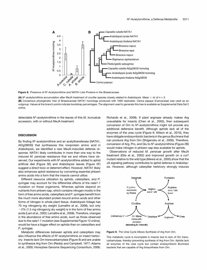
Nd-Acetylornithine in Other Plant Species
Amino acid analysis of 25 Arabidopsis accessions (see Sup-
plemental Table 1 online) showed MeJA-induced production of
Nd-acetylornithine in every case. We also found MeJA-induced
Nd-acetylornithine accumulation in two other Arabidopsis spe-
cies, as well as in Capsella rubella and Boechera stricta (Figure
8A). No Nd-acetylornithine production was found in other tested
Brassicaceae: Barbarea vulgaris, Brassica oleracea, Brassica
rapa, Camelina sativa, Crucihimalaya lasiocarpa, Olimarabi-
dopsis pumila, Sisymbrium irio, Thellungiella parvula, and
Thellungiella salsuginea (see Supplemental Table 2 online).
Analysis of publicly available sequence data (see Supplemen-
tal Data Set 2 online) and construction of a phylogenetic tree
based on the predicted protein sequences showed likely
At2g39020 homologs in several other Brassicaceae, but NATA1
homologs were found only in the genomes of C. rubella and
Arabidopsis lyrata (Figure 8B). As in Arabidopsis, the NATA1 and
At2g39020 homologs in theC. rubella and A. lyrata genomeswere
found to be tandem duplications, with the two genes ;80%
identical in their predicted amino acid sequences.
Although Nd-acetylornithine has been reported in some
grasses (Fowden, 1958), we were unable to detect it in Zea
mays,Oryza sativa,Brachypodium distachion, andSetaria viridis.
Tested solanaceous species (Solanum tuberosum, Solanum
lycopersicum,N. tabacum, andN. benthamiana) did not produce
Nd-acetylornithine, with or without MeJA treatment. Consistent
with reports of Nd-acetylornithine in the seeds of other legume
species (Brown and Fowden, 1966; Zacharius, 1970; Kite and
Ireland, 2002), Medicago truncatula cv Jemalong had 0.035 60.003 nmol/gram Nd-acetylornithine in the seeds. There was no
Figure 7. Metabolite Changes Induced 4 d after MeJA Treatment of Wild-Type Col-0 and nata1-1 Mutant Plants.
(A) Comparison of metabolites in Col-0 and nata1-1 in the absence of MeJA treatment.
(B) Comparison of metabolites in Col-0 with and without MeJA treatment.
(C) Comparison of metabolites in nata1-1 with and without MeJA treatment.
(D) Comparison of MeJA-induced metabolites in Col-0 and nata1-1. Each data point represents the average of eight independent measurements. x and
y axis units are arbitrary (mass spectrometer counts). Symbols represent the same group of metabolites in each figure, indicating those that are
unaltered (diamonds), significantly induced (circles), or significantly repressed (triangles) by MeJA treatment of wild-type Col-0 in (B).
[See online article for color version of this figure.]
3310 The Plant Cell
detectable Nd-acetylornithine in the leaves of this M. truncatula
accession, with or without MeJA treatment.
DISCUSSION
By finding Nd-acetylornithine and an acetyltransferase (NATA1,
At2g39030) that synthesizes this nonprotein amino acid in
Arabidopsis, we identified a new MeJA-inducible defense re-
sponse. NATA1 likely contributes in more than one way to the
induced M. persicae resistance that we and others have ob-
served. Our experiments with Nd-acetylornithine added to aphid
artificial diet (Figure 5E) and Arabidopsis leaves (Figure 5F)
suggest a direct toxic or deterrent effect. However, NATA1 likely
also enhances aphid resistance by converting essential phloem
amino acids into a form that the insects cannot utilize.
Different resource utilization by aphids, caterpillars, and P.
syringae may account for the differential effects of the nata1-1
mutation on these organisms. Whereas aphids depend on
nutrients from phloem sap, which contains nitrogenmostly in the
form of free amino acids, caterpillars andP. syringae benefit from
the much more abundant protein-bound amino acids and other
forms of nitrogen in whole plant tissue. Arabidopsis foliage has
70 mg nitrogen/g dry weight (Lemaıtre et al., 2008), but only
;2% (1.5 mg nitrogen/g dry weight) is in the form of free amino
acids (Lamet al., 2003; Lemaıtre et al., 2008). Therefore, changes
in the abundance of free amino acids, such as those observed
due to the nata1-1mutation (see Supplemental Figure 12 online),
would be have a bigger effect on aphids than on caterpillars and
P. syringae.
Metabolic differences between aphids and caterpillars may
also influence the effects of Nd-acetylornithine on insect herbiv-
ory. Insects lack Orn transcarbamylase (Figure 9) and are unable
to synthesize Arg from Orn (Reddy and Campbell, 1977; Adams
et al., 2000; Honeybee Genome Sequencing Consortium, 2006;
Richards et al., 2008). If plant arginase already makes Arg
unavailable for insects (Chen et al., 2005), then subsequent
conversion of Orn to Nd-acetylornithine might not provide any
additional defensive benefit. Although aphids lack all of the
enzymes of the urea cycle (Figure 9; Wilson et al., 2010), they
have obligate endosymbiotic bacteria in the genusBuchnera that
can produce Arg from Orn (Shigenobu et al., 2000). Therefore,
conversion of Arg, Pro, and Glu to Nd-acetylornithine (Figure 2B)
would make nitrogen in phloem sap less available for aphids.
Observations of reduced M. persicae growth after MeJA
treatment (Ellis et al., 2002) and improved growth on a coi1
mutant relative to the wild type (Mewis et al., 2005) show that the
JA signaling pathway contributes to aphid defense in Arabidop-
sis. However, although caterpillar herbivory strongly induces
Figure 8. Presence of Nd-Acetylornithine and NATA1-Like Proteins in the Brassicaceae.
(A) Nd-acetylornithine accumulation after MeJA treatment of crucifer species closely related to Arabidopsis. Mean 6 SE of n = 3.
(B) Consensus phylogenetic tree of Brassicaceae NATA1 homologs produced with 1000 replicates. Carica papaya (Caricaceae) was used as an
outgroup. Values at the branch points indicate bootstrap percentages. The alignment used to generate this tree is available as Supplemental Data Set 2
online.
Figure 9. The Urea Cycle Allows Synthesis of Arg from Orn.
This metabolic cycle is incomplete in insects due to lack of Orn trans-
carbamylase, thereby preventing synthesis of Arg from Orn. Aphids lack
all enzymes of the urea cycle but contain endosymbiont Buchnera
bacteria that are capable of Arg biosynthesis from Orn.
Nd-Acetylornithine, a Defense Metabolite 3311
JA-related genes, this is not the case for aphid feeding (Bidart-
Bouzat and Kliebenstein, 2011). These differences in JA in-
duction likely cause greater Nd-acetylornithine accumulation in
response to caterpillars (Figure 6B) than aphids (Figure 5D) and
may represent active suppression of JA-regulated defenses by
phloem-feeding insects (Zarate et al., 2007). ABA-regulated
NATA1 expression also causes elevated Nd-acetylornithine ac-
cumulation (Figure 1D). Publicly available microarray data show
overlapping gene expression patterns induced by aphid feeding
and ABA (www.genevestigator.com; Zimmermann et al., 2004),
and heavier aphid infestation of Arabidopsis induces wilting that
could lead to ABA-regulated gene expression changes.
Several prior studies show that coronatine promotes P.
syringae growth on Arabidopsis and tomato (Feys et al., 1994;
Kloek et al., 2001; Zhao et al., 2003; Cui et al., 2005; Laurie-Berry
et al., 2006; Uppalapati et al., 2007, 2008, 2011; Fernandez-
Calvo et al., 2011). Although induction of JA-regulated gene
expression by coronatine likely suppresses SA-mediated plant
defenses, other molecular mechanisms that connect COI1 to
increased P. syringae growth remain unknown. Our observation
of improved P. syringae growth in the nata1-1 mutant back-
ground (Figure 4D) suggests that some of the observed NATA1-
requiring metabolic changes (Figure 7) contribute to pathogen
resistance. However, more direct effects on Orn-related metab-
olism also may be relevant for P. syringae virulence. WIN1
(At1g80600), a predicted Na-acetylornithine transaminase, inter-
acts directly with the P. syringae effector HopW1-1, and WIN1
overexpression prevents elicitation of Arabidopsis defenses by
HopW1-1 and other P. syringae effectors (Lee et al., 2008).
In proceeding from the discovery of Nd-acetylornithine, a
previously unknown Arabidopsis metabolite, to the elucidation
of a biosynthetic pathway and defense-related functions, we
applied several of the excellent genetic and genomic resources
that are available for Arabidopsis. Many recent studies of
Arabidopsis defense mechanisms started with the identification
of defense-related gene expression or mutants to identify new
metabolites and signaling pathways. However, given the well-
annotated Arabidopsis genome and extensive gene expression
data sets, it now may be easier to move from identifying a novel
plant metabolite to the discovery of a previously unknown
enzymatic activity than vice versa. Our results, which have
implications for research on both plant–pathogen and plant–
herbivore interactions, provide new insight into the induction of
biotic stress responses in plants.
METHODS
Plants and Growth Conditions
Seeds of Arabidopsis thaliana accessions (see Supplemental Table
1 online) and other Brassicaceae (see Supplemental Table 2 online)
were obtained from the ABRC (www.Arabidopsis.org), T. Mitchell-Olds
(Duke University, Durham, NC), and J. Jaworski (Danforth Center, St.
Louis, MO). Seeds of other species were supplied by T. Brutnell (Boyce
Thompson Institute, Ithaca, NY; Zea mays, Brachypodium distachion,
and Setaria viridis), M. Kovach (Cornell University, Ithaca, NY; Oryza
sativa), and N. Pumplin (Boyce Thompson Institute, Ithaca, NY;Medicago
truncatula). Seeds of homozygous mutant argah1-1 (SALK_057987;
Alonso et al., 2003) and argah2-1 (SAIL_181_C11; Sessions et al., 2002)
were from C. Todd (University of Saskatchewan, Saskatoon, Canada;
Flores et al., 2008). Homozygous fls2 mutant seeds were supplied by S.
He (Michigan State University, East Lansing, MI; Zeng and He, 2010).
Arabidopsis coi1 (Feys et al., 1994), npr1 (Cao et al., 1994), sid1 (Nawrath
et al., 2002), and pad4 (Glazebrook et al., 1996) mutants were obtained
from F. Ausubel (Massachusetts General Hospital, Boston, MA). The
T-DNA insertion lines GK-256F07 (nata1-1; Rosso et al., 2003) and
SALK_092319 (At2g39020 mutation; Alonso et al., 2003) were obtained
from the ABRC. All plants were grown in Cornell mix (by weight 56% peat
moss, 35% vermiculite, 4% lime, 4% Osmocoat slow-release fertilizer
[Scotts], and 1% Unimix [Peters]) in 203 40-cm nursery flats in Conviron
growth chambers with a photosynthetic photon flux density of 200 mmol
m22 s21 and a 16-h photoperiod at 238C with a 50% relative humidity.
Isolation and Purification of Nd-Acetylornithine
Five-week-old Arabidopsis plants were sprayed with 0.45mMMeJA, and
;620 g leaves were harvested, frozen with liquid nitrogen, and homog-
enized in 1.5 liters of 80% methanol. The extract was concentrated by
evaporation and partitioned with dichloromethane. The aqueous fraction
was passed through DEAE-Sephadex A-25 column to extract the
glucosinolate fraction for a separate experiment. The eluate from the
Sephadex A-25 was then passed through a packed column of Dowex-
50WX8-200 ion-exchange resin. The columnwaswashed repeatedly with
water, and amino acids were eluted with 200 mL 4 N NH4OH followed by
100 mL water. Pooled eluate was lyophilized to leave 237 mg extract,
which was then dissolved in 3 mL triethylamine (TEA) solution (2:2:1 of
ethanol:water:TEA). The extract was then derivatized for 30 min in 1.26
mL phenylisothiocyanate (PITC) solution (980 mL ethanol, 70 mL water, 70
mL TEA, and 140 mL PITC). The reaction mixture was concentrated to
dryness, taken up in 400 mL TEA solution, and concentrated to dryness
again, leaving 680 mg residue. The extract was successively fractionated
with Sephadex-LH-20 and RP18 packed columns and eluted with 95:5
water:methanol. The fraction containing the target compound, retention
time = 29.90min, was tracked by HPLC-DAD (Waters 2695 pump system
and 996 diode array detector) using an RP18 column (Nova-Pak). HPLC
Solvent Awas 140mm sodium acetate with 0.05%TEA (final pH adjusted
to 6.33 with acetic acid). Solvent B was 60% acetonitrile. The gradient
used was as follows: 0 to 10 min, 99% A; 10 to 35 min, linear gradient to
65%A; 35 to 40min, 65%A; 40 to 42min, linear gradient to 100%B; 42 to
44 min, 100% B. Further purification was achieved using a Lichrosphere
column with water and acetonitrile as solvents to produce 1.1 mg of the
target compound. For the HPLC runs (Lichrosphere column: 5 RP-18,
ECAP 250, 4.6-mm inside diameter, 5-mm particle size) the mobile
phases were A, water, and B, 90% acetonitrile, at a flow rate of 1.1 mL
min21 at 238C. Column linear gradients for the samples were as follows: 0
to 1 min 75% A; 1 to 10 min, 60% A; 10 to 20 min 40% A; 20 to 30 min,
25% A; 30 to 35 min, 0% A; 35 to 40 min, 75% A.
The purified compound was analyzed by NMR. The (+ve)-ESI-MS
spectrum showed an abundant molecular ion [M+PITC+Na]+ peak at
332, in addition to 175 [M+H-PITC]+ and 310 [M+H+PITC]+. In (–ve)-ESI-MS
mode, the molecular ion was also detected with a peak [M+PITC-H]2 at
mass-to-charge ratio (m/z) 308 in addition to 173 [M-H-PITC]2. Therefore, a
molecular weight of 174 was assigned to the isolated compound. From the1HNMR, HMBC, and ESI-MS data, the isolated compound was assigned
the structure of Nd-acetylornithine. To further confirm the assigned struc-
ture, the isolated compound was compared with all possible acetylated
products obtained after the treatment of L-Orn with glacial acetic acid and
acetic acid anhydride after PITC derivatization. The isolated compound
coeluted with Nd-acetylornithine (retention time = 29.90 min.). Optical
rotation measurements to determine whether Arabidopsis Nd-acetylorni-
thine has the L or D configuration were performed with a Polarimeter 241
(Perkin-Elmer) at 589 nm at 208C using a sodium lamp.
3312 The Plant Cell
Synthesis of Nd-Acetylornithine
Nd-Acetylornithine was synthesized from L-Orn and 4-nitrophenyl acetate
(Leclerc and Benoiton, 1968). L-Orn monohydrochloride (0.5 g, 5.95
mmol) was dissolved in 5 mL water, and the pH was adjusted to 11 with
2MNaOH. 4-Nitrophenyl acetate (1.07 g, 11.9mmol) was then added and
the mixture was stirred on ice. The mixture was stirred at room temper-
ature for 4 h, concentrated to dryness with a Rotavap evaporator (Buchi),
dissolved in 2 mL water, and partitioned repeatedly with ethyl acetate.
The aqueous fraction containing the target compound was concentrated
to dryness and redissolved in water followed by treatment with 0.1 M HCl
to pH 4, until the mixture became colorless. Ethanol was added to the
mixture and the precipitate was filtered with Buchner funnel to give a
white precipitate (0.40 g). The precipitate was washed repeatedly with
ethanol. The identity of synthesized Nd-acetylornithine was confirmed by
1D- and 2D-NMR and ESI-MS. The corresponding ESI-MS spectral data
are as follows: ESI-MS (negative,m/z, relative abundance): [M-H]2 (m/z);
[173] (100).
GC-MS (Varian 1200L; Agilent Technologies) analysis of N-methyl-N-
(trimethylsilyl)trifluoroacetamide + 1% trichloromethylsilane derivatized
synthetic Nd-acetylornithine (retention time = 23.5 min [M+2TMS] and
retention time = 23.95 min [M+1TMS]) was identical to the induced
endogenous Arabidopsis Nd-acetylornithine. Coelution of the synthetic
and the naturally occurring compounds both on HPLC and GC further
confirmed its identity. The 1H-NMR spectral data of the synthesized Nd-
acetylornithine were identical to those of earlier reports (Wohlfarth et al.,
1993). The 1H-NMR (D2O) spectrum of Nd-acetylornithine established the
following: 1H-NMR (500 MHz, D2O) d: 3.59 (1H, t, J = 6 Hz), 3.09 to 3.02
(2H, m), 1.82 (3H, s), 1.76 to 1.66 (2H, m), 1.49 to 1.36 (2H, m).The 13C-
NMR extracted from the HMBC data revealed seven carbon atoms:
174.33, 174.08, 54.57, 38.63, 27.83, 24.26, and 22.02. GC-MS data of
Nd-acetylornithine with 2-TMS derivatives gave an EI spectrum with m/z
(%) 318 [1.7, M+2TMSi], 201 (22, M–CO2TMSi), 112 (100, M-C2NH4O2-
2TMSi), 75(76), and 73(55).
Amino Acid Assays
For analysis of leaf amino acids, 100mg of plant tissuewas frozen in liquid
nitrogen in 2-mL microcentrifuge tubes and ground to fine powder with
3-mm steel beads using a Harbil model 5G-HD paint shaker. Ground
tissue was taken up in 20 mM HCl (10 mL/mg of tissue), the extracts were
centrifuged at 3800g for 20 min at 238C, and the supernatant was saved
for analysis. Phloem sap was collected from rosettes of 3-week-old Col-0
plants sprayed with water or 0.45 mM MeJA treatment. Roots were cut
from the rosettes, leaving 1mmof the shoot tissue, and put in 100mL of 15
mM EDTA solution, pH 7.7, for 4 h in the dark with high humidity. Fifteen
samples were pooled for each replicate of the experiment. For analysis of
Nd-acetylornithine uptake, nata1-1 leaf petioles were immersed in Nd-
acetylornithine solutions in a 1.5-mLmicrocentrifuge tube for 24 h prior to
tissue harvest. The petiole and other portions of the leaf that were
immersed in the Nd-acetylornithine solution were not used for the amino
acid analysis. For themeasurement ofNd-acetylornithine in aphid bodies,
aphids were collected from Col-0 plants treated with water or MeJA and
transferred into a 2 mL centrifuge tube containing three 3-mm steel balls.
The aphids were homogenized and extracted with 20 mM HCl. For aphid
honeydew collection, ;50 aphids were placed on artificial diet with and
without Nd-acetylornithine for 4 d, and honeydew was collected on
aluminum foil. The aluminum foil was washed with 80% methanol to
extract amino acids. Themethanolic extract was concentrated to dryness
a SpeedVac rotary evaporator and then redissolved in 40 mL 20 mM HCl.
Amino acids were derivatized with 6-aminoquinolyl-N-hydroxysuccini-
midylcarbamate using an AccQ-Fluor reagent kit (Waters). For derivati-
zation, 5mL extracts weremixedwith 35mL borate buffer and the reaction
was initiated by the addition of 10 mL 6-aminoquinolyl-N-hydroxysucci-
nimidyl carbamate reagent, followed by immediatemixing and incubation
for10 min at 558C. Ten microliters of each sample were injected onto a
Nova-Pak C18 column using a Waters 2695 pump system, and the data
were recorded usingWaters’ Empower Software. L-Norleucine was used
as an internal standard. Eluted amino acid derivatives were detected
using a Waters model 2475 fluorescence detector with an excitation
wavelength of 250 nm and an emission wavelength of 395 nm. Solvent A
(containing sodium acetate and TEA at pH 5.05) was purchased premixed
from Waters; Solvent B was acetonitrile:water (60:40). The gradient used
was 0 to 0.01min, 100%A; 0.01 to 0.5 min, linear gradient to 3%B; 0.5 to
12min, linear gradient to 5%B; 12 to 15min, linear gradient to 8%B; 15 to
45min, 35%B; 45 to 49min, linear gradient to 35%B; 50 to 60min, 100%
B. Flow rate was 1.0 mL min21.
Glucosinolate Assays
Leaves of 3-week old Col-0 wild-type nata1-1 Arabidopsis, with or
without MeJA treatment, were collected and lyophilized. Extraction of
plant tissue and preparation of desulphoglucosinolates was done as
described previously (Kim et al., 2004; Barth and Jander, 2006). Desul-
phoglucosinolates were separated using a Waters 2695 HPLC and
detected using a Waters 2996 photodiode array detector. For HPLC
separation, the mobile phases were A, water, and B, 90% acetonitrile,
with a flow rate of 1 mL/min at 238C. Column linear gradients for samples
were: 0 to 1 min, 98%A; 1 to 6 min 94%A; 6 to 8 min, 92%A; 8 to 16min,
77% A; 16 to 20 min, 60%A; 20 to 25 min, 0% A; 25 to 27 min hold 0% A;
27 to 28 min, 98% A; 28 to 37 min, 98% A.
MeJA, SA, ACC, ABA, and Coronatine Induction
The leaves of 4-week-old Arabidopsis plants were sprayed with aqueous
solution containing 0.01% (v/v) Tween 20, supplemented with each of 0.45
mMSA, 0.45mMMeJA, 0.45mMACC, and 0.1mMABA, a combination of
both SA and MeJA, 1 mM coronatine, or 10 mM coronatine. Control plants
were treated with 0.01% Tween 20 only. Controls for MeJA treatment also
contained 0.03% acetone. Plants were covered and harvested 4 d after
elicitation (or at multiple times for a time course of induction) and imme-
diately frozen in liquid nitrogen. Pulverized plants were extracted with 20
mM HCl, and amino acids were analyzed as described above.
For time-course experiments, 2-week-old Arabidopsis plants were
treated with 0.45 mM MeJA, and leaves were harvested after 0 to 15 d.
Leaves were frozen in liquid nitrogen and stored at 2808C before amino
acid analysis as described above. For a separate experiment, the whole
aerial parts were cut just above the root and weighed.
Stable Isotope Labeling Experiments
Petioles of a detached Col-0 leaves from 4-week-old plants were inserted
into 1.5 mL microcentrifuge tubes containing 5 mM labeled amino acid
([13C5]Glu, [13C6]Arg, and [13C5]Pro) in water. In the case of Glu, 0.01%
NaOH was used to aid solubility. Control leaves were treated with water
only and allowed to stand for 12 h. After 12 h, leaveswere sprayedwith 0.45
mMMeJA. Leaf samples were covered and allowed to stand for 3 d. Three
leaves receiving the same treatment were combined as one replicate for
derivatization and GC-MS analysis of amino acids, as described above.
Amino acid analysis byGC-MSwas performed as described previously
(Joshi and Jander, 2009) with minor modifications. Single leaves har-
vested from Arabidopsis Col-0 wild type and nata1-1mutant at the same
developmental stages were used. Leaves were frozen in liquid nitrogen in
2-mL tubes and ground to fine powder with 3-mm steel beads using a
Harbil model 5G-HD paint shaker. Ground tissue was taken up in 20 mM
HCl (1 mL per leaf tissue), the extracts were centrifuged at 4000g for 20
min at 238C, and the supernatant leaf acid extracts were applied to
Dowex-50 columns (0.6 mL). These were eluted individually with 6 M
Nd-Acetylornithine, a Defense Metabolite 3313
NH4OH (1 mL). Fractions were combined, and solutions were concen-
trated to 200 mL with a rotary evaporator. These extracts were then
completely dried under nitrogen flow, with heating at 708C. Methoxy-
amine hydrochloride in anhydrous pyridine (50 mL, 15 mg mL21) was
added to the residue and the sample was heated at 308C for 90 min. A
volume of 90 mL N-methyl-N-(trimethylsilyl)trifluoroacetamide + 1%
trimethylchlorosilane was then added, and the sample was heated for a
further 30 min at 708C. GC-MS analysis was performed using a Varian
1200L GC-MS with a DB-17 ms capillary column. Spectra of known
amino acids were assigned by reference to an in-house spectral library
and the NIST library.
Confirmation of T-DNA Insertions and Expression of Transgenes
Homozygous T-DNA insertions inNATA1 (GK-256F07; Rosso et al., 2003)
and At2g39020 (Salk_092319; Alonso et al., 2003) were confirmed by
PCR using a previously outlined strategy using three primers to amplify
both the insertion junction and the genomic DNA without an insertion
(http://signal.salk.edu/tdnaprimers.2.html) and primers described in Sup-
plemental Table 3 online. PCR reactions were run using Taq DNA
polymerase (Fisher) under the following conditions: 958C for 5 min, 40
cycles of 958C for 1 min, 558C for 1 min and 728C for 1 min, followed by
728C for 10 min.
For transgenic expression,NATA1 and At2g39020 were cloned behind
the cauliflower mosaic virus 35S promoter in the T-DNA binary vector
pMDC32 (Curtis and Grossniklaus, 2003) using the Gateway recombina-
tion system (Invitrogen), and inserts were confirmed by DNA sequencing.
Stable Arabidopsis p35S:NATA1 transgenics were made by dipping
transformation with Agrobacterium tumefaciens strain GV3101 (Clough
and Bent, 1998). Seeds collected from transformed Arabidopsis plants
were selected on agar containing 40 mg mL21 hygromycin, and trans-
gene expression was confirmed by qRT-PCR. The same T-DNA plasmids
in GV3010were used for transient expression ofNATA1 and At2g39020 in
tobacco (Nicotiana benthamiana and Nicotiana tabacum) by Agrobacte-
rium infiltration (Voinnet et al., 1998). Agrobacterium was cultured over-
night at 308C in Luria-Bertani (LB) broth supplemented with 50 mg mL21
rifampicin, 25 mg mL21 gentamycin, and 100 mg mL21 kanamycin.
Bacterial cultures were centrifuged at 8000g, washed three times with
sterile water, and resuspended in sterile water to 0.23 108 colony-forming
units/mL. Cultures were mixed with Agrobacterium (0.1 3 108/mL)
carrying T-DNA constructs expressing the turnip crinkle virus capsid
protein (P38; Thomas et al., 2003) to reduce expression silencing. N.
benthamiana or N. tabacum leaves (four leaves per plant) were infiltrated
with 1 mL bacterial solution using a 1 mL syringe. Excess bacterial
solution was wiped off with paper towel. Three days after infiltration, leaf
plugs (8-mm diameter) were collected to confirm expression of the
transgenes by qRT-PCR.
qRT-PCR
Total RNA was extracted from frozen tissue samples using the SV Total
RNA Isolation system with on-column DNase treatment (Promega). RNA
integrity was verified using a 1.2% formaldehyde agarose gel (Sambrook
et al., 1989). Transcript abundance of NATA1 was analyzed with qRT-
PCR, using eEF1-a (elongation factor 1-a, At5g60390) as an internal
standard. eEF1-a was identified from publicly available microarray data
as constitutively expressed before and after herbivory, and stable ex-
pression was verified across samples using qRT-PCR. After RNA extrac-
tion and DNase treatment, 1mg of total RNAwas reverse transcribed with
SMART MMLV reverse transcriptase (Clonetech) using oligo-dT12-18 as a
primer. Gene-specific primers used for qRT-PCR were designed using
Primer-Blast (http://www.ncbi.nlm.nih.gov/tools/primer-blast/) with the
following criteria: temperature of 608C, PCR amplicon lengths of 90 to 150
bp, primer sequences with lengths of 18 to 24 nucleotides with an
optimum at 21 nucleotides, and GC contents of 40 to 60%. Primers for
amplifying NATA1 and eEF1-a are described in Supplemental Table 3
online. Reactions were performed using 5 mL of the SYBR Green PCR
master mix (Applied Biosystems), with 800 nM of primer, in the 7900HT
instrument (Applied Biosystems). The PCR was initiated by incubation at
958C for 10 min to activate the enzyme. Then the following cycle was
repeated 40 times: 958C for 15 s, 608C for 15 s, and 728C for 15 s. The CT
values were quantified and analyzed according to the standard curve
method.
GC-MSMetabolomics
For metabolomic analysis, 3-week-old wild-type and nata1-1 mutant
plants treated with water or 0.45 mMMeJA. After 4 d, 6.5-mm disks was
taken from the central part of the first and second adult leaves of each
plant. The two disks were combined and stored into a 2-mL micro-
centrifuge tubes put on ice. One milliliter of prechilled extraction buffer
(acetonitrile:isopropanol:water [3:3:2]) and one metal ball were added to
the sample. Plant tissue was macerated briefly for 30 s using a Harbil
model 5G-HD paint shaker. Extracts were centrifuged at 4000g for 2 min
at 48C. Supernatants from each sample (400 mL) were transferred into
new 2-mL tubes and stored at 2808C until automated derivatization and
GC-TOF-MS analysis at the UC Davis Genome Center Metabolomics
Facility (http://metabolomics-core.ucdavis.edu/; Fiehn et al., 2005). Me-
tabolite identity was determined by comparing retention time andmass to
the UC Davis Genome Center Metabolomics Facility metabolites data-
base (http://fiehnlab.ucdavis.edu/Metabolite-Library-2007/; Fiehn et al.,
2005). This library contains reference spectra for 713 knownmetabolites,
generated by the analysis of purified reference compounds. Metabolites
not contained within this library are listed as unknown or unidentified
metabolites. After analysis, samples were subject to quality control by
further analysis of samples with internal standard values >3 s from the
experimental mean within a given experiment, suggesting faulty deriva-
tization or GC-TOF analysis of that specific sample.
Bacterial Bioassays
Pseudomonas syringae strains were supplied by H.G. Kang (Boyce
Thompson Institute, Ithaca, NY; DC3000AvrRpt2 andDC3000AvrRpm1),
C. Danna (Massachusetts General Hospital, Boston, MA; Psm4326 and
Psm4326Cor-), andN. Clay (Yale University, NewHaven, CT; PstDC3000
and PstDC3000 Cor-). For assays of Nd-acetylornithine production in
Arabidopsis, P. syringae was cultured at 308C in LB broth. Bacteria were
centrifuged and resuspended in water at an OD600 of 0.2. Bacterial
suspensions containing 0.01% Tween 20 were sprayed on Arabidopsis
plants. Control plants are sprayed with water containing 0.01%Tween 20
only. After 4 d, infected leaves were collected for amino acid analysis as
described above.
For in vitro growth experiments,P. syringae strain DC3000was cultured
at 308C in modified mannitol-glutamate medium (Kean et al., 1970; per
liter: 10 g mannitol, 2 g L21 Glu, 0.5 g KH2PO4, 0.2 g NaCl, 0.2 g MgSO4-
7H2O; pH adjusted to 7.0 with 3 N NaOH prior to autoclaving). To test
growth with other amino acids as nitrogen sources, Glu was replaced
successively with 2 g L21 Arg, Pro, Orn, Na-acetylornithine, and Nd-
acetylornithine. Cultures were inoculated by diluting a fresh overnight
culture of P. syringae 1:100 (50 mL into 5 mL). OD600 was measured at 0,
14, 48, and 72 h after treatment. For growth inhibition experiments,
cultures were grown in minimal medium with 2 g L21 Glu and 0, 10, 100,
250, or 500 mM Nd-acetylornithine.
For in planta growth experiments, P. syringae PstDC3000 was cultured
at 308C in LB broth supplemented with 50 mg mL21 rifampicin, centri-
fuged, resuspended in sterile water, and diluted to a concentration of 105
colony-forming units/mL. Col-0 wild-type and nata1-1 leaves were
3314 The Plant Cell
infiltrated with 0.2 mL bacterial solution using a 1-mL syringe. Excess
bacterial solution was wiped off with paper towel. Leaf plugs (8-mm
diameter) were collected at 0, 2, and 4 d after infiltration. Leaf discs were
floated in 1 mL sterile water with 0.01% Tween 20 on a shaker for;2 h.
Ten-microliter samples of a bacterial dilution series were spotted on an
LB agar plate containing 50 mg mL21 rifampicin and incubated at 308C.
Bacterial colonies were counted after 2 d, and the concentration of
bacteria in the leaf plugs was calculated.
Caterpillar Bioassays
The Pieris rapaewere from a colony maintained by the Jander laboratory,
which is descended from 20 adult insects collected in the wild on the
Cornell University campus in July, 2008. Plutella xylostella eggs were
obtained from Benzon Research. Neonate caterpillars were confined on
the leaves of 3-week-old wild-type and nata1-1 plants using mesh-
covered cups. P. rapae were allowed to feed on plants for 7 d before
harvesting. P. xylostella were harvested after pupation. Caterpillars and
pupae were lyophilized and dry weight was determined using a precision
balance (Sartorius). Mutant and wild-type leaves infested by caterpillar
were also collected for amino acid analysis as described above.
Aphid Bioassays
Artificial diet forMyzus persicae fecundity assays consisted of Suc (440mM)
and 20 amino acids (Ala, 10mM; Arg, 16mM; Asn, 20mM; Asp, 10mM;Cys,
3.3 mM; Glu, 10mM; Gln, 10mM; Gly, 10 mM; His, 10mM; Ile, 6 mM; Leu, 6
mM;Lys, 10mM;Met, 5mM;Phe, 3mM;Pro,7mM;Ser, 10mM;Thr, 12mM;
Trp, 4 mM; Tyr, 2 mM; Val, 7 mM). Zero, 0.1, 0.5, 5, or 10 mM Nd-
acetylornithine was added to the diet. For experiments with a single amino
acid as the nitrogen source, Orn, Glu, and Nd-acetylornithine were added at
10 mM concentration to 440 mM Suc. One wingless adult aphid was placed
in a 30-mL plastic cup that was covered with a Parafilm sachet containing
100 mL of the liquid diet. Aphid nymphs were counted after 4 d.
M. persicae reproduction was compared on Arabidopsis Col-0 wild
type and nata1-1, with and without MeJA treatment. Plants were covered
after MeJA application for 48 h. The cover was then removed 48 h prior to
the caging aphids on Arabidopsis flower stalks or rosette leaves. Plant
trays with and without MeJA were separated and covered to prevent
volatiles induced byMeJA from eliciting defenses in control plants. Aphid
progeny production was recorded after 7 d. ForN. tabacum experiments,
aphids were caged on individual leaves in the area where Agrobacterium
constructs had been infiltrated 3 d previously, and progeny production
was assessed 7 d later.
To assess effects of exogenously added Nd-acetylornithine, petioles
frommature leaves of 4-week-old nata1-1 plants were inserted in 1.5-mL
microcentrifuge tubes containing 0, 0.1, or 0.5 mM Nd-acetylornithine in
water. After 24 h, one aphid was placed on each individual leaf, and aphid
progeny production was recorded after 4 d.
Sequence Alignment and Phylogenetic Tree Generation
Arabidopsis NATA1 homologs in the Brassicaceae (Arabidopsis lyrata,
Brassica rapa, Brassica napus, Raphanus raphanistrum, and Capsella
rubella) were obtained by comparison to genomic and EST data in
GenBank. The closest NATA1 homolog of Carica papaya in the Caricaeae,
a related family in the Brassicales, was used as an outgroup. Multiple
sequence alignment of translated protein sequences was performed with
ClustalW (Larkin et al., 2007). Programs in the PHYLIP version 3.6 software
package (Felsenstein, 2005) were used to create a phylogenetic tree. The
SEQBOOT function was used to make 1000 bootstrapped data sets.
PROMLK was used to construct rooted phylogenetic trees, using the
parameters search for the best tree, Jones-Taylor-Thornton probability
model, one category of substitution rates, constant rate variation among
sites, no global rearrangement, no weighted sites, and randomization of
input sequences. A rooted consensus tree was made with CONSENSE
using the majority rule (extended) parameter.
Accession Numbers
Sequence data from this article can be found in the Arabidopsis Genome
Initiative or GenBank/EMBL databases under the following accession num-
bers: At2g39030 (NATA1), At2g39020, At4g08900 (ARGAH1), At4g08870
(ARGAH2), At3g30775 (ProDH1), At5g38710 (ProDH2), At2g39800 (P5CS1),
At3g55610 (P5CS2), At5g46180 (OAT), At1g80600 (WIN1), At5g46330
(FLS2), At4g39030 (SID1), At1g64280 (NPR1), and At2g39940 (COI1). Na-
tional Center for Biotechnology Information accession numbers for the
sequences used for the phylogenetic analysis are as follows: Arabidopsis
(NP_565898 andNP_181435),A. lyrata (XP_002879780 and XP_002879781),
B. rapa (EE421453 and EX097874), B. napus (EE409067), R. raphanistrum
(FD557329), and C. papaya (EX280263). C. rubella sequences were kindly
supplied by Y. Guo and D. Weigel in advance of publication (C. rubella raw
sequence data can be found at http://www.ncbi.nlm.nih.gov/bioproject/
13878).
Supplemental Data
The following materials are available in the online version of this article.
Supplemental Figure 1. Identification of Nd-Acetylornithine by MS
and NMR.
Supplemental Figure 2. Nd-Acetylornithine Is Distinct from the
Common Plant Metabolite Na-Acetylornithine.
Supplemental Figure 3. Nd-Acetylornithine Accumulation in Arabi-
dopsis Foliage after Elicitation with Abscisic Acid.
Supplemental Figure 4. Pathways for Incorporation of [U-13C]Arg,
[U-13C]Pro, and [U-13C]Glu into Nd-Acetylornithine.
Supplemental Figure 5. Accumulation of Free Amino Acids in Foliage
of nata-1 Mutant and nata1-1 p35S:NATA1 Plants.
Supplemental Figure 6. Nd-Acetylornithine Accumulation in fls2
Mutant Arabidopsis after P. syringae Treatment.
Supplemental Figure 7. Abundance of Transport Amino Acids in
Phloem Exudates after MeJA Treatment.
Supplemental Figure 8. Free Amino Acids in nata1-1 Mutant Leaves
with and without Added Nd-Acetylornithine.
Supplemental Figure 9. Aphid Reproduction on an Artificial Diet with
a Single Amino Acid as the Nitrogen Source.
Supplemental Figure 10. Glucosinolate Accumulation in Col-0 and
nata1-1, with and without MeJA Treatment.
Supplemental Figure 11. Polyamine Accumulation in Col-0 and
nata1-1, with and without MeJA Treatment.
Supplemental Figure 12. Free Amino Acids in Control and MeJA-
Induced Col-0 Wild Type and nata1-1 Mutants.
Supplemental Figure 13. Growth of Col-0 Wild Type and nata1-1,
with and without MeJA Treatment.
Supplemental Table 1. Arabidopsis Isolates That Show MeJA-
Induced Production of Nd-Acetylornithine.
Supplemental Table 2. Crucifers That Were Tested for the Presence
of Nd-Acetylornithine.
Supplemental Table 3. PCR Primers Used in This Study.
Supplemental Data Set 1. Metabolite Profiling of Col-0 Wild-Type
and nata1-1 Mutant Leaves, with and without MeJA Treatment.
Nd-Acetylornithine, a Defense Metabolite 3315
Supplemental Data Set 2. ClustalW Sequence Alignment of NATA1
Homologs in the Brassicaceae.
ACKNOWLEDGMENTS
We thank T. Mitchell-Olds, N. Pumplin, J. Jaworski, T. Brutnell, M.
Kovach, C. Todd, H. G. Kang, C. Danna, S. He, F. Ausubel, and N. Clay for
seeds and bacterial strains, Y. Guo and D. Weigel for providing C. rubella
genome sequence data (U.S. Department of Energy Joint Genome
Institute, supported by the Office of Science of the U.S. Department of
Energy under Contract DE-AC02-05CH11231), F. Schroeder for assis-
tance in identifying Nd-acetylornithine, and H.G. Kang for advice with P.
syringae assays. This research was funded through National Science
Foundation Grant IOS-0718733 and National Institutes of Health Grant
3R37GM048707-17S1 to G.J. as well as National Science Foundation
Grants IOS-1021861 and DBI-0820580 to D.J.K.
AUTHOR CONTRIBUTIONS
A.M.A. andG.J. designed the research. A.M.A., C.L.S., M.D.V., J.H.K., V.J.,
B.L., C.J., J.D., andG.J. performed research.A.M.A., C.L.C.,M.D.V., J.H.K.,
V.J., D.J.K., and G.J. analyzed data. A.M.A. and G.J. wrote the article.
Received July 14, 2011; revised August 19, 2011; accepted August 26,
2011; published September 13, 2011.
REFERENCES
Adams, M.D., et al. (2000). The genome sequence of Drosophila
melanogaster. Science 287: 2185–2195.
Agrawal, A.A. (1998). Induced responses to herbivory and increased
plant performance. Science 279: 1201–1202.
Agrawal, A.A. (2000). Overcompensation of plants in response to
herbivory and the by-product benefits of mutualism. Trends Plant
Sci. 5: 309–313.
Alonso, J.M., et al. (2003). Genome-wide insertional mutagenesis of
Arabidopsis thaliana. Science 301: 653–657.
Barth, C., and Jander, G. (2006). Arabidopsis myrosinases TGG1 and
TGG2 have redundant function in glucosinolate breakdown and insect
defense. Plant J. 46: 549–562.
Bednarek, P., Schneider, B., Svatos, A., Oldham, N.J., and Hahlbrock,
K. (2005). Structural complexity, differential response to infection, and
tissue specificity of indolic and phenylpropanoid secondary metabolism
in Arabidopsis roots. Plant Physiol. 138: 1058–1070.
Bell, E.A. (2003). Nonprotein amino acids of plants: significance in med-
icine, nutrition, and agriculture. J. Agric. Food Chem. 51: 2854–2865.
Bidart-Bouzat, M.G., and Kliebenstein, D. (May 31, 2011). An eco-
logical genomic approach challenging the paradigm of differential
plant responses to specialist versus generalist insect herbivores.
Oecologia http://dx.doi.org/10.1007/s00442-011-2015-z.
Bino, R.J., et al. (2004). Potential of metabolomics as a functional
genomics tool. Trends Plant Sci. 9: 418–425.
Brooks, D.M., Hernandez-Guzman, G., Kloek, A.P., Alarcon-Chaidez,
F., Sreedharan, A., Rangaswamy, V., Penaloza-Vazquez, A., Bender,
C.L., and Kunkel, B.N. (2004). Identification and characterization of a
well-defined series of coronatine biosynthetic mutants of Pseudomonas
syringae pv. tomato DC3000. Mol. Plant Microbe Interact. 17: 162–174.
Brown, D.H., and Fowden, L. (1966). Characterization of d-acetyl-L-
ornithine isolated from Onobrychis vicifolia Scop. Phytochemistry 5:
881–886.
Brownfield, D.L., Todd, C.D., and Deyholos, M.K. (2008). Analysis of
Arabidopsis arginase gene transcription patterns indicates specific
biological functions for recently diverged paralogs. Plant Mol. Biol. 67:
429–440.
Cao, H., Bowling, S.A., Gordon, A.S., and Dong, X. (1994). Charac-
terization of an Arabidopsis mutant that Is nonresponsive to inducers
of systemic acquired resistance. Plant Cell 6: 1583–1592.
Century, K.S., Holub, E.B., and Staskawicz, B.J. (1995). NDR1, a
locus of Arabidopsis thaliana that is required for disease resistance to
both a bacterial and a fungal pathogen. Proc. Natl. Acad. Sci. USA 92:
6597–6601.
Chen, H., Wilkerson, C.G., Kuchar, J.A., Phinney, B.S., and Howe,
G.A. (2005). Jasmonate-inducible plant enzymes degrade essential
amino acids in the herbivore midgut. Proc. Natl. Acad. Sci. USA 102:
19237–19242.
Chini, A., Fonseca, S., Fernandez, G., Adie, B., Chico, J.M., Lorenzo,
O., Garcıa-Casado, G., Lopez-Vidriero, I., Lozano, F.M., Ponce,
M.R., Micol, J.L., and Solano, R. (2007). The JAZ family of repressors
is the missing link in jasmonate signalling. Nature 448: 666–671.
Clay, N.K., Adio, A.M., Denoux, C., Jander, G., and Ausubel, F.M.
(2009). Glucosinolate metabolites required for an Arabidopsis innate
immune response. Science 323: 95–101.
Clough, S.J., and Bent, A.F. (1998). Floral dip: a simplified method for
Agrobacterium-mediated transformation of Arabidopsis thaliana. Plant
J. 16: 735–743.
Cui, J., Bahrami, A.K., Pringle, E.G., Hernandez-Guzman, G.,
Bender, C.L., Pierce, N.E., and Ausubel, F.M. (2005). Pseudomonas
syringae manipulates systemic plant defenses against pathogens and
herbivores. Proc. Natl. Acad. Sci. USA 102: 1791–1796.
Curtis, M.D., and Grossniklaus, U. (2003). A gateway cloning vector
set for high-throughput functional analysis of genes in planta. Plant
Physiol. 133: 462–469.
D’Auria, J.C., and Gershenzon, J. (2005). The secondary metabolism
of Arabidopsis thaliana: Growing like a weed. Curr. Opin. Plant Biol. 8:
308–316.
De Vos, M., and Jander, G. (2009). Myzus persicae (green peach aphid)
salivary components induce defence responses in Arabidopsis thali-
ana. Plant Cell Environ. 32: 1548–1560.
Dixon, R.A. (2001). Natural products and plant disease resistance.
Nature 411: 843–847.
Ellis, C., Karafyllidis, I., and Turner, J.G. (2002). Constitutive activation
of jasmonate signaling in an Arabidopsis mutant correlates with en-
hanced resistance to Erysiphe cichoracearum, Pseudomonas syringae,
and Myzus persicae. Mol. Plant Microbe Interact. 15: 1025–1030.
Fan, J., Crooks, C., Creissen, G., Hill, L., Fairhurst, S., Doerner, P.,
and Lamb, C. (2011). Pseudomonas sax genes overcome aliphatic
isothiocyanate-mediated non-host resistance in Arabidopsis. Science
331: 1185–1188.
Felsenstein, J. (2005). PHYLIP (Phylogeny Inference Package) Version
3.6. (Seattle, WA: University of Washington).
Fernandez-Calvo, P., et al. (2011). The Arabidopsis bHLH transcription
factors MYC3 and MYC4 are targets of JAZ repressors and act
additively with MYC2 in the activation of jasmonate responses. Plant
Cell 23: 701–715.
Feys, B., Benedetti, C.E., Penfold, C.N., and Turner, J.G. (1994).
Arabidopsis mutants selected for resistance to the phytotoxin coro-
natine are male sterile, insensitive to methyl jasmonate, and resistant
to a bacterial pathogen. Plant Cell 6: 751–759.
Fiehn, O., Wohlgemuth, G., and Scholz, M. (2005). Setup and anno-
tation of metabolomics experiments by integrating biological and
mass spectrometric metadata. Lect. Not. Comp. Sci. 3615: 224–239.
Flores, T., Todd, C.D., Tovar-Mendez, A., Dhanoa, P.K., Correa-
Aragunde, N., Hoyos, M.E., Brownfield, D.M., Mullen, R.T., Lamattina,
3316 The Plant Cell
L., and Polacco, J.C. (2008). Arginase-negative mutants of Arabidopsis
exhibit increased nitric oxide signaling in root development. Plant Physiol.
147: 1936–1946.
Fowden, L. (1958). d-Acetylornithine: A constituent of some common
grasses. Nature 182: 406–407.
Fowden, L. (2001). Plant amino acid research in retrospect: From
Chinball to Singh. Amino Acids 20: 217–224.
Glazebrook, J., Rogers, E.E., and Ausubel, F.M. (1996). Isolation of
Arabidopsis mutants with enhanced disease susceptibility by direct
screening. Genetics 143: 973–982.
Halkier, B.A., and Gershenzon, J. (2006). Biology and biochemistry of
glucosinolates. Annu. Rev. Plant Biol. 57: 303–333.
Honeybee Genome Sequencing Consortium (2006). Insights into
social insects from the genome of the honeybee Apis mellifera.
Nature 443: 931–949.
Howe, G.A., and Jander, G. (2008). Plant immunity to insect herbivores.
Annu. Rev. Plant Biol. 59: 41–66.
Joshi, V., and Jander, G. (2009). Arabidopsis methionine gamma-lyase
is regulated according to isoleucine biosynthesis needs but plays a
subordinate role to threonine deaminase. Plant Physiol. 151: 367–378.
Kean, P.J., Kerr, A., and New, P.B. (1970). Crown gall of stone fruit: II,
Identification and nomenclature of Agrobacterium isolates. Aust. J.
Biol. Sci. 23: 585–595.
Kessler, A., and Baldwin, I.T. (2002). Plant responses to insect her-
bivory: The emerging molecular analysis. Annu. Rev. Plant Biol. 53:
299–328.
Kim, J.H., Durrett, T.P., Last, R.L., and Jander, G. (2004). Character-
ization of the Arabidopsis TU8 glucosinolate mutation, an allele of
TERMINAL FLOWER2. Plant Mol. Biol. 54: 671–682.
Kim, J.H., and Jander, G. (2007). Myzus persicae (green peach aphid)
feeding on Arabidopsis induces the formation of a deterrent indole
glucosinolate. Plant J. 49: 1008–1019.
Kite, G.C., and Ireland, H. (2002). Non-protein amino acids of Bocoa
(Leguminosae; Papilionoideae). Phytochemistry 59: 163–168.
Kloek, A.P., Verbsky, M.L., Sharma, S.B., Schoelz, J.E., Vogel, J.,
Klessig, D.F., and Kunkel, B.N. (2001). Resistance to Pseudomonas
syringae conferred by an Arabidopsis thaliana coronatine-insensitive
(coi1) mutation occurs through two distinct mechanisms. Plant J. 26:
509–522.
Lam, H.M., Wong, P., Chan, H.K., Yam, K.M., Chen, L., Chow, C.M.,
and Coruzzi, G.M. (2003). Overexpression of the ASN1 gene en-
hances nitrogen status in seeds of Arabidopsis. Plant Physiol. 132:
926–935.
Larkin, M.A., et al. (2007). Clustal W and Clustal X version 2.0.
Bioinformatics 23: 2947–2948.
Laurie-Berry, N., Joardar, V., Street, I.H., and Kunkel, B.N. (2006).
The Arabidopsis thaliana JASMONATE INSENSITIVE 1 gene is required
for suppression of salicylic acid-dependent defenses during infection by
Pseudomonas syringae. Mol. Plant Microbe Interact. 19: 789–800.
Leclerc, J., and Benoiton, L. (1968). On the selectivity of acylation of
unprotected diamino acids. Can. J. Chem. 46: 1047–1051.
Lee, M.W., Jelenska, J., and Greenberg, J.T. (2008). Arabidopsis
proteins important for modulating defense responses to Pseudomo-
nas syringae that secrete HopW1-1. Plant J. 54: 452–465.
Lemaıtre, T., Gaufichon, L., Boutet-Mercey, S., Christ, A., and
Masclaux-Daubresse, C. (2008). Enzymatic and metabolic diagnos-
tic of nitrogen deficiency in Arabidopsis thaliana Wassileskija acces-
sion. Plant Cell Physiol. 49: 1056–1065.
Lipson, D.L., Raab, T.K., and Monson, R.K. (1996). d-Acetylornithine
as a major nitrogen storage compound in Bistorta bistortoides.
Phytochemistry 41: 29–30.
Manske, R.H.F. (1937). The natural occurence of acetyl-ornithine. Can.
J. Res. 15B: 84–87.
Marona, H.R.N., Schenkel, E.P., and Bergonoci, J.I. (2003). Phyto-
toxic activity of Ateleia glazioviana Baill. extracts on lettuce seeds.
Acta Farm. Bonaerense 22: 17–20.
Melotto, M., Underwood, W., Koczan, J., Nomura, K., and He, S.Y.
(2006). Plant stomata function in innate immunity against bacterial
invasion. Cell 126: 969–980.
Mewis, I., Appel, H.M., Hom, A., Raina, R., and Schultz, J.C. (2005).
Major signaling pathways modulate Arabidopsis glucosinolate accu-
mulation and response to both phloem-feeding and chewing insects.
Plant Physiol. 138: 1149–1162.
Mur, L.A., Kenton, P., Atzorn, R., Miersch, O., and Wasternack, C.
(2006). The outcomes of concentration-specific interactions between
salicylate and jasmonate signaling include synergy, antagonism, and
oxidative stress leading to cell death. Plant Physiol. 140: 249–262.
Nawrath, C., Heck, S., Parinthawong, N., and Metraux, J.P. (2002).
EDS5, an essential component of salicylic acid-dependent signaling
for disease resistance in Arabidopsis, is a member of the MATE
transporter family. Plant Cell 14: 275–286.
Norris, R.D., and Fowden, L. (1972). Substrate discrimination by prolyl-
tRNA synthetase from various higher plants. Phytochemistry 11:
2921–2935.
Peterson, P.J., and Fowden, L. (1963). Different specificities of proline-
activating enzymes from some plant species. Nature 200: 148–151.
Pfalz, M., Vogel, H., and Kroymann, J. (2009). The gene controlling the
indole glucosinolate modifier1 quantitative trait locus alters indole
glucosinolate structures and aphid resistance in Arabidopsis. Plant
Cell 21: 985–999.
Reddy, S.R., and Campbell, J.W. (1977). Enzymic basis for the nutri-
tional requirement of arginine in insects. Experientia 33: 160–161.
Richards, S., et al.; Tribolium Genome Sequencing Consortium
(2008). The genome of the model beetle and pest Tribolium casta-
neum. Nature 452: 949–955.
Rosenthal, G.A. (2001). L-Canavanine: A higher plant insecticidal
allelochemical. Amino Acids 21: 319–330.
Rosso, M.G., Li, Y., Strizhov, N., Reiss, B., Dekker, K., and Weisshaar,
B. (2003). An Arabidopsis thaliana T-DNA mutagenized population
(GABI-Kat) for flanking sequence tag-based reverse genetics. Plant
Mol. Biol. 53: 247–259.
Sambrook, J., Fritsch, E.F., and Maniatis, T. (1989). Molecular Clon-
ing: A Laboratory Manual, 2nd ed. (Cold Spring Harbor, NY: Cold
Spring Harbor Laboratory Press).
Sessions, A., et al. (2002). A high-throughput Arabidopsis reverse
genetics system. Plant Cell 14: 2985–2994.
Shigenobu, S., Watanabe, H., Hattori, M., Sakaki, Y., and Ishikawa,
H. (2000). Genome sequence of the endocellular bacterial symbiont of
aphids Buchnera sp. APS. Nature 407: 81–86.
Spoel, S.H., et al. (2003). NPR1 modulates cross-talk between salicylate-
and jasmonate-dependent defense pathways through a novel function
in the cytosol. Plant Cell 15: 760–770.
Stowe, K.A., Marquis, R.J., Hochwender, C.G., and Simms, E.L.
(2000). The evolutionary ecology of tolerance to consumer damage.
Annu. Rev. Ecol. Syst. 31: 565–595.
Thaler, J.S., Karban, R., Ullman, D.E., Boege, K., and Bostock,
R.M. (2002). Cross-talk between jasmonate and salicylate plant
defense pathways: Effects on several plant parasites. Oecologia
131: 227–235.
Thines, B., Katsir, L., Melotto, M., Niu, Y., Mandaokar, A., Liu, G.,
Nomura, K., He, S.Y., Howe, G.A., and Browse, J. (2007). JAZ
repressor proteins are targets of the SCF(COI1) complex during
jasmonate signalling. Nature 448: 661–665.
Thomas, C.L., Leh, V., Lederer, C., and Maule, A.J. (2003). Turnip
crinkle virus coat protein mediates suppression of RNA silencing in
Nicotiana benthamiana. Virology 306: 33–41.
Nd-Acetylornithine, a Defense Metabolite 3317
Tiffin, P. (2000). Mechanisms of tolerance to herbivore damage: What
do we know? Evol. Ecol. 14: 523–536.
Uppalapati, S.R., Ishiga, Y., Ryu, C.M., Ishiga, T., Wang, K., Noel, L.D.,
Parker, J.E., and Mysore, K.S. (2011). SGT1 contributes to coronatine
signaling and Pseudomonas syringae pv. tomato disease symptom
development in tomato and Arabidopsis. New Phytol. 189: 83–93.
Uppalapati, S.R., Ishiga, Y., Wangdi, T., Kunkel, B.N., Anand, A.,
Mysore, K.S., and Bender, C.L. (2007). The phytotoxin coronatine
contributes to pathogen fitness and is required for suppression of
salicylic acid accumulation in tomato inoculated with Pseudomonas
syringae pv. tomato DC3000. Mol. Plant Microbe Interact. 20: 955–965.
Uppalapati, S.R., Ishiga, Y., Wangdi, T., Urbanczyk-Wochniak, E.,
Ishiga, T., Mysore, K.S., and Bender, C.L. (2008). Pathogenicity of
Pseudomonas syringae pv. tomato on tomato seedlings: phenotypic
and gene expression analyses of the virulence function of coronatine.
Mol. Plant Microbe Interact. 21: 383–395.
Verslues, P.E., and Sharma, S. (2010). Proline metabolism and its
implications for plant-environment interaction. In The Arabidopsis
Book 8: e0140, doi/10.1199/tab.0140.
Vetting, M.W., de Carvalho, L.P., Roderick, S.L., and Blanchard, J.S.
(2005). A novel dimeric structure of the RimL Nalpha-acetyltransferase
from Salmonella typhimurium. J. Biol. Chem. 280: 22108–22114.
Virtanen, A.I., and Linko, P. (1955). The occurrence of free ornithine
and it N-acetyl derivative in plants. Acta Chem. Scand. 9: 531–554.
Voinnet, O., Vain, P., Angell, S., and Baulcombe, D.C. (1998). Sys-
temic spread of sequence-specific transgene RNA degradation in
plants is initiated by localized introduction of ectopic promoterless
DNA. Cell 95: 177–187.
Wenzel, C.L., Hester, Q., and Mattsson, J. (2008). Identification of
genes expressed in vascular tissues using NPA-induced vascular
overgrowth in Arabidopsis. Plant Cell Physiol. 49: 457–468.
Whalen, M.C., Innes, R.W., Bent, A.F., and Staskawicz, B.J. (1991).
Identification of Pseudomonas syringae pathogens of Arabidopsis and
a bacterial locus determining avirulence on both Arabidopsis and
soybean. Plant Cell 3: 49–59.
Wilson, A.C., Ashton, P.D., Calevro, F., Charles, H., Colella, S.,
Febvay, G., Jander, G., Kushlan, P.F., Macdonald, S.J., Schwartz,
J.F., Thomas, G.H., and Douglas, A.E. (2010). Genomic insight into
the amino acid relations of the pea aphid, Acyrthosiphon pisum, with
its symbiotic bacterium Buchnera aphidicola. Insect Mol. Biol. 19
(Suppl. 2): 249–258.
Wohlfarth, A., Severin, J., and Galinski, E.A. (1993). Identification of
N-delta-acetylornithine as a novel osmolyte in some gram-positive
halophilic eubacteria. Appl. Microbiol. Biotechnol. 39: 568–573.
Yan, Y., Stolz, S., Chetelat, A., Reymond, P., Pagni, M., Dubugnon,
L., and Farmer, E.E. (2007). A downstream mediator in the growth
repression limb of the jasmonate pathway. Plant Cell 19: 2470–
2483.
Zacharius, R.M. (1970). Composition of the nitrogenous components of
the bush bean seed (Phaseolus vulgaris) including isolation of d-N-
acetylornithine. Phytochemistry 9: 2047–2051.
Zarate, S.I., Kempema, L.A., and Walling, L.L. (2007). Silverleaf
whitefly induces salicylic acid defenses and suppresses effectual
jasmonic acid defenses. Plant Physiol. 143: 866–875.
Zeng, W., and He, S.Y. (2010). A prominent role of the flagellin receptor
FLAGELLIN-SENSING2 in mediating stomatal response to Pseudo-
monas syringae pv tomato DC3000 in Arabidopsis. Plant Physiol. 153:
1188–1198.
Zhao, Y., Thilmony, R., Bender, C.L., Schaller, A., He, S.Y., and
Howe, G.A. (2003). Virulence systems of Pseudomonas syringae pv.
tomato promote bacterial speck disease in tomato by targeting the
jasmonate signaling pathway. Plant J. 36: 485–499.
Zhu, X., Shaw, P.N., Pritchard, J., Newbury, J., Hunt, E.J., and
Barrett, D.A. (2005). Amino acid analysis by micellar electrokinetic
chromatography with laser-induced fluorescence detection: applica-
tion to nanolitre-volume biological samples from Arabidopsis thaliana
and Myzus persicae. Electrophoresis 26: 911–919.
Zimmermann, P., Hirsch-Hoffmann, M., Hennig, L., and Gruissem,
W. (2004). GENEVESTIGATOR. Arabidopsis microarray database and
analysis toolbox. Plant Physiol. 136: 2621–2632.
3318 The Plant Cell
DOI 10.1105/tpc.111.088989; originally published online September 13, 2011; 2011;23;3303-3318Plant Cell
Juéry, Josquin Daron, Daniel J. Kliebenstein and Georg JanderAdewale M. Adio, Clare L. Casteel, Martin De Vos, Jae Hak Kim, Vijay Joshi, Baohua Li, Caroline
MetaboliteArabidopsis-Acetylornithine, a Jasmonate-Induced δNBiosynthesis and Defensive Function of
This information is current as of August 25, 2019
Supplemental Data /content/suppl/2011/08/29/tpc.111.088989.DC1.html
References /content/23/9/3303.full.html#ref-list-1
This article cites 87 articles, 28 of which can be accessed free at:
Permissions https://www.copyright.com/ccc/openurl.do?sid=pd_hw1532298X&issn=1532298X&WT.mc_id=pd_hw1532298X
eTOCs http://www.plantcell.org/cgi/alerts/ctmain
Sign up for eTOCs at:
CiteTrack Alerts http://www.plantcell.org/cgi/alerts/ctmain
Sign up for CiteTrack Alerts at:
Subscription Information http://www.aspb.org/publications/subscriptions.cfm
is available at:Plant Physiology and The Plant CellSubscription Information for
ADVANCING THE SCIENCE OF PLANT BIOLOGY © American Society of Plant Biologists