39
B.E. DIXON B.D.S., M.Sc., D.P.D.S. Bitewing Radiography

B.E. DIXON B.D.S., M.Sc., D.P.D.S.
Bitewing Radiography
Main Indications
Detection of Dental Caries
Monitoring progression of caries
Assessment of existing restorations
Assessment of Periodontal status

Ideal Technique Requirements
The tab or bite platform should be positioned on the middle
of the film packet
And parallel to the upper and lower edges of the film

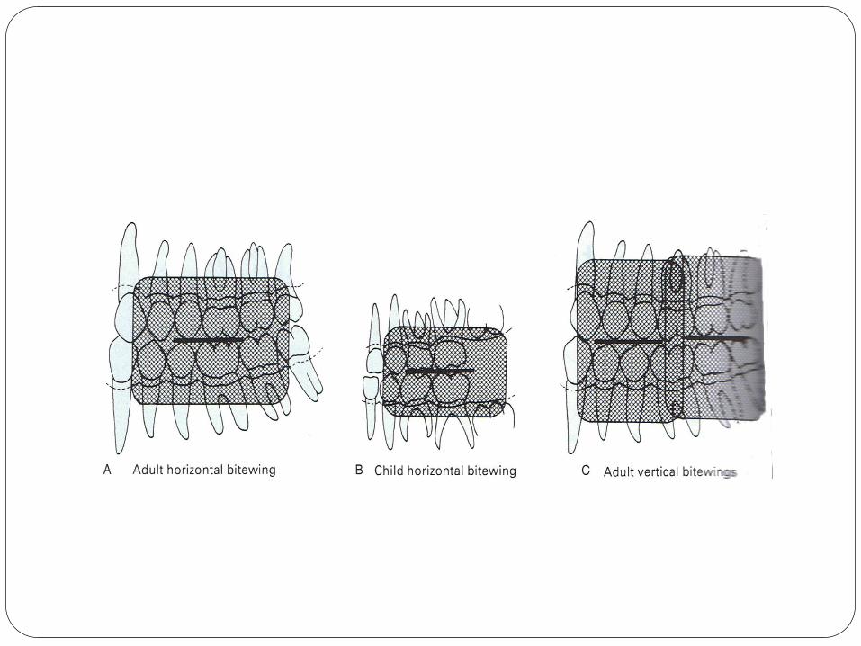
Position of Tab

The film packet should be positioned with its long axis
horizontally for a horizontal bitewing or vertically for a
vertical bitewing

The posterior teeth and the film packet should be in contact
or as close together as possible whilst avoiding over bending
of the film
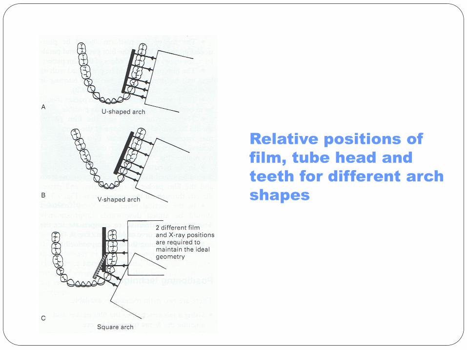
Relative positions of
film, tube head and
teeth for different arch
shapes
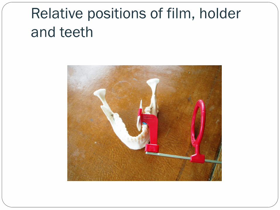
Relative positions of film, holder
and teeth
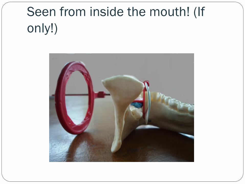
Seen from inside the mouth! (If
only!)

In the horizontal plane, the X-ray tubehead should be aimed
so that the beam meets the teeth and the film packet at right
angles, and passes directly through all the contact areas.

In the vertical plane, the X-ray tubehead should be aimed
downwards ( approximately 5-8 degrees to the horizontal) to
compensate for the upwardly rising curve of Monson.

Angulation for curve of Monson
The positioning should be
reproducible !

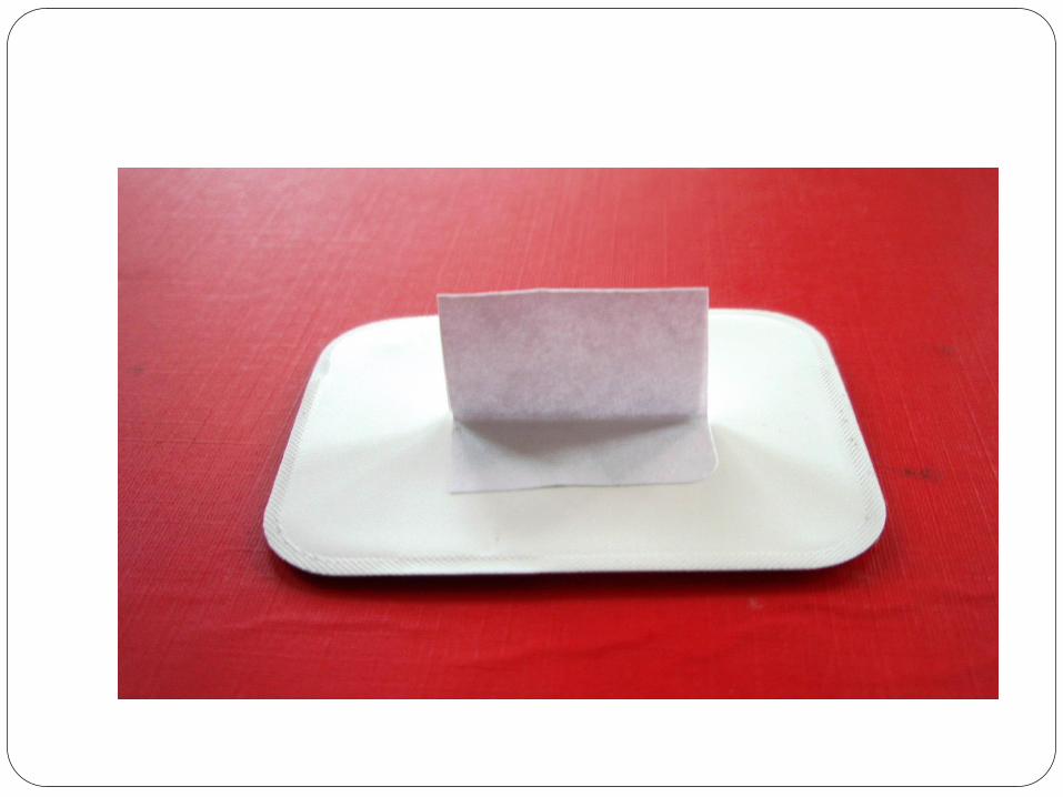
Using a tab attached to the film
packet
1. The appropriate size of film is selected and the tab
attached as shown

Film Sizes Large film 31 x 41 mm for adults
Small film packet 22 x 35 mm for children under 12.Once the
second permanent molars have erupted the adult size film is
required.
Occasionally a longer film packet
( 53 x 26 mm ) is used for adults

2
The patient is positioned with the head supported and with
the occlusal plane horizontal.

3
The shape of the dental arch and the number of films
required is assessed

4
The operator holds the tab between thumb and forefinger
and inserts the film packet into the lingual sulcus opposite
the posterior teeth

5
The anterior edge of the film packet should be positioned
opposite the distal aspect of the lower canine – in this
position, the posterior edge of the film packet extends
usually just beyond the mesial aspect of the lower third molar

6
The tab is placed on to the occlusal surfaces of the lower
teeth.
7
The patient is asked to close the teeth firmly together on to
the tab.
8
As the patient closes the teeth together, the operator pulls
the tab firmly between the teeth to ensure that the film
packet and the teeth are in contact.

9
The operator releases the tab and positions the tube head.

When positioning the tubehead, after the
patient closes the mouth the film can no
longer be seen.
To ensure that the anterior part of the film
is exposed and to avoid coning off or cone
cutting , a simple guide to remember is
that the front edge of the open-ended
spacer cone should be positioned
adjacent to the corner of the patient’s
mouth

10
The X-ray beam is aimed directly through the contact areas,
at right angles to the teeth and the film packet , with an
approximate 5 – 8 degree downward vertical angulation.

11
The exposure is made

12
The procedure is repeated for the premolar teeth if required,
with a new film packet and
X-ray tubehead position.

Advantages
Simple to use
Inexpensive
Tabs are disposable, so no extra cross infection control
procedures required.
Can be easily used for children.

Disadvantages Arbitrary, operator- dependent assessment of the horizontal and vertical
angulations of the X-ray tubehead.
Radiographs not accurately reproducible, so not suitable for monitoring the progression of caries.
Coning off or Cone cutting of anterior part of film is common.
The tongue can easily displace the film packet
Using simple film packet holders
Many types are available
They can eliminate many of the disadvantages of the tab
method.
Holders vary in cost and design

Three Basic components
A mechanism for holding the film packet parallel to the teeth
A bite platform that replaces the wing ( tab)
An X-ray beam aiming device.

Advantages
Simple
Film packet held firmly and not displaced by tongue
Position of tubehead determined by holder
Avoids coning off or cone cutting.
Holders are autoclavable or disposable

Disadvantages
Position of the holder is operator dependent
Not 100% reproducible
Positioning of film holder can be uncomfortable for the
patient
Some holders are relatively expensive
Holders not suitable for children.

Conclusion
Traditional bitewing techniques,
using detachable tabs, although
simple to perform are operator
dependent and inaccurate
Film holders are more accurate
and are recommended

Normal Appearance of Bitewing
Radiographs

Ideal Exposure Factors
The clinical reasons for taking bitewing radiographs should determine the
exposure factors.
Assessment of caries – well exposed
Assessment of periodontal status- underexposed
In Practice
A typical pair of bitewings involves a compromise with
regard to exposure factors. In this way the radiation dose to
the patient is kept to a minimum.


