CLINICAL MICROBIOLOGY REVIEWS, 0893-8512/01/$04.0010 DOI: 10.1128/CMR.14.3.513–527.2001 July 2001, p. 513–527 Vol. 14, No. 3 Borna Disease Virus and Human Disease KATHRYN M. CARBONE* FDA/CBER, HFM 460, Bethesda, Maryland 20892 INTRODUCTION .......................................................................................................................................................513 HISTORY .....................................................................................................................................................................513 EPIDEMIOLOGY .......................................................................................................................................................514 VIROLOGY..................................................................................................................................................................514 BIOLOGY AND PATHOGENESIS OF BORNA DISEASE .................................................................................514 Encephalitic Borna Disease ...................................................................................................................................515 Behavioral Borna Disease......................................................................................................................................515 HUMAN BDV INFECTION ......................................................................................................................................515 Diagnostic Tests for BDV Infection in Humans.................................................................................................515 Anti-BDV antibody detection.............................................................................................................................516 (i) Immunofluorescence assay .......................................................................................................................516 (ii) Immunoblot assay ....................................................................................................................................518 (iii) Enzyme-linked immunosorbent assay ..................................................................................................518 Methods to detect virus protein in tissue or cells .........................................................................................518 Virus nucleic acid detection ..............................................................................................................................520 Infectious-virus isolation ...................................................................................................................................520 BDV—a Human Pathogen? ...................................................................................................................................521 Evidence for BDV-Induced Disease in Humans: Possible Clinical Syndromes of BDV Infection .............522 Treatment .................................................................................................................................................................523 CONCLUSIONS .........................................................................................................................................................523 REFERENCES ............................................................................................................................................................524 INTRODUCTION For over a century, a fatal encephalitis, Borna disease, has been diagnosed in horses and sheep in Central Europe (120). In 1929, Borna disease was found to be caused by an infectious agent, and in 1990, this agent was determined to be a negative- sense, single-stranded RNA virus (25, 39). Borna disease virus (BDV) persistently infects the nervous system of many animal species, from primate to avian (120). Indeed, by natural or experimental inoculation, the ability of BDV to replicate in the nervous system of virtually every warm-blooded animal strongly suggests that BDV-like viruses are very unlikely to spare the human host. Over the years, information has also accumulated about unusual features of BDV-induced disease in experimental an- imals such as rats, mice, and tree shrews. In these animals, BDV can induce behavioral disease (e.g., anxiety, aggression, cognitive defects, and hyperactivity) without obvious physio- logical signs of viral encephalitis (e.g., fever, neurological signs, and decreased level of consciousness) (7, 48, 58, 66, 68, 99, 111, 112, 114, 115, 122, 136). Studies of behavioral disease in BDV-infected animals have sparked reasonable speculation that BDV infection in humans might also lead to psychiatric disease. It is tempting to specu- late that BDV might be linked to some psychiatric disease syndromes such as affective disorders (e.g., depression) or psy- chosis (e.g., schizophrenia) or to idiopathic acute or chronic encephalitis. In the 1980s, the first significant serological evi- dence for BDV infection of humans was reported in the sci- entific literature (1). However, despite two decades of study and published serological, pathological, or virological evidence of BDV infection in humans, complete medical and scientific acceptance of the human as a natural target of BDV has yet to be achieved. Even more controversial are the specific human disease syndromes for which BDV has been proposed to be an etiologic agent. Much of the controversy in the study of human BDV infec- tion is linked to technical difficulties in developing and validat- ing a uniform test for diagnosis of BDV infection in humans. Clearly, without a validated test for diagnosing BDV infection in humans, data from a clinical study to identify possible hu- man diseases linked to BDV infection should be evaluated with proper caution. HISTORY Borna disease and, later, BDV were named after the town of Borna in Saxony, Germany, where an epidemic of infectious encephalitis caused a large number of equine deaths in 1885 (120). Veterinary scientists spent many years carefully describ- ing the natural history of BDV infection in animals in Central Europe, including infections of horses, sheep, rabbits, and birds, and subsequently started working with the virus in tissue culture (120). BDV research spread from laboratories in Cen- tral Europe to the United States in the mid-1980s (99), where projects in disease pathogenesis and animal models were ini- tiated (28). Later, the first cDNA clones were isolated that identified the Borna disease agent as an RNA virus (88, 143) and the sequencing of the BDV genome was reported (26, 39). Since that time, researchers all over the world, including * Mailing address: FDA/CBER, HFM 460, 8800 Rockville Pike, Bethesda, MD 20892. Phone: (301) 827-1973. Fax: (301) 480-5679. E-mail: [email protected]. 513 Downloaded from https://journals.asm.org/journal/cmr on 13 October 2021 by 2.50.176.164.
Borna Disease Virus and Human DiseaseKATHRYN M. CARBONE*
FDA/CBER, HFM 460, Bethesda, Maryland 20892
INTRODUCTION .......................................................................................................................................................513HISTORY.....................................................................................................................................................................513EPIDEMIOLOGY.......................................................................................................................................................514VIROLOGY..................................................................................................................................................................514BIOLOGY AND PATHOGENESIS OF BORNA DISEASE .................................................................................514
HUMAN BDV INFECTION ......................................................................................................................................515Diagnostic Tests for BDV Infection in Humans.................................................................................................515
Methods to detect virus protein in tissue or cells .........................................................................................518Virus nucleic acid detection ..............................................................................................................................520Infectious-virus isolation ...................................................................................................................................520
BDV—a Human Pathogen?...................................................................................................................................521Evidence for BDV-Induced Disease in Humans: Possible Clinical Syndromes of BDV Infection .............522Treatment.................................................................................................................................................................523
For over a century, a fatal encephalitis, Borna disease, hasbeen diagnosed in horses and sheep in Central Europe (120).In 1929, Borna disease was found to be caused by an infectiousagent, and in 1990, this agent was determined to be a negative-sense, single-stranded RNA virus (25, 39). Borna disease virus(BDV) persistently infects the nervous system of many animalspecies, from primate to avian (120). Indeed, by natural orexperimental inoculation, the ability of BDV to replicate in thenervous system of virtually every warm-blooded animalstrongly suggests that BDV-like viruses are very unlikely tospare the human host.
Over the years, information has also accumulated aboutunusual features of BDV-induced disease in experimental an-imals such as rats, mice, and tree shrews. In these animals,BDV can induce behavioral disease (e.g., anxiety, aggression,cognitive defects, and hyperactivity) without obvious physio-logical signs of viral encephalitis (e.g., fever, neurological signs,and decreased level of consciousness) (7, 48, 58, 66, 68, 99, 111,112, 114, 115, 122, 136).
Studies of behavioral disease in BDV-infected animals havesparked reasonable speculation that BDV infection in humansmight also lead to psychiatric disease. It is tempting to specu-late that BDV might be linked to some psychiatric diseasesyndromes such as affective disorders (e.g., depression) or psy-chosis (e.g., schizophrenia) or to idiopathic acute or chronicencephalitis. In the 1980s, the first significant serological evi-
dence for BDV infection of humans was reported in the sci-entific literature (1). However, despite two decades of studyand published serological, pathological, or virological evidenceof BDV infection in humans, complete medical and scientificacceptance of the human as a natural target of BDV has yet tobe achieved. Even more controversial are the specific humandisease syndromes for which BDV has been proposed to be anetiologic agent.
Much of the controversy in the study of human BDV infec-tion is linked to technical difficulties in developing and validat-ing a uniform test for diagnosis of BDV infection in humans.Clearly, without a validated test for diagnosing BDV infectionin humans, data from a clinical study to identify possible hu-man diseases linked to BDV infection should be evaluated withproper caution.
HISTORY
Borna disease and, later, BDV were named after the town ofBorna in Saxony, Germany, where an epidemic of infectiousencephalitis caused a large number of equine deaths in 1885(120). Veterinary scientists spent many years carefully describ-ing the natural history of BDV infection in animals in CentralEurope, including infections of horses, sheep, rabbits, andbirds, and subsequently started working with the virus in tissueculture (120). BDV research spread from laboratories in Cen-tral Europe to the United States in the mid-1980s (99), whereprojects in disease pathogenesis and animal models were ini-tiated (28). Later, the first cDNA clones were isolated thatidentified the Borna disease agent as an RNA virus (88, 143)and the sequencing of the BDV genome was reported (26, 39).Since that time, researchers all over the world, including
groups in Asia, Scandinavia, and Australia, have joined in thestudy of BDV.
EPIDEMIOLOGY
BDV infection was originally believed to be limited to farmanimals (e.g., horses and sheep) and some wild animals (e.g.,rabbits) in areas of endemic infection in Germany and Swit-zerland (120). With the advent of more modern tools for di-agnosis of BDV infection (e.g., in situ hybridization, reversetranscriptase PCR [RT-PCR]) and with the increasing inter-national research interest in BDV, reports of susceptible spe-cies and the geographic location of cases of natural infectionhave expanded (120). Animals at risk for natural or experi-mental infection include rhesus monkeys, horses, sheep, cattle,goats, rabbits, deer, llamas, alpacas, cats, rats, mice, gerbils,dogs, and ostriches (2, 10, 27, 35, 72, 77, 89, 95, 102, 104, 120;Y. Weisman, D. Huminer, M. Malkinson, R. Meir, S. Kliche,W. I. Lipkin, and S. Pitlik, Letter, Lancet 344:1232–1233,1994). Evidence for natural BDV infection of animals has nowspread beyond the confines of Central Europe to the UnitedKingdom, Israel, Japan, Sweden, Australia, and the UnitedStates (11, 76, 82, 89, 116, 120; Weisman et al., Letter). Sincethe first subjects studied in the United States and Germany,evidence of human BDV infection has been reported in othercountries in the Eurasian continent including Taiwan, Thai-land, Iran, and Japan (3, 5, 36; Weisman et al., Letter).
VIROLOGY
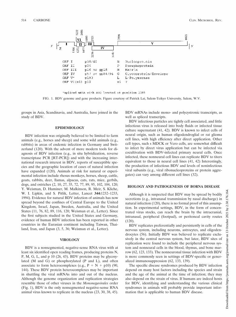
BDV is a nonsegmented, negative-sense RNA virus with atleast six identified open reading frames, producing proteins N,P, M, G, L, and p 10 (26, 43). BDV proteins may be glycosy-lated (M and G) or phosphorylated (P and L), and oftenassociate to form heterocomplexes (e.g., P 1 N 1 p10) (90,144). These BDV protein heterocomplexes may be importantin shuttling the viral mRNAs into and out of the nucleus.Although the genome organization and replication strategiesresemble those of other viruses in the Mononegavirales order(Fig. 1), BDV is the only nonsegmented negative-sense RNAanimal virus known to replicate in the nucleus (23, 30, 38, 45).
BDV mRNAs include mono- and polycystronic transcripts, aswell as spliced transcripts.
BDV infectious particles are tightly cell associated, and littleinfectious virus is released into body fluids or infected tissueculture supernatant (41, 42). BDV is known to infect cells ofneural origin, such as human oligodendroglial or rat gliomacell lines, with high efficiency after direct application. Othercell types, such s MDCK or Vero cells, are somewhat difficultto infect by direct virus application but can be infected viacocultivation with BDV-infected primary neural cells. Onceinfected, these nonneural cell lines can replicate BDV to titersequivalent to those in neural cell lines (41, 42) Interestingly,the production of infectious BDV and levels of noninfectiousviral subunits (e.g., viral ribonucleoproteins or protein aggre-gates) can vary among different cell lines (32).
BIOLOGY AND PATHOGENESIS OF BORNA DISEASE
Although it is suspected that BDV may be spread by bodilysecretions (e.g., intranasal transmission by nasal discharge) innatural infection (120), there is no formal proof of this assump-tion. In experimental settings, BDV, in the form of concen-trated virus stocks, can reach the brain by the intracranial,intranasal, peripheral (footpad), or peritoneal cavity routes(120).
BDV replicates preferentially and persistently in cells of thenervous system, including neurons, astrocytes, and oligoden-drocytes (56). Initially BDV was believed to replicate exclu-sively in the central nervous system, but later, BDV sites ofreplication were found to include the peripheral nervous sys-tem and nonneural cells in the blood, thymus, and bone mar-row (62, 123, 133). The nonneuronal tissue infection with BDVis more commonly seen in settings of BDV-specific or gener-alized immunosuppression (62, 133, 139).
The specific disease syndromes produced by BDV infectiondepend on many host factors including the species and strainand the age of the animal at the time of infection; they mayalso depend on the strain of virus. If humans are indeed hostsfor BDV, identifying and understanding the various clinicalsyndromes in animals will probably provide important infor-mation that is applicable to human BDV disease.
FIG. 1. BDV genome and gene products. Figure courtesy of Patrick Lai, Salem-Teikyo University, Salem, W.V.
514 CARBONE CLIN. MICROBIOL. REV.
Dow
nloa
ded
from
http
s://j
ourn
als.
asm
.org
/jour
nal/c
mr
on 1
3 O
ctob
er 2
021
by 2
.50.
176.
164.
Encephalitic Borna Disease
Unlike most viruses, much of the morbidity and mortalityfollowing BDV infection stems from the host immune responseto the virus and the ensuing immune-mediated death of in-fected and nearby uninfected cells rather than from directvirus-mediated lysis of infected cells (137). Encephalitic Bornadisease (EBD) typically develops following infection of anadult animal and is associated with a massive mononuclear cellimmune infiltration into the brain parenchyma (137). In rats,initial immune cell infiltrates in the perivascular spaces areCD81 and CD41 T cells, NK cells, and macrophages (12, 60,119). Over time, B cells, NK cells, and activated microgliadominate in a parenchymal reaction (60). Astrocyte and mi-croglia cell activation is also seen (34, 94, 149). Disease may beexpressed initially as behavioral abnormalities including hyper-activity, hyperreactivity, and aggression and subsequently as arapidly progressive, often fatal, neurological impairment, in-cluding seizures, ataxia, and paraplegia. Horses, sheep, manyadult-infected rat strains, and, to some degree, cats fall victimto this form of EBD.
For reasons that are not yet clear, some animals survive theacute disease and, after several weeks of EBD, begin to showsigns of chronic Borna disease (CBD) (78). CBD is sometimesassociated with near resolution of central nervous system(CNS) inflammatory infiltrates (100). Lewis rats surviving withCBD have significant, permanent brain destruction (e.g., cor-tical thinning and hydrocephalus) and chronic signs of neuro-logical disease, (e.g., chronic apathy, blindness) (28, 100).
Behavioral Borna Disease
Unlike adult Lewis rats, adult black-hooded rats and adultBALB/c and SJL mice have limited susceptibility to EBD (61,125). While these animals replicate BDV in the nervous systemand may even have severe encephalitis, they fail to exhibit thesigns of serious neurological disease. Some animals fail todevelop fatal encephalitis and exhibit significant behavioralabnormalities in a form of behavioral Borna disease (BBD).BBD is seen following BDV infection of some species orstrains of immature animals (e.g., newborn Lewis rats), adultanimals with suppressed immune systems (e.g., athymic orthymectomized rats or rats immunosuppressed via drug treat-ment), and certain species or strains of adult animals (e.g.,tupiais glis or MRL strain mice) (7, 48, 65, 66, 99, 125, 136, 140,141).
Of all the animal models of experimental BBD, the mostextensively studied is the neonatally infected Lewis rat, firstdescribed in the early 1980s (66, 99). In neonatally BDV-inoculated rats, the lack of significant immune cell infiltrationin the brain is believed to stem from infection of the thymusduring immune system maturation, leading to BDV-specific“immune tolerance,” although direct proof of this hypothesis islacking (31).
Although animals with BBD appear normal to the casualobserver, they have documented behavioral abnormalities as-sociated with neuroanatomical, neurochemical, and neuroim-mune deficits. Some of these behavioral abnormalities havebeen measured by formal behavioral testing, which revealedhyperactivity, cognitive deficits, social behavior (play) abnor-malities, and chronic anxiety (7, 48, 68, 114, 115, 122, 124).
Animals with an immature CNS at the time of BDV infectionmay show evidence of developmental neuroanatomical dam-age, including dropout of specific neurons in the cerebellum(granule cells and Purkinje cells), dentate gyrus of the hip-pocampus and cerebral cortex (6, 32, 49, 68), and alterations insynaptic plasticity (55). Interestingly, despite little, if any, cel-lular inflammation in these animals, abnormal levels of cyto-kines (e.g., tumor necrosis factor, interleukin-1b, and trans-forming growth factor b) and chemokines (68, 109, 128, 129)and of serotonin, norepinephrine, dopamine, and other neu-rotransmitters (68, 113) have been reported.
HUMAN BDV INFECTION
Diagnostic Tests for BDV Infection in Humans
Scientists have been searching for evidence of human BDVinfection and associated disease states for over 15 years. In-deed, there are reports of the recovery of anti-BDV antibodies,BDV RNA, BDV proteins, and infectious BDV from humantissues and body fluids from normal humans and humans witha wide variety of psychiatric disorders. Using a variety of test-ing methods and clinical study designs, many investigators haveused evidence for and against human BDV infection to postu-late an association, or lack thereof, of BDV infection withspecific human diseases. The numerous tests used to diagnoseBDV infection have evolved over the years, coincident with theavailability of BDV-specific reagents such as cDNA clones (88,143) and with general scientific technological advancements,such as RT-PCR (133). Therefore, before continuing the dis-cussion of reported evidence of human BDV infection, it isimportant to understand the advantages and limitations of thenumerous different tests used to obtain evidence of BDV in-fection in humans.
Reliable and accurate diagnosis of human infection withBDV, or a BDV-related virus, is a prerequisite for the confir-mation of a BDV-induced human disease. Generally, BDVassays are modeled after tests used in animal studies and in-clude tests for anti-BDV antibody, immunologically based testsfor BDV proteins, RT-PCR assays for BDV RNA, and in vitroor in vivo assays for infectious BDV. To validate the ability ofan assay to identify subjects with BDV infection as “positive”and subjects without BDV infection as “negative,” resultsshould be provided from experiments using this assay with asufficient type and number of samples from animals whoseBDV infection state is clearly known, e.g., from several sub-jects in different species of experimentally inoculated animalsor naturally infected animals with independent confirmation ofBDV infection.
The validity of a diagnostic test can be determined by mea-suring the rate of sensitivity (true-negative rate) and specificity(true-positive rate), and, in general, the more sensitive the testthe lower the specificity and vice versa. In assay developmentand validation, the weighted emphasis on the specificity orsensitivity of a specific assay also depends on the use for whichthe assay is intended (101). In cases where false-negative re-sults might cause significant harm (e.g., missing a virus-infectedunit of blood intended for transfusion), a highly sensitive test isused, even if specificity is somewhat compromised, i.e., aslightly higher false-positive rate, resulting in the disposal of
VOL. 14, 2001 BORNA DISEASE VIRUS AND HUMAN DISEASE 515
Dow
nloa
ded
from
http
s://j
ourn
als.
asm
.org
/jour
nal/c
mr
on 1
3 O
ctob
er 2
021
by 2
.50.
176.
164.
some good units of blood. These highly sensitive tests are oftenused as “screening” assays. In clinical settings where false-positive results might lead to unnecessary treatment, a positiveresult on a sensitive screening assay is often confirmed by a testof higher specificity.
In designing a BDV assay for human samples, one mustconsider the question to be answered. To answer the question“Does BDV infect humans?” a highly specific test is preferred,even if sensitivity is reduced, to ensure the maximum possibletrue-positive rate. In this situation, confirmatory testing of theindividuals who test BDV positive by the assay in question willuniformly show further evidence of infection (e.g., cultivationof infectious virus). Thus, confidence in the accuracy of the testfor identifying a BDV-infected subject will be high. To answerthe question “Is disease state ‘X’ caused by BDV infection?” ahighly sensitive and specific determination of the BDV infec-tion status is required. To perform a meaningful clinical studyto causally associate BDV with a specific disease, it is critical tohave a test, or series of tests, to place the subjects in the correctcategory of BDV-infected and control subjects. This may ne-cessitate the combined use of a highly sensitive screening test,e.g., an enzyme-linked immunosorbent assay (ELISA), fol-lowed by a more specific confirmatory test for those testingBDV positive by ELISA, e.g., Western blot.
Anti-BDV antibody detection. Serological tests evaluate thepresence of a humoral immune response to a virus infection,e.g., that antibodies bind to specific virus antigens. When theindividual contracts a virus infection, the first serological evi-dence of virus infection is often the immunoglobulin M (IgM)antibody. As the immune response matures, an antiviral IgGantibody response is detected. When the onset of infection canbe identified, typically acute-phase (early in infection) andconvalescent-phase (several weeks into the infection) sera aresampled and tested as a pair. A significant increase in the titerof virus-specific IgG from the acute-phase to the convalescent-phase serum sample is indicative of infection.
In natural BDV infections, acute-phase serum is not alwaysavailable and, as a result, most serological diagnostic tests fornatural BDV infection are a single test for anti-BDV IgG frompresumed convalescent-phase serum. In many animal species,BDV infection is associated with persistence of anti-BDV an-tibodies. It has been debated whether the anti-BDV antibodytiter in humans is consistent or variable on repeated measures,but in several individuals anti-BDV antibodies have been de-tected by repeated testing (14). Notably, virus may or may notbe present in individuals who test positive by serological assays.In some virus infections, a positive serological result indicatescure or “clearance” of the virus, e.g., mumps virus. In othercases of persistent virus infection, a positive antibody test mayindicate latent (e.g., herpes simplex virus) or persistent (e.g.,
human immunodeficiency virus [HIV]) virus replication. BDVis a persistent virus infection in many species and may persistat low (e.g., detectable in mononuclear white blood cells byRT-PCR) or high (i.e., comparable to acute infection in brain)levels (133). It is unknown whether humans might becomepersistently infected with BDV or clear the virus infection.Thus, we do not know how the detection of anti-BDV antibodyrelates to the presence or clearance of BDV in humans.
All serological tests for BDV use natural BDV antigens(from infected cells) or recombinant antigens (from BDVstrains recovered from animals) (Table 1). Using BDV derivedfrom nonhuman sources as the antigen in serological studieshas caused some investigators to question the validity of low-avidity, low-titer binding of antibodies in human sera. Whetherthese findings are due to cross-reactivity necessitated by usinga nonhuman strain of BDV or suggest the nonspecificity ofhuman antibody binding to BDV antigens is not known.
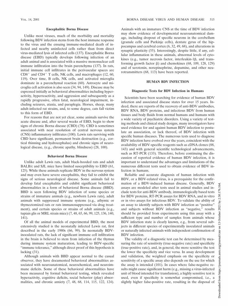
(i) Immunofluorescence assay. Diagnostic tests for anti-BDV antibodies were the first method by which BDV infectionwas diagnosed in animals (120). The first serological diagnostictest for BDV infection in humans was the indirect immunoflu-orescence assay (IFA) (1). In this test, human serum is overlaidon a slide covered with fixed BDV-infected cells to allow anti-BDV antibodies in the serum to bind to the viral antigensexpressed by the cells. BDV-infected MDCK (64) or C6 (32)cells are most commonly used in the IFA. After the cells arewashed, a fluorescence-labeled anti-human IgG antibody isadded to signal the presence of human anti-BDV antibodies.Using a fluorescent light-equipped microscope, infected cellsare detected on the basis of brightly lit inclusions in the nu-cleus, often associated with a more finely patterned signal inthe cytoplasm (Fig. 2A). To control for false-positive signals,e.g., the artifactual binding of autoantibodies to the cell anti-gens, the same human serum should be overlaid on uninfectedcells. Positive controls for the IFA include the use of anti-BDVserum from experimentally infected animals on duplicate cells.Some researchers have incorporated a “double-label” tech-nique using a monoclonal mouse anti-BDV antibody mixedwith the human serum sample and a second-color fluorescentlylabeled anti-mouse IgG added to the anti-human IgG, to iden-tify colocalization of the human serum signal with the mouseanti-BDV antibody signal (19, 147). While the double-labeltechnique can identify false-positive signals from human sera(when human serum binds to cell locations that are distantfrom binding sites of positive-control animal BDV antiserum)at the light microscope level, the technique has insufficientresolution to clearly prove that human sera and animal controlsera are binding to identical antigens in the infected cell.
The IFA is a rapid, sensitive assay in controlled, experimen-tal BDV infections. However, when screening human sera
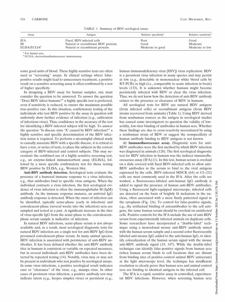
IFA Fixed, BDV-infected cells Poor GoodIB Natural or recombinant BDV proteins Good ModerateELISA/ECLIAb Natural or recombinant proteins Moderate to good Moderate to low
a For human sera.b ECLIA, electrochemiluminescence immunoassay.
516 CARBONE CLIN. MICROBIOL. REV.
Dow
nloa
ded
from
http
s://j
ourn
als.
asm
.org
/jour
nal/c
mr
on 1
3 O
ctob
er 2
021
by 2
.50.
176.
164.
FIG. 2. (A and B) OL cells mock infected or persistently infected with HuP2br were immunohistochemically stained with polyclonal rabbitanti-BDV p40 and then with fluorescein isothiocyanate-conjugated goat anti-rabbit IgG (A) and were subjected to in situ hybridization usingantisense digoxigenin-conjugated riboprobe directed to BDV p40 region and then stained with fluorescent-labeled anti-digoxigenin Ig (B). (C)Newborn gerbils were mock infected or infected with HuP2Br. The sections prepared from the cerebral cortex (frontal lobe) of the gerbils on day30 postinfection were subjected to in situ hybridization using antisense riboprobe directed to the BDV p40 region. Figure courtesy of KazuyoshiIkuta, Osaka University, Osaka, Japan.
VOL. 14, 2001 BORNA DISEASE VIRUS AND HUMAN DISEASE 517
Dow
nloa
ded
from
http
s://j
ourn
als.
asm
.org
/jour
nal/c
mr
on 1
3 O
ctob
er 2
021
by 2
.50.
176.
164.
where exposure or infection with BDV is unknown, the IFAmay be insufficiently specific (147). The major technical con-cerns with the IFA technique are specificity and the variabilityintroduced by reader expertise (i.e., correct recognition of thespecific, characteristic pattern of BDV antigens in the infectedcell). Many laboratories performing BDV serological testinghave a great deal of expertise in IFA techniques; however, foraccurate determination of BDV seropositivity by IFA, reader-to-reader variability in testing makes it difficult or impossibleto replicate serology results among independent laboratories.Moreover, unless careful use of negative controls is incorpo-rated into the assay (e.g., duplicate serum samples run in par-allel using uninfected cells), there is a risk of confusion be-tween signals from binding of cell-specific autoantibodies andanti-BDV antibodies. Sensitivity concerns are raised, for ex-ample, when trying to determine if a faint signal is specific forBDV or represents artifactual “background” staining. This lat-ter situation is often a problem in testing human sera, whereantibody levels are generally quite low.
(ii) Immunoblot assay. Like IFA, the immunoblot assay (IB)is a serological technique that also relies on detection of hu-man antibody bound to virus antigens. In IB, virus antigens areseparated by electrophoresis through a gel matrix and thentransferred to a specialized blotting membrane. Strips of theblot are incubated in human serum and washed, and binding ofhuman antibodies is detected by a secondary, enzyme-labeledanti-human IgG antibody. Binding of human anti-BDV anti-bodies is indicated by an enzymatic reaction resulting in avisible dye stain or a light signal captured on X-ray film orimage equipment. The image can be evaluated qualitatively (byeye) or by more quantitative methods using computer-basedimaging systems.
Molecular weight markers are included in the blot to con-firm the location of the known sizes of BDV proteins. Positive-control sera from infected animals are applied to duplicateblots to confirm the appropriate technical performance andprovide the location of the BDV antigens on the blot. Typi-cally, negative-control lanes with uninfected material are run induplicate with lanes containing BDV antigens. The sources ofBDV antigens in IB include experimentally infected animalbrain, lysates of infected neuronal or nonneuronal cell lines,baculovirus recombinant proteins, and prokaryotic recombi-nant proteins (37, 53, 70, 74, 82, 107, 130, 145).
There are some general aspects of IB that may affect thesensitivity of the technique. While baculovirus and prokaryoticrecombinant BDV proteins can be synthesized in large quan-tities and are easily purified, normal human glycosylation ofvirus proteins is altered (70). Although not proven for BDVproteins, both nonhuman glycosylation patterns of virus anti-gens as well as the typically protein-reducing and -denaturingcharacteristics of the gel can destroy or alter conformationalvirus epitopes.
Although some reader expertise is involved in interpretingIB results, the specificity of this technique is believed to beexcellent. In part, the ability of IB to show which virus anti-gen(s) is recognized by the serum sample provides increasedspecificity for the IB compared to the IFA (146). Seropositivitycriteria have included the recognition of a single BDV protein(53) or of multiple BDV proteins by the serum (146). Whilethere is a risk that a single virus protein will be recognized by
“nonspecific” antibodies from an uninfected subject, there is areduced probability of nonspecific recognition of multiple virusproteins by antibodies in a single serum. The specificity ofantibody binding in the IB may be improved by the use of asecond IB run in tandem using soluble BDV antigens to showspecific inhibition of antibody binding to BDV antigens. Draw-backs of the IB include the time-consuming and costly natureof this technique and the disadvantage that the high specificityseen with IB may be accompanied by some decrease in sensi-tivity of the test (146).
(iii) Enzyme-linked immunosorbent assay. ELISAs arecommonly used serological tests designed for high-throughputscreening; unlike IFA and IB, they are designed to give non-reader-dependent quantitative results. Several different typesof ELISAs have been developed to perform BDV serologicalassays, using native or recombinant antigens singly or in com-binations (24, 67, 81, 150; Weisman et al., Letter). Antigens arebound directly to the ELISA plate or “captured” by BDV-specific antibodies bound to the plate. The human serum to betested is overlaid on the bound BDV antigens, and a second-ary, enzyme-labeled anti-human IgG antibody is added. Fol-lowing enzymatic reactions that produce a visible pigment orfluorescent product, the amount of bound BDV-specific anti-body is measured in a specialized spectrophotometer.
Although ELISA is generally believed to be a highly sensi-tive serological assay system, BDV-specific ELISAs have beenreported to have some difficulty with sensitivity. For example,ELISAs that are capable of reliably detecting anti-BDV anti-body in one species (e.g., rats) have been unable to detectanti-BDV antibody reliably in other species (e.g., rabbits andhorses) (67, 78). Therefore, it is unclear whether the inabilityof these ELISAs to detect anti-BDV antibody in humans,where anti-BDV antibody titers are generally low, represents afalse-negative due to species-specific variability in the sensitiv-ity of the ELISA or a true-negative result.
Specificity concerns with these ELISAs are demonstratedwhen human sera shown seronegative by IB give a positiveresult in ELISA due to nonspecific reactivity (50). As with IB,some ELISA protocols have incorporated a tandem “blocking”step using soluble BDV antigens to confirm the specificity ofthe antibodies binding to the BDV antigens on the ELISAplate in order to improve BDV specificity (150).
Methods to detect virus protein in tissues or cells. Methodsfor detecting virus protein expression in human tissues (immu-nohistochemistry) are most often used on postmortem brainspecimens. Anti-BDV-specific antisera are applied to tissueand bind virus antigens, and enzymatic reactions are used toindicate areas where BDV proteins are recognized by the an-tisera (Fig. 3). Although flow cytometry has been reported todetect BDV antigens in peripheral blood cells (L. Bode, F.Steinbach, and H. Ludwig, Letter, Lancet 343:297–298, 1994),this technique has not gained wide acceptance.
Sensitivity in these assays is dependent on the amount ofBDV proteins expressed in the examined tissues and cells. Innonhuman, experimentally infected species, BDV antigens canbe expressed at relatively high (rat) or low (mouse) levels (28,125). The sensitivity of the immunohistochemical assay canalso be affected by the binding properties of the BDV antibodyused to detect BDV antigens. In addition, the specificity ofthese assays depends on the cross-reactivity with non-BDV
518 CARBONE CLIN. MICROBIOL. REV.
Dow
nloa
ded
from
http
s://j
ourn
als.
asm
.org
/jour
nal/c
mr
on 1
3 O
ctob
er 2
021
by 2
.50.
176.
164.
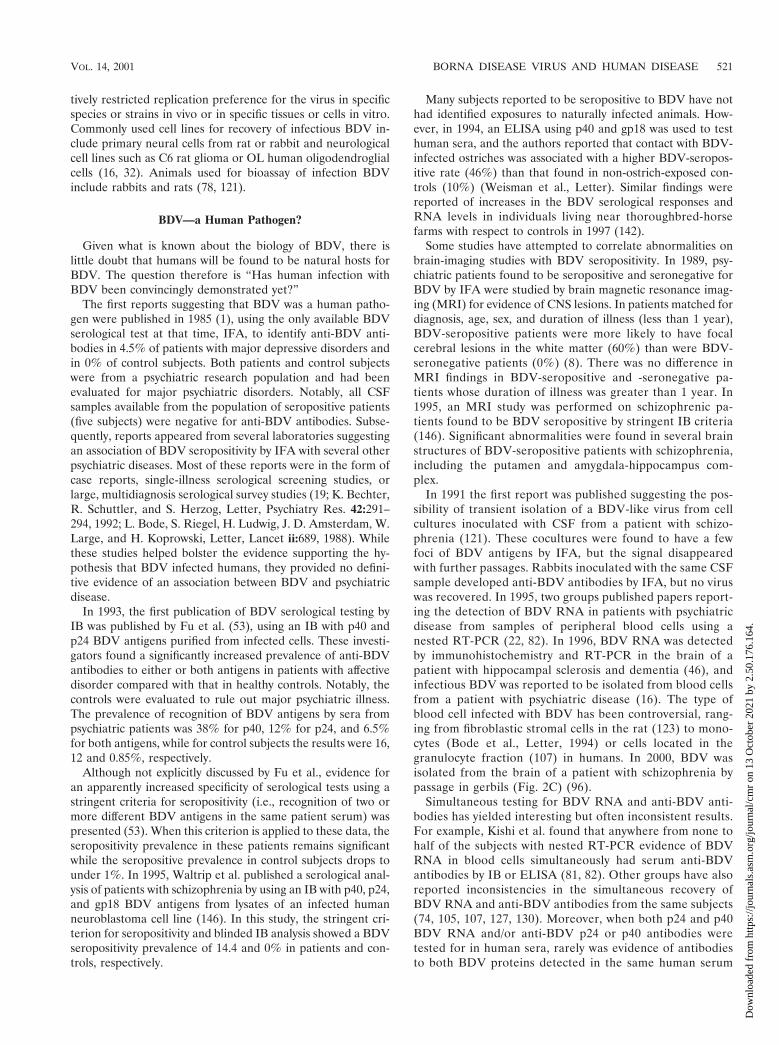
FIG. 3. Expression of BDV N (p40) antigen and BDV RNA in autopsy tissue from the human brain. (A) For detection of viral antigen, hip-pocampal sections from brains of representative patients with hippocampal sclerosis (HS) or Alzheimer’s disease (AD), immunolabeled with eithera rabbit anti-BDV N serum or an anti-GFAP antibody, were used. (B) For detection of viral nucleic acid, RNA was isolated from frozen brainsamples of hippocampal sclerosis or Alzheimer’s disease patients and analyzed by RT-PCR using specific primers to amplify a 528-nucleotide seg-ment of the BDV N ORF. An aliquot of cDNA from each sample was also used to amplify glyceraldehyde-3-phosphate dehydrogenase (GAPDH)sequences by RT-PCR as a control of RNA quality. The BDV specificity of the PCR products was confirmed by Southern blot hybridization usinga BDV N probe internal to the predicted PCR product. Figure courtesy of Juan Carlos de la Torre, Scripps Research Institute, La Jolla, Calif.
519
Dow
nloa
ded
from
http
s://j
ourn
als.
asm
.org
/jour
nal/c
mr
on 1
3 O
ctob
er 2
021
by 2
.50.
176.
164.
proteins of the anti-BDV antibody that is used to detect theBDV antigen in tissues or cells. Artifacts can occur throughnonspecific antibody binding or other technical problems, andreader expertise is needed to interpret the results.
Virus nucleic acid detection. In 1990, with the advent ofcDNA clones for BDV, techniques to locate BDV RNA be-came feasible. In situ hybridization uses a radioactively orenzymatically labeled complementary strand of BDV se-quence-specific nucleic acid probe that binds to BDV RNA ininfected cells. In situ hybridization can be a sensitive tech-nique, especially if the infected cells are localized or are fewand inhomogeneously distributed in the tissue. In situ hybrid-ization provides excellent histological information, especiallywhen combined with immunohistochemistry techniques thatidentify the infected cell type (34). On the other hand, in situhybridization can yield false-negative results if low concentra-tions of BDV RNA are present or if the BDV RNA is de-graded (as might occur in human autopsy specimens), or it canyield false-positive results if non-BDV-specific probe bindingoccurs. BDV RNA has also been detected in RNA extractedfrom cells in a soluble form via capture by antibodies or bycomplementary nucleic acid sequences bound to microtiterplates (54, 133). RT-PCR assays have been used extensively tolocate BDV RNA in human samples. In RT-PCR, purifiedRNA is reverse transcribed and the cDNA is amplified repeat-edly with specific primers, and subjected to a sequence-specificconfirmation method, e.g., Southern blotting.
RT-PCR for BDV was initially developed to identify verysmall amounts of BDV RNA in circulating peripheral bloodmononuclear cells in neonatally infected rats (133). Prior tothe report by Sierra-Honigmann et al., it was believed thatBDV did not circulate in the peripheral blood. Thus, recoveryof BDV from humans was believed to require invasive sam-pling techniques (e.g., lumbar puncture for cerebrospinal fluid(CSF) or tissue biopsy). After the discovery of BDV in ratblood cells, it became common to use RT-PCR to test samplesof human peripheral blood cells for evidence of BDV RNA.Since experimental rat studies showed that the number ofinfected cells and level of infectious virus per cell are estimatedto be very low, the sensitivity was increased by the use of a“nested” RT-PCR technique with two sequential rounds ofamplification using two separate sets of BDV primers (127,133). Using nested RT-PCR, between 10 and 100 copies ofBDV genome per sample can be detected.
Although RT-PCR is generally believed to be highly sensi-tive, the presence of RT-PCR inhibitors, such as heparin orhemoglobin in a blood sample, may result in false-negativeresults (133). Moreover, the low level of BDV RNA in periph-eral blood may fall below the detection sensitivity of RT-PCRand produce a false-negative result. With such a low level ofviral RNA in blood samples, poor specimen handling, such asfreeze-thawing, can produce false-negative results (A. M. Si-erra-Honigmann, unpublished results). Based on the numberof BDV-infected white blood cells in rats (around 1 in 105 to106 cells), avoiding false-negative results in humans due tosampling error may require at least 5 ml of blood to obtain asufficient number of infected cells for testing (127). Finally, theability of BDV primers designed from animal virus sequencesto recognize a putative human BDV also needs to be takeninto account.
In addition to the primers chosen for amplification of theBDV RNA, the specificity of RT-PCR is further documentedby Southern blot or sequencing analysis of the amplified DNAarising from the RT-PCR. The specificity of the RT-PCR re-sults is also supported by the use of multiple controls, includingthe amplification of normal negative control cells in parallelwith positive control BDV-infected cells; however, typically thecells chosen are tissue culture lines that are not from the sametissue areas as the test samples from humans (e.g., dog kidneytissue culture cells used as controls for human brain or bloodcell samples). A mismatch of sample and control cells can beproblematic for RT-PCR assays in which cellular mRNA mayvary from cell type to cell type.
Most concerns regarding false-positive results obtained withthe RT-PCR technique have revolved around fears of contam-ination by BDV cDNA or PCR products. This contaminationproblem was clearly demonstrated in a multiple-laboratorystudy (117). Several technical adjustments have been used byBDV researchers to reduce the risk of BDV contamination,including running the RT reaction without the RT enzyme(wherein a positive signal identifies contaminating BDV cDNAin the sample or reagents), utilizing a single tube RT-PCRmethod to minimize opportunities for contamination withBDV cDNA, and “tagging” BDV cDNAs so that contamina-tion of those products can be easily identified in future reac-tions (92, 93, 127).
Nucleic acid sequencing can be used to confirm that thematerial is BDV. In many viruses, sequence differences amongwild-type and laboratory strains can be used to ensure thatlaboratory virus strains are not contaminating the human spec-imens being tested. However, for the most part, the sequencevariations of the BDV strains isolated from any source arelimited, on the order of 0 to 5% of the genome, and “human-specific” sequence changes are limited and controversial, (13,44, 63, 80, 131). Thus, using the sequence to confirm the sourceof the recovered strain, e.g., to distinguish a human BDV strainfrom a laboratory contaminant, is not feasible at present. How-ever, a recent report of a horse strain of BDV with substantialRNA (more than 15%) and protein (almost 20% in the p10protein) sequence variation from previous horse strains pro-vides hope that a unique strain might be recovered from hu-mans as well (103). Notably, this new horse strain was noteasily detected by some standard BDV RT-PCR primers, in-timating that using standard BDV primers designed to detectvirus RNA in animals may not be the best approach for screen-ing for the presence of BDV in human tissue.
Infectious-virus isolation. The “gold standard” for diagnosisof BDV infection is the isolation of infectious virus from thesubject. BDV is largely a cell-associated virus, and it is difficultto recover infectious virus from bodily fluids. In general, infec-tious-BDV isolation is performed by inoculating tissue homog-enates into cell culture or by in vivo testing (i.e., animal infec-tivity assays). Depending on the virus titer in the inoculatedtest material, evidence of infectious BDV in tissue homoge-nates can be detected as early as 24 h (e.g., high-titer virusdetected by expression of BDV antigens in cell culture) or aslate as several months (e.g., low-titer virus detected by expres-sion of Borna disease in inoculated animals). Limitations ofthis technique include the low level of infectious BDV repli-cation in some species (probably including humans), the rela-
520 CARBONE CLIN. MICROBIOL. REV.
Dow
nloa
ded
from
http
s://j
ourn
als.
asm
.org
/jour
nal/c
mr
on 1
3 O
ctob
er 2
021
by 2
.50.
176.
164.
tively restricted replication preference for the virus in specificspecies or strains in vivo or in specific tissues or cells in vitro.Commonly used cell lines for recovery of infectious BDV in-clude primary neural cells from rat or rabbit and neurologicalcell lines such as C6 rat glioma or OL human oligodendroglialcells (16, 32). Animals used for bioassay of infection BDVinclude rabbits and rats (78, 121).
BDV—a Human Pathogen?
Given what is known about the biology of BDV, there islittle doubt that humans will be found to be natural hosts forBDV. The question therefore is “Has human infection withBDV been convincingly demonstrated yet?”
The first reports suggesting that BDV was a human patho-gen were published in 1985 (1), using the only available BDVserological test at that time, IFA, to identify anti-BDV anti-bodies in 4.5% of patients with major depressive disorders andin 0% of control subjects. Both patients and control subjectswere from a psychiatric research population and had beenevaluated for major psychiatric disorders. Notably, all CSFsamples available from the population of seropositive patients(five subjects) were negative for anti-BDV antibodies. Subse-quently, reports appeared from several laboratories suggestingan association of BDV seropositivity by IFA with several otherpsychiatric diseases. Most of these reports were in the form ofcase reports, single-illness serological screening studies, orlarge, multidiagnosis serological survey studies (19; K. Bechter,R. Schuttler, and S. Herzog, Letter, Psychiatry Res. 42:291–294, 1992; L. Bode, S. Riegel, H. Ludwig, J. D. Amsterdam, W.Large, and H. Koprowski, Letter, Lancet ii:689, 1988). Whilethese studies helped bolster the evidence supporting the hy-pothesis that BDV infected humans, they provided no defini-tive evidence of an association between BDV and psychiatricdisease.
In 1993, the first publication of BDV serological testing byIB was published by Fu et al. (53), using an IB with p40 andp24 BDV antigens purified from infected cells. These investi-gators found a significantly increased prevalence of anti-BDVantibodies to either or both antigens in patients with affectivedisorder compared with that in healthy controls. Notably, thecontrols were evaluated to rule out major psychiatric illness.The prevalence of recognition of BDV antigens by sera frompsychiatric patients was 38% for p40, 12% for p24, and 6.5%for both antigens, while for control subjects the results were 16,12 and 0.85%, respectively.
Although not explicitly discussed by Fu et al., evidence foran apparently increased specificity of serological tests using astringent criteria for seropositivity (i.e., recognition of two ormore different BDV antigens in the same patient serum) waspresented (53). When this criterion is applied to these data, theseropositivity prevalence in these patients remains significantwhile the seropositive prevalence in control subjects drops tounder 1%. In 1995, Waltrip et al. published a serological anal-ysis of patients with schizophrenia by using an IB with p40, p24,and gp18 BDV antigens from lysates of an infected humanneuroblastoma cell line (146). In this study, the stringent cri-terion for seropositivity and blinded IB analysis showed a BDVseropositivity prevalence of 14.4 and 0% in patients and con-trols, respectively.
Many subjects reported to be seropositive to BDV have nothad identified exposures to naturally infected animals. How-ever, in 1994, an ELISA using p40 and gp18 was used to testhuman sera, and the authors reported that contact with BDV-infected ostriches was associated with a higher BDV-seropos-itive rate (46%) than that found in non-ostrich-exposed con-trols (10%) (Weisman et al., Letter). Similar findings werereported of increases in the BDV serological responses andRNA levels in individuals living near thoroughbred-horsefarms with respect to controls in 1997 (142).
Some studies have attempted to correlate abnormalities onbrain-imaging studies with BDV seropositivity. In 1989, psy-chiatric patients found to be seropositive and seronegative forBDV by IFA were studied by brain magnetic resonance imag-ing (MRI) for evidence of CNS lesions. In patients matched fordiagnosis, age, sex, and duration of illness (less than 1 year),BDV-seropositive patients were more likely to have focalcerebral lesions in the white matter (60%) than were BDV-seronegative patients (0%) (8). There was no difference inMRI findings in BDV-seropositive and -seronegative pa-tients whose duration of illness was greater than 1 year. In1995, an MRI study was performed on schizophrenic pa-tients found to be BDV seropositive by stringent IB criteria(146). Significant abnormalities were found in several brainstructures of BDV-seropositive patients with schizophrenia,including the putamen and amygdala-hippocampus com-plex.
In 1991 the first report was published suggesting the pos-sibility of transient isolation of a BDV-like virus from cellcultures inoculated with CSF from a patient with schizo-phrenia (121). These cocultures were found to have a fewfoci of BDV antigens by IFA, but the signal disappearedwith further passages. Rabbits inoculated with the same CSFsample developed anti-BDV antibodies by IFA, but no viruswas recovered. In 1995, two groups published papers report-ing the detection of BDV RNA in patients with psychiatricdisease from samples of peripheral blood cells using anested RT-PCR (22, 82). In 1996, BDV RNA was detectedby immunohistochemistry and RT-PCR in the brain of apatient with hippocampal sclerosis and dementia (46), andinfectious BDV was reported to be isolated from blood cellsfrom a patient with psychiatric disease (16). The type ofblood cell infected with BDV has been controversial, rang-ing from fibroblastic stromal cells in the rat (123) to mono-cytes (Bode et al., Letter, 1994) or cells located in thegranulocyte fraction (107) in humans. In 2000, BDV wasisolated from the brain of a patient with schizophrenia bypassage in gerbils (Fig. 2C) (96).
Simultaneous testing for BDV RNA and anti-BDV anti-bodies has yielded interesting but often inconsistent results.For example, Kishi et al. found that anywhere from none tohalf of the subjects with nested RT-PCR evidence of BDVRNA in blood cells simultaneously had serum anti-BDVantibodies by IB or ELISA (81, 82). Other groups have alsoreported inconsistencies in the simultaneous recovery ofBDV RNA and anti-BDV antibodies from the same subjects(74, 105, 107, 127, 130). Moreover, when both p24 and p40BDV RNA and/or anti-BDV p24 or p40 antibodies weretested for in human sera, rarely was evidence of antibodiesto both BDV proteins detected in the same human serum
VOL. 14, 2001 BORNA DISEASE VIRUS AND HUMAN DISEASE 521
Dow
nloa
ded
from
http
s://j
ourn
als.
asm
.org
/jour
nal/c
mr
on 1
3 O
ctob
er 2
021
by 2
.50.
176.
164.
sample (36, 150). This “mismatch” of BDV serological withrespect to RNA and infectious-virus test results may be aexplained by one or all of the following scenarios: (i) BDVis cleared from human tissues through an immune response(e.g., anti-BDV antibody positive and BDV RNA negative),(ii) BDV is persistently replicating in the human host with anonneutralizing immune response (e.g., anti-BDV antibodypositive and BDV RNA positive), or (iii) BDV is replicatingin the human host without a measurable immune response(e.g., anti-BDV antibody negative and BDV RNA positive).
In contrast to findings in humans, many animals infectedwith BDV (those that survived acute infection) have antibodiesto several BDV proteins coincident with evidence of BDVRNA and/or infectious-virus replication (120). Because muchof the human evidence is not consistent with this finding inanimals, it is possible that (i) BDV detection in human samplesis an artifact and humans are not infected with BDV, (ii)humans are infected with BDV but our current tests are notwell designed to detect human BDV strains, or (iii) humans areinfected with BDV but the human immune response to BDVmay be capable of clearing the virus infection.
Questions continue to be raised about technical artifactsin RT-PCR assays or sequencing that may have introducedinaccuracies in reported BDV sequences (127). For exam-ple, there is a report of sequence similarities between hu-man BDV strains and the BDV laboratory strains used insome laboratories, and the authors of the report suggestedthat most, if not all, “human” BDV strains recovered werecontaminated by laboratory strains (M. Schwemmle, C.Jehle, S. Formella, and P. Staeheli, Letter, Lancet 354:1973–1974, 1999). However, it has also been argued that thishypothesis was based on comparison of limited sequencesand is inaccurate (O. Planz, H. J. Rziha, and L. Stitz, Letter,Lancet 355:656–657, 2000). Despite the continued contro-versy, it is encouraging that since the inception of the searchfor human BDV in 1985, publications have appropriatelyprovided increased technical details and control proceduresused in the assays for BDV RNA, e.g., the inclusion of“RT-negative” and water samples to demonstrate the ab-sence of contaminating BDV PCR products.
While the failure to detect BDV in some human sampleshas been interpreted as evidence of lack of human infectionwith BDV, these interpretations should be viewed with ap-propriate caution. As we learn more about BDV infection indifferent species of animals, information is accumulating tosupport the hypothesis that evidence of BDV infection maybe difficult to obtain for some species, such as cats (104) andexperimentally infected ponies (78); it may, therefore alsobe difficult to obtain for humans. Since BDV is likely to bea relatively rare infection in humans, problems could lie withthe clinical design, e.g., the proper selection of appropriatehuman subjects. For example, based on the findings in therat model of hippocampal degeneration (31), a more tar-geted patient selection approach was used by screening sam-ples of hippocampus from 600 non-Alzheimer’s dementiapatients (46). Of the five patients with evidence of hip-pocampal degeneration, four were found to have evidenceof BDV RNA and protein expression (Fig. 3).
Evidence for BDV-Induced Disease in Humans: PossibleClinical Syndromes of BDV Infection
To date, BDV has not been identified as a clear etiology ofany known human disease. However, based on what is knownabout BDV biology and disease pathogenesis in many nonhu-man species, it is tempting to speculate about the clinicaloutcomes of human BDV infection.
Although BDV might be the etiologic agent of idiopathicacute or chronic inflammatory encephalitis in humans, nolarge-scale studies have evaluated such a connection and so nodata are available to support or refute this hypothesis. None-theless, a clinical picture of human EBD might be similar tothat of a hospitalized patient with idiopathic acute encephalitisassociated with changes in the level of consciousness, alongwith fever and significant neurological impairment. As withrabies or herpes simplex encephalitis, damage to the neuro-anatomical targets of BDV, such as the limbic system, mightlead to erratic personality changes or even violent behavior.Individuals who survived EBD and developed CBD mightshow evidence of chronic neurological damage, even in theface of a receding inflammatory response in the brain. Im-paired cognitive function, apathy, and emotional instabilitywould probably be exhibited by the survivors of the infection.
An alternative expression of human BDV infection maymore closely resemble BBD as seen in the neonatally BDV-infected rat, with a preponderance of behavioral disease symp-toms. Humans with BDV infection might not have fever,changes in mental alertness, or other typical signs of viralencephalitis but instead might express signs of psychiatric dis-ease, such as depression, mania, anxiety, cognitive disorders,tardive dyskinesia, social dysfunction, eating disorders, andidiopathic seizures (7, 48, 114, 115, 124, 135, 136). Moreover, ifinfection occurred in utero or during the first 3 years of life,BDV infection might result in autistic spectrum disorder, withabnormal social interactions, chronic anxiety, cognitive deficits,and evidence of abnormal development of the cerebellum andhippocampus (6, 31, 48, 49, 110–112, 122).
Some post-virus infection sequelae are not due to directvirus damage but to autoimmune responses stimulated by virusinfection (e.g., post-viral encephalitis and Guillain-Barre syn-drome). Therefore, some consideration must be given to therole of autoimmunity in BDV-associated disease as well. Insome experimental systems, BDV infection induces autoanti-bodies, e.g., antibodies to cellular proteins seen in post-BDVinfection sera but not in preinfection sera (70). As with othervirus infections, it is possible that autoantibodies are the resultof BDV-associated cell lysis and release of host proteins, towhich an autoimmune response develops. Alternatively, auto-antibodies might be the result of “molecular mimicry” of hostantigens by BDV proteins. Finally, the possibility that antibod-ies that coincidentally recognize BDV antigens may be presentin the serum of individuals who have not been infected withBDV should also be considered. These autoantibodies may ormay not be relevant to the clinical disease presentation of theindividual but are only coincidentally detected in tests includ-ing BDV antigens.
Since there is no agreed upon, validated assay for diagnosingBDV infection in humans, it is not possible to causally associ-ate any disease with BDV infection. Nonetheless, below are
522 CARBONE CLIN. MICROBIOL. REV.
Dow
nloa
ded
from
http
s://j
ourn
als.
asm
.org
/jour
nal/c
mr
on 1
3 O
ctob
er 2
021
by 2
.50.
176.
164.
data summarized from publications from many internationalgroups supporting and refuting the hypothesis of human BDVinfection associated with numerous disease outcomes. Thetechniques used in these publications are varied and some-times are not accompanied by the technical informationneeded to evaluate the assay specificity and sensitivity. Simi-larly, many clinical studies of BDV do not clearly indicate theincorporation of one or more significant clinical trial designelements required to demonstrate a causal association betweenBDV infection and specific disease. These missing or unre-ported clinical design elements include appropriate patientand control screening and selection techniques, blinding oflaboratory workers to clinical status, and collection and anal-ysis of associated non-BDV-related factors that may influencethe trial outcome (e.g., hospitalizations, pharmacotherapy, age,sex, race, socioeconomic status, and exposure to animals).Thus, when examining the conclusions of BDV associationwith specific diseases, care should be taken not to overinterpretthe presented evidence.
In the past decade, BDV RNA, BDV proteins, anti-BDVantibody, and/or infectious virus have been found in the blood,CSF, and/or brains of patients with chronic fatigue syndrome(83, 97, 98), blood donors (81), HIV-infected patients (3),patients with schizophrenia or with deficit syndrome schizo-phrenia subtype (36, 37, 73, 74, 96, 145, 146; M. Salvatore, S.Morzunov, M. Schwemmle, and W. I. Lipkin, Letter, Lancet349:1813–1814, 1997), normal humans (57), patients with mul-tiple sclerosis and/or depression (52; M. Deuschle, L. Bode, I.Heuser, J. Schmider, and H. Ludwig, Letter, Lancet 352:1828–1829, 1998), patients with non-Alzheimer’s dementia and hip-pocampal degeneration (46), and patients with various psychi-atric disorders (150).
Reports of the failure to find evidence of BDV infectionassociated with specific disease syndromes have been increas-ing. The inability to find evidence of significant BDV infectionin blood, CSF, and/or brain has been reported for studies ofpatients with chronic fatigue syndrome (50; L. Bode, A. L.Komaroff, and H. Ludwig, Letter, Clin. Infect. Dis. 15:1049,1992), patients with schizophrenia (133), patients with multiplesclerosis (B. Kitze, S. Herzog, P. Rieckmann, S. Poser, andJ. Richt, Letter, J. Neurol. 243:660–662, 1996), HIV-infectedpatients (4), and various psychiatric patients (79, 85, 117; K.Lieb, W. Hallensleben, M. Czygan, L. Stitz, and P. Staeheli,Letter, Lancet 350:1002, 1997)
Notably, the positive and negative studies have been re-ported from assays performed on samples from the same pa-tient cohorts and sometimes by the same research group. Insum, it is not possible to discern the diseases, if any, that maybe caused by BDV infection of humans. Much more work isneeded in this area, since many technical factors, as well as ourlack of understanding of the biology of BDV infection in theanimal and human, probably have contributed to the contro-versy.
Treatment
Our inability to connect human BDV infection to specificdisease states also limits our ability to substantiate the use ofunapproved “anti-BDV” treatment in persons in whom unvali-dated research assays suggest BDV infection. Nonetheless,
proposed therapies for BDV infection have been reported (51;L. Bode, D. E. Dietrich, R. Stoyloff, H. M. Emrich, and H.Ludwig, Letter, Lancet 3:178–179, 1997). As with any virusinfection and, perhaps, especially with persistent virus infec-tions, prevention of infection through vaccination is likely to bethe preferred approach over treatment following establishedinfection.
Experimental passive transfer of humoral immunity has notbeen shown to cure established BDV infection or preventinfection of the immunized rat, nor has vaccination with killedvirus been shown to be protective against disease (137). Thereis a report that vaccination with high-titer tissue culture-pas-saged BDV can offer incomplete protection of rats from Bornadisease following challenge with brain-derived virus (106).Transfer of BDV-specific immune T lymphocytes prior to in-fection can limit or prevent BDV infection and/or Borna dis-ease in Lewis rats (118).
Amantadine has been reported as being effective (51; Bodeet al., Letter, 1997) and ineffective (59, 138) at reducing BDVreplication in vitro, in vivo (rats), and/or in treating patients.Since there are some reports of amantadine having direct an-tidepressive effects (71) and since no controlled, blinded clin-ical trials have been performed, the use of amantadine to treatBDV infection has not been adequately supported. Mizutani etal. reported that ribavirin inhibited BDV transcription in vitro(91), a finding that has been replicated by an independentgroup (75). There are no reports of ribavirin use in humanswith evidence of BDV infection. Notably, ribavirin has signif-icant adverse effects, and its use for treatment of putative BDVinfection in patients is not an approved indication.
CONCLUSIONS
The biology of BDV strongly supports the likelihood ofhuman infection with BDV or a variant of BDV. Thus far, theevidence supporting BDV infection in humans has initiatedmuch controversy among basic and clinical scientists; only timeand additional research will support or refute the hypothesis ofhuman BDV infection.
What is needed first is a validated assay or series of assaysthat are capable of reliably identifying BDV infection in hu-mans. Difficulties that must be overcome include species vari-ability in replication of virus and development of anti-BDVantibodies, e.g., the apparent low titer and low affinity of hu-man antibodies against BDV; the use of animal virus compo-nents (proteins and RNA sequences) rather than human viruscomponents in these assays; possible differences in the naturalhistory of BDV infection in humans and animals (e.g., persis-tence versus clearance of virus); and the low infectious-virusrecovery from human samples.
Validation of the reproducibility of the assay(s) requires thateach assay be performed in several independent laboratoriesusing uniform reagents and reference samples (blinded). Inaddition, the sensitivity and specificity of the assay should bedetermined using documented reference samples from a vari-ety of species. For example, for a serological assay, referencesamples would include sera from animals from a variety ofspecies of low-avidity (e.g., horse) and high-avidity (e.g., rat)anti-BDV antibodies as well as low- and high-titer samples.These reference sera should be unquestionably from BDV-
VOL. 14, 2001 BORNA DISEASE VIRUS AND HUMAN DISEASE 523
Dow
nloa
ded
from
http
s://j
ourn
als.
asm
.org
/jour
nal/c
mr
on 1
3 O
ctob
er 2
021
by 2
.50.
176.
164.
infected animals, recovered either from animals with experi-mental infection with BDV or from animals in which the BDVinfection has been independently confirmed (e.g., by cultiva-tion of infectious virus). With this approach, reproducible as-says of predictable sensitivity and specificity can be developed.
After assays of acceptable specificity and sensitivity havebeen developed and validated using animal virus and postin-fection sera, the next step would be to use the assays on humansera with the intention of identifying possibly BDV-infectedindividuals. Attempts to document human infection with BDVshould precede any attempt to associate BDV positivity withspecific clinical diseases. Human BDV infection can be suc-cessfully identified using a small number of carefully chosensubjects. It is likely that a series of tests for BDV infectionstatus will perform more reliably than a single test; thus, ex-pectation of a sensitive serological screening test (e.g., ELISA)followed by a confirmatory test (e.g., IB or RT-PCR) would bereasonable. When a cohort of likely BDV-infected individualsis identified, careful evaluation of these individuals by addi-tional methods (e.g., infectious-virus recovery) would beneeded to confirm the findings.
Once BDV has been reliably detected in humans, scientistscan reasonably begin to study an association of BDV infectionwith specific disease syndromes. For clinical studies seekingcausal associations between BDV infection and specific dis-eases, a reliable, validated test for BDV infection in humansmust be in place in order to ensure proper identification of theBDV infection status of patients and control subjects. Of ut-most importance is careful patient and control cohort selectionand screening. Control subjects must be carefully matched tothe patient population on the basis of social, economic, racial,gender, and other critical features. The control populationmust be carefully screened by active testing to document theabsence of the disease condition being evaluated in the patientpopulation. Sufficient numbers of patients and control subjectsshould be evaluated to provide a reasonable “power” of thestudy to detect differences between the two populations inBDV positivity rates. Success of these studies might be im-proved by wise preselection of patients based on known BDVdisease pathogenesis in animals.
Causal association of BDV with human disease will be hin-dered by the current lack of documentation of the pathogen-esis and etiology of many psychiatric syndromes. Many psychi-atric diseases are described “syndromes,” i.e., constellations ofsymptoms and signs of disease that tend to group into a rou-tinely defined syndrome. Thus, it would not be unreasonable toexpect that even if BDV is identified in humans with a specifictype of disease syndrome, it may be the etiologic agent in onlya small proportion of patients with this syndrome. In addition,given the clear effects of genetic background (e.g., strain andspecies) on BDV replication and expression of disease, itwould not be surprising to see a constellation of differentdisease symptoms, signs, and syndromes all associated withBDV infection. However, over the years, BDV researchershave faced many unique and difficult problems on the way toscientific discovery in BDV research, and with concerted effortand continued work, these thorny issues that surround BDVhuman infection will be resolved.
REFERENCES
1. Amsterdam, J. D., A. Winokur, W. Dyson, S. Herzog, F. Gonzalez, R. Rott,and H. Koprowski. 1985. Borna disease virus. A possible etiologic factor inhuman affective disorders? Arch. Gen. Psychiatry 42:1093–1096.
2. Ashash, E., M. Malkinson, R. Meir, S. Perl, and Y. Weisman. 1996. Causesof losses including a Borna disease paralytic syndrome affecting youngostriches of one breeding organization over a five-year period (1989–1993).Avian Dis. 40:240–245.
3. Auwanit, W., P. I. Ayuthaya, T. Nakaya, S. Fujiwara, T. Kurata, K. Yama-nishi, and K. Ikuta. 1996. Unusually high seroprevalence of Borna diseasevirus in clade E human immunodeficiency virus type 1-infected patientswith sexually transmitted diseases in Thailand. Clin. Diagn. Lab. Immunol.3:590–593.
4. Bachmann, S., P. Caplazi, M. Fischer, F. Ehrensperger, and R. W. Cone.1999. Lack of association between Borna disease virus infection and neu-rological disorders among HIV-infected individuals. J. Neurovirol. 5:190–195.
5. Bahmani, M. K., I. Nowrouzian, T. Nakaya, Y. Nakamura, K. Hagiwara, H.Takahashi, M. A. Rad, and K. Ikuta. 1996. Varied prevalence of Bornadisease virus infection in Arabic, thoroughbred and their cross-bred horsesin Iran. Virus Res. 45:1–13.
6. Bautista, J. R., S. A. Rubin, T. H. Moran, G. J. Schwartz, and K. M.Carbone. 1995. Developmental injury to the cerebellum following perinatalBorna disease virus infection. Dev. Brain Res. 90:45–53.
7. Bautista, J. R., G. J. Schwartz, J. C. de la Torre, T. H. Moran, and K. M.Carbone. 1994. Early and persistent abnormalities in rats with neonatallyacquired Borna disease virus infection. Brain Res. Bull. 34:31–40.
8. Bechter, K., S. Herzog, R. Schuttler, and R. Rott. 1989. MRI in psychiatricpatients with serum antibodies against Borna disease virus. Psychiatry Res.29:281–282.
9. Reference deleted.10. Berg, A. L., and M. Berg. 1998. A variant form of feline Borna disease. J.
Comp. Pathol 119:323–331.11. Berg, A. L., R. Reid-Smith, M. Larsson, and B. Bonnett. 1998. Case control
study of feline Borna disease in Sweden. Vet. Rec. 142:715–717.12. Bilzer, T., and L. Stitz. 1994. Immune-mediated brain atrophy. CD81 T
cells contribute to tissue destruction during borna disease. J. Immunol.153:818–823.
13. Binz, T., J. Lebelt, H. Niemann, and K. Hagenau. 1994. Sequence analysesof the p24 gene of Borna disease virus in naturally infected horse, donkeyand sheep. Virus Res. 34:281–289.
14. Bode, L. 1995. Human infections with Borna disease virus and potentialpathogenic implications. Curr. Top. Microbiol. Immunol. 190:103–130.
15. Reference deleted.16. Bode, L., R. Durrwald, F. A. Rantam, R. Ferszt, and H. Ludwig. 1996. First
isolates of infectious Borna disease virus from patients with mood disor-ders. Mol. Psychiatry 1:200–212.
17. Reference deleted.18. Reference deleted.19. Bode, L., S. Riegel, W. Lange, and H. Ludwig. 1992. Human infections with
Borna disease virus: seroprevalence in patients with chronic diseases andhealthy individuals. J. Med. Virol 36:309–315.
20. Reference deleted.21. Reference deleted.22. Bode, L., W. Zimmermann, R. Ferszt, F. Steinbach, and H. Ludwig. 1995.
Borna disease virus genome transcribed and expressed in psychiatric pa-tients. Nat. Med. 1:232–236.
23. Briese, T., J. C. de la Torre, A. Lewis, H. Ludwig, and W. I. Lipkin. 1992.Borna disease virus, a negative-strand RNA virus, transcribes in the nucleusof infected cells. Proc. Natl. Acad. Sci. USA 89:11486–11489.
24. Briese, T., C. G. Hatalski, S. Kliche, Y. S. Park, and W. I. Lipkin. 1995.Enzyme-linked immunosorbent assay for detecting antibodies to Bornadisease virus-specific proteins. J. Clin. Microbiol. 33:348–351.
25. Briese, T., W. I. Lipkin, and J. C. de la Torre. 1995. Molecular biology ofBorna disease virus. Curr. Top. Microbiol. Immunol. 190:1–16.
26. Briese, T., A. Schneemann, A. J. Lewis, Y. S. Park, S. Kim, H. Ludwig, andW. I. Lipkin. 1994. Genomic organization of Borna disease virus. Proc.Natl. Acad. Sci. USA 91:4362–4366.
27. Caplazi, P., A. Waldvogel, L. Stitz, U. Braun, and F. Ehrensperger. 1994.Borna disease in naturally infected cattle. J. Comp. Pathol. 111:65–72.
28. Carbone, K. M., C. S. Duchala, J. W. Griffin, A. L. Kincaid, and O.Narayan. 1987. Pathogenesis of Borna disease in rats: evidence that intra-axonal spread is the major route for virus dissemination and the determi-nant for disease incubation. J. Virol. 61:3431–3440.
29. Reference deleted.30. Carbone, K. M., T. R. Moench, and W. I. Lipkin. 1991. Borna disease virus
replicates in astrocytes, Schwann cells and ependymal cells in persistentlyinfected rats: location of viral genomic and messenger RNAs by in situhybridization. J. Neuropathol. Exp. Neurol. 50:205–214.
31. Carbone, K. M., S. W. Park, S. A. Rubin, R. W. Waltrip, and G. B.Vogelsang. 1991. Borna disease: association with a maturation defect in the
524 CARBONE CLIN. MICROBIOL. REV.
Dow
nloa
ded
from
http
s://j
ourn
als.
asm
.org
/jour
nal/c
mr
on 1
3 O
ctob
er 2
021
by 2
.50.
176.
164.
cellular immune response. J. Virol. 65:6154–6164.32. Carbone, K. M., S. A. Rubin, A. M. Sierra-Honigmann, and H. M. Leder-
man. 1993. Characterization of a glial cell line persistently infected withBorna disease virus (BDV): influence of neurotrophic factors on BDVprotein and RNA expression. J. Virol. 67:1453–1460.
33. Reference deleted.34. Carbone, K. M., B. D. Trapp, J. W. Griffin, C. S. Duchala, and O. Narayan.
1989. Astrocytes and schwann cells are virus host cells in the nervous systemof rats with Borna disease. J. Neuropathol. Exp. Neurol. 48:631–644.
35. Cervos-Navarro, J., W. Roggendorf, H. Ludwig, and H. Stitz. 1981. Dieborna-Krankheit beim Affen unter besonderer Berucksichtigung der en-cephalitischen Reaktion. Verh. Dtsch. Ges. Pathol. 65:208–212.
36. Chen, C. H., Y. L. Chiu, C. K. Shaw, M. T. Tsai, A. L. Hwang, and K. J.Hsiao. 1999. Detection of Borna disease virus RNA from peripheral bloodcells in schizophrenic patients and mental health workers. Mol. Psychiatry4:566–571.
37. Chen, C. H., Y. L. Chiu, F. C. Wei, F. J. Koong, H. C. Liu, C. K. Shaw, H. G.Hwu, and K. J. Hsiao. 1999. High seroprevalence of Borna virus infectionin schizophrenic patients, family members and mental health workers inTaiwan. Mol. Psychiatry 4:33–38.
38. Cubitt, B., and J. C. de la Torre. 1994. Borna disease virus (BDV), anonsegmented RNA virus, replicates in the nuclei of infected cells whereinfectious BDV ribonucleoproteins are present. J. Virol. 68:1371–1381.
39. Cubitt, B., C. Oldstone, and J. C. de la Torre. 1994. Sequence and genomeorganization of Borna disease virus. J. Virol. 68:1382–1396.
40. Reference deleted.41. Danner, K., D. Heubeck, and A. Mayr. 1978. In vitro studies on Borna virus.
I. The use of cell cultures for the demonstration, titration and productionof Borna virus. Arch. Virol. 57:63–75.
42. Danner, K., and A. Mayr. 1979. In vitro studies on Borna virus. II. Prop-erties of the virus. Arch. Virol. 61:261–271.
43. de la Torre, J. C. 1994. Molecular biology of borna disease virus: prototypeof a new group of animal viruses. J. Virol. 68:7669–7675.
44. de la Torre, J. C., L. Bode, R. Durrwald, B. Cubitt, and H. Ludwig. 1996.Sequence characterization of human Borna disease virus. Virus Res. 44:33–44.
45. de la Torre, J. C., K. M. Carbone, and W. I. Lipkin. 1990. Molecularcharacterization of the Borna disease agent. Virology 179:853–856.
46. de la Torre, J. C., D. Gonzalez-Dunia, B. Cubitt, M. Mallory, N. Mueller-Lantzsch, F. A. Grasser, L. A. Hansen, and E. Masliah. 1996. Detection ofBorna disease virus antigen and RNA in human autopsy brain samples fromneuropsychiatric patients. Virology 223:272–282.
47. Reference deleted.48. Dittrich, W., L. Bode, H. Ludwig, M. Kao, and K. Schneider. 1989. Learning
deficiencies in Borna disease virus-infected but clinically healthy rats. Biol.Psychiatry 26:818–828.
49. Eisenman, L. M., R. Brothers, M. H. Tran, R. B. Kean, G. M. Dickson, B.Dietzschold, and D. C. Hooper. 1999. Neonatal Borna disease virus infec-tion in the rat causes a loss of Purkinje cells in the cerebellum. J. Neuro-virol. 5:181–189.
50. Evengard, B., T. Briese, G. Lindh, S. Lee, and W. I. Lipkin. 1999. Absenceof evidence of Borna disease virus infection in Swedish patients withchronic fatigue syndrome. J. Neurovirol. 5:495–499.
51. Ferszt, R., K. P. Kuhl, L. Bode, E. W. Severus, B. Winzer, A. Berghofer, G.Beelitz, B. Brodhun, B. Muller-Oerlinghausen, and H. Ludwig. 1999.Amantadine revisited: an open trial of amantadine sulfate treatment inchronically depressed patients with Borna disease virus infection. Pharma-copsychiatry 32:142–147.
52. Ferszt, R., E. Severus, L. Bode, M. Brehm, K.-P. Kuhl, H. Berzewski, andH. Ludwig. 1999. Activated Borna disease virus in affective disorders. Phar-macopsychiatry 32:93–98.
53. Fu, Z. F., J. D. Amsterdam, M. Kao, V. Shankar, H. Koprowski, and B.Dietzschold. 1993. Detection of Borna disease virus-reactive antibodiesfrom patients with affective disorders by western immunoblot technique.J. Affective Disord. 27:61–68.
54. Fujiwara, S., H. Takahashi, T. Nakaya, Y. Nakamura, K. Nakamura, K.Iwahashi, H. Kazamatsuri, S. Iritani, N. Kuroki, K. Ikeda, and K. Ikuta.1997. Microplate hybridization for Borna disease virus RNA in humanperipheral blood mononuclear cells. Clin. Diagn. Lab. Immunol. 4:387–391.
55. Gonzalez-Dunia, D., M. Watanabe, S. Syan, M. Mallory, E. Masliah, andJ. C. de la Torre. 2000. Synaptic pathology in Borna disease virus persistentinfection. J. Virol. 74:3441–3448.
56. Gosztonyi, G., and H. Ludwig. 1995. Borna disease—neuropathology andpathogenesis. Curr. Top. Microbiol. Immunol. 190:39–73.
57. Haga, S., M. Yoshimura, Y. Motoi, K. Arima, T. Aizawa, K. Ikuta, M.Tashiro, and K. Ikeda. 1997. Detection of Borna disease virus genome innormal human brain tissue. Brain Res. 770:307–309.
58. Hallensleben, W., M. Schwemmle, J. Hausmann, L. Stitz, B. Volk, A. Pa-genstecher, and P. Staeheli. 1998. Borna disease virus-induced neurologicaldisorder in mice: infection of neonates results in immunopathology. J. Vi-rol. 72:4379–4386.
59. Hallensleben, W., M. Zocher, and P. Staeheli. 1997. Borna disease virus is
not sensitive to amantadine. Arch. Virol. 142:2043–2048.60. Hatalski, C. G., W. F. Hickey, and W. I. Lipkin. 1998. Evolution of the
immune response in the central nervous system following infection withBorna disease virus. J. Neuroimmunol. 90:137–142.
61. Herzog, S., K. Frese, and R. Rott. 1991. Studies on the genetic control ofresistance of black hooded rats to Borna disease. J. Gen. Virol 72:535–540.
62. Herzog, S., C. Kompter, K. Frese, and R. Rott. 1984. Replication of Bornadisease virus in rats: age-dependent differences in tissue distribution. Med.Microbiol. Immunol. 173:171–177.
63. Herzog, S., I. Pfeuffer, K. Haberzettl, H. Feldmann, K. Frese, K. Bechter,and J. A. Richt. 1997. Molecular characterization of Borna disease virusfrom naturally infected animals and possible links to human disorders.Arch. Virol. Suppl. 13:183–190.
64. Herzog, S., and R. Rott. 1980. Replication of Borna disease virus in cellcultures. Med. Microbiol. Immunol. 168:153–158.
65. Herzog, S., K. Wonigeit, K. Frese, H. J. Hedrich, and R. Rott. 1985. Effectof Borna disease virus infection on athymic rats. J. Gen. Virol 66:503–508.
66. Hirano, N., M. Kao, and H. Ludwig. 1983. Persistent, tolerant or subacuteinfection in Borna disease virus-infected rats. J. Gen. Virol. 64:1521–1530.
67. Horimoto, T., H. Takahashi, M. Sakaguchi, K. Horikoshi, S. Iritani, H.Kazamatsuri, K. Ikeda, and M. Tashiro. 1997. A reverse-type sandwichenzyme-linked immunosorbent assay for detecting antibodies to Borna dis-ease virus. J. Clin. Microbiol. 35:1661–1666.
68. Hornig, M., H. Weissenbock, N. Horscroft, and W. I. Lipkin. 1999. Aninfection-based model of neurodevelopmental damage. Proc. Natl. Acad.Sci. USA 96:12102–12107.
69. Reference deleted.70. Hsu, T. A., K. M. Carbone, S. A. Rubin, S. L. Vonderfecht, and J. J. Eiden.
1994. Borna disease virus p24 and p38/40 synthesized in a baculovirusexpression system: virus protein interactions in insect and mammalian cells.Virology 204:854–859.
71. Huber, T. J., D. E. Dietrich, and H. M. Emrich. 1999. Possible use ofamantadine in depression. Pharmacopsychiatry 32:47–55.
72. Ihlenburg, H. 1996. Experimentelle Prufung der Empfanglichkeit der Katzefur das Virus der Bornaschen Krankheit. Arch. Exp. Veterinaermed. 20:859–864.
73. Iwahashi, K., M. Watanabe, K. Nakamura, H. Suwaki, T. Nakaya, Y.Nakamura, H. Takahashi, and K. Ikuta. 1997. Clinical investigation of therelationship between Borna disease virus (BDV) infection and schizophre-nia in 67 patients in Japan. Acta Psychiatr. Scand. 96:412–415.
74. Iwahashi, K., M. Watanabe, K. Nakamura, H. Suwaki, T. Nakaya, Y.Nakamura, H. Takahashi, and K. Ikuta. 1998. Positive and negative syn-dromes, and Borna disease virus infection in schizophrenia. Neuropsycho-biology 37:59–64.
75. Jordan, I., T. Briese, D. R. Averett, and W. I. Lipkin. 1999. Inhibition ofBorna disease virus replication by ribavirin. J. Virol. 73:7903–7906.
76. Kao, M., A. N. Hamir, C. E. Rupprecht, Z. F. Fu, V. Shankar, H. Ko-prowski, and B. Dietzschold. 1993. Detection of antibodies against Bornadisease virus in sera and cerebrospinal fluid of horses in the USA. Vet. Rec.132:241–244.
77. Kao, M., H. Ludwig, and G. Gosztonyi. 1984. Adaptation of Borna diseasevirus to the mouse. J. Gen. Virol. 65:1845–1849.
78. Katz, J. B., D. Alstad, A. L. Jenny, K. M. Carbone, S. A. Rubin, and R. W.Waltrip. 1998. Clinical, serologic, and histopathologic characterization ofexperimental Borna disease in ponies. J. Vet. Diagn. Investig. 10:338–343.
79. Kim, Y. K., S. H. Kim, S. H. Choi, Y. H. Ko, L. Kim, M. S. Lee, K. Y. Suh,D. I. Kwak, K. J. Song, Y. J. Lee, R. Yanagihara, and J. W. Song. 1999.Failure to demonstrate Borna disease virus genome in peripheral bloodmononuclear cells from psychiatric patients in Korea. J. Neurovirol. 5:196–199.
80. Kishi, M., Y. Arimura, K. Ikuta, Y. Shoya, P. K. Lai, and M. Kakinuma.1996. Sequence variability of Borna disease virus open reading frame IIfound in human peripheral blood mononuclear cells. J. Virol. 70:635–640.
81. Kishi, M., T. Nakaya, Y. Nakamura, M. Kakinuma, T. A. Takahashi, S.Sekiguchi, M. Uchikawa, K. Tadokoro, K. Ikeda, and K. Ikuta. 1995. Prev-alence of Borna disease virus RNA in peripheral blood mononuclear cellsfrom blood donors. Med. Microbiol. Immunol. 184:135–138.
82. Kishi, M., T. Nakaya, Y. Nakamura, Q. Zhong, K. Ikeda, M. Senjo, M.Kakinuma, S. Kato, and K. Ikuta. 1995. Demonstration of human Bornadisease virus RNA in human peripheral blood mononuclear cells. FEBSLett. 15:293–297.
83. Kitani, T., H. Kuratsune, I. Fuke, Y. Nakamura, T. Nakaya, S. Asahi, M.Tobiume, K. Yamaguti, T. Machii, R. Inagi, K. Yamanishi, and K. Ikuta.1996. Possible correlation between Borna disease virus infection and Jap-anese patients with chronic fatigue syndrome. Microbiol. Immunol. 40:459–462.
84. Reference deleted.85. Kubo, K., T. Fujiyoshi, M. M. Yokoyama, K. Kamei, J. A. Richt, B. Kitze,
S. Herzog, M. Takigawa, and S. Sonoda. 1997. Lack of association of Bornadisease virus and human T-cell leukemia virus type 1 infections with psy-
VOL. 14, 2001 BORNA DISEASE VIRUS AND HUMAN DISEASE 525
Dow
nloa
ded
from
http
s://j
ourn
als.
asm
.org
/jour
nal/c
mr
on 1
3 O
ctob
er 2
021
by 2
.50.
176.
164.
chiatric disorders among Japanese patients. Clin. Diagn. Lab. Immunol.4:189–194.
86. Reference deleted.87. Lipkin, W. I., K. M. Carbone, M. C. Wilson, C. S. Duchala, O. Narayan,
and M. B. Oldstone. 1988. Neurotransmitter abnormalities in Borna dis-ease. Brain Res. 475:366–370.
88. Lipkin, W. I., G. H. Travis, K. M. Carbone, and M. C. Wilson. 1990.Isolation and characterization of Borna disease agent cDNA clones. Proc.Natl. Acad. Sci. USA 87:4184–4188.
89. Lundgren, A. L., and H. Ludwig. 1993. Clinically diseased cats with non-suppurative meningoencephalomyelitis have Borna disease virus-specificantibodies. Acta Vet. Scand. 34:101–103.
90. Malik, T. H., M. Kishi, and P. K. Lai. 2000. Characterization of the Pprotein-binding domain on the 10-kilodalton protein of Borna disease virus.J. Virol. 74:3413–3417.
91. Mizutani, T., H. Inagaki, K. Araki, H. Kariwa, J. Arikawa, and I.Takashima. 1998. Inhibition of Borna disease virus replication by ribavirinin persistently infected cells. Arch. Virol. 143:2039–2044.
92. Mizutani, T., Y. Nishino, H. Kariwa, and I. Takashima. 1999. Reversetranscription-nested polymerase chain reaction for detecting p40 RNA ofBorna disease virus, without risk of plasmid contamination. J. Vet. Med.Sci. 61:77–80.
93. Mizutani, T., M. Ogino, Y. Nishino, T. Kimura, H. Inagaki, D. Hayasaka,H. Kariwa, and I. Takashima. 1999. Single-step reverse transcriptase-poly-merase chain reaction for detection of Borna disease virus RNA in vitroand in vivo. Jpn. J. Vet. Res. 46:165–169.
94. Morimoto, K., D. Hooper, A. Bornhorst, M. Corisdeo, M. Bette, Z. F. Fu,M. Schafer, H. Koprowski, and E. Weihe. 1996. Intrinsic responses to Bornadisease virus infection of the central nervous system. Proc. Natl. Acad. Sci.USA 93:13345–13350.
95. Nakamura, Y., T. Nakaya, K. Hagiwara, N. Momiyama, Y. Kagawa, H.Taniyama, C. Ishihara, T. Sata, T. Kurata, and K. Ikuta. 1999. Highsusceptibility of Mongolian gerbil (Meriones unguiculatus) to Borna diseasevirus. Vaccine 17:480–489.
96. Nakamura, Y., H. Takahashi, Y. Shoya, T. Nakaya, M. Watanabe, K.Tomonaga, K. Iwahashi, K. Ameno, N. Momiyama, H. Taniyama, T. Sata,T. Kurata, J. C. de la Torre, and K. Ikuta. 2000. Isolation of Borna diseasevirus from human brain tissue. J. Virol. 74:4601–4611.
97. Nakaya, T., H. Takahashi, Y. Nakamur, H. Kuratsune, T. Kitani, T.Machii, K. Yamanishi, and K. Ikuta. 1999. Borna disease virus infection intwo family clusters of patients with chronic fatigue syndrome. Microbiol.Immunol. 43:679–689.
98. Nakaya, T., H. Takahashi, Y. Nakamura, S. Asahi, M. Tobiume, H. Kurat-sune, T. Kitani, K. Yamanishi, and K. Ikuta. 1996. Demonstration of Bornadisease virus RNA in peripheral blood mononuclear cells derived fromJapanese patients with chronic fatigue syndrome. FEBS Lett. 378:145–149.
99. Narayan, O., S. Herzog, K. Frese, H. Scheefers, and R. Rott. 1983. Behav-ioral disease in rats caused by immunopathological responses to persistentborna virus in the brain. Science 220:1401–1403.
100. Narayan, O., S. Herzog, K. Frese, H. Scheefers, and R. Rott. 1983. Patho-genesis of Borna disease in rats: immune-mediated viral ophthalmoen-cephalopathy causing blindness and behavioral abnormalities. J. Infect. Dis.148:305–315.
101. Nielsen, K., and Y. J. Bryson. 2000. Diagnosis of HIV infection in children.Pediatr. Clin. North Am. 47:39–63.
102. Nishino, Y., M. Funaba, R. Fukushima, T. Mizutani, T. Kimura, R. Iizuka,H. Hirami, and M. Hara. 1999. Borna disease virus infection in domesticcats: evaluation by RNA and antibody detection. J. Vet. Med. Sci. 61:1167–1170.
103. Nowotny, N., J. Kolodziejek, C. O. Jehle, A. Suchy, P. Staeheli, and M.Schwemmle. 2000. Isolation and characterization of a new subtype of Bornadisease virus. J. Virol. 74:5655–5658.
104. Nowotny, N., and H. Weissenbock. 1995. Description of feline nonsuppu-rative meningoencephalomyelitis (“staggering disease”) and studies of itsetiology. J. Clin. Microbiol. 33:1668–1669.
105. Ogino, M., K. Yoshimatsu, K. Tsujimura, T. Mizutani, J. Arikawa, and I.Takashima. 1998. Evaluation of serological diagnosis of Borna disease virusinfection using recombinant proteins in experimentally infected rats. J. Vet.Med. Sci. 60:531–534.
106. Oldach, D., M. C. Zink, J. M. Pyper, S. Herzog, R. Rott, O. Narayan, andJ. E. Clements. 1995. Induction of protection against Borna disease byinoculation with high-dose-attenuated Borna disease virus. Virology 206:426–434.
107. Planz, O., C. Rentzsch, A. Batra, T. Winkler, M. Buttner, H. J. Rziha, andL. Stitz. 1999. Pathogenesis of borna disease virus: granulocyte fractions ofpsychiatric patients harbor infectious virus in the absence of antiviral anti-bodies. J. Virol. 73:6251–6256.
108. Reference deleted.109. Plata-Salaman, C. R., S. E. Ilyin, D. Gayle, A. Romanovitch, and K. M.
Carbone. 1999. Persistent Borna disease virus infection of neonatal ratscauses brain regional changes of mRNAs for cytokines, cytokine receptorcomponents and neuropeptides. Brain Res. Bull. 49:441–451.
110. Pletnikov, M., S. A. Rubin, G. J. Schwartz, K. M. Carbone, and T. Moran.2000. Effects of neonatal rat Borna disease virus (BDV) infection on thepostnatal development of brain monoaminergic systems. Dev. Brain Res.119:179–185.
111. Pletnikov, M., S. A. Rubin, M. Schwemmle, T. Moran, T. Sobatka, andK. M. Carbone. 1999. Persistent neonatal Borna disease virus (BDV) in-fection of the brain causes chronic emotional abnormalities in adult rats.Physiol. Behav. 66:823–831.
112. Pletnikov, M., S. A. Rubin, K. Vasudevan, T. Moran, and K. M. Carbone.1999. Developmental brain injury associated with abnormal play behaviorin neonatally Borna disease virus (BDV) infected Lewis rats: a model ofautism. Behav. Brain Res. 100:30–45.
113. Pletnikov, M. V., S. A. Rubin, G. J. Schwartz, K. M. Carbone, and T. H.Moran. 2000. Effects of neonatal rat Borna disease virus (BDV) infectionon the postnatal development of the brain monoaminergic systems. Dev.Brain Res. 119:179–185.
114. Pletnikov, M. V., S. A. Rubin, G. J. Schwartz, T. H. Moran, T. J. Sobotka,and K. M. Carbone. 1999. Persistent neonatal Borna disease virus (BDV)infection of the brain causes chronic emotional abnormalities in adult rats.Physiol. Behav. 66:823–831.
115. Pletnikov, M. V., S. A. Rubin, K. Vasudevan, T. H. Moran, and K. M.Carbone. 1999. Developmental brain injury associated with abnormal playbehavior in neonatally Borna disease virus-infected Lewis rats: a model ofautism. Behav. Brain Res. 100:43–50.
116. Reeves, N. A., C. R. Helps, D. A. Gunn-Moore, C. Blundell, P. L.Finnemore, G. R. Pearson, and D. A. Harbour. 1998. Natural Borna diseasevirus infection in cats in the United Kingdom. Vet. Rec. 143:523–526.
117. Richt, J. A., R. C. Alexander, S. Herzog, D. C. Hooper, R. Kean, S. Spitsin,K. Bechter, R. Schuttler, H. Feldmann, A. Heiske, Z. F. Fu, B. Dietzschold,R. Rott, and H. Koprowski. 1997. Failure to detect Borna disease virusinfection in peripheral blood leukocytes from humans with psychiatric dis-orders. J. Neurovirol. 3:174–178.
118. Richt, J. A., A. Schmeel, K. Frese, K. M. Carbone, O. Narayan, and R. Rott.1994. Borna disease virus-specific T cells protect against or cause immuno-pathological Borna disease. J. Exp. Med. 179:1467–1473.
119. Richt, J. A., L. Stitz, H. Wekerle, and R. Rott. 1989. Borna disease, aprogressive meningoencephalomyelitis as a model for CD41 T cell-medi-ated immunopathology in the brain. J. Exp. Med. 170:1045–1050.
120. Rott, R., and H. Becht. 1995. Natural and experimental Borna disease inanimals. Curr. Top. Microbiol. Immunol. 190:17–30.
121. Rott, R., S. Herzog, K. Bechter, and K. Frese. 1991. Borna disease, apossible hazard for man? Arch. Virol. 118:143–149.
122. Rubin, S. A., J. R. Bautista, T. H. Moran, G. J. Schwartz, and K. M.Carbone. 1999. Viral teratogenesis: brain developmental damage associ-ated with maturation state at time of infection. Dev. Brain Res. 112:237–244.
123. Rubin, S. A., A. M. Sierra-Honigmann, H. M. Lederman, R. W. Waltrip,J. J. Eiden, and K. M. Carbone. 1995. Hematologic consequences of Bornadisease virus infection of rat bone marrow and thymus stromal cells. Blood85:2762–2769.
124. Rubin, S. A., P. Sylves, M. Vogel, M. Pletnikov, T. H. Moran, G. J.Schwartz, and K. M. Carbone. 1999. Borna disease virus-induced hippo-campal dentate gyrus damage is associated with spatial learning and mem-ory deficits. Brain Res. Bull. 48:23–30.
125. Rubin, S. A., R. W. Waltrip, J. R. Bautista, and K. M. Carbone. 1993. Bornadisease virus in mice: host-specific differences in disease expression. J.Virol. 67:548–552.
126. Reference deleted.127. Sauder, C., and J. C. de la Torre. 1998. Sensitivity and reproducibility of
RT-PCR to detect Borna disease virus (BDV) RNA in blood: implicationsfor BDV epidemiology. J. Virol. Methods 71:229–245.
128. Sauder, C., and J. C. de la Torre. 1999. Cytokine expression in the ratcentral nervous system following perinatal Borna disease virus infection.J. Neuroimmunol. 96:29–45.
129. Sauder, C., W. Hallensleben, A. Pagenstecher, S. Schneckenburger, L. Biro,D. Pertlik, J. Hausmann, M. Suter, and P. Staeheli. 2000. Chemokine geneexpression in astrocytes of Borna disease virus-infected rats and mice in theabsence of inflammation. J. Virol. 74:9267–9280.
130. Sauder, C., A. Muller, B. Cubitt, J. Mayer, J. Steinmetz, W. Trabert, B.Ziegler, K. Wanke, N. Mueller-Lantzsch, J. C. de la Torre, and F. A.Grasser. 1996. Detection of Borna disease virus (BDV) antibodies andBDV RNA in psychiatric patients: evidence for high sequence conservationof human blood-derived BDV RNA. J. Virol. 70:7713–7724.
131. Schneider, P. A., T. Briese, W. Zimmermann, H. Ludwig, and W. I. Lipkin.1994. Sequence conservation in field and experimental isolates of Bornadisease virus. J. Virol. 68:63–68.
132. Reference deleted.133. Sierra-Honigmann, A. M., S. A. Rubin, M. G. Estafanous, R. H. Yolken,
and K. M. Carbone. 1993. Borna disease virus in peripheral blood mono-nuclear and bone marrow cells of neonatally and chronically infected rats.J. Neuroimmunol. 45:31–36.
134. Reference deleted.
526 CARBONE CLIN. MICROBIOL. REV.
Dow
nloa
ded
from
http
s://j
ourn
als.
asm
.org
/jour
nal/c
mr
on 1
3 O
ctob
er 2
021
by 2
.50.
176.
164.
135. Solbrig, M. V., G. F. Koob, J. H. Fallon, and W. I. Lipkin. 1994. Tardivedyskinetic syndrome in rats infected with Borna disease virus. Neurobiol.Dis. 1:111–119.
136. Sprankel, H., K. Richarz, H. Ludwig, and R. Rott. 1978. Behavior alter-ations in tree shrews (Tupaia glis, Diard 1820) induced by Borna diseasevirus. Med. Microbiol. Immunol. 165:1–18.
137. Stitz, L., B. Dietzchold, and K. M. Carbone. 1995. Immunopathogenesis ofBorna disease. Curr. Top. Microbiol. Immunol. 190:75–92.
138. Stitz, L., O. Planz, and T. Bilzer. 1998. Lack of antiviral effect of amanta-dine in Borna disease virus infection. Med Microbiol. Immunol. 186:195–200.
139. Stitz, L., D. Schliken, and K. Frese. 1990. Atypical dissemination of thehighly neurotropic Borna disease virus during persistent infection in cyclo-sporin A-treated, immunosuppressed rats. J. Virol. 65:457–460.
140. Stitz, L., M. Sobbe, and T. Bilzer. 1992. Preventive effects of early anti-CD4or anti-CD8 treatment on Borna disease in rats. J. Virol. 66:3316–3323.
141. Stitz, L., D. Soeder, U. Deschl, K. Frese, and R. Rott. 1989. Inhibition ofimmune-mediated meningoencephalitis in persistently Borna disease virus-infected rats by cyclosporine A. J. Immunol. 143:4250–4256.
142. Takahashi, H., T. Nakaya, Y. Nakamura, S. Asahi, Y. Onishi, K. Ikebuchi,T. A. Takahashi, T. Katoh, S. Sekiguchi, M. Takazawa, H. Tanaka, andK. Ikuta. 1997. Higher prevalence of Borna disease virus infection in blooddonors living near thoroughbred horse farms. J. Med. Virol. 52:330–335.
143. VandeWoude, S., J. A. Richt, M. C. Zink, R. Rott, O. Narayan, and J. E.Clements. 1990. A borna virus cDNA encoding a protein recognized by
antibodies in humans with behavioral diseases. Science 250:1278–1281.144. Walker, M. P., I. Jordan, T. Briese, N. Fischer, and W. I. Lipkin. 2000.
Expression and characterization of the Borna disease virus polymerase.J. Virol. 74:4425–4428.
145. Waltrip, R. W., R. W. Buchanan, W. T. J. Carpenter, B. Kirkpatrick, A.Summerfelt, A. Breier, S. A. Rubin, and K. M. Carbone. 1997. Bornadisease virus antibodies and the deficit syndrome of schizophrenia. Schizo-phr. Res. 23:253–257.
146. Waltrip, R. W., R. W. Buchanan, A. Summerfelt, A. Breier, W. T. J. Car-penter, N. L. Bryant, S. A. Rubin, and K. M. Carbone. 1995. Borna diseasevirus and schizophrenia. Psychiatry Res. 56:33–44.
147. Waltrip, R. W., II, R. W. Buchanan, A. Summerfelt, A. Breier, W. T.Carpenter, N. L. Bryant, S. A. Rubin, and K. M. Carbone. 1995. Bornadisease virus and schizophrenia. Psychiatry Res. 56:33–44.
148. Reference deleted.149. Weissenbock, H., M. Hornig, W. F. Hickey, and W. I. Lipkin. 2000. Micro-
glial activation and neuronal apoptosis in Bornavirus infected neonatalLewis rats. Brain Pathol. 10:260–272.
150. Yamaguchi, K., T. Sawada, T. Naraki, R. Igata-Yi, H. Shiraki, Y. Horii,T. Ishii, K. Ikeda, N. Asou, H. Okabe, M. Mochizuki, K. Takahashi, S.Yamada, K. Kubo, S. Yashiki, R. W. Waltrip, and K. M. Carbone. 1999.Detection of borna disease virus-reactive antibodies from patients withpsychiatric disorders and from horses by electrochemiluminescence immu-noassay. Clin. Diagn. Lab. Immunol. 6:696–700.
VOL. 14, 2001 BORNA DISEASE VIRUS AND HUMAN DISEASE 527