A. K. Mariappan (*) · P. Munuswamy · M. R. Reddy · S. D. SinghDivision of Pathology, ICAR-Indian Veterinary Research Institute (ICAR-IVRI), Izatnagar, Bareilly, Uttar Pradesh, India
K. Dhama Avian Disease Section, Division of Pathology, ICAR-Indian Veterinary Research Institute (ICAR-IVRI), Izatnagar, Uttar Pradesh, India
AbstractMarek’s disease (MD) is caused by an oncogenic alphaherpesvirus, a common lymphoproliferative inducing agent usually characterized by mononuclear cellular infiltrates, mostly T-cell lymphomas in various visceral organs and peripheral nerves. The genome is linear and made up of double-stranded DNA molecules of nearly 160–180 kb in size. This was first reported by Dr. József Marek in the year 1907. Various pathotypes exist, and pathotyping is generally done based on the pathology the particular isolate induces in vaccinated and unvaccinated chickens and on their ability to overcome the effects of vaccination. Several avian species including both domesticated and wild birds are susceptible to Marek’s disease, and genetic susceptibility/resistance to MD is well characterized in chickens. The dis-ease is highly contagious, and the transmission occurs mainly by the airborne route. The host responds to MDV infection by mounting both innate and adaptive immune mechanisms. The incidence of Marek’s disease is variable depending upon the pathotype and host susceptibility. Nerve lesions and visceral lymphomas are the prime pathologic changes noticed in MD. In the field, diagnosis is primarily based on the clinical signs and postmortem lesions. Apart from the above methods, virus isolation, identification of various viral markers in tissues, genomic detection assays (PCR, qPCR, nested PCR), and antibody detection (ELISA) aid in diagno-sis of MD. Some of the strains used for vaccination are HVT, SB-1, and CVI988. Vaccination against MDV using these strains offers good protection. Despite effec-tive vaccination regime, MD continues to be a threat to the industry due to the evolution of newer pathotypes. Thus, genetic resistance and strict biosecurity mea-sures will be very critical adjuncts to vaccination in controlling the disease.
Marek’s disease (MD) is caused by an oncogenic alphaherpesvirus, a common lym-phoproliferative inducing agent usually characterized by mononuclear cellular infil-trates, mostly T-cell lymphomas in various visceral organs and peripheral nerves. The disease causes strong immunosuppression and neurological disorders, leading directly to death and/or health implications in susceptible domesticated and wild avian species.
In the year 1907, the maiden report of the disease was made by a distinguished veterinary pathologist Dr. József Marek working at the Royal Hungarian Veterinary School in Budapest through his publication entitled Multiple Nervenentziindung (Polyneuritis) beiHiihnern (Marek 1907), wherein he described thickening of the sacral plexuses in four adult cockerels leading to paralysis of the wings and legs. Microscopically, the nerves of the affected birds showed infiltration by mononu-clear cells that lead him to name the disease as a “neuritis interstitialis/polyneuritis.” After a span of 14 years, Kaupp in the USA (1921) and Van Der Walle and Winkler- Junius (1924) in the Netherlands described and reported similar disease conditions affecting the central and peripheral nervous system; thus, the terms “neurolympho-matosis gallinarum, fowl paralysis, and range paralysis” were given. Until 1929, it was thought that the disease only affects peripheral nerves and spinal ganglia, until Pappenheimer et al. (1929) reported that the disease not only affects nerves but also causes lymphoid tumors in the ovary and other visceral organs, thus deciphering its lymphoproliferative nature. Jungherr and colleagues in 1941 recommended that the term lymphomatosis be further sectioned into visceral, neural, and ocular forms based on the system affected. Between 1950s and 1960s, the disease was divided into two entities, viz., acute and classical forms, wherein the term acute Marek’s disease was used to describe visceral lymphomas and classical Marek’s disease for nervous lymphomas (Biggs 1966).
6.2 Taxonomy of MDV
MDV belongs to Group I (dsDNA) (order, Herpesvirales; family, Herpesviridae; subfamily, Alphaherpesvirinae; genus, Mardivirus; species, Gallid alphaherpesvirus 2. Other relevant species apart from Gallid alphaherpesvirus 2 in Mardivirus are Gallid herpesvirus 3 (serotype 2) and Meleagris herpesvirus 1 (serotype 3/herpesvi-rus of turkey, HVT). Gallid alphaherpesvirus 2 causes oncogenic Marek’s disease, and Gallid herpesvirus 3 and Meleagris herpesvirus 1 are found to be nononcogenic species and were isolated from apparently healthy chickens and turkeys, respectively.
A. K. Mariappan et al.
101
Pathotyping of serotype 1 Gallid alphaherpesvirus 2 is done on the basis of their viru-lence, and they are further divided into different pathotypes, as mild MDV (m MDV), virulent MDV (v MDV), very virulent MDV(vv MDV), and very virulent plus MDV (vv+ MDV) strains (Sun et al. 2017).
6.3 Physicochemical Properties of MDV
Genome is linear and made up of double-stranded DNA molecules of nearly 160–180 kb in size. The buoyant density of the MDV genome in neutral CsCl is 1.706 g/mL with guanine plus cytosine (G+C) ratio ranges from 43.9% to 53.6% (Izumiya et al. 2001). Pulse-field gel electrophoresis used to obtain pure viral DNA as the density of viral DNA is close to that of chicken DNA (646). MDV is infective at pH 7, but there is loss of infectivity at mild changes above and below pH 7, but, at pH 3 and 11, there is a complete loss of infectivity. Storage temperature plays a major role in the maintenance of virus titer wherein the virus is stable at −65 °C for mini-mum 7 months, but loses its titer when stored for 7 months at −20 °C. Infectivity is lost when stored at 4 °C in 2 weeks, 25 °C in 4 days, 37 °C in 18 h, 56 °C in 30 min, and 60 °C in 10 min. Lyophilization in the presence of glutamate, phosphate, sucrose, albumin, and EDTA causes no loss of titer. The survival of virus remains unaffected even up to four repeated cycles of freezing and thawing and even short cycles of sonic vibration. Cell-free MDV is found sensitive to solvent like ether and fixative like formalin (Nazerian 1973). Interestingly, feather materials and poultry dander infected with virus retain their infectivity for many months even at room temperature (Hlozanek et al. 1973).
6.4 Genomic Organization
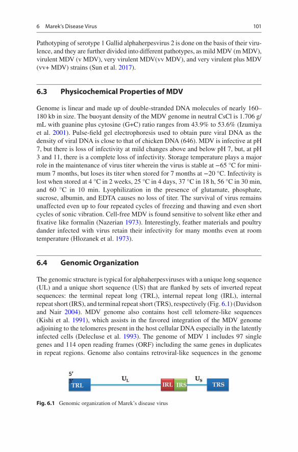
The genomic structure is typical for alphaherpesviruses with a unique long sequence (UL) and a unique short sequence (US) that are flanked by sets of inverted repeat sequences: the terminal repeat long (TRL), internal repeat long (IRL), internal repeat short (IRS), and terminal repeat short (TRS), respectively (Fig. 6.1) (Davidson and Nair 2004). MDV genome also contains host cell telomere-like sequences (Kishi et al. 1991), which assists in the favored integration of the MDV genome adjoining to the telomeres present in the host cellular DNA especially in the latently infected cells (Delecluse et al. 1993). The genome of MDV 1 includes 97 single genes and 114 open reading frames (ORF) including the same genes in duplicates in repeat regions. Genome also contains retroviral-like sequences in the genome
Fig. 6.1 Genomic organization of Marek’s disease virus
6 Marek’s Disease Virus
102
especially in serotype I which is absent in other serotypes which plays a role in transcriptional regulation (Schat and Nair 2008). Complete genome sequences of serotype 1 strains, viz., Md5, GA, BAC clone of Md11, and BAC clone of CV1988, are available in GenBank, which can be retrieved using accession numbers AF147806, AF147806, AY510475, and DQ530348, respectively (Hirai 2001). There exists a very meager difference in the genome of the above sequences, and these changes are mostly limited to the numbers of the direct repeats in the repeat regions of the genome (Schat and Nair 2008). The two serotype 1 strains vMDV GA and vvMDV Md5 strains showed little structural differences (Hirai 2001). The unique long sequence (UL) regions of GA and Md5 are very comparable in length, whereas the unique short sequence (US) regions in GA was found quite longer than Md5 (Schat and Nair 2008). The difference in the genome is due to the presence of one additional copy of small ORF2 (SORF2) in GA and a second small ORF2-like gene in Md5. The SORF1 is truncated in Md5 and is located in the repeats. The vac-cine strain CV1988 BAC contains 14 copies of the 132 bp repeat, which is the rea-son behind the viral attenuation (Silva and Gimeno, 2006). There exist differences in the two of the three promoter regions of ICP4 genes between the GA RB-1B and CV1988 strains, which leads to the difference in the transcription level of ICP4 genes between these strains. Complete analysis of the genomes of 13 strains of varying virulence of the terminal repeat long (TRL) and internal repeat long (IRL) regions showed several single-nucleotide polymorphisms (SNPs) which enables to differentiate non-attenuated and attenuated strains (Spatz and Silva 2006). In con-clusion, based on cross-hybridization studies, the genomes for all the three different serotypes are found to be collinear (Igarashi et al. 1987). Further, all these three serotypes vary significantly in their restriction endonuclease digestion patterns (Silva and Barnett 1991).
6.5 Spread
The disease is highly contagious, and the transmission occurs by the airborne route by direct or indirect contact to other susceptible hosts. The infective material is dander/feather follicle epithelium consisting of fully infective enveloped virus. The shedding of the infected material/dander occurs 2–4 weeks post infection, and shedding can continue all the way through the bird’s life span. The infectivity of the virus associated with feather debris and dander remains for months. Once the virus enters into a susceptible chicken flock, the infection spreads quickly from bird to bird despite vaccination. Indirect transmission by darkling beetles (Alphitobius diaperinus) also plays a minor role in transmission apart airborne mode of transmission. Vertical transmission of MDV through the egg is not evi-dent. Several factors influence the spread of Marek’s disease within a flock, viz., the level of initial exposure, the concentration of susceptible birds, handling, change of housing, and vaccination, and females tend to develop more tumors.
A. K. Mariappan et al.
103
6.6 Pathotyping
Pathotyping of MDV isolates is done based on the pathology the particular isolate induces in vaccinated and unvaccinated chickens and also based on their ability to overcome the effects of vaccination. This method of pathotyping described as Avian Diseases and Oncology Laboratory assay (ADOL assay) was developed at the United States Department of Agriculture (USDA). In short, ADOL assay measures the ability of the isolate to induce lymphoproliferative lesions in chickens that are vaccinated. Based on the ADOL assay, the isolates can be classified as mild MDV (m MDV), virulent MDV (v MDV), very virulent MDV(vv MDV), and very viru-lent plus MDV (vv+ MDV) strains (Witter et al. 2005). Table 6.1 mentions various reference pathotypes of MDV.
6.7 Susceptible Hosts
Several avian species including both domesticated and wild birds are susceptible to Marek’s disease. The species include chickens (Gallus gallus), domestic Japanese quail (Coturnix japonica), European common quail (Coturnix coturnix), domestic turkey (Meleagris gallopavo), common buzzard (Buteo buteo), red jungle fowl (Gallus gallus bankiva), sparrow hawk (Accipiter nisus), Ceylon jungle fowl (Gallus gallus lafayettii), mallard (Anas platyrhynchos), little owl (Athene noctua), mute swan (Cynusolor), eagle owl (Bubo bubo), and domestic goose (Anser anser).
6.8 Genetic Resistance and Susceptibility to MD
Genetic makeup of the birds especially the alleles that are blood group linked pre-cipitates the severity of the disease. Other gallinacious species, wild turkeys, and birds that belong to other orders resist infection due to their unique genetic resis-tance to MDV infection. The chicken genotype proven was associated with suscep-tibility and resistance to Marek’s disease. Owing to its genetic susceptibility/resistance to MD infection, genetic approaches to control MD are being attempted. Thus, poultry breeders have incorporated MD resistance in the selection programs for breeder stocks. Moreover, genetic resistance could be surpassed by challenge with highly virulent MDVs. MD resistance is seen in chickens expressing the B21 allele, which is linked with the major histocompatibility complex class I genes, for
Table 6.1 Various pathotypes and their respective reference strains of MDV
whose mechanism is not understood still. Contrary to the above findings, expres-sion of the major histocompatibility complex class I allele (B2) is seen in line 61 and line 71, but line 61 is resistant to MD and line 71 is susceptible to MD (Burgess et al. 2001).
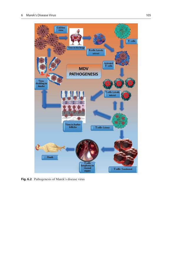
6.9 Virus Replication and Pathogenesis
Airborne route is the primary route of MDV transmission by both direct and indirect contacts between chickens. Dander consisting of feather follicular epithelial cells with fully infectious virus serves as a potential source of contamination to the envi-ronment and to other susceptible hosts (Schat and Nair 2008). The pathogenesis of MD in a susceptible host occurs in four distinct phases: (a) early cytolytic infection, (b) latent infection, (c) late cytolytic infection/immunosuppression phase, and (d) transformation phase (Calnek 1986, Schat 2000) (Fig. 6.2). Following inhalation of the virus, the virus could be detected in the spleen as early as 2 days and then get released via the feather follicle epithelium to the environment in the form of dander at around 2 weeks. The virus-encoded gB glycoprotein expressed by MDV binds to heparin sulfate of the host cell surface and is found to be the potential cellular recep-tor molecule for MDV entry (Lee et al. 2001).
Post inhalation of MD virus, early cytolytic phase occurs in B cells of the bursa of Fabricius, spleen, and thymus. Following the early cytolytic phase, latent infec-tion starts wherein the MDV becomes latent in activated T cells at 6–7 days post MDV infection, and the virus spreads throughout the entire body by the MDV- infected lymphocytes resulting in cell-associated viremia (Silva et al. 2003). The virus is disseminated to various organs including feather follicles. Then, late cyto-lytic infection occurs in the feather follicular epithelium, which disseminates infec-tious cell-free virus to the environment via feather follicle debris and dander. Few latently infected T cells consequently are transformed, leading all the way to the development of lymphoma in peripheral nerves and visceral organs (Schat 2004, Schermuly et al. 2015). The principal target cells that undergo transformation in MDV infection are the CD4+ T cells. Apart from CD4+ T cells, the virus has the ability to transform CD8+ T cells.
6.10 Genes Specific to MDV 1
Various proteins are expressed during specific phases of the infection as mentioned in the pathogenesis. The viral proteins and their potential roles are given in Table 6.2.
A. K. Mariappan et al.
105
Fig. 6.2 Pathogenesis of Marek’s disease virus
6 Marek’s Disease Virus
106
6.11 Immunobiology
6.11.1 The Innate Immune Response to MDV
The host responds to MDV infection by mounting both innate and adaptive immune mechanisms, in turn controlling the MDV infection. Interferon production plays a major protective role against MDV infection and found to be the chief mode of innate immune response. MDV is taken up by the antigen-presenting cells like den-dritic cells or macrophages present within the respiratory system, and further the virus is recognized by TLR21 leading to a cascade of events by stimulation and expression of type I interferons. An in vitro model proved that interferons could control MDV replication by reducing the plaque formation. Contrary to the mam-malian counterparts where type I interferons stimulate NK cells and enhance its cytotoxic function causing reduction in viral replication, chicken interferon-α does
Table 6.2 Genes specific to MDV and their functions (Boodhoo et al. 2016)
MDV gene (protein) Function Infection stageMDV003/078 (vIL-8)
Recruitment of immune system cells to site of viral replication
Lytic replication
MDV010 (vLIP) Forms covalent bonds with lipidsLytic replication
Lytic replication
MDV011/012 Downregulates cell surface and in turn immune evasion
Lytic replication
MDV012 ORF012 Necessary for both in vivo and in vitro viral growth
Necessary for both in vivo and in vitro replication Lytic replication
MDV073 (pp38) Early protein expressed during cytolytic infection Lytic replicationMDV092 (Us3p) Serine/threonine protein kinase that
phosphorylates pp38Lytic replication
MDV084/100 (ICP4) Viral gene transactivation function Lytic replicationMDV001a (vTR) Required for integration of viral genome into host
DNA for immune evasion, neoplastic transformation, and viremia
Latency
MDV006 (pp14) Neurovirulence factor required for PNS neuropathy
Latency
MDV062 ( VP22) Tegument protein essential for viral replication and modulates host cell cycle
Latency
MDV057 (gC/UL44) Type 1 transmembrane protein required for horizontal transmission/shedding from feather follicle epithelium
Feather follicle shedding
MDV005/076 (MEQ)
Viral oncogenic protein involved in T-cell neoplastic
Neoplastic transformation
MDV029(pUL17) Colocalizes with VP5 and VP13/14 tegument protein and essential for in vivo viral growth, capsid maturation, and DNA packaging
Neoplastic transformation
A. K. Mariappan et al.
107
not increase natural killer cell cytotoxicity. Apart from inhibition of viral replica-tion, type I IFNs are shown to be involved in stimulating the latency infection of MDV. IFN-γ induces nitric oxide production leading to inhibitory effects on MDV replication.
Dendritic cells are the proposed immune cells that mount both innate and adap-tive immunities against MDV exposure in the hosts. They initiate adaptive immu-nity by presenting MDV antigens to both MHC class I and II molecules. Macrophages, on the other hand, play a vital role in controlling viral replication and MDV-induced tumor formation. Thus, macrophages are directly involved in the inhibition of viral replication and tumor development in Marek’s disease by their potent phagocytic ability. It has proven that MDV replication inhibited effectively by macrophages collected from MDV-infected chickens than macrophages obtained from noninfected chickens. Production of inducible nitric oxide (iNOS) is the prime mechanism by which macrophages inhibit MDV infection progressing to tumor formation. Experimental evidences prove that nitric oxide production occurs in vari-ous organs like the spleen, brain, and lungs of MDV-infected chickens. In vivo experimentation of iNOS inhibition in chickens increased viral load proving the pivotal role of macrophages in MDV pathogenesis. Another main function of mac-rophages is tumoricidal activity of virus-transformed tumor cells. Macrophages from tumor tissues of MDV-infected chickens have comparable functional abilities of tumor-associated macrophages that could suppress T-cell proliferation, factors that promote in vitro tumor growth and factors that cause immunosuppression (Boodhoo et al. 2016).
Natural killer (NK) cells have functions like production of IFN-γ with potent antiviral activity and sensing of virus-infected cells and tumor cells through down-regulation of cell surface markers such as MHC I. NK cells are associated with MD resistance as MDV-susceptible chickens have greater cytotoxic capacity than the susceptible chickens.
6.11.2 The Adaptive Immune Response to MDV
As far as MDV infection is concerned, protective immunity is not conferred by antibodies and thus do not play a crucial role in the MDV infection. Among all antibodies raised against MDV glycoproteins by chicken, anti-glycoprotein B neu-tralizing antibodies block the entry of the virus into the host cells and thus play a protective role against MDV infection in chickens. Studies proved that maternal antibodies deferred both clinical signs and tumor development, but, on the other side, maternal antibody neutralizes the vaccine virus by interfering with the live replicating vaccine strains.
Cell-mediated immunity (CMI) plays a key role in combating the intracellular cell-associated virus and also offers protection against MDV after vaccination. T cells are efficient in controlling replication of virus but proved inefficient in control-ling tumor growth. Infected chickens produce CD8+ T cells against various viral proteins, viz., gB, Meq, pp38, and ICP4. Apart from CD8+ T-cell activation by viral
6 Marek’s Disease Virus
108
proteins, they also act on cytotoxic T cells to release perforins and granzymes. The activated T cells express Marek’s disease tumor-associated surface antigens (MATSA) on its surface but once reported to be MD specific but later proved that MATSA does not exclusively express on the transformed T cells. CD30 molecule, a co-stimulatory molecule, was found to be one of the MATSA antigens and had been identified with pleiotropic effects. CD30 molecule plays a major role in MDV pathogenesis by its involvement in apoptosis, cytotoxicity, T-cell activation, and regulation of T-cell migration. Certain pathways like programmed death-1 (PD1), programmed death-ligand (PD-L1), and cytotoxic T-lymphocyte-associated pro-tein- 4 (CTLA-4) get upregulated in chicken immune system cells during MDV infection. In MD pathogenesis, there is increased expression of PD-1 and CTLA-4 in the early cytolytic phase, while, at the latent phase, there is increased expression of PD-L1. In the tumorigenic phase, expression increases in both PD-1 and PD-L1 (Boodhoo et al. 2016).
6.12 Clinical Signs
The first indication of infection is the development of several nonspecific signs like weight loss, pallor, reduced feed intake, and diarrhea. The birds suffering from Marek’s disease exhibit various clinical signs. Some chickens die without showing any clinical signs. In neurolymphomatosis, most of the affected birds show varying degrees of paralysis, and asymmetric progressive paralysis of one or more of the extremities, drooping of wings, torticollis of the neck, dilatation of the crop, and/or gasping in case of vagal nerve involvement can also be seen (Singh et al. 2012). Paralyzed birds develop anorexia and die because the birds cannot reach feed and water. The eyes of birds with ocular lymphomatosis (gray eye) lose its ability to accommodate light intensity and blindness occurs. In the cutaneous form of MD, broilers are condemned at slaughter, predominantly due to the presence of numer-ous cutaneous nodules/tumors. Acute lymphomatosis do not show paralytic signs, and the tumors in the internal organs cause significant damage to the immune sys-tem leading to suboptimal performance and clinical outbreaks of other diseases such as coccidiosis, worm infestations, and Gumboro disease.
6.13 Gross and Microscopical Lesions
6.13.1 Gross Pathology
Nerve lesions and visceral lymphomas are the prime pathologic changes noticed in MD. Peripheral nerves enlarge in the affected birds. The brain does not show any gross changes, but the spinal ganglia may show gross enlargements. Almost all nerves and plexi are involved, but prominent lesions are seen in sciatic and brachial nerves (Pappenheimer et al. 1929; Payne and Biggs 1967). Almost all visceral
A. K. Mariappan et al.
109
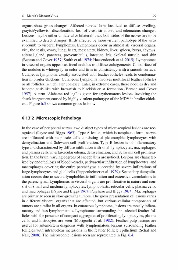
organs show gross changes. Affected nerves show localized to diffuse swelling, grayish/yellowish discoloration, loss of cross-striations, and edematous changes. Lesions may be either unilateral or bilateral; thus, both sides of the nerves are to be examined to detect changes. Birds affected by more virulent pathotype of the virus succumb to visceral lymphomas. Lymphomas occur in almost all visceral organs, viz., the testis, ovary, lung, heart, mesentery, kidney, liver, spleen, bursa, thymus, adrenal gland, pancreas, proventriculus, intestine, iris, skeletal muscle, and skin (Benton and Cover 1957; Smith et al. 1974; Haesendonck et al. 2015). Lymphomas in visceral organs appear as focal nodules to diffuse enlargements. Cut surface of the nodules is white/gray in color and firm in consistency with a smooth surface. Cutaneous lymphoma usually associated with feather follicles leads to condemna-tion in broiler chickens. Cutaneous lymphoma involves multifocal feather follicles or all follicles, which later coalesce. Later, in extreme cases, these nodules dry and become scab-like with brownish to blackish crust formation (Benton and Cover 1957). A term “Alabama red leg” is given for erythematous lesions involving the shank integument caused by highly virulent pathotype of the MDV in broiler chick-ens. Figure 6.3 shows common gross lesions.
6.13.2 Microscopic Pathology
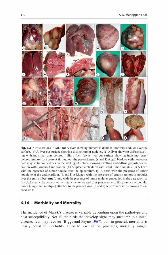
In the case of peripheral nerves, two distinct types of microscopical lesions are rec-ognized (Payne and Biggs 1967). Type A lesion, which is neoplastic form, nerves are infiltrated with neoplastic cells consisting of pleomorphic lymphocytes with demyelination and Schwann cell proliferation. Type B lesion is of inflammatory type and characterized by diffuse infiltration with small lymphocytes, macrophages, and plasma cells, interfascicular edema, demyelination, and Schwann cell prolifera-tion. In the brain, varying degrees of encephalitis are noticed. Lesions are character-ized by endotheliosis of blood vessels, perivascular infiltration of lymphocytes, and macrophages covering the entire parenchyma succeeded by severe infiltrations of large lymphocytes and glial cells (Pappenheimer et al. 1929). Secondary demyelin-ation occurs due to severe lymphoblastic infiltration and extensive vacuolations in the parenchyma. Lymphomas in visceral organs are proliferative in nature and con-sist of small and medium lymphocytes, lymphoblasts, reticular cells, plasma cells, and macrophages (Payne and Biggs 1967; Purchase and Biggs 1967). Macrophages are primarily seen in slow-growing tumors. The gross presentation of lesions varies in different visceral organs that are affected, but various cellular components of tumors are similar in all organs. In cutaneous lymphoma, lesions are mostly inflam-matory and less lymphomatous. Lymphomas surrounding the infected feather fol-licles with the presence of compact aggregates of proliferating lymphocytes, plasma cells, and histiocytes are seen (Moriguchi et al. 1982). Feather pulp lesions are useful for antemortem diagnosis with lymphomatous lesions surrounding feather follicles with intranuclear inclusions in the feather follicle epithelium (Schat and Nair, 2008). The microscopic lesions seen are represented in Fig. 6.4.
6 Marek’s Disease Virus
110
6.14 Morbidity and Mortality
The incidence of Marek’s disease is variable depending upon the pathotype and host susceptibility. Not all the birds that develop signs may succumb to clinical disease; few may recover (Biggs and Payne 1967), but, in general, mortality is nearly equal to morbidity. Prior to vaccination practices, mortality ranged
Fig. 6.3 Gross lesions in MD. (a) A liver showing numerous distinct tumorous nodules over the surface. (b) A liver cut surface showing distinct tumor nodules. (c) A liver showing diffuse swell-ing with indistinct gray-colored miliary foci. (d) A liver cut surface showing indistinct gray- colored miliary foci present throughout the parenchyma. (e and f) A gall bladder with numerous pale grayish tumor nodules on the wall. (g) A spleen showing swelling and diffuse grayish discol-oration with lymphoid infiltration. (h) A spleen embedded with solid tumor nodules. (I) A heart with the presence of tumor nodule over the epicardium. (j) A heart with the presence of tumor nodule over the endocardium. (k and l) A kidney with the presence of grayish tumorous nodules over the entire lobes. (m) A lung with the presence of tumor nodules embedded in the parenchyma. (n) Unilateral enlargement of the sciatic nerve. (o and p) A pancreas with the presence of nodular tumor (single and multiple) attached to the parenchyma. (q and r) A proventriculus showing thick-ened walls
A. K. Mariappan et al.
111
between 30% and occasionally as high as 60%. Presently, due to stringent vac-cination practices, mortality reduced less than 5% in most of the countries. Broiler industry succumbs to severe economic losses despite vaccination not due to mortality but due to condemnations.
6.15 Diagnosis of MD
In the field, diagnosis is done primarily based on the clinical signs, postmortem lesions like thickened peripheral nerves, visceral lymphoid tumors, iris/pupillary changes, and atrophy of the bursa and the thymus. Apart from conventional diagnos-tic techniques, various techniques are used to arrive at an apt and final diagnosis. Test methods available for the diagnosis of Marek’s disease and their purpose are depicted in Table 6.3.
6.15.1 Virus Isolation
Common materials used for the virus isolation are white blood cells isolated from blood samples, suspensions made from visceral lymphoma and splenocytes. Blood lymphocytes or single-cell lymphoid organ suspensions are the preferred source of
Fig. 6.4 Microscopic lesions in MD showing multifocal to diffuse areas of pleomorphic lymphoid cell infiltration in (a) the liver, (b) spleen, (c) proventriculus, (d) kidney, (e) lung, (f) heart, (g) pancreas, (h) nerve, (i) ovary (H&E, X10)
6 Marek’s Disease Virus
112
the virus from infected chickens. Chicken kidney cells (CKC) and duck embryo fibroblasts (DEF) cultures are the two preferred cell culture systems for initial isola-tion of serotype 1 MDV, whereas serotypes 2 and 3 grow well in CEF. The first attempt of MDV was first isolated in CKC and DEF cultures (Churchill and Biggs 1967, Solomon et al. 1968). Later, isolation of MDV has been carried out in MD-derived tumor transplant cells (JMCT), chicken embryonal fibroblasts (CEF), chicken embryonal liver (CEL) cell, chicken embryonal lung cell, and chicken embryonal kidney cell (CEKC) (Schat 2005). Characteristic cytopathic effects (CPE) caused by MDV include focal degeneration of infected cells owing to direct cell-to-cell transfer of the virus. The spreading of the virus occurs concentrically, so that the cells peel off with enlarged, rounded cells, surrounded by healthy tissue. Infected cells become enlarged, rounded, and highly refractile, which in due course detach from the surface. Typical plaque formation develops in inoculated cultures within 3–12 days. These plaques induced by all three serotypes are distinguished morphologically and by immunofluorescent staining.
6.15.2 Viral Markers in Tissues
Detection of the MDV antigen “Meq” is a suitable marker in the diagnosis of MD, as induced only by MDV-induced tumors and not in avian leukosis or reticuloendo-theliosis induced.
Table 6.3 Test methods available for the diagnosis of Marek’s disease and their purpose
Purpose
Method
Agent identification
Detection of immune response
HistopathologyVirus isolation
Antigen detection
Real- time qPCR AGID IFA
Population freedom from infection
n/a n/a n/a n/a n/a n/a
Individual animal freedom from infection prior to movement
n/a n/a n/a n/a n/ n/a
Contribute to eradication policies
n/a n/a n/a n/a n/a n/a
Confirmation of clinical cases
+++ + + ++ − −
Prevalence of infection surveillance
+ − − + + +
Immune status in individual animals or populations post-vaccination
− − − − − +
A. K. Mariappan et al.
113
6.15.3 Viral Antigen Detection
MD viral antigen is demonstrated in the feather tip by the use of monoclonal antibody by employing agar gel immunodiffusion (AGID) (Scholten et al. 1990). Birds infected with Marek’s disease virus express viral oncoprotein “Meq” in tissues; a dot-ELISA technique that could detect Meq protein in tissues was developed which was found to have better specificity than conventional polymerase chain reaction (Kumar et al. 2016).
6.15.4 Genomic Detection Assays
Polymerase chain reaction (PCR) and the real-time quantitative PCR (qPCR) allow the distinction between vaccine and field isolates (Handberg et al. 2001). Polymerase chain reaction (PCR)-based diagnosis from the lungs, liver, spleen, kidneys, skin, pancreas, and ovary was standardized. Serotype-specific PCRs play a vital role in differentiating serotype 1 pathogenic herpesviruses from serotype 3 HVT vaccine strain (Handberg et al. 2001). Nested PCR not only detects Meq gene of MDV in infected chickens but also could aid in differentiating highly virulent pathotypes from attenuated/vaccine MDV strain (Murata et al. 2007a, b). Apart from simple and nested PCR, quantitative real-time PCR can differentially quantify CVI988/Rispens virus vaccine strain and virulent RB-1B strain in chicken tissues, in turn understanding vaccinal efficiency (Baigent et al. 2016). MDV genome copies are quantified by real-time quantitative PCR (qPCR) (Islam et al. 2004; Baigent et al. 2005; Abdul-Careem et al. 2006). Loop-mediated isothermal amplification (LAMP) targeting the meq gene was proved to be a rapid method of diagnosis especially from feather follicles (Wei et al. 2012; Angamuthu et al. 2011). Another variation of quantitative real-time PCR is the SYBR green duplex q-PCR assay targeting the Eco-Q protein gene (meq) that could detect and quantify viral loads and distribution patterns of the virus in various organs (Zhang et al. 2015).
6.15.5 Antibody Detection
Agar gel immunodiffusion (AGID) is the most commonly used test to detect anti-body in infected chickens. Detection of antibodies to MDV was described using enzyme-linked immunosorbent assay (ELISA) (Cheng et al. 1984; Zelnik 2004). An antigen-detecting ELISA for detecting the MDV antigens in feather tips of infected chickens with better sensitivity than AGID was devised (Davidson et al. 1986).
6.16 Differential Diagnosis
Several criteria have been used by Gimeno et al. (2005) to differentially diagnose dif-ferent lymphoma-inducing viral agents, viz., Marek’s disease virus (MDV), avian leu-kosis virus (ALV), and reticuloendotheliosis virus (REV). Viral-induced lymphomas
6 Marek’s Disease Virus
114
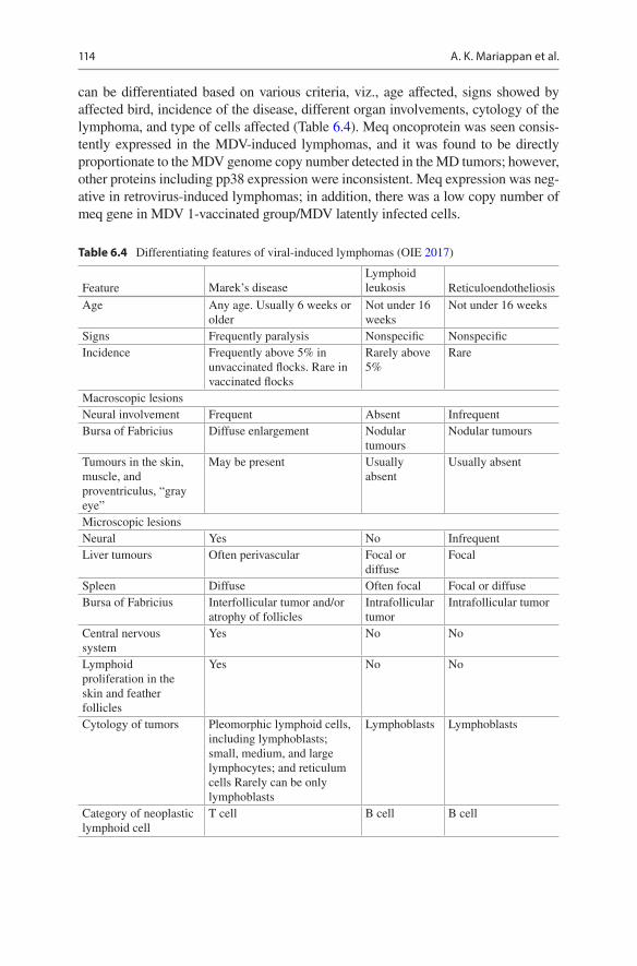
can be differentiated based on various criteria, viz., age affected, signs showed by affected bird, incidence of the disease, different organ involvements, cytology of the lymphoma, and type of cells affected (Table 6.4). Meq oncoprotein was seen consis-tently expressed in the MDV-induced lymphomas, and it was found to be directly proportionate to the MDV genome copy number detected in the MD tumors; however, other proteins including pp38 expression were inconsistent. Meq expression was neg-ative in retrovirus-induced lymphomas; in addition, there was a low copy number of meq gene in MDV 1-vaccinated group/MDV latently infected cells.
Table 6.4 Differentiating features of viral-induced lymphomas (OIE 2017)
Tumours in the skin, muscle, and proventriculus, “gray eye”
May be present Usually absent
Usually absent
Microscopic lesionsNeural Yes No InfrequentLiver tumours Often perivascular Focal or
diffuseFocal
Spleen Diffuse Often focal Focal or diffuseBursa of Fabricius Interfollicular tumor and/or
atrophy of folliclesIntrafollicular tumor
Intrafollicular tumor
Central nervous system
Yes No No
Lymphoid proliferation in the skin and feather follicles
Yes No No
Cytology of tumors Pleomorphic lymphoid cells, including lymphoblasts; small, medium, and large lymphocytes; and reticulum cells Rarely can be only lymphoblasts
Lymphoblasts Lymphoblasts
Category of neoplastic lymphoid cell
T cell B cell B cell
A. K. Mariappan et al.
115
6.17 MDV Vaccination
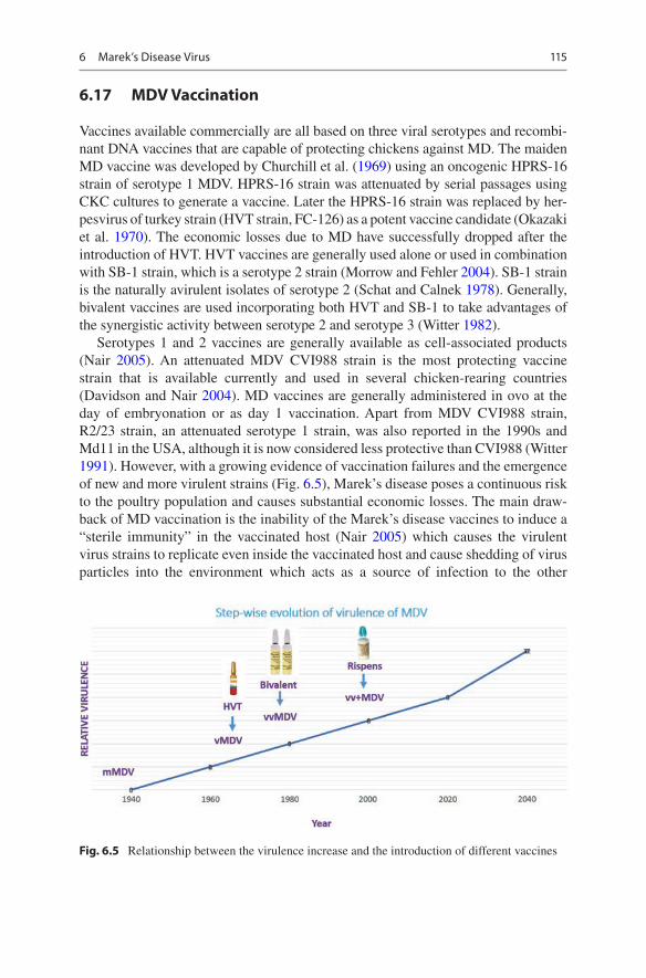
Vaccines available commercially are all based on three viral serotypes and recombi-nant DNA vaccines that are capable of protecting chickens against MD. The maiden MD vaccine was developed by Churchill et al. (1969) using an oncogenic HPRS-16 strain of serotype 1 MDV. HPRS-16 strain was attenuated by serial passages using CKC cultures to generate a vaccine. Later the HPRS-16 strain was replaced by her-pesvirus of turkey strain (HVT strain, FC-126) as a potent vaccine candidate (Okazaki et al. 1970). The economic losses due to MD have successfully dropped after the introduction of HVT. HVT vaccines are generally used alone or used in combination with SB-1 strain, which is a serotype 2 strain (Morrow and Fehler 2004). SB-1 strain is the naturally avirulent isolates of serotype 2 (Schat and Calnek 1978). Generally, bivalent vaccines are used incorporating both HVT and SB-1 to take advantages of the synergistic activity between serotype 2 and serotype 3 (Witter 1982).
Serotypes 1 and 2 vaccines are generally available as cell-associated products (Nair 2005). An attenuated MDV CVI988 strain is the most protecting vaccine strain that is available currently and used in several chicken-rearing countries (Davidson and Nair 2004). MD vaccines are generally administered in ovo at the day of embryonation or as day 1 vaccination. Apart from MDV CVI988 strain, R2/23 strain, an attenuated serotype 1 strain, was also reported in the 1990s and Md11 in the USA, although it is now considered less protective than CVI988 (Witter 1991). However, with a growing evidence of vaccination failures and the emergence of new and more virulent strains (Fig. 6.5), Marek’s disease poses a continuous risk to the poultry population and causes substantial economic losses. The main draw-back of MD vaccination is the inability of the Marek’s disease vaccines to induce a “sterile immunity” in the vaccinated host (Nair 2005) which causes the virulent virus strains to replicate even inside the vaccinated host and cause shedding of virus particles into the environment which acts as a source of infection to the other
Fig. 6.5 Relationship between the virulence increase and the introduction of different vaccines
6 Marek’s Disease Virus
116
susceptible hosts. Meq gene-deleted mutant vvMDV, a genetically modified vaccine for MD, showed effective protection in the vaccinated birds by controlling the for-mation of tumors (Lupiani et al. 2004; Li et al. 2011).
Recombinant fowl poxvirus (rFPV) and herpesvirus of turkey strain vaccines incorporated with various MDV genes showed some protective efficacy (Ross et al. 1993; Nazerian et al. 1996; Hirai and Sakaguchi 2001; Lee et al. 2005), and rFPV vaccines were also found to be effective in chickens with maternal antibodies against MDV (Lee et al. 2003). Avian myelomonocytic growth factor gene has been incorporated as a source of cytokine in rFPV, and its inclusion was found to improve vaccine efficacy against MDV (Djeraba et al. 2002). Apart from these conventional vaccines, pBAC20-engineered DNA vaccine conferred partial protection against challenge with virulent MDV (Tischer et al. 2002). Several candidate vaccines have been developed in the past, but none proved more efficacious than CV1988 vaccines (Witter and Kreager 2004).
6.18 Biosecurity in Farms Against Marek’s Disease
There are two hazard points to be taken cared of to control the MDV: (i) reducing the initial viral load on a farm by preventing the entry of MDV into the poultry rear-ing premises and (ii) preventing the exit of MDV to the environment from the affected flock (Schat and Nair 2008). Reducing the initial viral load on a farm can be possible if practicing all-in-all-out systems, avoiding multiage flocks, etc. Measures to avoid the exit of MDV to the environment from the affected flock include air management and the disposal of dead/infected chicken carcasses and manure. Biofilters are used to decrease the viral load level in the effluent air from affected poultry farms. Carcasses should be disposed of by either burning or burial. Composting of the manures has shown to be an effective method to discard both carcasses and manure (Schat and Nair 2008).
6.19 Future Perspectives
Marek’s disease is an important lymphoproliferative/neoplastic disease of poultry and can be effectively controlled by vaccination. MD serves as a potential model system to study vaccinology, viral oncogenesis, and tumor immunology. Despite effective vaccines being used to prevent this disease, the virus continues to evolve into more virulent pathotypes and, in turn, causes a serious health threat to the poul-try industry; thus, it is very much necessary to develop viable vaccine strategies that could prevent the generation of newer pathotypes. Thus, natural genetic resistance of the birds against MD should be taken into account while breeding which would help in preventing newer pathotype emergence. Apart from the conventional vacci-nation regime, strict biosecurity measures are needed on the farms, which is a criti-cal adjunct to vaccination in controlling the disease.
A. K. Mariappan et al.
117
Acknowledgments All the authors of the manuscript thank and acknowledge their respective universities and institutes.
Conflict of Interest There is no conflict of interest.
References
Abdul-Careem M, Hunter B, Nagy É et al (2006) Development of a real-time PCR assay using SYBR green chemistry for monitoring Marek’s disease virus genome load in feather tips. J Virol Methods 133:34–40. https://doi.org/10.1016/j.jviromet.2005.10.018
Angamuthu R, Baskaran S, Gopal D et al (2011) Rapid detection of the Marek’s disease viral genome in chicken feathers by loop-mediated isothermal amplification. J Clin Microbiol 50:961–965. https://doi.org/10.1128/jcm.05408-11
Baigent S, Petherbridge L, Howes K et al (2005) Absolute quantitation of Marek’s disease virus genome copy number in chicken feather and lymphocyte samples using real-time PCR. J Virol Methods 123:53–64. https://doi.org/10.1016/j.jviromet.2004.08.019
Baigent S, Nair V, Le Galludec H (2016) Real-time PCR for differential quantification of CVI988 vaccine virus and virulent strains of Marek’s disease virus. J Virol Methods 233:23–36. https://doi.org/10.1016/j.jviromet.2016.03.002
Benton W, Cover M (1957) The increased incidence of visceral lymphomatosis in broiler and replacement birds. Avian Dis 1:320. https://doi.org/10.2307/1587746
Biggs PM (1966) Avian leucosis and Marek’s disease. XIIIth World’s Poultry Congress sympo-sium papers, Kiev USSR, pp 91–118
Biggs PM, Payne LN (1967) Studies on Marek’s disease. Experimental transmission. J Natl Cancer 39:267–280
Boodhoo N, Gurung A, Sharif S et al (2016) Marek’s disease in chickens: a review with focus on immunology. Vet Res 47:119. https://doi.org/10.1186/s13567-016-0404-3
Burgess S, Basaran B, Davison T (2001) Resistance to Marek’s disease herpesvirus-induced lym-phoma is multiphasic and dependent on host genotype. Vet Pathol 38:129–142. https://doi.org/10.1354/vp.38-2-129
Calnek BW (1986) Marek’s disease: a model for herpesvirus oncology. CRC Crit Rev Microbiol 12:293–320
Cheng Y, Lee L, Smith E, Witter R (1984) An enzyme-linked immunosorbent assay for the detec-tion of antibodies to Marek’s disease virus. Avian Dis 28:900. https://doi.org/10.2307/1590266
Churchill A, Biggs P (1967) Agent of Marek’s disease in tissue culture. Nature 215:528–530. https://doi.org/10.1038/215528a0
Churchill A, Baxendale W, Chubb R (1969) the attenuation, with loss of oncogenicity, of the herpes-type virus of Marek’s disease (Strain hprs-16) on passage in cell culture. J Gen Virol 4:557–564. https://doi.org/10.1099/0022-1317-4-4-557
Davidson F, Nair V (2004) Marek’s disease-an evolving problem. Elsevier Academic Press, OxfordDavidson I, Malkinson M, Strenger C, Becker Y (1986) An improved ELISA method, using a
streptavidin- biotin complex, for detecting Marek’s disease virus antigens in feather-tips of infected chickens. J Virol Methods 14:237–241. https://doi.org/10.1016/0166-0934(86)90025-x
Delecluse HJ, Schuller S, Hammerschmidt W (1993) Latent Marek’s disease virus can be activated from its chromosomally integrated state in herpesvirus-transformed lymphoma cells. EMBO J 12:3277–3286
Djeraba A, Kut E, Rasschaert D, Quéré P (2002) Antiviral and antitumoral effects of recombinant chicken myelomonocytic growth factor in virally induced lymphoma. Int Immunopharmacol 2:1557–1566. https://doi.org/10.1016/s1567-5769(02)00115-7
Gimeno I, Witter R, Fadly A, Silva R (2005) Novel criteria for the diagnosis of Marek’s disease virus- induced lymphomas. Avian Pathol 34:332–340. https://doi.org/10.1080/03079450500179715
Haesendonck R, Garmyn A, Dorrestein G et al (2015) Marek's disease virus associated ocular lymphoma in Roulroul partridges (Rollulusrouloul). Avian Pathol 44:347–351. https://doi.org/10.1080/03079457.2015.1056088
Handberg K, Nielsen O, J⊘rgensen P (2001) The use of serotype 1- and serotype 3-specific poly-merase chain reaction for the detection of Marek’s disease virus in chickens. Avian Pathol 30:243–249. https://doi.org/10.1080/03079450120054659
Hirai K (2001) Marek’s disease. Springer, Berlin/Heidelberg/Berlin/HeidelbergHirai K, Sakaguchi M (2001) Polyvalent recombinant Marek’s disease virus vaccine against poul-
try disease. Curr Top Microbiol Immunol 255:261–287Hlozanek I, Mach O, Jurajda V (1973) Cell-free preparations of Marek’s disease virus from poul-
try dust. Folia Biol (Praha) 19:118–123Igarashi T, Takahashi M, Donovan J et al (1987) Restriction enzyme map of herpesvirus of Turkey
DNA and its collinear relationship with Marek’s disease virus DNA. Virology 157:351–358. https://doi.org/10.1016/0042-6822(87)90277-7
Islam A, Harrison B, Cheetham BF et al (2004) Differential amplification and quantitation of Marek’s disease viruses using real-time polymerase chain reaction. J Virol Methods 119:103–113. https://doi.org/10.1016/s0166-0934(04)00084-9
Izumiya Y, Jang HK et al (2001) A complete genomic DNA sequence of Marek’s disease virus type 2, strain HPRS24. Curr Top Microbiol Immunol 255:191–221
Jungherr E, Doyle LP et al (1941) Tentative pathologic nomenclature for the disease and/or for the disease complex variously designated as fowl leukemia, fowl leucosis, etc. Am J Vet Res 2:116
Kaupp B (1921) Paralysis of the domestic fowl. Poult Sci 2:25–31Kishi M, Bradley G et al (1991) Inverted repeat regions of Marek’s disease virus DNA possess a
structure similar to that of a sequence of herpes simplex virus DNA and contain host cell telo-mere sequence. J Virol 65:2791–2797
Kumar M, Barathidasan R, Palanivelu M et al (2016) A novel recombinant Meq protein based dot- ELISA for rapid and confirmatory diagnosis of Marek’s disease induced lymphoma in poultry. J Virol Methods 236:271–280. https://doi.org/10.1016/j.jviromet.2016.08.007
Lee S, Ohashi K, Sugimoto C, Onuma M (2001) Heparin inhibits plaque formation by cell- free Marek’s disease viruses in vitro. J Vet Med Sci 63:427–432. https://doi.org/10.1292/jvms.63.427
Lee L, Witter R, Reddy S et al (2003) Protection and synergism by recombinant fowl pox vac-cines expressing multiple genes from Marek’s disease virus. Avian Dis 47:549–558. https://doi.org/10.1637/6073
Lee L, Cui X, Cui Z et al (2005) Characterization of a very virulent Marek’s disease virus mutant expressing the pp38 protein from the serotype 1 vaccine strain CVI988/Rispens. Virus Genes 31:73–80. https://doi.org/10.1007/s11262-005-2202-2
Li Y, Sun A, Su S et al (2011) Deletion of the meq gene significantly decreases immunosup-pression in chickens caused by pathogenic Marek’s disease virus. Virol J 8:2. https://doi.org/10.1186/1743-422x-8-2
Lupiani B, Lee L, Cui X et al (2004) Marek’s disease virus-encoded Meq gene is involved in transformation of lymphocytes but is dispensable for replication. P Natl Acad Sci 101:11815–11820. https://doi.org/10.1073/pnas.0404508101
Marek J (1907) Multiple neuritis (polyneuritis) in chickens. Ger Vet Wkly 15:417–421Moriguchi R, Fujimoto Y, Izawa H (1982) Chronological observations of feather pulp
lesions in chickens inoculated with Marek’s disease virus. Avian Dis 26:375. https://doi.org/10.2307/1590108
Morrow C, Fehler F (2004) Marek’s disease a worldwide problem. In: Davison F, Nair V (eds) Marek’s disease. An evolving problem. Elsevier Academic Press, London, pp 8–16
Murata S, Chang K, Lee S et al (2007a) Development of a nested polymerase chain reaction method to detect oncogenic Marek’s disease virus from feather tips. J Vet Diagn Invest 19:471–478. https://doi.org/10.1177/104063870701900503
Murata S, Chang K, Yamamoto Y et al (2007b) Detection of the virulent Marek’s disease virus genome from feather tips of wild geese in Japan and the Far East region of Russia. Arch Virol 152:1523–1526. https://doi.org/10.1007/s00705-007-0982-5
Nair V (2005) Evolution of Marek’s disease – A paradigm for incessant race between the pathogen and the host. Vet J 170:175–183. https://doi.org/10.1016/j.tvjl.2004.05.009
Nazerian K (1973) Studies on intracellular and membrane antigens induced by Marek’s disease virus. J Gen Virol 21:193–195. https://doi.org/10.1099/0022-1317-21-1-193
Nazerian K, Witter R, Lee L, Yanagida N (1996) Protection and synergism by recombinant fowl pox vaccines expressing genes from Marek's disease virus. Avian Dis 40:368. https://doi.org/10.2307/1592234
Office International des Epizooties (2017). Chapter 2.3.13. Marek’s disease. In: Manual of diag-nostic tests and vaccines for terrestrial animals
Okazaki W, Purchase H, Burmester B (1970) Protection against Marek’s disease by vaccination with a Herpesvirus of Turkeys. Avian Dis 14:413. https://doi.org/10.2307/1588488
Pappenheimer A (1929) Studies on fowl paralysis (neurolymphomatosis gallinarum): i. Clinical features and pathology. J Exp Med 49:63–86. https://doi.org/10.1084/jem.49.1.63
Pappenheimer AM, Dunn LC, Cone V (1929) Studies on fowl paralysis (Neurolymphomatosis gallinarum): I. Clinical features and pathology. J Exp Med 49:63–86
Payne LN, Biggs PM (1967) Studies on Marek’s disease. II. Pathogenesis. J Natl Cancer 39:281–302
Purchase HG, Biggs PM (1967) Characterization of five isolates of Marek’s disease. Res Vet Sci 8:440–449
Ross L, Binns M, Tyers P et al (1993) Construction and properties of a turkey herpesvirus recom-binant expressing the Marek's disease virus homologue of glycoprotein B of herpes simplex virus. J Gen Virol 74:371–377. https://doi.org/10.1099/0022-1317-74-3-371
Schat K (2000) Specific and nonspecific immune responses to Marek’s disease virus. Dev Comp Immunol 24:201–221. https://doi.org/10.1016/s0145-305x(99)00073-7
Schat K (2004) Understanding Marek’s disease immunity: a continuing challenge. Int J Poult Sci 3:89–95. https://doi.org/10.3923/ijps.2004.89.95
Schat K (2005) Isolation of Marek’s disease virus: revisited. Avian Pathol 34:91–95. https://doi.org/10.1080/03079450500059289
Schat K, Calnek B (1978) Characterization of an apparently nononcogenic Marek’s disease virus. J Natl Cancer Inst 60:1075–1082. https://doi.org/10.1093/jnci/60.5.1075
Schat AK, Nair V (2008) Marek’s disease. In: Saif YM, Fadly AM, Glisson JR, McDougald LR, Nolan LK, Swayne DE (eds) Diseases of poultry, 12th edn. Iowa, Iowa State University Press/Blackwell Publishing, pp 452–512
Schermuly J, Greco A, Härtle S et al (2015) In vitro model for lytic replication, latency, and transformation of an oncogenic alphaherpesvirus. P Natl Acad Sci 112:7279–7284. https://doi.org/10.1073/pnas.1424420112
Scholten R, Hilgers L, Jeurissen S, Weststrate M (1990) Detection of Marek's disease virus antigen in chickens by a novel immunoassay. J Virol Methods 27:221–226. https://doi.org/10.1016/0166-0934(90)90138-6
Silva R, Barnett J (1991) Restriction endonuclease analysis of Marek’s disease virus DNA: dif-ferentiation of viral strains and determination of passage history. Avian Dis 35:487. https://doi.org/10.2307/1591212
Silva R, Gimeno I (2006) Oncogenic Marek’s disease viruses lacking the 132 base pair repeats can still be attenuated by serial in vitro cell culture passages. Virus Genes 34:87–90. https://doi.org/10.1007/s11262-006-0022-7
Silva R, Reddy S, Lupiani B (2003) Expansion of a unique region in the Marek’s disease virus genome occurs concomitantly with attenuation but is not sufficient to cause attenuation. J Virol 78:733–740. https://doi.org/10.1128/jvi.78.2.733-740.2004
Singh S, Barathidasan R, Kumar A et al (2012) Recent trends in diagnosis and control of Marek’s Disease (MD) in poultry. Pak J Biol Sci 15:964–970. https://doi.org/10.3923/pjbs.2012.964.970
Smith TW, Albert DM et al (1974) Ocular manifestations of Marek’s disease. Invest Ophthalmol 13(8):586–592
Solomon J, Witter R, Nazerian K, Burmester B (1968) Studies on the etiology of Marek’s dis-ease. I. propagation of the agent in cell culture. Exp Biol Med 127:173–177. https://doi.org/10.3181/00379727-127-32649
Spatz S, Silva R (2006) Polymorphisms in the repeat long regions of oncogenic and attenu-ated pathotypes of Marek’s disease virus 1. Virus Genes 35:41–53. https://doi.org/10.1007/s11262-006-0024-5
Sun G, Zhang Y, Lv H et al (2017) A Chinese variant Marek’s disease virus strain with divergence between virulence and vaccine resistance. Viruses 9:71. https://doi.org/10.3390/v9040071
Tischer B, Fehler F, Osterrieder K et al (2002) A DNA vaccine containing an infectious Marek’s disease virus genome can confer protection against tumorigenic Marek’s disease in chickens. J Gen Virol 83:2367–2376. https://doi.org/10.1099/0022-1317-83-10-2367
Van der Walle N, Winkler-Junius E (1924). De-neuritis epizoötiebijte Barneveld tilt in the 1921stWei X, Shi X, Zhao Y et al (2012) Development of a rapid and specific loop-mediated isothermal
Witter R (1982) Protection by attenuated and polyvalent vaccines against highly virulent strains of Marek’s disease virus1. Avian Pathol 11:49–62. https://doi.org/10.1080/03079458208436081
Witter R (1991) Attenuated revertant Serotype 1 Marek’s disease viruses: safety and protective efficacy. Avian Dis 35:877. https://doi.org/10.2307/1591624
Witter R, Kreager K (2004) Serotype 1 mutagenesis: approaching the threshold of vaccine efficacy in Marek’s disease. Avian Dis 48:768–782. https://doi.org/10.1637/7203-050304r
Witter RL, Calnek BW et al (2005) Classification of Marek’s disease viruses according to pathot-ype -philosophy and methodology. Avian Pathol 34:75–90
Zelnik V (2004) Diagnosis of Marek’s disease. In: Davison F, Nair V (eds) Marek’s disease-an evolving problem. Elsevier Academic Press, London, pp 157–167
Zhang Z, Liu S, Ma C et al (2015) Absolute quantification of a very virulent Marek's disease virus dynamic quantity and distributions in different tissues. Poult Sci 94:1150–1157. https://doi.org/10.3382/ps/pev063