Assisting the cattle industry in solving challenging herd health problems using cutting-edge technology and personalized customer support. Bovine Disease Diagnostic Manual A FIELD GUIDE TO DIAGNOSING COMMON BOVINE DISEASES FIRST EDITION Editor: David H. Zeman, DVM, PhD, DACVP Custom Made Vaccines
Transcript
Assisting the cattle industry in solving challenging herd health problems using cutting-edge technology and personalized customer support.
Bovine Disease Diagnostic Manual
A FIELD GUIDE TO DIAGNOSING COMMON BOVINE DISEASES
FIrST EDITION
Editor: David H. Zeman, DVM, PhD, DACVP
Custom Made Vaccines
3 |www.newportlabs.com | 800-220-2522
Table of Contents
As the nation’s largest manufacturer of custom made vaccines, Newport Laboratories is a highly focused, technology–based company dedicated to providing timely, science–based solutions to food animal disease problems. We do this by providing customers with industry-leading science backed by PINPOINT® Technologies. Our products and services are delivered and supported by a dedicated and experienced sales staff and Veterinary Technical Service team.
Copyright Newport Laboratories, Inc. All rights reserved.
For bacterial culture, we recommend swabs with transport media to prevent desiccation.For virus isolation, swabs should be placed into viral transport media; call us for information.
Tissues-FreshAseptically collect approximately 2x4 inch samples and place in a plastic bag. Sample visible lesions with adjacent normal tissue. Double bag in Whirl-pak® bags. Do not mix swabs, intestines, or brains with other tissues in one single bag. Transport tissues with 2-3 cold packs in an insulated container. It is important that the tissue samples arrive at the laboratory before the cold packs expire.
Collect sections of small and large intestine. The selected, clearly identified samples should be double bagged and sealed in Whirl-pak bags to prevent spillage. Do not longitudinally cut the loops of intestines open. The intestine, approximately 2 inches long, should be refrigerated and cooled thoroughly prior to shipping. Avoid shipping over the weekends or holidays.
SwabsAerobic CultureCommercial swabs with Stuart’s or Amies transport media is recommended to prevent desiccation.
Anaerobic CulturePort-A Cult® (BBL) or other anaerobic transport system. (The Port-A Cult® tube can be used for anaerobic, facultative, and aerobic bacteria.) For abscesses or exudates use a capped syringe with needle removed or a tube with a snug cap.
Nasal Swabs-Bacterial SuspectClean the external nares and internal nostrils with a moist towel to remove common contaminants. (Use swabs with transport media such as Amies or Stuart’s). Insert swab into the pre-cleaned nasal cavity and rotate. Upon successful sample collection, the swab is inserted into the accompanying sterile plastic sheath. The ampule located at the end of the sheath is gently crushed, releasing transport medium.
Nasal Swabs-Viral SuspectPrepare nostrils and sample as in bacterial suspect. For viral swabs use Viral Culturette® (Becton Dickinson #4361514) or equivalent.
Use of the incorrect swab and media may jeopardize the ability to detect or culture the offending pathogen. For bacterial isolation, avoid using Mycoplasma sp. or viral media which contain antimicrobials and may inhibit growth of the desired pathogen. Avoid using bacterial culture media to isolate viruses or Mycoplasma sp. organisms.
Identify all swabs with the following:• Farm ID, including site and pasture/lot
where appropriate• Animal identification number
HistopathologyPreparation of Tissue for FixationMultiple sites or types of lesions, to include both normal and diseased tissue and a sample at the line of demarcation, should be taken. The sections should be no more than 1 inch thick. The small size of the tissue results in rapid and complete penetration of the fixative.
Selected tissues should be cut with a sharp knife or scalpel since the squeezing action of scissors crushes and tears tissue. Autolysis or freezing will make samples unsuitable for histopathological evaluation. Place formalin and tissues in double Whirl-paks. Identify bags if multiple animals are submitted. Do not use narrow mouth bottles to submit fixed tissues.
Whenever possible, animals selected for laboratory analysis should be free from antibiotic therapy and in an early or acute disease stage. Selected tissues should be collected as aseptically as possible. A meaningful history of the disease outbreak and a tentative diagnosis, based upon clinical evaluation and necropsy findings, should be included. Laboratory test results are directly affected by animal selection, necropsy technique, specimen selection and specimen handling, including preservation and shipment to the laboratory. Contact Newport Laboratories if you have any questions regarding sample collection or the diagnostic process.
Preparation & Collection of Tissues/Samples
For any materials submitted to Newport Laboratories for analysis, Newport Laboratories solely owns the work developed or derived from the materials submitted as unique work product and an invention by Newport Laboratories. All written materials and other works which may be subject to copyright, and all patentable and unpatentable inventions, ideas, improvements, or discoveries conceived or made by Newport Laboratories arising out of the developments shall be the sole and entire property of Newport Laboratories. Any and all intellectual property rights related to the vaccine and the development of the vaccine belong solely to Newport Laboratories.
Volume of FixativeThe selected tissues should be fixed in 10% neutral buffered formalin. Use 10 times the volume of the tissues being fixed to assure good perfusion of the sample and to maintain the tissue architecture. After 24 hours fixation, excess formalin can be poured off, and a smaller formalin volume can then be used for shipping.
Formula to make 10% Neutral Buffered Formalin37-40% formaldehyde 100 mL Distilled water 900 mL Sodium phosphate, monobasic monohydrate 4.0 g Sodium phosphate, dibasic anhydrous 6.5 g
Tissue Selection for HistopathologyCheck the recommended samples in the guideline table on pages 4 & 5. If the cause of death is unknown or the clinical syndrome is vague, then submit samples exhibiting suspected gross lesions and the following tissues: heart, liver, lung, kidney, spleen, various levels of the gastrointestinal tract, mesenteric lymph nodes, and brain.
If hollow organs (gut or uterus) retain significant amounts of content, then they should be gently flushed with 10% formalin without disturbing the mucosal lining before placing in the formalin bag. Be sure to take proper precautions when handling formalin.
I.D. & Handling of Blood Samples:Collection of Blood Samples
• Collect in sterile tubes. Serum separator tubes work well. Follow the manufacturer’s directions. Based on the number of tests requested, 1 mL – 3 mL of nonhemolyzed serum is required.
• Fill vacutainer tubes 3/4 full and allow to stand at room temperature for an hour to permit a solid clot to form and retract.
• Pipette the serum into sterile tubes with snap caps (3 mL plastic tubes with snap caps, Falcon #2054, are recommended). Make sure caps are securely closed.
• Use permanent markers and underline the I.D. numbers (e.g. 16 vs. 91).
• Do not freeze whole blood or samples with the clot remaining.
• Contaminated or toxic samples cannot be used in virus isolation tests. Many serology tests are adversely effected by hemolysis.
I.D. Samples on Submission Forms• Using one form per client and site, identify
the tubes on the submission request form by different barns, or age groups as logical for the diagnostic investigation.
• Clearly specify the test(s) requested on the submission form.
• When sending paired sera, identify the acute samples from the convalescent samples on the tube and on the request form.
Diagnostic submission forms can be downloaded from our website: www.newportlabs.com, or by calling Newport customer service at 800-220-2522.
Packing SpecimensTo avoid leaking in transit, double bag ALL samples. Whirl-pak bags or equivalent are recommended. Wrap sample bags and 2-4 ice packs in absorbent paper (e.g. newspaper). Place the package into a styrofoam container. Completed submission forms should be inserted in a separate bag in case of leakage and clearly attached to the matching specimens. This is especially important if your container contains specimens from multiple clients or sites. Avoid mixing intestinal samples with other tissues. If you need more information about shipping specimens to Newport’s Diagnostic Laboratory, please call us at 800-220-2522.
MailingNewport Laboratories provides free diagnostic kits for sample submission. Call us at 800-220-2522 to request submission form(s) or shipping containers. Submission forms are also available online at www.newportlabs.com. Samples should be submitted by the fastest means possible to avoid deterioration of specimens. Next day or overnight delivery is preferred. The most reliable services that we have found are listed below:
• Fed Ex• United Parcel Service (UPS)• Spee-Dee• U.S. Parcel Post (only as a final option)
Laboratory HoursThe Newport Diagnostic Laboratory is open for service from 8:00 A.M. to 5:00 P.M. (CST) Monday through Friday, with the exception of holidays.
The Dx-rEPOrTS is a secure site which allows veterinarians to view diagnostic testing results where they want and when they want. This system allows veterinarians to easily organize and distribute diagnostic results. Livestock producers receive pertinent information from their veterinarian in a precise and understandable format.
Dx-REPORTS provides numerous features and benefits:• Password protected site maintains confidentiality of all diagnostics information• Complies with producer’s and attending veterinarian’s privacy requirements • Accessible from any computer location at any time • Easy to navigate interface • Applications for all food animals and cervidae• Ease of collecting, distributing and banking individual production site data• Data analysis to identify disease trends within groups and between groups • Left margins icons and header action button provides data management menu options • real-time database allows you to see results and status of submissions
Call Newport Laboratories at 800-220-2522
for more information about Dx-REPORTS capabilities.
Diagnostic Reporting & Management Software
Online Web Portal
Report Date:
Accession No: 0000001Final Report
Case Coordinator: Dr. Randy ShirbrounDate Received: 01/01/2014Collection Date: 01/01/2014
Lab FindingsSpecimen Test Name Result
Associated Parties: Vet Practice:Veterinarian:Producer:System User:
Reference Data: Site:
Animal Information: Bovine, 10-11 months
Report TypePreliminaryInterim
Delivery MethodFaxFax
Date Sent2/1/2014 9:29:22 AM2/24/2014 9:29:22 AM
Client Report History
Company NameAddressCity, ST, ZIP
Page 1 of 1 - Final (3/4/2014) Newport Laboratories, Inc. Accessin No: 0000001
Feedlot bovine necropsies will require ribs to be cut from the thorax with a rib cutter. Cut each rib free along the back and along the sternum, and then reflect the rib cage away to expose the lungs. The necropsy can be continued by dissecting free the trachea and larynx, and then removing the entire pluck (lung, heart, trachea, larynx) as a unit.
Cut the muscle between each rib and snap them one at a time towards the back, to expose the thoracic organs.
Thoracic organs are now easily examined and sampled.
Carefully open the abdominal wall, and puncture the peritoneum. Use the butt of the knife to push the guts out of the way as you extend it towards the ventral midline, then towards the pelvic canal.
Dissect the flap of abdominal wall you just created away towards the back. The abdominal organs are now exposed. To expose the thoracic organs cut through the sternum.
Clinical Signs & History• Sudden death is often the first indication of H. somni infection in a
feedlot animal.• Thromboembolic Meningoencephalitis (TEME) is the result of septic
bacterial infarcts within the brain, especially the cerebral cortex and brainstem. Animals with the nervous form exhibit profound depression; often the most noticeable clinical sign of histophilosis.
• Fever is also a common finding; however, animals diagnosed with undifferentiated fever may be suffering from Mannheimia pneumonia, Histophilosis, or both.
• Specifically, animals affected with the fibrinous pleuritic form of the disease may exhibit extreme labored breathing.
• Animals with inflammation of the heart (myocarditis) may exhibit sudden collapse and death on exertion (eg, being moved through a handling facility).
• Other findings are determined by the system(s) involved and may include rapid respiration, stiffness, lameness, muscle weakness, lack of coordination, paralysis, and eye twitching.
• Animals found dead and confirmed with H. somni infection often have a history of having been treated for undifferentiated fever or depression in the previous 14 days.
Consolidated lung with severe pleuritis in a chronic case. Note small abscesses in lung (yellow dots) which represent pus filled airways, very typical of H. somni.
Consolidated lung - early case of H. somni pneumonia with pleuritis.
H. somni laryngitis and proximal tracheitis. Note fibrinous exudate on surface with ulceration. This lesion is seen in many cattle that become septicemic and develop TEME, myocarditis or polyarthritis. Can grossly be confused with IBr.
H. S
OM
NI
Tissues to Submit• Affected lung• Myocardium• Joint swabs (if involved)• Brain including
Age of Occurrence• Histophilosis is a common disease in North America, primarily northern
cattle feedlots. • All feedlot cattle are at risk of histophilosis for the duration of the
feeding period.• It also is seen sporadically in individual beef and dairy cattle worldwide.
Diagnosis• Be suspicious if the herd has cattle showing one or more of the following:
pneumonia, CNS disease, sudden death related to heart failure, and lameness.
• Necropsy and organ examination for the following: bronchopneumonia with small abscesses and pleuritis; purulent myocarditis; hemorrhagic septic infarcts in brain stem and brain (TEME is Thromboembolic Meningoencephalitis).
• Isolation of the organism from lung, CSF (Cerebral Spinal Fluid), brain, blood, urine, joint fluid, or other sterile, internal organs or fluids confirms the diagnosis.
Necropsy view of two TEME lesions (arrows) in the cerebral cortex. These necrotic septic infarcts appear on the surface as red foci, usually less than 1cm. Confirm these are real lesions and not just agonal hemorrhage by sectioning into the lesion which should extend into the parenchyma. Meningeal hemorrhages are superficial only.
Clinical Signs & History• BrSV infection usually begin 3 to 5 days after cattle are exposed
to the virus.• BrSV commonly occurs with secondary bacteria, as is often the case
with other respiratory virus infections in cattle.• Infected cattle have a watery to thick mucous discharge from the
nose and eyes.• Decreased appetites or go off feed, and appear slightly depressed.• In pastured cattle that are not seen daily, sudden death may be the first
sign of BrSV infection.• If the disease progresses cattle may develop a dry cough and have
difficulty breathing.• Frequently breath with open mouths, with tongues hanging to the
side of the mouth.• Saliva may be frothy and blood tinged.
Age of Occurrence• Occurs predominately in young beef and dairy cattle .• BrSV can infect and cause disease in all ages of cattle, although
suckling calves often experience the most severe disease.
Diagnosis• Increased temperatures between (104º - 108.5° F) with increased
breathing rates (>40 per minute).• Affected cattle are frequently seen near water troughs, but have trouble
drinking due to difficult breathing.• The course of clinical disease may last 1 to 2 weeks.• A diagnosis of BRSV requires laboratory confirmation.• Improve chances of virus isolation when cattle are sampled during
incubation or acute phase.
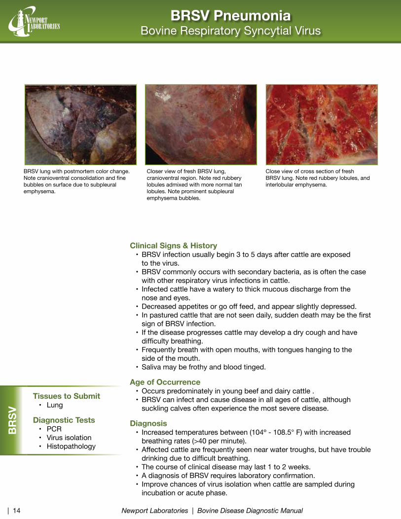
Close view of cross section of fresh BrSV lung. Note red rubbery lobules, and interlobular emphysema.
BrSV lung with postmortem color change. Note cranioventral consolidation and fine bubbles on surface due to subpleural emphysema.
Closer view of fresh BrSV lung, cranioventral region. Note red rubbery lobules admixed with more normal tan lobules. Note prominent subpleural emphysema bubbles.
Clinical Signs & History• Calf diphtheria (necrotic laryngitis) usually occurs sporadically and
typically affects a small percentage of cattle.• Found primarily in feedlot cattle.• Fusobacterium necrophorum is the primary pathogen; occasionally other
organisms may be present, most often Trueperella pyogenes.• Clinical presentation consists of dyspnea and a harsh, painful cough.• The cough, as well as efforts to inhale, may produce loud
“honking” sounds.• Exertion may result in sudden death due to upper airway obstruction.• Laryngeal contact ulcers, which commonly occur in feedlot cattle due
to a variety of causes (sever coughing, irritation from forages, etc.) may predispose them to infection.
Diagnosis• Lesions noted at necropsy are typically associated with the larynx.• Lesions may vary from edema and hyperemia in the acute form, to areas
of necrosis and caseous granulation in chronic cases.• Differential diagnoses include pharyngeal trauma and infectious bovine
Clinical Signs & History• respiratory form clinical signs range from mild to severe depending on
the presence of secondary bacterial pneumonia.• Coughing, difficulty inhaling, breathes rapidly, profuse watery
nasal discharge.• Nasal discharge becomes thicker and darker as the infection progresses.• If encrusted nostrils are rubbed off, the underlying tissue appears very
red and inflamed hence the term “red nose”.• Progression of the disease is marked by increasing nasal encrustation.• Depression, high body temperature (104º to 108º F).• Decreased appetite, rapid weight loss and may have diarrhea.• Incubation period is 2 to 6 days.• Other forms are pinkeye, abortion, and infectious pustular
vulvovaginitis (IPV).• IBr virus is one of the most common agents in the Bovine respiratory
Disease Complex of feedlot cattle.• Keeping cattle in close contact is an ideal situation for virus to
spread rapidly.
Age of Occurrence• Susceptible cattle of all ages but can be severe in young calves.
Diagnosis• Lesions are restricted to upper respiratory tract and trachea appearing
as small hemorrhages. • Lesions are found in the mucous membrane of nasal cavity and the
paranasal sinuses. • Samples taken for virus isolation should be taken early in the disease.• Lesions: Acute - tracheal pseudomembrane and ulcerative rhinitis.
Chronic - secondary bacterial bronchopneumonia.
Hyperemic (red) inflamed necrotic tracheal mucosa with thick adherent mucopurulent exudate on surface.
Hyperemic (red) inflamed nasal turbinates mucosa with adherent mucopurulent exudate on surface.
Clinical Signs & History• The first clinical signs observed in calves affected by Mannheimia
haemolytica are vague and often limited to a slight depression and lack of interest in eating. As the disease rapidly progresses, the calf refuses to eat, becomes depressed, exhibits lowered or drooped head and ears, and suffers increasing nasal discharge which changes in consistency from thin and clear to thick yellow and viscous.
• Body temperature may rise to as high as 107°F, with breathing often rapid and labored.
• Mannheimia haemolytica, serotype 1 and serotype 6, is the bacterium most frequently isolated from the lungs of cattle with the Bovine respiratory Disease complex (BrD).
• A cough may be noted early in the disease; however, as lung damage increases, coughing and breathing become very painful for the animal.
• If the disease process is not stopped, the lungs become irreversibly damaged, the body temperature drops to below normal and the animal usually dies.
• Commonly seen in stressed high risk calves infected with BrD viruses or severe stress such as recent transport.
Age of Occurrence• Young and growing cattle.
Diagnosis• The diagnosis is confirmed by culture from the lung with characteristic
lesions; also check lung for common viral copathogens such as IBr, BVD, BrSV or PI3.
• Lung specimens can be collected for culture at postmortem. If possible, specimens for culture should be collected from animals that have not been treated with antibiotics.
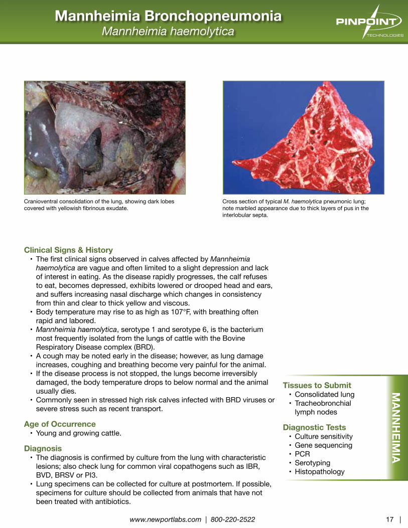
Cranioventral consolidation of the lung, showing dark lobes covered with yellowish fibrinous exudate.
Cross section of typical M. haemolytica pneumonic lung; note marbled appearance due to thick layers of pus in the interlobular septa.
Clinical Signs & History• Mannheimia haemolytica, serotype 1 is the bacterium most
frequently isolated from the lungs of cattle with BrD. Although less frequently cultured, Pasteurella multocida is also an important cause of bacterial pneumonia usually preceded by signs of viral infection of the respiratory tract.
• Fever (104-106°F) serous to mucopurulent nasal discharge; moist cough; and a rapid, shallow respiratory rate may be noted.
Age of Occurrence• Cattle of all ages affected with BrD virus.• Young dairy calves especially, but also pasture calves.
Diagnosis• Diagnosis relies on bacterial culture. Because the bacteria involved
are normal inhabitants of the upper respiratory tract, the specificity of culture can be increased by collecting antemortem specimens from the lower respiratory tract by tracheal swab, transtracheal wash, or bronchoalveolar lavage.
• P. multocida is associated with purulent bronchopneumonia, only small amounts of fibrin exudation, some thrombosis, limited lung necrosis, and suppurative bronchitis and bronchiolitis.
• Lung specimens can be collected for culture at postmortem. If possible, specimens for culture should be collected from animals that have not been treated with antibiotics.
Cranioventral consolidation of the lung, with minimal pleuritis
AIP in feedlot bovine. Note dorsal and caudal lung is puffed up, not collapsed, tends to fill the thorax. Note rounded caudal margins. Cranioventral lung has collapsed and palpates rubbery, not as firm as bacterial bronchopneumonia.
Emphysema & Edema syndrome (ABPEE) are not infectious diseases, but are often clinically confused with infectious pneumonias.
• Sudden onset of respiratory distress or sudden death within a few days of new feed source.
• Can be associated with sudden dietary changes to lush green forages such as alfalfa, rape, kale, or turnip tops, certain toxic plants (such as Purple Mint), moldy feeds (such as moldy sweet potato).
• The tryptophan in forages is metabolized to a pulmonary toxin, 3-methylindole.
Age of Occurrence• Cattle with functional rumens are susceptible.
Diagnosis• The lungs are markedly distended, do not collapse; and feel rubbery, not
hard consolidation.• The pleura is normal, but interlobular septa are distended with gas
vacuoles or edematous fluid.
Cross section of AIP lung. Note all lobules are expanded, some collapsed and congested red lobules, some air- filled tan lobules. Note interlobular gas bubbles and diffusely edematous and heavy lung.
AIP lung. Note the entire lung sits high, fills the thorax, and has not collapsed like a normal lung would during necropsy. The dorsal lung feels spongy. The ventral lung is semi-firm and rubbery; not hard consolidation.
Bovine Virus Diarrhea and Mucosal DiseaseBVD Virus
Clinical Signs & History• BVD virus infection can be involved with many disease syndromes
including pneumonia, severe ulcerative alimentary disease commonly called Mucosal Disease in Persistently Infected animals, abortions and birth defects.
• Acute transient (not PI) BVD can present as a mild or severe disease syndrome. In the mild form cattle may show depression, decreased milk production, transient inappetence, rapid respiration, excessive nasal secretion, excessive lacrimation, and diarrhea.
• In severe acute transient BVD, cattle may show high fever (~107°F), oral ulcers, weeping dermatitis of the coronary band and interdigital cleft, diarrhea, dehydration, leukopenia, thrombocytopenia and associated petechiation on mucosal surfaces, swollen lymph nodes, erosions and ulcerations of the GI tract. Morbidity and mortality may be high.
• Mucosal disease is a highly fatal form of BVD that may be acute or chronic and occurs when persistently infected cattle become superinfected with cytopathic BVD virus. Acute mucosal disease is characterized by fever, leukopenia, diarrhea, anorexia, dehydration, erosive lesions of the nares and mouth, and death within a few days of onset. Erosions and ulcers may be found throughout the GI tract with extensive necrosis of lymphoid tissues, especially Peyer’s patches.
• Transmission of the pathogenic virus can occur through secretions from a transiently or a persistently infected animal.
Age of Occurrence• Can affect cattle of all ages.• Commonly associated with young cattle with pneumonia. • Mucosal Disease cattle are usually under 2 years old.
Diagnosis• Diagnosis is confirmed by compatible gross and microscopic lesions and
virus identification via isolation, PCR, or IHC tests.• Samples taken for virus isolation should be taken early in the disease.
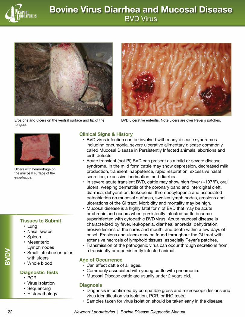
Erosions and ulcers on the ventral surface and tip of the tongue.
Ulcers with hemorrhage on the mucosal surface of the esophagus.
BVD ulcerative enteritis. Note ulcers are over Peyer’s patches.
BVD
V
Tissues to Submit• Lung • Nasal swabs• Spleen• Mesenteric
• Calves may appear unthrifty and have fecal-stained perineal areas. In light infections, the most characteristic sign of clinical coccidiosis is watery feces, with little or no blood, and the animal shows only slight discomfort for a few days.
• Severely affected cattle develop thin, bloody diarrhea that may continue for >1 wk, or thin feces with streaks or clots of blood, shreds of epithelium, and mucus. They may develop a fever; become anorectic, depressed, and dehydrated; and lose weight.
• During the acute period, some cattle die; others die later from secondary complications (e.g., pneumonia).
• Outbreaks usually occur within the first month of confinement.• Incubation period is 17-21 days.• range cattle outbreaks related to severe weather stress and crowding
around a limited water source. • Severe epidemics have been reported in feedlot cattle.
Cryptosporidiosis• Diarrhea – profuse water, mucoid, and green, occasionally bloody.• Colic and pain.• Depression, loss of appetite, weight loss.
Age of OccurrenceCoccidiosis
• Young cattle (1-2 mo to 1 yr) and usually is sporadic during the wet seasons of the year.
Cryptosporidiosis• Calves between 5 days and two weeks of age.• Inconsequential in animals older than a month old, because by this age
most animals will have become immune to infection.
DiagnosisCoccidiosis
• Finding oocysts on fecal flotation or direct smear or by the McMaster’s technique.
• Differential diagnoses include salmonellosis, bovine virus diarrhea, malnutrition, toxins, or other intestinal parasites.
Cryptosporidiosis• Examination of diarrhea for the presence of cryptosporidia.• Caution must be taken when evaluating animals because it is zoonotic.
Cryptosporidiosis: green loose mucoid stool, with streak of blood
Calf with diarrhea contaminated hide; note blood clot adhered to perineum
An acid fast stained fecal smear showing red cryptosporidium organisms.
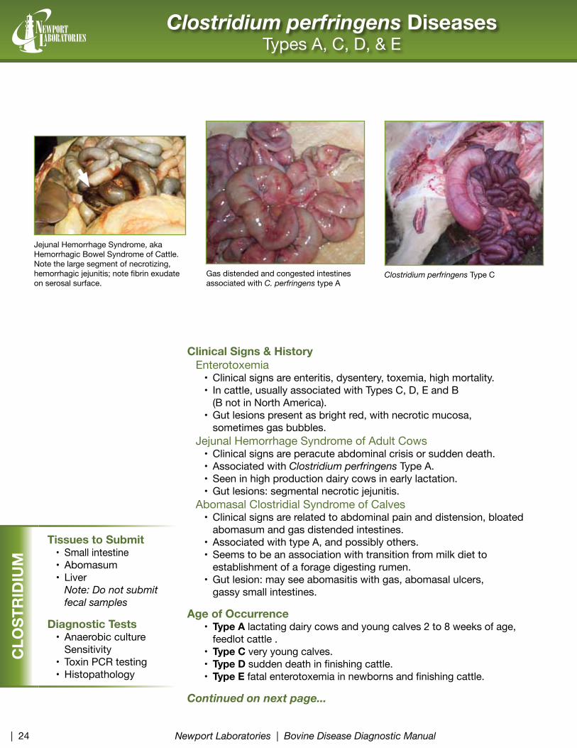
Gas distended and congested intestines associated with C. perfringens type A
Clostridium perfringens Type C
Clostridium perfringens DiseasesTypes A, C, D, & E
Clinical Signs & HistoryEnterotoxemia
• Clinical signs are enteritis, dysentery, toxemia, high mortality.• In cattle, usually associated with Types C, D, E and B
(B not in North America).• Gut lesions present as bright red, with necrotic mucosa,
sometimes gas bubbles.Jejunal Hemorrhage Syndrome of Adult Cows
• Clinical signs are peracute abdominal crisis or sudden death.• Associated with Clostridium perfringens Type A.• Seen in high production dairy cows in early lactation.• Gut lesions: segmental necrotic jejunitis.
Abomasal Clostridial Syndrome of Calves• Clinical signs are related to abdominal pain and distension, bloated
abomasum and gas distended intestines.• Associated with type A, and possibly others.• Seems to be an association with transition from milk diet to
establishment of a forage digesting rumen.• Gut lesion: may see abomasitis with gas, abomasal ulcers,
gassy small intestines.
Age of Occurrence• Type A lactating dairy cows and young calves 2 to 8 weeks of age,
feedlot cattle .• Type C very young calves.• Type D sudden death in finishing cattle.• Type E fatal enterotoxemia in newborns and finishing cattle.
Continued on next page...
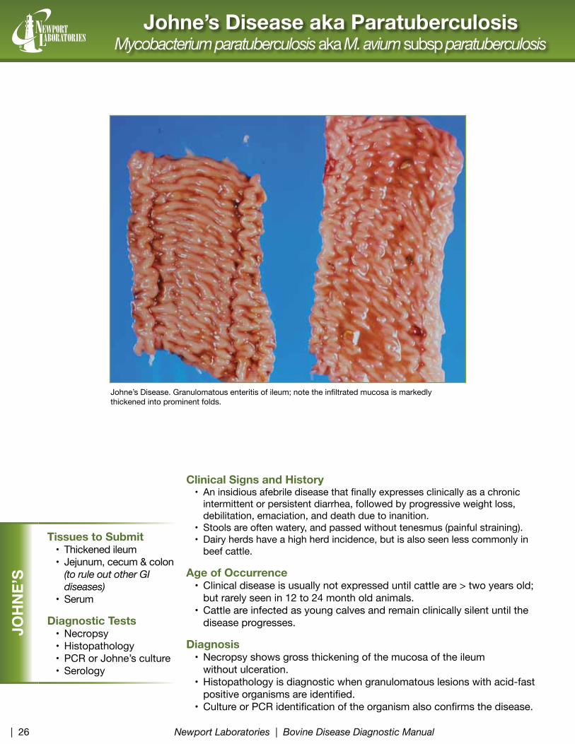
Jejunal Hemorrhage Syndrome, aka Hemorrhagic Bowel Syndrome of Cattle. Note the large segment of necrotizing, hemorrhagic jejunitis; note fibrin exudate on serosal surface.
CLO
STR
IDIU
M
Tissues to Submit• Small intestine• Abomasum• Liver
Diagnosis• The organism is often found in the large intestine of normal cattle. A simple culture of the
organism from the animal is not sufficient by itself to confirm a diagnosis.• Culture results are matched with clinical signs, lesions in the tissues, and, in some cases,
toxin identification, to obtain a true diagnosis.• Type A: Postmortem examinations of calves affected with Type A abomasitis will often
show inflammation, ulceration, and hemorrhage of the lining of the rumen and abomasum. Enterotoxemia diagnosis can be difficult due to the fact that it is a very common inhabitant of the normal intestinal tract; therefore, culture results need to be matched to clinical signs and lesions in the tissues.
• Type C: Tissue samples from calves suspected of having clostridial enterotoxemia should be collected soon after death and kept well-preserved (after the death of the calf, normal populations of clostridial organisms can overgrow and confuse diagnosis).
• Type E: Enterotoxemia causes a severe local intestinal necrosis and systemic toxemia similar to the syndrome described with Type C.
Clostridium perfringens DiseasesTypes A, C, D, & E
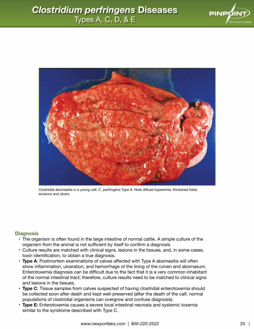
Clostridial abomasitis in a young calf, C. perfringens Type A. Note diffuse hyperemia, thickened folds, erosions and ulcers.
Clinical Signs & History• This disease is caused by enterotoxigenic E. coli with the fimbrial
antigens K99 (F5) or F41.• Hypersecretory diarrhea in newborn calves.• Dehydration, weakness, death.
Age of Occurrence• This disease only occurs in calves less than 7 days old.
Diagnosis• Histopathology is diagnostic, showing prominent colonization of the
jejunum or ileum brush border by non-invasive small bacilli.• Culture and fimbrial identification of the E. coli.
Enteric Colibacillosis in a calf less than 1 week old. Note loops of small intestine are distended with watery yellowish fluid, as is the spiral colon.
Clinical Signs & History• Salmonellosis can present as septicemia or enteritis.• Newborn Septicemia:
• S. dublin, S. newport or S. typhimurium are most common.• May be preceded by enteritis, but not always detected
in newborns.• fever, depression, rapid progression.• high mortality.
• Enteritis:• Often associated with S. typhimurium or S. dublin.• Fever, depression, straining, diarrhea begins loose, then putrid,
mucoid with necrotic flecks of sloughed tissues, sometimes bloody dysentery, emaciation.
• Usually progresses to septicemia.• S. dublin is often endemic on a farm; S. typhimurium is often recently
introduced when outbreaks occur.
Age of Occurrence• Septicemia in newborn to 12 week old calves.• Enteritis in 2 – 12 week old calves and adults.
Diagnosis• Isolation of S typhimurium, S dublin, and S newport.• Gross lesions of necrotizing ulcerative enteritis.• Histopathology of gut, liver, and lung.
Multifocal ulcers on the mucosal surface of the gut. Note tan necrotic debris covering the ulcer; note the rim of red inflammation around the ulcer.
Multifocal ulcers on the surface of the cecal mucosa covered by necrotic tissue and exudate; surrounded by hemorrhage.
SALM
ON
ELLA
Tissues to Submit• Gut• Feces• Liver• Lung• Mesenteric lymph node
(collected near gut lesions)
Diagnostic Tests• Bacterial cultures• Typing of positive
Clinical Signs & History• Calves from 5 to 21 days old experiencing sudden onset of diarrhea.• Diarrhea caused by rotavirus is watery pale yellow at first, changing to pasty
as dehydration sets in, sometimes with mucus and blood flecks. Calves are dull and reluctant to drink.
• The differences between rotavirus and coronavirus diarrhea are subtle with coronavirus usually more severe and leading rapidly to dehydration and acidosis.
• Coronavirus diarrhea is initially fluid yellow diarrhea, later milk clots and mucus are passed and the diarrhea becomes very watery. Depression, fever and anorexia are common.
• Clinical signs of either disease usually last 4 to 8 days.• Calves become severely dehydrated and emaciated.• Combined rotavirus and coronavirus infections are common, and
sometimes associated with cryptosporidiosis also.
Age of Occurrence• rotavirus and coronavirus infections are usually seen in calves between
one and three weeks old, although sometimes a bit younger or older.
Diagnosis• Clinical signs are not enough to make a diagnosis.• Examination of feces for presence of virus. However, it is important to
remember that viruses are found in healthy calves also, so examination of feces from more than one calf is necessary.
• Definitive diagnosis requires correlation of clinical signs (diarrhea), histopathological evidence of viral enteritis, and organism identification.
Diarrhea, dehydration, and yellow stool consistent with enteritis. rotavirus enteritis in a calf during the 2nd week of life. Note ileum and cecum
are distended (rounded) due to accumulation of large amounts of watery diarrhea fluids. Otherwise the serosa appears normal, not inflamed.
Bovine SalmonellosisSalmonella spp
VIRAL EN
TERITIS
Tissues to Submit• Jejunum• Ileum• Cecum• Spiral colon• Cecal/spiral
Clinical Signs & History• A common disease of neonatal calves, especially dairy calves, but
also beef calves.• Calves that have failure or partial failure of passive transfer (FPT) of
colostrum antibodies are highly susceptible to colisepticemia.• Clinical signs of acute colisepticemia: depression, weakness, sudden
shock, recumbency and coma without fever or diarrhea.• Clinical signs of subacute colisepticemia: a sequel to bacteremia leading
to polyarthritis, meningitis, hypopyon, omphalophlebitis, peritonitis.
Age of Occurrence• Acute colisepticemia is seen in calves the first week of life; subacute or
localized colisepticemia is generally seen the second week of life.
Diagnosis• Acute form diagnosis requires a history of FPT of colostral antibody and
isolation of E. coli from multiple fresh parenchymal organs (lung, liver, kidney, spleen) in a calf less than one week old.
• Subacute form diagnosis requires a history of FPT and isolation of E. coli from purulent lesions in multiple sites such as omphalophlebitis, polyarthritis, meningitis, peritonitis, or hypopyon in a calf generally 7 to 14 days old .
Meningitis in a young calf with E. coli septicemia. Note white exudate in sulci of the cerebral cortex (arrows) and over brain stem and cerebellum.
Abdominal side of umbilical region with umbilical vessels. Note abscess (arrow).
Hypopyon. Note large pus clot (arrow) floating in the anterior chamber of the eye.
CO
LISE
PTIC
EMIA
Tissues to Submit• Lung• Liver• Spleen• Kidney• Tissues with
Clinical Signs and History• Cattle develop massive, granulomas and abscesses involving the bony
tissues of the head, especially the mandible and maxilla.• Large mandibular or maxillary lumps develop, often with draining tracks
through ulcerated skin.• Fetid oral odor, difficulty in mastication, loss of teeth in later stages.• May be a concurrent history of Hardware Disease or cattle being fed
rough coarse poor forage (puncture wounds in the mouth may initiate the infection).
Age of Occurrence• Mature dairy or beef cattle, feedlot cattle.
Diagnosis• Gross lesions are highly suggestive; sulfur granules may be visible as
specks in the exudate.• Histopathology or anaerobic culture can confirm the nature of the lesion
and infection.
Actinomycosis. Note large firm mass arising from the lateral and caudal aspect of the left mandible. On section, the mass is filled with multiple coalescing abscesses (arrow).
Clinical Signs & History• Lameness in any limb, but more than one foot is rarely involved at the
same time in mature cows. Footrot can occasionally develop in multiple feet in calves.
• First sign is swelling and redness of the soft tissues of the interdigital space between toes and the adjacent coronary band.
• Inflammation may extend to the pastern and fetlock. Typically, the claws are markedly separated, and the inflammatory edema is uniformly distributed between the 2 digits.
• Onset of the disease is rapid, and the extreme pain leads to increasing lameness.
• The incidence varies according to weather, season of year, grazing periods, and housing system. On average, footrot accounts for ~15% of claw diseases.
Age of Occurrence• Cows, heifers and bulls of all ages.• Occasionally calves.
Diagnosis• It is frequently assumed that every cow with a swollen foot has foot rot.
However, many other conditions, such as infected sandcracks, white line disease, retroarticular abscesses, foreign bodies in the interdigital space, and infection of the distal interphalangeal joint can have a similar appearance if viewed from a distance.
• Despite the difficulties encountered in lifting a hindlimb, a detailed examination should be performed in every case. An incorrect diagnosis can have disastrous results.
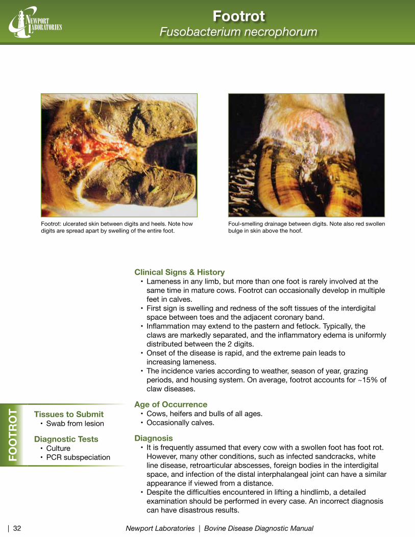
Foul-smelling drainage between digits. Note also red swollen bulge in skin above the hoof.
Footrot: ulcerated skin between digits and heels. Note how digits are spread apart by swelling of the entire foot.
Hardware Disease Traumatic reticuloperitonitis or Traumatic reticulopericarditis
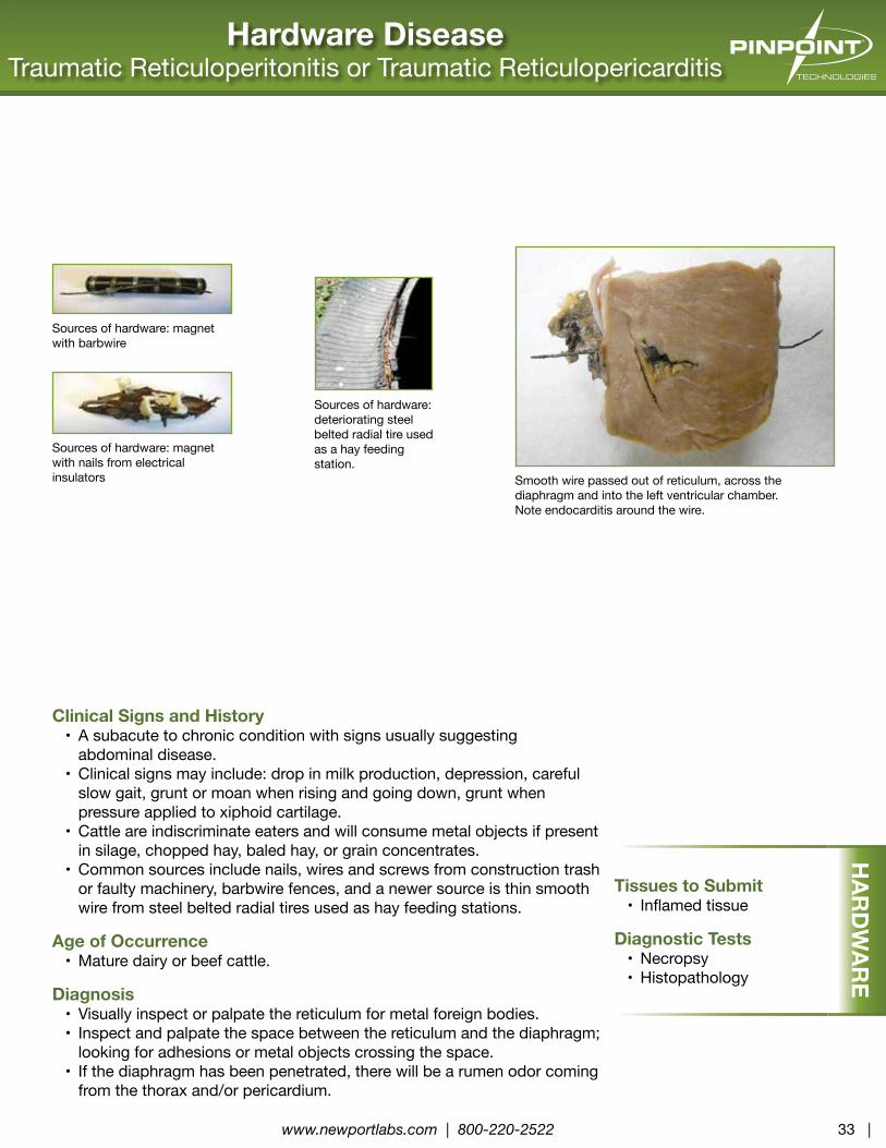
Smooth wire passed out of reticulum, across the diaphragm and into the left ventricular chamber. Note endocarditis around the wire.
Sources of hardware: magnet with barbwire
Sources of hardware: magnet with nails from electrical insulators
Sources of hardware: deteriorating steel belted radial tire used as a hay feeding station.
Clinical Signs and History• A subacute to chronic condition with signs usually suggesting
abdominal disease.• Clinical signs may include: drop in milk production, depression, careful
slow gait, grunt or moan when rising and going down, grunt when pressure applied to xiphoid cartilage.
• Cattle are indiscriminate eaters and will consume metal objects if present in silage, chopped hay, baled hay, or grain concentrates.
• Common sources include nails, wires and screws from construction trash or faulty machinery, barbwire fences, and a newer source is thin smooth wire from steel belted radial tires used as hay feeding stations.
Age of Occurrence• Mature dairy or beef cattle.
Diagnosis• Visually inspect or palpate the reticulum for metal foreign bodies.• Inspect and palpate the space between the reticulum and the diaphragm;
looking for adhesions or metal objects crossing the space.• If the diaphragm has been penetrated, there will be a rumen odor coming
Clinical Signs & History• Seen in feedlot and dairy cattle as a sequel to rumenitis.• Clinical signs are often absent, or general depression and a decrease in
growth rate or milk production.
Age of Occurrence• Feedlot and dairy cattle on high energy rations.• In young calves can be an extension from omphalophlebitis
(naval infection).
Diagnosis• History or gross indication of previous ulcerative rumenitis is supportive;
the organisms come to the liver via blood flow, following microbial invasion of rumen ulcers.
• root cause is anything that predisposes the cattle to ulcerative rumenitis. • Fusobacterium necrophorum necrophorum is the primary
etiological agent.• F. necrophorum funduliforme, Trueperella pyogenes, streptococci,
staphylococci, and Bacteroides spp may be found in mixed infections.• Gross lesions and culture of typical organisms from abscess are
diagnostically definitive.
Liver AbscessFusobacterium necrophorum
A large abscess has ruptured releasing white liquid pus into the abdominal cavity.
Multiple liver abscesses, containing tan caseous pus. Note adherence to the abdominal wall.
Pinkeye: lift the eyelids to see the red inflamed conjunctiva and sclera.Pinkeye: note white ulcer in center of cornea; inflamed red sclera and conjunctiva, and wet skin below eye from chronic tearing.
Clinical Signs & History• The disease usually is acute and tends to spread rapidly.• One or both eyes may be affected.• In cattle, dry, dusty environmental conditions; shipping stress; bright
sunlight; and irritants such as pollens, grasses, and flies tend to predispose to or exacerbate the disease. Flies also serve as vectors.
• The initial signs are photophobia, spasm of the orbicular muscle of the eyelids, and overflow of tears due to obstruction of tear duct.
• Conjunctivitis, with or without varying degrees of keratitis, is always present.
• Appetite may be depressed due to ocular discomfort or visual disturbance that results in inability to locate food.
• The clinical course varies from a few days to several weeks unless complicated by other diseases.
Age of Occurrence• Calves and yearlings are more prone to pinkeye.• Older cattle are susceptible with physical irritation of the eye coupled
with heavy face fly populations.
Diagnosis• Presumptive diagnosis is based on ocular signs and concurrent systemic
disease. It is important to distinguish that the lesions are not due to foreign bodies or parasites.
• Microbial culture or PCr testing may identify involved organisms. Moraxella bovis is often isolated. The role of other organisms is less clear, but may include IBr virus, Mycoplasma spp, and Moraxella bovoculi.
• In IBr, upper respiratory signs and conjunctivitis predominate, while keratitis accompanied by ulceration is rare.
Pyelonephritis Bacterial Cystitis and Pyelonephritis
Clinical Signs and History• A chronic disease that begins in the urinary bladder as bacterial cystitis, and
progresses by ascending the ureters to cause pyelonephritis.• Usually appears weeks after parturition.• rare in male cattle.• A variety of bacteria may be involved including Corynebacterium spp,
E. coli, or Trueperella pyogenes.
Age of Occurrence• Mature cows; risk seems to increase with parity.
Diagnosis• Clinically see increased frequency of urination, blood in urine, pus in
urine, straining to urinate, twitching of tail, fever, loss of condition and production drop.
• Gross lesions include thickened urinary bladder, thickened ureters, pus pockets in the kidney.
• Histopathology shows cystitis and pus in renal tubules.• Culture of the organisms from renal or bladder tissues.
Pyelonephritis. Note pus mixed with urine creates a thin white fluid filling many chronic abscesses. Note the extensive white tissue is fibrous scarring. Note the distended ureters.
Pyelonephritis. Cross section of kidney reveals prominent multifocal abscessation throughout. The lesions develop from infection ascending via individual nephron units.
Newport Laboratories would like to extend appreciation to the following organizations and individuals that have contributed valuable content and/or input for the first edition of the
Bovine Disease Diagnostic Manual:
University of Minnesota - Veterinary Diagnostic Laboratory
Iowa State University - Veterinary Diagnostic Laboratory
South Dakota State University - Animal Disease research & Diagnostic Laboratory
Professional Animal Health - Newcastle, OK
Northwest Veterinary Supply - Parkston, SD
Edward G. (Ted) Clark, DVM, MVSc, Diagnostic Pathologist - Calgary AB
Brian Dorcey, DVM, Veterinary Medical Center - Worthington, MN
Feedlot Health Management Services Ltd. - Okotoks, AB, Canada
Paul Lawrence MS, PhD, Newport Laboratories - Worthington, MN