Bovine Herpesvirus 1 Glycoprotein M Forms a Disulfide-LinkedHeterodimer with the UL49.5 Protein
S. X. WU, X. P. ZHU, AND G. J. LETCHWORTH*
Department of Animal Health and Biomedical Sciences, University of Wisconsin—Madison,Madison, Wisconsin 53706
Received 27 October 1997/Accepted 16 December 1997
Nine glycoproteins (gB, gC, gD, gE, gG, gH, gI, gK, and gL) have been identified in bovine herpesvirus 1(BHV-1). gM has been identified in many other alpha-, beta-, and gammaherpesviruses, in which it appears toplay a role in membrane penetration and cell-to-cell fusion. We sought to express BHV-1 open reading frameUL10, which encodes gM, and specifically identify the glycoprotein. We corrected a frameshift error in thepublished sequence and used the corrected sequence to design coterminal peptides from the C terminus. Thesewere expressed as glutathione S-transferase fusion proteins in Escherichia coli. The fusion protein containingthe 63 C-terminal amino acids from the corrected gM sequence engendered antibodies that immunoprecipi-tated a 30-kDa protein from in vitro translation reactions programmed with the UL10 gene. Proteins immu-noprecipitated by this antibody from virus-infected cells ran at 36 and 43 kDa in reducing sodium dodecylsulfate-polyacrylamide gel electrophoresis (SDS-PAGE) and 43 and 48 kDa in nonreducing SDS-PAGE. Onlythe larger of the pair was present in virions. A 7-kDa protein was released from gM by reducing agents. The7-kDa protein was not recognized in Western blots probed with the anti-gM antibody but reacted specificallywith antibodies prepared against BHV-1 UL49.5, previously reported to be a 9-kDa protein associated with anunidentified 39-kDa protein (X. Liang, B. Chow, C. Raggo, and L. A. Babiuk, J. Virol. 70:1448–1454, 1996). Thisis the first report of a small protein covalently bound to any herpesvirus gM. Similar patterns of hydrophobicdomains and cysteines in all known gM and UL49.5 homologs suggest that these two proteins may be linkedby disulfide bonds in all herpesviruses.
Bovine herpesvirus 1 (BHV-1) is a common pathogen caus-ing respiratory, ocular, and reproductive disease in cattleworldwide. Nine BHV-1 glycoproteins (gB, gC, gD, gE, gG,gH, gI, gK, and gL) have been identified and partially charac-terized. The BHV-1 sequence (27, 28) also contains open read-ing frames (ORFs) UL10 and UL49.5, encoding homologs ofgM (2) and gN (10), respectively.
The BHV-1 UL10 gene sequence (28) predicts a 411-amino-acid protein with all of the features of the gMs found in everyother herpesvirus sequenced to date. Herpesvirus gM genesequences predict proteins about 350 to 475 amino acids longwith little amino acid identity but nearly identical patterns oflarge hydrophobic domains separated by 20- to 40-amino-acidhydrophilic domains, a proline-cysteine pair 40 to 70 aminoacids from the N terminus, and an N-linked glycosylation siteexactly 12 amino acids to the right of the conserved proline-cysteine (13). The major physical difference between gMs ap-pears to be the length of the C-terminal hydrophilic region,which varies from 20 to 110 amino acids. The gM genes thathave been studied are expressed late (3, 13, 26). Transcripts (3)and gM protein (23) are described as abundant. The herpessimplex virus (HSV), human cytomegalovirus, pseudorabiesvirus (PRV), and equine herpesvirus 1 (EHV-1) glycoproteinshave been detected by antipeptide or monoclonal antibodies as46- to 63-kDa glycoproteins in infected cells and virions (2, 7,11, 16, 21, 23). Consistent with the presence of many hydro-phobic domains, gM is associated with cellular and virionmembranes. Deletion of the majority of the gene or insertionalmutagenesis shows that the gM genes of HSV, EHV-1, and
PRV are not essential for replication in vitro (1, 6, 7, 17, 21) orin vivo (17), although the deletants grow to lower titers, pen-etrate cells more slowly than wild type virus (7, 21), and aredefective for cell-to-cell fusion (6), suggesting that gM mayplay a role in membrane penetration.
The BHV-1 UL49.5 gene codes for a nonglycosylated virionsurface protein (14, 15). It appears to have a homolog in everyherpesvirus (5). Typical examples include HSV types 1 and 2(HSV-1 and -2) UL49a or UL49.5, PRV UL49.5, EHV-1 gene10, human and murine cytomegalovirus UL73, human herpes-virus 6 and 7 protein U46, Epstein-Barr virus BLRF1, andhuman herpesvirus 8 ORF 53. Sequences vary widely, but allcode for proteins 84 to 138 amino acids in length, having aputative signal sequence approximately 20 amino acids long, a15- to 81-amino-acid hydrophilic, presumably extracellular, do-main containing a single cysteine 4 to 13 amino acids N ter-minal to a 19- to 35-amino-acid putative membrane anchor,and a 5- to 17-amino-acid cytoplasmic tail containing one ortwo cysteines immediately C terminal to the membrane an-chor. The UL49.5 proteins are nonessential in BHV-1 (15),HSV-1 (24), and varicella-zoster virus (25).
In this study, we sought to identify the BHV-1 gM protein.We attempted to make antibodies against the predicted C-terminal domain and generated antibodies that failed to reactwith any viral protein. Therefore, we resequenced the 39 end ofUL10 and discovered a single nucleotide that was omitted fromthe original sequence. Using the corrected sequence, we ex-pressed three C-terminal predicted peptides as glutathioneS-transferase (GST) fusion proteins in Escherichia coli, madeantibodies against the fusion proteins, and used antibodiesagainst one fusion protein to immunoprecipitate a 30-kDaprotein from in vitro translation reactions programmed withthe UL10 gene and glycoproteins from virus-infected cells thatran at 36 and 43 kDa in reducing sodium dodecyl sulfate-
* Corresponding author. Mailing address: Department of AnimalHealth and Biomedical Sciences, University of Wisconsin—Madison,1655 Linden Dr., Madison, WI 53706. Phone: (608) 262-8616. Fax:(608) 262-7420. E-mail: [email protected].
3029
Dow
nloa
ded
from
http
s://j
ourn
als.
asm
.org
/jour
nal/j
vi o
n 18
Jan
uary
202
2 by
218
.51.
48.2
20.
polyacrylamide gel electrophoresis (SDS-PAGE) and 43 and48 kDa in nonreducing SDS-PAGE. The larger of the pair wasprecipitated from virion envelopes. Reducing conditions re-leased a 7-kDa protein from gM immunoprecipitated fromvirus-infected cells and virion envelopes, suggesting that it waslinked to gM by a disulfide bond. The 7-kDa protein wasidentified by specific antibodies as the BHV-1 UL49.5 protein.
MATERIALS AND METHODS
Virus, cells, and media. BHV-1 (Cooper strain; ATCC VR-864) was replicatedin Madin-Darby bovine kidney (MDBK; ATCC CCL22) cells in minimum es-sential medium (Gibco Laboratories, Life Technologies, Inc.) supplemented with5% fetal bovine serum (HyClone Laboratories, Inc.) at 35°C in a 5% CO2humidified atmosphere. The virus was semipurified by centrifugation through a30% sucrose cushion and stored at 280°C. Virus for one experiment was furtherpurified by isopycnic centrifugation on a potassium tartrate gradient (20).
E. coli JM109 (Promega) was used for plasmid maintenance and transforma-tion, and E. coli BL-21 (Novagen) was used for GST fusion protein expression.
Production of antibodies against GST-gM C-terminal fusion proteins. Weresequenced nucleotides 17942 to 17708 from plasmid pSD58 (19) and con-firmed the published sequence (28) except that we found a single extra Gbetween positions 17836 and 17835, causing a reading frameshift and resulting ina predicted protein of 438 amino acids, 27 amino acids longer than the publishedgM, with an unglycosylated size of 43 kDa. The N-terminal primer TGGATCCCCGCTCGAAGGCGACGCA and C-terminal primer GGAGAATTCTTTATTTGACGTGCGCGG were used to amplify the corrected gM C-terminal 63codons from plasmid pSD58. The gene fragment was amplified with Pfu poly-merase and cloned into pGEX-KG (8) in frame with the GST gene to create theconstruct gMC-63. The recombinant plasmid was transformed into E. coli BL-21and induced by isopropylthiogalactopyranoside at a final concentration of 0.2mM overnight with gentle shaking at room temperature to restrict the formationof inclusion bodies. The cells were suspended in phosphate-buffered saline (PBS)and lysed by sonication. Triton X-100 was added at a final concentration of 1%to aid in solubilization of the fusion proteins. A 50% slurry of glutathione-Sepharose 4B equilibrated with 13 PBS was added and incubated with gentleagitation at room temperature for 30 min. The glutathione-Sepharose pellet waswashed twice with 10 bed volumes of PBS. The fusion protein was eluted inbuffer (10 mM glutathione, 50 mM Tris-HCl [pH 8.0]) and analyzed by SDS-PAGE. A preparation of GST lacking a fusion partner was similarly prepared.The proteins were emulsified in Freund’s complete adjuvant and injected sub-cutaneously into BALB/c mice. Mice were boosted twice at 3-week intervals withfusion protein emulsified with Freund’s incomplete adjuvant. Sera were sampled2 weeks following the final dose.
Production of antibodies against GST-UL49.5 full-length and truncated fusionproteins. Primers TGAGGATCCATGCCGCGGTCGCCGCTCATC and TCATCTAGATCAGCCCCGCCCCCGCGACT were used to amplify the entire 96-codon UL49.5 ORF from plasmid pSD57 (19). Primers ACTGGATCCATGGCCATCGTGCGCGGCCGCGA and TCATCTAGATCAGCCCCGCCCCCGCGACT were used to amplify codons 17 to 96. Both the full-length and truncated(UL49.5T) products were digested with BamHI and XbaI and cloned into simi-larly digested pGEX and pCDNA3 (Invitrogen). The fusion proteins were ex-pressed as inclusion bodies which were solubilized in 7 M urea or 0.3% sarcosylbefore injection into mice as described above.
In vitro transcription and translation of the gM and UL49.5 genes. Based onthe corrected sequence, we amplified the entire gM gene, bases 17628 to 18948,from plasmid pSD58, using primers CAGAATTCATGGCGGGCTCCGCGCAGCCT and GGAGAATTCTTTATTTGACGTGCGCGG and Pfu polymerase.The amplified fragment was ligated to itself, cut with EcoRI, and cloned into theEcoRI site in pcDNA3 downstream of the T7 promoter. The resulting plasmidwas confirmed by restriction enzyme analysis.
The in vitro transcription was carried out according to the AmpliScribe pro-tocol (Epicentre Technologies). Briefly, the following components are combinedin a 20-ml total volume: 1 mg of EcoRI-cleaved plasmid, 2 ml of 103 T7 reactionbuffer, 1.5 ml of 100 mM each ATP, CTP, GTP, and UTP, 2 ml of 100 mMdithiothreitol (DTT), and 2 ml of AmpliScribe T7 enzyme. The reaction mixturewas incubated at 37°C for 2 h.
Transcripts were translated in an in vitro translation reaction mixture thatincluded 8 ml of 12.53 translation mixture, 2 ml of 25 mM magnesium acetate, 2ml of 2.5 M potassium acetate, 40 ml of reticulocyte lysate (Promega), 10 mg ofgM RNA transcript, 10 ml of [35S]methionine, and water to 100 ml. The mixturewas incubated at 30°C for 60 min. A sample was treated at 56°C for 10 min withsample preparation buffer containing 40 mM DTT as a reducing agent, subjectedto SDS-PAGE in a 12 or 18% acrylamide gel, and autoradiographed at 270°C.
Radioimmunoprecipitation. Radiolabeled uninfected and BHV-1-infectedMDBK cells and virions were prepared as described by Marshall et al. (18).MDBK cells were infected at a multiplicity of infection (MOI) of 10 and labeledwith [35S]methionine and -cysteine (ICN Pharmaceuticals Inc.) from 6 to 22 hafter infection. The 35S-labeled cells and virions were lysed in NET buffer (150mM NaCl, 5 mM EDTA, 50 mM Tris [pH 8]) containing 0.2 mM phenylmeth-
ylsulfonyl fluoride (PMSF), 0.5% Nonidet P-40 (NP-40), and 0.5% deoxycholate.Radiolabeled virion envelopes were prepared by lysing purified virions with 0.5%NP-40 and 0.5% deoxycholate and centrifuging the lysate over a 30% sucrosecushion to remove nucleocapsids. Radiolabeled BHV-1-infected MDBK cellmembranes were prepared by Dounce homogenizing cells in the presence ofhomogenizing buffer (20 mM Tris-HCl [pH 8.0], 10 mM MgCl2, 0.5 mM EDTA,2 mM DTT, 1 mM PMSF), centrifuging the cells for 1 min at 2,000 rpm toremove cell debris, and centrifuging the supernatant for 20 min at 12,000 rpm topellet membranes. The membrane pellet was washed once in homogenizingbuffer and centrifuged at 40,000 rpm, producing the 40K supernatant. To test thestrength of gM binding to membranes, some membrane pellets were washedonce in 1 M NaCl. Washed membranes were resuspended in lysing buffer (NET,0.5% NP-40, 0.5% deoxycholate). Immunoprecipitations were done with 10 ml ofantiserum for each 106 cells.
Proteins were immunoprecipitated from in vitro translation reactions or fromlysates of BHV-1-infected and uninfected MDBK cells or cell membranes,BHV-1 virions, or BHV-1 envelopes on Staphylococcus aureus (Gibco Labora-tories, Life Technologies, Inc.) coated successively with rabbit anti-mouse anti-bodies (Cappel) and murine polyclonal antibodies. Precipitates were treated at56°C with SDS-PAGE sample buffer with or without reducing agents, analyzedby reducing or nonreducing SDS-PAGE, and autoradiographed at 270°C.
Analysis of N-linked glycosylation. N-linked glycosylation was analyzed asdescribed previously (30). Briefly, radiolabeled gM immunoprecipitated frominfected cell membranes was eluted from S. aureus with 0.8% SDS at 56 or 100°Cand digested with various amounts of endo-b-N-acetylglucosaminidase H (endoH; Boehringer Mannheim) in 100 mM sodium acetate (pH 5)–150 mM NaCl–1%Triton X-100–1% 2-mercaptoethanol–0.2% SDS–0.5 mM PMSF for 18 h at 37°or digested with various amounts of peptide:N-glycosidase F (PNGase F; NewEngland BioLabs) in 50 mM sodium phosphate (pH 7.5)–1% NP-40 for 5 h at37°C. Proteins were analyzed by reducing SDS-PAGE and autoradiography.
To monitor the glycosylation of gM, cells were pulse-labeled with [35S]methi-onine and -cysteine from 7.5 to 8 h after infection in methionine- and cysteine-deficient medium and chased for 0 to 60 min with unlabeled methionine andcysteine. Uninfected cells were labeled at the same time. The glycosylationinhibitor tunicamycin was added to some infected cultures 30 min prior tolabeling and was not removed for the 60-min chase. The cells were lysed, cen-trifuged to remove debris and nuclei, and immunoprecipitated as describedabove. The immunoprecipitated products were incubated at 56° in SDS-PAGEsample buffer with and without 100 mM DTT and analyzed by SDS-PAGE in 10to 20% gradient gels with or without reducing agents.
Western blotting. Western blotting was carried out as described previously(30). Lysates were incubated at 56°C for 10 min in SDS-PAGE sample bufferwith or without 100 mM DTT. Samples were analyzed by SDS-PAGE in 10 to20% gradient gels and transferred to nitrocellulose paper (Bio-Rad) at 500 mAfor 30 to 60 min. The nitrocellulose was blocked for 30 min with 20 mM Tris-HCl(pH 7.5)–150 mM NaCl–0.05% Tween 20–5% powdered skim milk, incubatedfor 60 min with antiviral antibody diluted 1:500 in blocking buffer, and thenreacted with peroxidase-labeled anti-mouse antibody (1:5,000; Amersham). Thefinal result was visualized with an enhanced chemiluminescence (ECL) reaction(Amersham). For reprobing, blots were incubated at 55°C for 30 min in strippingbuffer (100 mM 2-mercaptoethanol, 2% SDS, 62.5 mM Tris-HCl [pH 6.7]) andwashed fully to remove residual reducing agent before repetition of the Westernblotting with a second antibody.
RESULTS
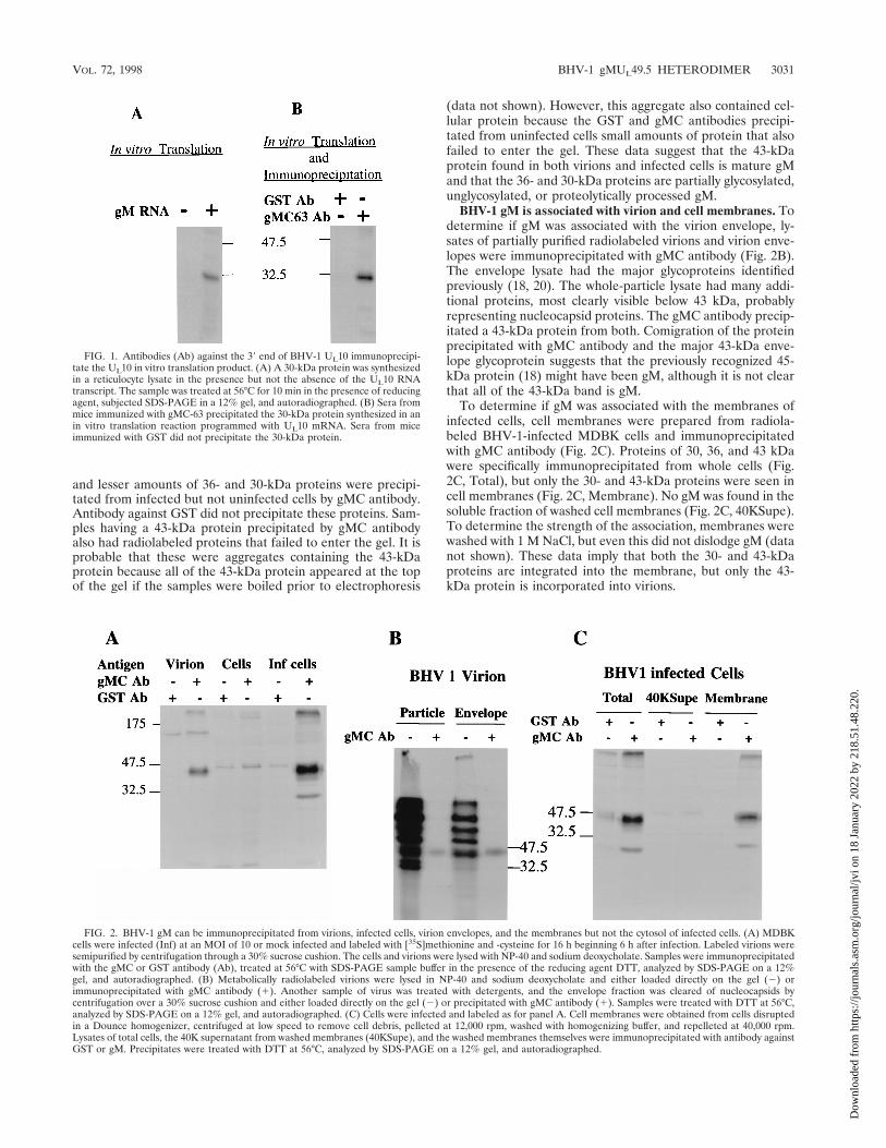
In vitro translation of BHV-1 gM. The entire gM gene wasamplified by PCR using Pfu polymerase and inserted intopcDNA3 downstream of the T7 promoter. The gM mRNAtranscript from this construct was translated in a rabbit reticu-locyte lysate in the absence of membranes. A protein of 30 kDawas detected in reactions programmed with gM mRNA but notin control reactions (Fig. 1A). Antibody from mice immunizedwith gMC-63 but not GST precipitated the 30-kDa gM from invitro translation reactions (Fig. 1B). Purified gMC-63, but notGST, blocked the immunoprecipitation (data not shown). ThegMC-63 antibody was designated gMC antibody and was usedfor all subsequent experiments.
Immunoprecipitation of gM from BHV-1-infected cells andvirions. To identify gM in viral materials, detergent-solubilizedvirions and lysates of uninfected and BHV-1-infected cellswere immunoprecipitated with gMC or GST antibody. A 43-kDa protein was precipitated from virions by gMC but notGST antibody (Fig. 2A). A 100-kDa protein was precipitatedfrom virions by both gMC and GST antibodies, suggesting thatit was not precipitated specifically. A major 43-kDa protein
3030 WU ET AL. J. VIROL.
Dow
nloa
ded
from
http
s://j
ourn
als.
asm
.org
/jour
nal/j
vi o
n 18
Jan
uary
202
2 by
218
.51.
48.2
20.
and lesser amounts of 36- and 30-kDa proteins were precipi-tated from infected but not uninfected cells by gMC antibody.Antibody against GST did not precipitate these proteins. Sam-ples having a 43-kDa protein precipitated by gMC antibodyalso had radiolabeled proteins that failed to enter the gel. It isprobable that these were aggregates containing the 43-kDaprotein because all of the 43-kDa protein appeared at the topof the gel if the samples were boiled prior to electrophoresis
(data not shown). However, this aggregate also contained cel-lular protein because the GST and gMC antibodies precipi-tated from uninfected cells small amounts of protein that alsofailed to enter the gel. These data suggest that the 43-kDaprotein found in both virions and infected cells is mature gMand that the 36- and 30-kDa proteins are partially glycosylated,unglycosylated, or proteolytically processed gM.
BHV-1 gM is associated with virion and cell membranes. Todetermine if gM was associated with the virion envelope, ly-sates of partially purified radiolabeled virions and virion enve-lopes were immunoprecipitated with gMC antibody (Fig. 2B).The envelope lysate had the major glycoproteins identifiedpreviously (18, 20). The whole-particle lysate had many addi-tional proteins, most clearly visible below 43 kDa, probablyrepresenting nucleocapsid proteins. The gMC antibody precip-itated a 43-kDa protein from both. Comigration of the proteinprecipitated with gMC antibody and the major 43-kDa enve-lope glycoprotein suggests that the previously recognized 45-kDa protein (18) might have been gM, although it is not clearthat all of the 43-kDa band is gM.
To determine if gM was associated with the membranes ofinfected cells, cell membranes were prepared from radiola-beled BHV-1-infected MDBK cells and immunoprecipitatedwith gMC antibody (Fig. 2C). Proteins of 30, 36, and 43 kDawere specifically immunoprecipitated from whole cells (Fig.2C, Total), but only the 30- and 43-kDa proteins were seen incell membranes (Fig. 2C, Membrane). No gM was found in thesoluble fraction of washed cell membranes (Fig. 2C, 40KSupe).To determine the strength of the association, membranes werewashed with 1 M NaCl, but even this did not dislodge gM (datanot shown). These data imply that both the 30- and 43-kDaproteins are integrated into the membrane, but only the 43-kDa protein is incorporated into virions.
FIG. 1. Antibodies (Ab) against the 39 end of BHV-1 UL10 immunoprecipi-tate the UL10 in vitro translation product. (A) A 30-kDa protein was synthesizedin a reticulocyte lysate in the presence but not the absence of the UL10 RNAtranscript. The sample was treated at 56°C for 10 min in the presence of reducingagent, subjected SDS-PAGE in a 12% gel, and autoradiographed. (B) Sera frommice immunized with gMC-63 precipitated the 30-kDa protein synthesized in anin vitro translation reaction programmed with UL10 mRNA. Sera from miceimmunized with GST did not precipitate the 30-kDa protein.
FIG. 2. BHV-1 gM can be immunoprecipitated from virions, infected cells, virion envelopes, and the membranes but not the cytosol of infected cells. (A) MDBKcells were infected (Inf) at an MOI of 10 or mock infected and labeled with [35S]methionine and -cysteine for 16 h beginning 6 h after infection. Labeled virions weresemipurified by centrifugation through a 30% sucrose cushion. The cells and virions were lysed with NP-40 and sodium deoxycholate. Samples were immunoprecipitatedwith the gMC or GST antibody (Ab), treated at 56°C with SDS-PAGE sample buffer in the presence of the reducing agent DTT, analyzed by SDS-PAGE on a 12%gel, and autoradiographed. (B) Metabolically radiolabeled virions were lysed in NP-40 and sodium deoxycholate and either loaded directly on the gel (2) orimmunoprecipitated with gMC antibody (1). Another sample of virus was treated with detergents, and the envelope fraction was cleared of nucleocapsids bycentrifugation over a 30% sucrose cushion and either loaded directly on the gel (2) or precipitated with gMC antibody (1). Samples were treated with DTT at 56°C,analyzed by SDS-PAGE on a 12% gel, and autoradiographed. (C) Cells were infected and labeled as for panel A. Cell membranes were obtained from cells disruptedin a Dounce homogenizer, centrifuged at low speed to remove cell debris, pelleted at 12,000 rpm, washed with homogenizing buffer, and repelleted at 40,000 rpm.Lysates of total cells, the 40K supernatant from washed membranes (40KSupe), and the washed membranes themselves were immunoprecipitated with antibody againstGST or gM. Precipitates were treated with DTT at 56°C, analyzed by SDS-PAGE on a 12% gel, and autoradiographed.
VOL. 72, 1998 BHV-1 gMUL49.5 HETERODIMER 3031
Dow
nloa
ded
from
http
s://j
ourn
als.
asm
.org
/jour
nal/j
vi o
n 18
Jan
uary
202
2 by
218
.51.
48.2
20.
BHV-1 gM has N-linked but not O-linked glycosylation. Todetermine if the 43-kDa protein was the glycosylated form ofthe 30-kDa protein and determine the type of glycosylation, weimmunoprecipitated the 43-kDa protein from infected cellswith the gMC antibody and digested it with glycolytic enzymes.Both endo H, which cleaves high-mannose structures, andPNGase F, which removes all N-linked oligosaccharides, re-duced the size of the 43-kDa gM to 30 kDa, where it comi-grated with gM synthesized in the in vitro translation system(Fig. 3), although endo H digestion was incomplete. Furtherevidence was obtained by treating infected cells with tunica-mycin, an inhibitor of N-linked glycosylation, and immunopre-cipitating gM with the gMC antibody. Only a molecule of 30kDa was precipitated (see Fig. 5, lane 5), suggesting either thatno O-linked carbohydrate is normally added or that tunicamy-cin inhibited O-linked glycosylation by blocking transport outof the endoplasmic reticulum. Thus, all unglycosylated gM,whether synthesized in a reticulocyte lysate in the absence ofmembranes, made in MDBK cells in the presence of tunica-
mycin, or deglycosylated by endo H or PNGase F, had anapparent molecular mass of 30 kDa. These data and the pres-ence of only one N-linked glycosylation site strongly suggestthat BHV-1 gM is synthesized as a protein with an apparentmolecular mass of 30 kDa that is decorated with a singlecomplex N-linked oligosaccharide to an apparent molecularmass of 43 kDa.
A 7-kDa protein coprecipitates with BHV-1 gM. To definethe kinetics of gM synthesis, infected cells were pulse-labeledfor 1 h at 2, 5, 8, 11, and 23 h after infection. Labeled cells werewashed, lysed, and immunoprecipitated with gMC antibody.Precipitated proteins were analyzed by SDS-PAGE in 10 to20% gradient gels under reducing conditions (Fig. 4). Smallamounts of the 43-kDa gM first appeared at 9 h, suggestingthat BHV-1 gM is a late protein like other herpesvirus gMs (3,12, 13, 26). Three other proteins with apparent molecularmasses of 36, 30, and 7 kDa appeared at the same time. Thedensity of the 43-kDa protein band increased with time. The36- and 30-kDa proteins peaked at 12 h and then decreaseddramatically, suggesting that the 36-kDa protein was a partiallyglycosylated intermediate of the 30-kDa unglycosylated gM.The 7-kDa protein appeared with the same kinetics as gM,suggesting that it either coprecipitated with gM or shared anepitope with the C-terminal 63 amino acids of gM. A 10-kDaprotein in the UL10 in vitro translation reaction is probably theproduct of a contaminating RNA because it does not immu-noprecipitate with the gMC antibody (data not shown).
Evidence that the association of the 43-kDa gM with the7-kDa protein requires disulfide bonding. To clearly charac-terize the relationship between the various proteins immuno-precipitated by the gMC antibody, we compared immunopre-cipitated proteins from pulse-chase-labeled BHV-1-infectedMDBK cells, tunicamycin-treated BHV-1-infected cells, and invitro-translated UL10 RNA by SDS-PAGE in the presence(Fig. 5, left panel) and absence (Fig. 5, right panel) of DTT. Inthe presence of DTT, a 36-kDa protein present at the begin-ning of the chase (lane 1) decreased as a 43-kDa proteinincreased, implying that the 36-kDa protein may be a precursor
FIG. 3. Deglycosylation of gM by PNGase F and endo H. Lysates of 35S-labeled infected cell membranes were immunoprecipitated with gMC antibody.Precipitates were treated with 0.8% SDS at 100 or 56°C and digested with 0 to5 kU of PNGase F or 0 to 2 mU of endo H. The gM in the left lane of each panelwas synthesized in an in vitro translation system programmed with the BHV-1UL10 RNA transcript.
FIG. 4. Coprecipitation of gM and a 7-kDa protein by gMC antibody atvarious times postinfection (p.i.). MDBK cells were infected at an MOI of 10 andlabeled with [35S]methionine and -cysteine for 1 h at the times indicated. Cellswere lysed and immunoprecipitated with gMC antibody. The samples wereanalyzed by SDS-PAGE in a 10 to 20% gradient gel alongside in vitro-translatedBHV-1 gM (gM). The 43-, 36-, 30-, 10-, and 7-kDa protein bands are indicated.
FIG. 5. Pulse-chase and immunoprecipitation to show the maturation ofBHV-1 gM. Proteins from 35S-labeled BHV-1-infected cells without (lanes 1 to4 and 8 to 11) or with (lanes 5 and 12) tunicamycin treatment (Tunic), andsimilarly labeled uninfected cells (lanes 6 and 13), were chased into matureproteins for 0 to 60 min with unlabeled amino acids. gM was immunoprecipitatedwith gMC antibody and analyzed by SDS-PAGE on 10 to 20% gradient gels inthe presence (lanes 1 to 7) or absence (lanes 8 to 14) of the reducing agent DTT.Unprecipitated UL10 in vitro translation products were analyzed in lanes 7 and14. The strong signal at 8 to 10 kDa in the UL10 in vitro translation reaction wasnot present in reactions done in other reticulocyte lysates.
3032 WU ET AL. J. VIROL.
Dow
nloa
ded
from
http
s://j
ourn
als.
asm
.org
/jour
nal/j
vi o
n 18
Jan
uary
202
2 by
218
.51.
48.2
20.
of the 43-kDa protein. The 36-kDa protein was obviouslylarger than the unglycosylated gM made in the presence oftunicamycin (lane 5) or by in vitro translation (lane 7), whichsuggests it may be partially glycosylated gM. No unglycosylatedgM was detected in infected cells even without the chase,implying that oligosaccharide was transferred to the sole gly-cosylation site at amino acids 57 to 59 before synthesis of theC terminus, the site of the gMC epitope(s) between aminoacids 375 and 438. The 7-kDa protein was present with glyco-sylated (lanes 1 to 4) and unglycosylated (lane 5) gM, suggest-ing that gM glycosylation is not required for formation of thegM–7-kDa protein heterodimer. In the absence of DTT, asmall amount of a 36-kDa protein present after the pulsedisappeared during the chase, perhaps being chased into alarger protein, either the 43-kDa band that was present duringthe entire chase or the 48-kDa protein that increased steadilyduring the chase. Again, the 30-kDa protein detected in tuni-camycin-treated infected cells (lane 12) and the in vitro-trans-lated UL10 (lane 14) was not detected during the chase (lanes8 to 11). The 7-kDa protein is nearly undetectable in all lanes.The majority of the gMC-precipitable protein in the tunicamy-cin-treated culture was 43 kDa, a protein absent from themock-infected culture (lane 13) and the tunicamycin-treatedsample analyzed in the presence of DTT (lane 5) and perhapsidentical to the 43-kDa protein detected in reducing conditions(lanes 1 to 4). The probable interpretation of lanes 8 to 11,therefore, is that the 36-kDa protein is partially glycosylatedgM, the 43-kDa band is monomeric fully glycosylated gM, andthe 48-kDa band is a fully glycosylated, disulfide-linked het-erodimer of 43-kDa gM and the 7-kDa protein. An alternativeinterpretation is that gM and the 7-kDa protein may requireintrachain disulfide bonding of one or both proteins for asso-ciation. Most gM in tunicamycin-treated cells was linked to the7-kDa protein, as shown by the presence of a 40-kDa band innonreducing conditions (lane 12) and 30- and 7-kDa bands inreducing conditions (lane 5), suggesting that glycosylation isnot required for dimerization. Again, the band at about 10 kDain the in vitro translation reaction was not seen when the sameRNA was translated in other reticulocyte lysates and was notprecipitated by the gMC antibody. We concluded it was theproduct of a contaminating RNA.
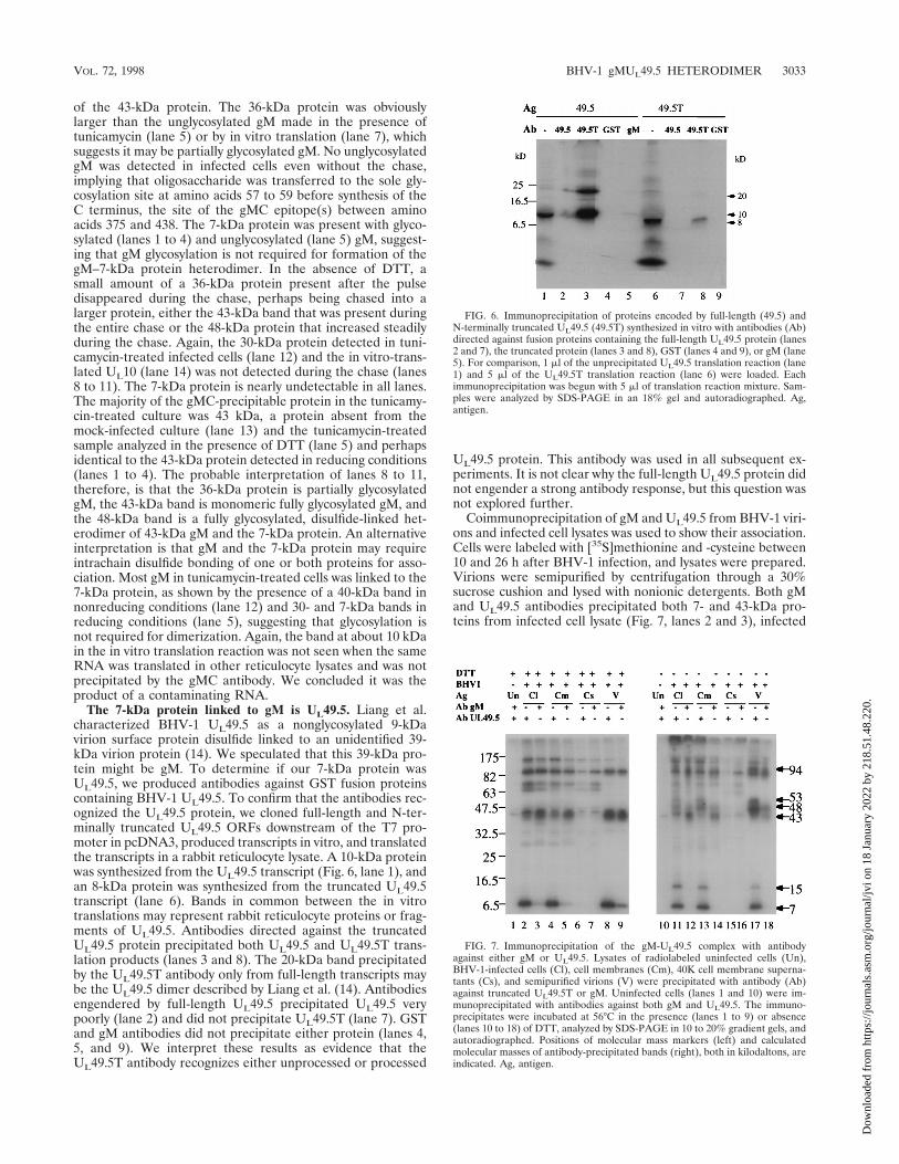
The 7-kDa protein linked to gM is UL49.5. Liang et al.characterized BHV-1 UL49.5 as a nonglycosylated 9-kDavirion surface protein disulfide linked to an unidentified 39-kDa virion protein (14). We speculated that this 39-kDa pro-tein might be gM. To determine if our 7-kDa protein wasUL49.5, we produced antibodies against GST fusion proteinscontaining BHV-1 UL49.5. To confirm that the antibodies rec-ognized the UL49.5 protein, we cloned full-length and N-ter-minally truncated UL49.5 ORFs downstream of the T7 pro-moter in pcDNA3, produced transcripts in vitro, and translatedthe transcripts in a rabbit reticulocyte lysate. A 10-kDa proteinwas synthesized from the UL49.5 transcript (Fig. 6, lane 1), andan 8-kDa protein was synthesized from the truncated UL49.5transcript (lane 6). Bands in common between the in vitrotranslations may represent rabbit reticulocyte proteins or frag-ments of UL49.5. Antibodies directed against the truncatedUL49.5 protein precipitated both UL49.5 and UL49.5T trans-lation products (lanes 3 and 8). The 20-kDa band precipitatedby the UL49.5T antibody only from full-length transcripts maybe the UL49.5 dimer described by Liang et al. (14). Antibodiesengendered by full-length UL49.5 precipitated UL49.5 verypoorly (lane 2) and did not precipitate UL49.5T (lane 7). GSTand gM antibodies did not precipitate either protein (lanes 4,5, and 9). We interpret these results as evidence that theUL49.5T antibody recognizes either unprocessed or processed
UL49.5 protein. This antibody was used in all subsequent ex-periments. It is not clear why the full-length UL49.5 protein didnot engender a strong antibody response, but this question wasnot explored further.
Coimmunoprecipitation of gM and UL49.5 from BHV-1 viri-ons and infected cell lysates was used to show their association.Cells were labeled with [35S]methionine and -cysteine between10 and 26 h after BHV-1 infection, and lysates were prepared.Virions were semipurified by centrifugation through a 30%sucrose cushion and lysed with nonionic detergents. Both gMand UL49.5 antibodies precipitated both 7- and 43-kDa pro-teins from infected cell lysate (Fig. 7, lanes 2 and 3), infected
FIG. 6. Immunoprecipitation of proteins encoded by full-length (49.5) andN-terminally truncated UL49.5 (49.5T) synthesized in vitro with antibodies (Ab)directed against fusion proteins containing the full-length UL49.5 protein (lanes2 and 7), the truncated protein (lanes 3 and 8), GST (lanes 4 and 9), or gM (lane5). For comparison, 1 ml of the unprecipitated UL49.5 translation reaction (lane1) and 5 ml of the UL49.5T translation reaction (lane 6) were loaded. Eachimmunoprecipitation was begun with 5 ml of translation reaction mixture. Sam-ples were analyzed by SDS-PAGE in an 18% gel and autoradiographed. Ag,antigen.
FIG. 7. Immunoprecipitation of the gM-UL49.5 complex with antibodyagainst either gM or UL49.5. Lysates of radiolabeled uninfected cells (Un),BHV-1-infected cells (Cl), cell membranes (Cm), 40K cell membrane superna-tants (Cs), and semipurified virions (V) were precipitated with antibody (Ab)against truncated UL49.5T or gM. Uninfected cells (lanes 1 and 10) were im-munoprecipitated with antibodies against both gM and UL49.5. The immuno-precipitates were incubated at 56°C in the presence (lanes 1 to 9) or absence(lanes 10 to 18) of DTT, analyzed by SDS-PAGE in 10 to 20% gradient gels, andautoradiographed. Positions of molecular mass markers (left) and calculatedmolecular masses of antibody-precipitated bands (right), both in kilodaltons, areindicated. Ag, antigen.
VOL. 72, 1998 BHV-1 gMUL49.5 HETERODIMER 3033
Dow
nloa
ded
from
http
s://j
ourn
als.
asm
.org
/jour
nal/j
vi o
n 18
Jan
uary
202
2 by
218
.51.
48.2
20.
cell membranes (lanes 4 and 5), and virions (lanes 8 and 9).These proteins were not precipitated from a 40K supernatantof infected cell membranes (lanes 6 and 7). More 7-kDa pro-tein than 43-kDa protein was precipitated by UL49.5 antibody(lanes 2, 4, and 8), and more 43-kDa protein than 7-kDaprotein was precipitated by gM antibody (lanes 3, 5, and 9).Different ratios of 43- and 7-kDa proteins may reflect thepresence of uncomplexed gM and UL49.5. Other bands ap-peared in all immunoprecipitations, including those from the40K supernatant which contained very little gM or UL49.5,suggesting they may be unrelated to gM and UL49.5.
To show that the 43-kDa gM and 7-kDa UL49.5 proteinswere linked by disulfide bonds, the same immunoprecipitationswere analyzed in the absence of DTT (Fig. 7, right panel). Twobands, 43 and 48 kDa, were precipitated from infected cells bygM antibody (lanes 12 and 14). This result was identical to thatin Fig. 5. Five bands, 7, 15, 48, 53, and 94 kDa, were specificallyprecipitated from infected cell lysates, cell membranes, andvirions by UL49.5 antibody (lanes 11, 13, and 17) but not fromuninfected cells (lane 10) or the 40K supernatant (lane 15). Weinterpret these data as evidence that some UL49.5 (7 kDa) andits dimer (15 kDa) and some gM (43 kDa) are independentand that some UL49.5 and gM are coprecipitated with antibodyto either. As reported by Liang et al. (14), the 15-kDa proteinband is seen only in the absence of DTT, suggesting that it isa disulfide-linked UL 49.5 dimer. The 48-kDa protein may bea complex of gM and UL49.5 because it is the only proteinprecipitated by both gM and UL49.5T antibodies. The 53-kDaprotein precipitated by UL49.5 antibody from all infected cellpreparations and virions (lanes 11, 13, and 17) may be a com-plex of gM and the UL49.5 dimer. A possible explanation for itnot being precipitated by gM antibody is that the UL49.5 dimerblocks the gM C-terminal epitope(s) or UL49.5 forms a het-erodimer with an as yet unidentified partner slightly largerthan gM. The linkage between gM and UL49.5 must depend ona disulfide bond because gM precipitated with UL49.5 antibodyresolved as a 43-kDa protein in the presence of DTT (lanes 2,4, and 8) and 48- and 53-kDa proteins in the absence of DTT(lanes 11, 13, and 17), and the UL49.5 precipitated with gMantibody appears as a 7-kDa protein in the presence (lanes 3,5, and 9) but not in the absence (lanes 12, 14, and 18) of DTT.
The identities of other bands in the immunoprecipitateswere less clear. A sharp band at 43 kDa was precipitated by allantibodies, including nonimmune sera (lanes 1 and 10), sug-gesting that it was a cellular protein precipitating nonspecifi-cally. All bands above the 47.5-kDa marker in DTT-treatedprecipitates appeared in the absence of gM and UL49.5 (lanes6 and 7), suggesting that they were unrelated to gM andUL49.5. Bands of similar size also were seen in nonreducedprecipitates, and at least three of these bands were seen in theabsence of gM and UL49.5 (lanes 15 and 16), suggesting thatthey also were unrelated to gM and UL49.5. An indistinct,DTT-sensitive band labeled 94 kDa is particularly prominentin precipitates from virions (lanes 17 and 18) and may repre-sent a dimer of the gM-UL49.5 dimer because it was precipi-tated by both antibodies.
To further confirm the identification of proteins seen inimmunoprecipitations, Western blots of uninfected cells, in-fected cell membranes, semipurified BHV-1, and highly puri-fied BHV-1 separated by SDS-PAGE in the presence andabsence of DTT were probed successively with antibodiesagainst UL49.5 and gM (Fig. 8). Infected cells and virus ana-lyzed in the presence of DTT contained a 7-kDa protein thatreacted with antibody against UL49.5 (lanes 6 to 8). When thesame blot was stripped and reprobed with antibody against gM,the 43-kDa protein reacted (lanes 2 to 4). This result showed
that the antibodies reacted only with the intended target pro-tein. When the same samples separated in nonreducing SDS-PAGE were probed with antibody against gM, a 48-kDa pro-tein was identified by both gM (lanes 10 to 12) and UL49.5(lanes 14 to 16) antibodies. Antibody against UL49.5 also iden-tified 15- and 53-kDa proteins in infected cells (lane 14) and a7-kDa protein in tartrate gradient-purified virions (lane 16).Antibody against UL49.5 also identified a larger protein la-beled 94 kDa (lanes 15 and 16). It is not clear that this is thesame protein labeled 94 kDa in Fig. 7. We interpret the reac-tivity of UL49.5 antiserum with a 7-kDa protein in the reducingblot and 48-kDa protein in the nonreducing blot as clear evi-dence that gM and UL49.5 are linked into a 48-kDa dimer byone or more disulfide bonds. Reactivity of UL49.5 antibodywith the 94-kDa protein that does not react with antibodyagainst gM suggests that UL49.5 may bind to some other pro-tein as well, but the appearance of the 94-kDa protein insamples run with DTT suggests that this does not require adisulfide bond. Further, the reactivity of UL49.5 antibody withthe 48- and 53-kDa proteins in the nonreducing blot of in-fected cell membranes (lane 14) but only the 48-kDa protein inthe blot of purified virions (lane 16) suggests a maturationprocess in which UL49.5 dimers are linked to gM and thendissociated, leaving only monomers of UL49.5 attached to gM.The presence of free UL49.5 in purified virions suggests thateither some UL49.5 is free in virions or the disulfide linkage togM was occasionally cleaved during the preparation of virions.
DISCUSSION
We interpret our data as evidence that BHV-1 UL10 en-codes gM, that nascent gM polypeptides obtain an immatureoligosaccharide before the C-terminal epitope is synthesized,and that by the time gM becomes reactive with the gMCantibody, some is glycosylated to a 43-kDa protein lacking aUL49.5 partner and some has been both glycosylated andlinked to UL49.5 to form the mature 48-kDa heterodimer.
FIG. 8. Analysis of the gM-UL49.5 complex by Western blotting. Cells wereinfected at an MOI of 10 and collected 24 h later, and membranes were prepared(Cm). Mock-infected cells were collected at the same time, and membranes weresimilarly prepared (U). Virions were prepared from cells infected for 60 h at anMOI of 0.5 and either semipurified (Vs) or banded on a potassium tartrategradient (Vt). Lysates were incubated at 56°C in the presence (lanes 1 to 8) orabsence (lanes 9 to 16) of 40 mM DTT for 10 min, analyzed by SDS-PAGE in 10to 20% gradient gels, and transferred to nitrocellulose paper. The blots wereprobed with UL49.5T antibody (Ab) first. Bound antibody was detected by ECL(Amersham). The blots were stripped, reprobed with gM antibody, and againdetected by ECL. Positions of molecular mass markers on the (left) and calcu-lated sizes of specific bands are indicated on the (right), both in kilodaltons, areindicated. Ag, antigen.
3034 WU ET AL. J. VIROL.
Dow
nloa
ded
from
http
s://j
ourn
als.
asm
.org
/jour
nal/j
vi o
n 18
Jan
uary
202
2 by
218
.51.
48.2
20.
Further, our data suggest that all of the gM present in the cellmembrane has been fully glycosylated but some lacks itsUL49.5 partner, and that the gM incorporated into the virionhas been fully glycosylated and most has been linked toUL49.5. Our data also suggest that UL49.5 is linked to the43-kDa gM prior to incorporation into virions. The lack of a53-kDa protein in complexes immunoprecipitated with thegMC antibody suggests that the UL49.5 dimer is cleaved beforeor shortly after linkage to gM.
The large disparity between the calculated molecular massof 45 kDa for unglycosylated BHV-1 gM and the apparent sizeof 30 kDa measured for gM synthesized in vitro was not un-expected. There are three possible explanations. First, trans-lation may have initiated after the first AUG or terminatedprematurely. The first AUG is used to initiate HSV-1 (2) andEHV-1 (22) gM. Since the second AUG in BHV-1 gM is atcodon 122 and would code for a protein of 33 kDa, our datamight suggest initiation at the second AUG. However, the onlyN-linked glycosylation site in BHV-1 gM is at amino acids 71to 73. Initiation at the second AUG would produce a proteinwith no N-linked glycosylation site, which is inconsistent withour data. Premature termination would result in a protein thatwould not react with our gMC antibody and also is inconsistentwith our results. Second, full-length gM could be proteolyti-cally cleaved near either the N or C terminus. It seems unlikelythat it is cleaved at the N terminus because gM synthesized inthe presence of tunicamycin (Fig. 5, lanes 5 and 12) has thesame mobility as gM synthesized in an in vitro translationsystem without membranes (Fig. 5, lanes 7 and 14), stronglysuggesting that no signal peptide is cleaved from gM. In addi-tion, no signal peptide has been found in other herpesvirusgMs studied to date. Again, it is unlikely that the C terminus iscleaved from the gM because this is the site of the epitoperecognized by the gMC antibody and only the C-terminal frag-ment would be precipitated. Third, anomalously rapid migra-tion in SDS-PAGE has been noted for other herpesvirus pro-teins with multiple membrane-spanning domains such as PRVdeglycosylated gM, which migrates with an apparent mass ofslightly over 30 kDa despite its predicted size of 42 kDa (7),and HSV gK, which has an apparent mass of 29 kDa but has apredicted size of 36 kDa (9). Therefore, we concluded thatBHV-1 gM was probably also migrating anomalously rapidly inSDS-PAGE and that the 30-kDa nonglycosylated proteinmade in in vitro translation reactions was probably the full-length 45-kDa BHV-1 gM. If this is true, the actual molecularmass of glycosylated gM may be around 60 kDa and the com-plex with the 7-kDa molecule may be 67 kDa. The anomalousmigration of these proteins in SDS-PAGE casts some doubt onthe conclusion that gM lacks O-linked carbohydrate because itis not certain that gM with and without O-linked glycosylationwould migrate to different positions in a gel.
The 7-kDa protein associated with gM is the 96-amino-acidUL49.5. The BHV-1 UL49.5 sequence predicts a protein with amolecular mass of 10 kDa that would be reduced to 8 kDa if itwere cleaved at amino acid 21 as predicted by the method ofvon Heijne (29). The first 16 amino acids constitute a potentialsignal sequence, and amino acids 35 to 51 of the putativemature protein constitute a probable membrane anchor, mak-ing it very similar to homologs in other herpesviruses (4, 5). Acorresponding O-glycosylated protein, designated gN, hasbeen identified in pseudorabies virion envelopes (10). Liang etal. (14) expressed BHV-1 UL49.5 as a fusion protein and madeantibodies that identified a nonglycosylated 9-kDa virion sur-face protein. They showed it was disulfide linked to an uniden-tified 39-kDa virion protein and showed that it was disulfide
linked to itself to make an 18-kDa dimer. This finding corre-sponds closely with our data.
How are gM and UL49.5 linked? All known gM homologshave a cysteine at the beginning of the second hydrophobicdomain 40 to 70 amino acids from the N terminus (13), another114 to 135 amino acids away from the first, near the end of thefifth hydrophobic domain, and others scattered throughout.Since each UL49.5 homolog has a cysteine about 14 aminoacids proximal (15) and another immediately distal to the pu-tative transmembrane anchor sequence, BHV-1 UL49.5 havingonly these cysteines, it is likely that one or both of the con-served cysteines in each molecule form disulfide bonds be-tween the two molecules. Linkage of the two proteins by twodisulfide bonds would likely constrain the conformation ofboth. An alternative explanation is that intrachain disulfidebonds are required to hold one or both of these molecules ina conformation that permits noncovalent interactions to bindthe two molecules together. Our data suggest that this is un-likely because the dimer remained intact when it was treatedwith 1% SDS and heated to 56°C prior to Western blotting(Fig. 8).
Since other proteins immunoprecipitated with gM andUL49.5, it could be argued that these two proteins may belinked via an intermediary. However, when unreduced proteinswere analyzed by Western blotting (Fig. 8), a band correspond-ing to the sum of the gM and UL49.5 molecular weights re-acted with antibodies against both proteins, suggesting anyadditional member(s) of the complex would have to be verysmall.
Where and when are gM and UL49.5 linked? Since ungly-cosylated gM made in the presence of tunicamycin was linkedto the UL49.5, the linkage must be possible prior to glycosyl-ation and therefore may occur in the endoplasmic reticulum.
Why did it take so long to identify BHV-1 gM and its UL49.5partner? The prominent 42-kDa protein seen in [3H]glu-cosamine-labeled virions (20) and 45-kDa glycoprotein seen inBHV-1 envelopes (18) were probably gM, but they have notbeen investigated. The fact that BHV-1 gM has only five me-thionines among its 438 amino acids suggests that if the 45-kDaprotein is gM, either there must be a large amount of it invirion membranes or there is a second protein that comigrateswith gM to account for the intensity of the 45-kDa band la-beled with [35S]methionine. Perhaps this very hydrophobicprotein escaped notice simply because it aggregates under thenormal conditions for SDS-PAGE or because it is a relativelypoor antigen.
Do the gMs of other herpesviruses have a smaller partner?gM has been particularly difficult to investigate because itsmany hydrophobic domains cause it to aggregate when boiledin SDS-PAGE sample buffer, and so it often fails to resolve ongels (2, 7, 21, 23). In addition, immunoprecipitates were oftenanalyzed on low-percentage gels where a small protein wouldnot have been resolved (2). In many studies, gM was reducedand analyzed by Western blotting, and so an associated proteincould not have been observed (23). Nevertheless, Osterriederet al. (22) noted that EHV-1 gM migrated more slowly inSDS-PAGE in the absence of 2-mercaptoethanol but reasonedthat this was caused by intramolecular disulfide bonds. How-ever, they observed a decrease in apparent mass from 59 to 61kDa to 50 to 55 kDa, which is exactly what would be expectedif the EHV-1 gene 10 protein was released by treatment with2-mercaptoethanol.
What is the function of the gM-UL49.5 dimer? Deletion andinsertion mutations in gM had minor effects on membranepenetration speed and replication efficiency (1, 7, 16, 17, 21).However, it is unclear if critical areas of gM were functional in
VOL. 72, 1998 BHV-1 gMUL49.5 HETERODIMER 3035
Dow
nloa
ded
from
http
s://j
ourn
als.
asm
.org
/jour
nal/j
vi o
n 18
Jan
uary
202
2 by
218
.51.
48.2
20.
these mutants because all changes were made distal to theconserved N-linked glycosylation site and the N-terminal pro-line-cysteine pair that may be the attachment site for UL49.5.Deletion of varicella-zoster virus ORF9A, the UL49.5 ho-molog, caused a slight decrease in viral replication and syncy-tium formation. Deletion of BHV-1 and HSV-1 UL49.5 hadminor effects on viral replication (15, 24). Osterrieder et al.(22) speculated that gM might create an ion channel but didnot observe functional ion channels when EHV-1 gM wasexpressed in Xenopus laevis oocytes (22). The absence of acovalently bound UL49.5 may help explain these negative data.Clearly, the gM-UL49.5 dimer requires further investigation.
ACKNOWLEDGMENTS
We thank Etienne Thiry and Eric Baranowski for their generous giftof the BH 44 monoclonal antibody, which was critical to preliminaryexperiments leading up to the present study. The assistance of ChadJohnson, Peter Evans, and Jing Wu is gratefully acknowledged.
This research was supported by USDA National Research Initiativegrants 92-37204-7900 and 94-37204-0857 and a Shaw Scholarship toG.J.L. from the Milwaukee Foundation.
ADDENDUM IN PROOF
After this article was accepted for publication, Jons et al. (A. Jons,J. M. Dijkstra, and T. C. Mettenleiter, J. Virol. 72:550–557, 1998)published their finding that pseudorabies virus gM and gN form adisulfide-linked dimer.
REFERENCES
1. Baines, J. D., and B. Roizman. 1991. The open reading frames UL3, UL4,UL10, and UL16 are dispensable for the replication of herpes simplex virus1 in cell culture. J. Virol. 65:938–944.
2. Baines, J. D., and B. Roizman. 1993. The UL10 gene of herpes simplex virus1 encodes a novel viral glycoprotein, gM, which is present in the virion andin the plasma membrane of infected cells. J. Virol. 67:1441–1452.
3. Baradaran, K., C. E. Dabrowski, and P. A. Schaffer. 1994. Transcriptionalanalysis of the region of the herpes simplex virus type 1 genome containingthe UL8, UL9, and UL10 genes and identification of a novel delayed-earlygene product, OBPC. J. Virol. 68:4251–4261.
4. Barker, D. E., and B. Roizman. 1992. The unique sequence of the herpessimplex virus 1 L component contains an additional translated open readingframe designated UL49.5. J. Virol. 66:562–566.
5. Barnett, B. C., A. Dolan, E. A. R. Telford, A. J. Davison, and D. J. McGeoch.1992. A novel herpes simplex virus gene (UL49A) encodes a putative mem-brane protein with counterparts in other herpesviruses. J. Gen. Virol. 73:2167–2171.
6. Davis-Poynter, N., S. Bell, T. Minson, and H. Browne. 1994. Analysis of thecontributions of herpes simplex virus type 1 membrane proteins to theinduction of cell-cell fusion. J. Virol. 68:7586–7590.
7. Dijkstra, J. M., N. Visser, T. C. Mettenleiter, and B. G. Klupp. 1996. Iden-tification and characterization of pseudorabies virus glycoprotein gM as anonessential virion component. J. Virol. 70:5684–5688.
8. Guan, K. L., and J. E. Dixon. 1991. Eukaryotic proteins expressed in Esch-erichia coli: an improved thrombin cleavage and purification procedure offusion proteins with glutathione S-transferase. Anal. Biochem. 192:262–267.
9. Hutchinson, L., K. Goldsmith, D. Snoddy, H. Ghosh, F. L. Graham, andD. C. Johnson. 1992. Identification and characterization of a novel herpessimplex virus glycoprotein, gK, involved in cell fusion. J. Virol. 66:5603–5609.
10. Jons, A., H. Granzow, R. Kuchling, and T. C. Mettenleiter. 1996. The UL49.5
gene of pseudorabies virus codes for an O-glycosylated structural protein ofthe viral envelope. J. Virol. 70:1237–1241.
11. Kari, B., W. Li, J. Cooper, R. Goertz, and B. Radeke. 1994. The humancytomegalovirus UL100 gene encodes the gC-II glycoproteins recognized bygroup 2 monoclonal antibodies. J. Gen. Virol. 75:3081–3086.
12. Lehner, R., H. Meyer, and M. Mach. 1989. Identification and characteriza-tion of a human cytomegalovirus gene coding for a membrane protein thatis conserved among human herpesviruses. J. Virol. 63:3792–3800.
13. Li, W., K. Eidman, R. C. Gehrz, and B. Kari. 1995. Identification andmolecular characterization of the murine cytomegalovirus homolog of thehuman cytomegalovirus UL100 gene. Virus Res. 36:163–175.
14. Liang, X., B. Chow, C. Raggo, and L. A. Babiuk. 1996. Bovine herpesvirus 1UL49.5 homolog gene encodes a novel viral envelope protein that forms adisulfide-linked complex with a second virion structural protein. J. Virol.70:1448–1454.
15. Liang, X., M. Tang, B. Manns, L. A. Babiuk, and T. J. Zamb. 1993. Identi-fication and deletion mutagenesis of the bovine herpesvirus 1 dUTPase geneand a gene homologous to herpes simplex virus UL49.5. Virology 195:42–50.
16. MacLean, C. A., S. Efstathiou, M. L. Elliott, F. E. Jamieson, and D. J.McGeoch. 1991. Investigation of herpes simplex virus type 1 genes encodingmultiply inserted membrane proteins. J. Gen. Virol. 72:897–906.
17. MacLean, C. A., L. M. Robertson, and F. E. Jamieson. 1993. Characteriza-tion of the UL10 gene product of herpes simplex virus type 1 and investi-gation of its role in vivo. J. Gen. Virol. 74:975–983.
18. Marshall, R. L., L. L. Rodriguez, and G. J. Letchworth. 1986. Characteriza-tion of envelope proteins of infectious bovine rhinotracheitis virus (bovineherpesvirus 1) by biochemical and immunological methods. J. Virol. 57:745–753.
19. Mayfield, J. E., P. J. Good, H. J. VanOort, A. R. Campbell, and D. E. Reed.1983. Cloning and cleavage site mapping of DNA from bovine herpesvirus 1(Cooper strain). J. Virol. 47:259–264.
20. Misra, V., R. M. Blumenthal, and L. A. Babiuk. 1981. Proteins specified bybovine herpesvirus 1 (infectious bovine rhinotracheitis virus). J. Virol. 40:367–378.
21. Osterrieder, N., A. Neubauer, C. Brandmuller, B. Braun, O. R. Kaaden, andJ. D. Baines. 1996. The equine herpesvirus 1 glycoprotein gp21/22a, theherpes simplex virus type 1 gM homolog, is involved in virus penetration andcell-to-cell spread of virions. J. Virol. 70:4110–4115.
22. Osterrieder, N., A. Neubauer, B. Fakler, C. Brandmuller, C. Seyboldt, O. R.Kaaden, and J. D. Baines. 1997. Synthesis and processing of the equineherpesvirus 1 glycoprotein M. Virology 232:230–239.
23. Pilling, A., A. J. Davison, E. A. Telford, and D. M. Meredith. 1994. Theequine herpesvirus type 1 glycoprotein homologous to herpes simplex virustype 1 glycoprotein M is a major constituent of the virus particle. J. Gen.Virol. 75:439–442.
24. Pyles, R. B., N. M. Sawtell, and R. L. Thompson. 1992. Herpes simplex virustype 1 dUTPase mutants are attenuated for neurovirulence, neuroinvasive-ness, and reactivation from latency. J. Virol. 66:6706–6713.
25. Ross, J., M. Williams, and J. I. Cohen. 1997. Disruption of the varicella-zoster virus dUTPase and the adjacent ORF9A gene results in impairedgrowth and reduced syncytia formation in vitro. Virology 234:186–195.
26. Scalzo, A. A., C. A. Forbes, N. J. Davis-Poynter, H. E. Farrell, and P. A.Lyons. 1995. DNA sequence and transcriptional analysis of the glycoproteinM gene of murine cytomegalovirus. J. Gen. Virol. 76:2895–2901.
27. Schwyzer, M., D. Styger, B. Vogt, D. E. Lowery, C. Simard, S. LaBoissiere,V. Misra, C. Vlcek, and V. Paces. 1996. Gene contents in a 31-kb segment atthe left genome end of bovine herpesvirus-1. Vet. Microbiol. 53:67–77.
28. Vlcek, C., V. Benes, Z. Lu, G. F. Kutish, V. Paces, D. Rock, G. J. Letchworth,and M. Schwyzer. 1995. Nucleotide sequence analysis of a 30-kb region ofthe bovine herpesvirus 1 genome which exhibits a colinear gene arrangementwith the UL21 to UL4 genes of herpes simplex virus. Virology 210:100–108.
29. von Heijne, G. 1986. A new method for predicting signal sequence cleavagesites. Nucleic Acids Res. 14:4683–4690.
30. Zhu, X. P., S. Wu, and G. J. Letchworth. 1997. Yeast-secreted bovine her-pesvirus type 1 glycoprotein D has authentic conformational structure andimmunogenicity. Vaccine 15:679–688.