1999, 73(10):8657. J. Virol. M. T. C. Winkler, A. Doster and C. Jones Death during Acute Infection of Cattle Lymphocytes and Induce Programmed Cell T + Bovine Herpesvirus 1 Can Infect CD4 http://jvi.asm.org/content/73/10/8657 Updated information and services can be found at: These include: REFERENCES http://jvi.asm.org/content/73/10/8657#ref-list-1 at: This article cites 53 articles, 23 of which can be accessed free CONTENT ALERTS more» articles cite this article), Receive: RSS Feeds, eTOCs, free email alerts (when new http://journals.asm.org/site/misc/reprints.xhtml Information about commercial reprint orders: http://journals.asm.org/site/subscriptions/ To subscribe to to another ASM Journal go to: on March 14, 2014 by guest http://jvi.asm.org/ Downloaded from on March 14, 2014 by guest http://jvi.asm.org/ Downloaded from
Transcript
1999, 73(10):8657. J. Virol.
M. T. C. Winkler, A. Doster and C. Jones Death during Acute Infection of CattleLymphocytes and Induce Programmed Cell
T+Bovine Herpesvirus 1 Can Infect CD4
http://jvi.asm.org/content/73/10/8657Updated information and services can be found at:
This article cites 53 articles, 23 of which can be accessed free
CONTENT ALERTS more»articles cite this article),
Receive: RSS Feeds, eTOCs, free email alerts (when new
http://journals.asm.org/site/misc/reprints.xhtmlInformation about commercial reprint orders: http://journals.asm.org/site/subscriptions/To subscribe to to another ASM Journal go to:
Bovine Herpesvirus 1 Can Infect CD41 T Lymphocytesand Induce Programmed Cell Death during
Acute Infection of CattleM. T. C. WINKLER, A. DOSTER, AND C. JONES*
Department of Veterinary and Biomedical Sciences, Center for Biotechnology,University of Nebraska, Lincoln, Lincoln, Nebraska 68583-0905
Received 1 April 1999/Accepted 29 June 1999
Acute infection of cattle with bovine herpesvirus 1 (BHV-1) represses cell-mediated immunity, which canlead to secondary bacterial infections. Since BHV-1 can induce apoptosis of cultured lymphocytes, we hypoth-esized that these virus-host interactions occur in cattle. To test this hypothesis, we analyzed lymph nodes andperipheral blood mononuclear cells (PBMC) after calves were infected with BHV-1. In situ terminal de-oxynucleotidyltransferase-mediated dUTP nick end-labeling (TUNEL) staining of lymphoid tissues (pharyn-geal tonsil, cervical, retropharyngeal, and inguinal) was used to detect apoptotic cells. Calves infected withBHV-1 for 7 days revealed increased apoptotic cells near the corticomedullary junction in lymphoid folliclesand in the subcapsular region. Increased frequency of apoptotic cells was also observed in the mucosa-associated lymphoid tissue lining the trachea and turbinate. Immunohistochemistry of consecutive sectionsfrom pharyngeal tonsil revealed that CD21 T lymphocytes were positive for the BHV-1 envelope glycoproteingD. The location of these CD21 T lymphocytes in the germinal center suggested that they were CD41 T cells.Electron microscopy and TUNEL also revealed apoptotic and herpesvirus-infected lymphocytes from this area.Fluorescence-activated cell sorting analyses demonstrated that CD41 and CD81 T cells decreased in lymphnodes and PBMC after infection. The decrease in CD41 T cells correlated with an increase in apoptosis. CD41
but not CD81 lymphocytes were infected by BHV-1 as judged by in situ hybridization and PCR, respectively.Immediate-early (bovine ICP0) and early (ribonucleotide reductase) transcripts were detected in PBMC andCD41 lymphocytes prepared from infected calves. In contrast, a late transcript (glycoprotein C) was notconsistently detected suggesting productive infection was not efficient. Taken together, these results indicatethat BHV-1 can infect CD41 T cells in cattle, leading to apoptosis and suppression of cell-mediated immunity.
Bovine herpesvirus 1 (BHV-1) is an important viral patho-gen of cattle that can cause severe respiratory infection, con-junctivitis, abortion, vulvovaginitis, balanopostitis, and sys-temic infection in neonate calves. Secondary bacterialinfections resulting in pneumonia and death are common (re-viewed in reference 58). BHV-1 belongs to the Alphaherpes-virinae subfamily and shares a number of biological propertieswith herpes simplex virus types 1 and 2 (HSV-1 and HSV-2)(54). BHV-1 establishes lifelong latency in ganglionic neuronsof the peripheral nervous system after initial replication in themucosal epithelia (reviewed in reference 27). Virus reactiva-tion and spread to other susceptible animals occur after natu-ral or corticosteroid-induced stress.
The mechanism of BHV-1-induced immunosuppression hasbeen studied following exposure of animals to virulent or vac-cine virus. Increased susceptibility to secondary infection cor-relates with depressed cell-mediated immunity after infection.Infection decreases interleukin-2 (IL-2) receptor expression(30), impairs IL-2 production, decreases mitogenic stimulationof peripheral blood mononuclear cells (PBMC) (5), impairscytotoxic responses (2), and decreases circulating T lympho-cytes (14, 15). Inhibition of lymphocyte proliferative responsesmay be the result of nonproductive infection (5). Compro-mised CD81 T-cell recognition of infected cells may, in part,
result from major histocompatibility complex class I repressionby HSV-1 (1, 40, 56, 60) and BHV-1 (12). Down-regulation ofthe transporter associated with antigen presentation by BHV-1also occurred (20). Finally, alphaherpesviruses may induce im-mune dysfunction by enhancing suppressor T-cell activity (re-viewed in reference 44).
In humans and mice, the cellular immune response is pre-dominantly CD41 and Th1-like after HSV-2 infection (37).CD41 T cells clear virus from cutaneous sites (35) and reduceestablishment of latency because HSV replication at the pri-mary site of infection is lower (38). CD41 T cells are also thelimiting cell type for antigen-induced proliferation in BHV-1infection (7). CD4-induced cytotoxicity is directed against in-fected cells expressing late HSV-1 glycoproteins, whereasCD8-induced cytotoxicity is directed against immediate-early(IE) and early (E) proteins (36). CD41 T lymphocytes arecrucial for generating primary cytolytic CD81 T cells againstsome HSV-1 antigens (26). CD81 T lymphocytes limit infec-tion in the peripheral nervous system, maintain the integrity ofneurons during primary HSV infection (53), and resolve HSVlesions (42). Finally, gamma/delta T cells may be the first lineof defense against HSV infection (34) and protect mice fromHSV-1 induced encephalitis (50).
Apoptosis, or programmed cell death, leads to chromatincondensation, nuclear fragmentation, and formation of apo-ptotic bodies. Apoptosis occurs during development and aftervirus infection (reviewed in references 19 and 52). Severalviruses have evolved mechanisms that block the host apoptoticpathway to maximize production of viral progeny and estab-lishment of latency. HSV-1 antigen-expressing cells and non-
* Corresponding author. Mailing address: Department of Veteri-nary and Biomedical Sciences, Center for Biotechnology, University ofNebraska, Lincoln Fair St. at East Campus Loop, Lincoln, NE 68583-0905. Phone: (402) 472-1890. Fax: (402) 472-9690. E-mail: [email protected].
infected cord blood T lymphocytes stimulated with phytohe-magglutinin undergo apoptosis (23). HSV-1 infection ofactivated T lymphocytes prepared from peripheral blood leadsto apoptosis of CD41 and HLA-DR-positive T lymphocytesbut not CD81 T cells (22). Live and inactivated BHV-1 caninduce apoptosis of cultured PBMC following mitogen stimu-lation (16–18). Cultured CD41 T lymphocytes that are acti-vated by standard procedures can be infected resulting in ap-optosis (9). Apoptosis of T lymphocytes may account fortransient lymphocytopenia and immunosupression followingBHV-1 infection. However, it is not known if lymphocytes areinfected by BHV-1 or if apoptosis occurs following infection ofcattle.
In this study, we demonstrate that reduction of CD41 Tlymphocytes occurs in PBMC and lymph nodes during acuteinfection of cattle. Relative to mock-infected calves, higherlevels of apoptosis were detected in lymphoid tissues. BHV-1DNA, bovine ICP0 (bICP0) RNA (IE), and ribonucleotidereductase (RR) RNA (E) were consistently detected in CD41
T lymphocytes. These studies demonstrate that BHV-1 infec-tion of CD41 T lymphocytes leads to apoptosis during acuteinfection of cattle. We hypothesize that this novel virus hostinteraction plays an important role in virus-induced immuno-suppression.
MATERIALS AND METHODS
Virus and cells. MDBK cells (American Type Culture Collection, Rockville,Md.) were maintained in Earle’s modified Eagle’s medium (EMEM) with 10%fetal calf serum (FCS). The Cooper strain of BHV-1, supplied by the NationalVeterinary Services Laboratory, Animal and Plant Health Inspection Services,Ames, Iowa, was propagated in MDBK cells with a multiplicity of infection of0.05. When cytopathic effect was evident, virus was harvested, titrated (43) inMDBK cells, aliquoted, and stored at 270°C.
Animals. Weanling dairy calves (5 to 6 months old) that were not vaccinatedagainst BHV-1 and were seronegative for BHV-1 were used for these studies.Calves were inoculated in the right and left conjunctival sacs and intranasally, 1ml per site, with 107 50% tissue culture infective doses of BHV-1 Cooper strainper ml. Trachea, turbinate membrane, pharyngeal tonsil, cervical, parotid, ret-ropharyngeal, and inguinal lymph nodes were obtained at 7 days postinfection(dpi) from BHV-1-infected or mock-infected calves (7 dpi is the time of the peakof clinical symptoms and maximal viral gene expression in trigeminal ganglia[47]). The tissue was fixed in 10% buffered formalin and processed by routinehistological methods. Experiments using animals were done in accordance withthe American Association of Laboratory Animal Care guidelines. Calves werehoused under strict isolation containment and given antibiotics before and afterinfection to prevent secondary bacterial infection.
Preparation of CD41 and CD81 lymphocytes from lymphoid organs andPBMC. Peripheral blood lymphocytes (PBL) and single-cell suspensions fromlymphoid tissue were prepared from mock-infected and BHV-1-infected calvesat 5, 7, 9, 12, and 16 dpi by density gradient centrifugation on Histopaque 1083(Sigma Chemical Co., St. Louis, Mo.). Cells located at the interface were col-lected, washed in CMF-PBS (phosphate-buffered saline) (150 mM NaCl, 5 mMKCl, 100 mM NaHCO3, 14 mM glucose [pH 7.4]), and adjusted to a concentra-tion of 2 3 107 cells/ml before primary antibody incubation.
To prepare single-cell suspensions from pharyngeal tonsil and retropharyngeallymph nodes, tissue fragments were minced with sterilized razor blades in glasspetri dishes. Minced tissue was predigested in 1% trypsin (Gibco) in EMEM(Sigma) supplemented with 25 mg of gentamicin (Gibco BRL, Grand Island,N.Y.) per ml for 30 min at 37°C. Trypsin was inactivated by adding 5% FCS, andsamples were centrifuged for 10 min at room temperature (500 3 g). Thesupernatant was discarded, and the pellet was suspended in EMEM containing0.25% of type IV clostridial collagenase (Worthington Biochemical Corp., Free-hold, N.J.) and digested at 37°C. The mixture was pipetted every 30 min for 2 to3 h until a single-cell suspension was obtained.
Cells located at the interface after density gradient centrifugation on His-topaque 1083 were collected, washed in CMF-PBS, and suspended in EMEMsupplemented with 10% FCS. Cell preparations from pharyngeal tonsil andlymph nodes (but not PBL) were plated on tissue culture dishes for 2 h in at 37°Cin a humidified CO2 incubator to remove adherent cells and debris. Nonadherentcells were removed by pipetting, and dishes were rinsed with medium. Cells werethen washed in CMF-PBS and adjusted to a concentration of 2 3 107 cells/ml.Mononuclear cells were incubated with primary monoclonal antibodies, mouseimmunoglobulin G1 (IgG1) anti-bovine CD4 (MCA834; Serotec, Oxford, UnitedKingdom) and mouse IgG2a anti-bovine CD8 (MCA837). CD41 and CD81
lymphocytes were purified by positive selection using goat anti-mouse IgG-coated Dynabeads (M-450; Dynal, Lake Success, N.Y.) according to the manu-facturers’ recommendation. For fluorescence-activated cell sorting (FACS) anal-ysis, the same secondary antibodies as described for flow cytometry were used.
Flow cytometry. Indirect immunofluorescence analysis was performed accord-ing to standard techniques. PBMC were incubated with primary monoclonalantibodies, mouse IgG1 anti-bovine CD4 and mouse IgG2a anti-bovine CD8, for1 h at 4°C. Antibodies were diluted according to the manufacturers’ recommen-dation in CMF-PBS with 1% bovine serum albumin (BSA). PBL were washed forthree times in CMF-PBS at 500 3 g at 4°C for 8 min. Single-color or double-colorimmunofluorescence was carried out by incubating with goat anti-mouse IgG1-phycoerythrin (PE) conjugate (Southern Biotechnology Associates, Inc., Bir-mingham, Ala.) and/or goat anti-mouse IgG2a-fluorescein isothiocyanate (FITC)conjugate (Southern Biotechnology Associates) for 1 h at 4°C. PBL were washedthree times and suspended in CMF-PBS. Nonspecific staining was determined byincubating cells with PE and FITC alone or with isotype-matched control anti-bodies. Initially, the monoclonal antibody mouse IgG2a anti-bovine CD2 (IL-A42; gift from S. Srikumaran, Department of Veterinary and Biomedical Sci-ences, University of Nebraska, Lincoln) was used to determine the gate settingsfor lymphocytes. Lymphocyte populations were gated by standard forward andside scatter properties, which excluded debris and monocytes. Flow cytometryanalysis and cell sorting for CD41 and CD81 lymphocytes were performed witha FACSVantage (Becton Dickinson Immunocytometry Systems, San Jose, Cal-if.); 2 3 104 events were acquired and analyzed with the CellQuest program.
The frequency of apoptosis was determined by identifying the number ofpropidium iodide (PI)-stained cells containing hypodiploid DNA. CD41 andCD81 T lymphocytes in PBL were labeled with goat anti-mouse IgG-FITCsecondary antibody (Sigma) and fixed in 70% ethanol for 30 min at 4°C. Thesecells were subsequently stained overnight at 4°C in the dark with Telford reagent(0.01 mM EDTA, 26.8 mg of RNase A per liter [93 U/mg], and 50 mg of PI perliter, 0.1% Triton X-100 in PBS). The PI fluorescence of individual cells wasmeasured with a flow cytometer (FACScan; Becton-Dickinson).
Cytospin preparation of CD41 and CD81 lymphocytes. CD41 and CD81
lymphocytes were prepared from mock-infected and BHV-1-infected calves (2, 5,7, and 9 dpi). These cells were applied to Superfrost Plus slides (Fisher Scientific,Pittsburgh, Pa.) by centrifugation at 200 3 g for 5 min in a cytocentrifuge at adensity of 5 3 104 per spot. Fixation was performed in 4% paraformaldehyde inPBS (pH 7.4) for 5 min at room temperature. The slides were then washed inCMF-PBS and dehydrated with graded ethanol. Dehydrated slides were stored at4°C. Cells were stained with hematoxylin and eosin for morphological evaluation.
In situ detection of apoptosis. Tissue sections, 4 to 5 mm thick on SuperfrostPlus slides, were deparaffinized in xylene for 10 min, rehydrated in a gradedethanol series, and treated with proteinase K (20 mg/ml; Gibco BRL) in Trisbuffer (100 mM Tris-HCl, 150 mM NaCl [pH 7.6]) for 20 min at 37°C. Nick endlabeling of DNA strand breaks in the tissue was performed by terminal de-oxynucleotidyltransferase-mediated dUTP-biotin nick end labeling (TUNEL)assay, which utilizes alkaline phosphatase (Boehringer Mannheim Corp., India-napolis, Ind.). Slides were counterstained with methyl green (Vector Laborato-ries) and then coverslipped with Permount (Fisher).
In situ hybridization (ISH). The DNA probe specific for BHV-1 glycoproteinC (gC) (229 bp) was contained in plasmid pKS92-2 and was obtained from S. I.Chowdhury (Kansas State University). The gC fragment was labeled by PCRwith digoxigenin-dUTP (DIG; Boehringer Mannheim). PCR conditions using gCupstream and downstream primers have been previously described (47). Depro-teinization of deparaffinized and rehydrated tissue sections of lymphocytes onslides was carried out in 0.2 N HCl for 20 min at room temperature. Permeabi-lization with proteinase K (4 mg/ml for cells and 20 mg/ml for tissue; BoehringerMannheim) was performed as described for tissue in PBS. After rinsing with PBS(three times for 5 min each), cells and tissues were fixed in 4% paraformaldehydein PBS for 5 min at room temperature. Acetylation was performed to reducenonspecific binding of the probe to other reactive groups in 0.1 M triethanol-amine-HCl buffer (pH 8.0) with 0.25% acetic anhydride. After 5 min of incuba-tion at room temperature, 0.25% acetic anhydride was added for an additional 5min. Slides were rinsed in 23 SSC (13 SSC is 150 mM NaCl plus 15 mM sodiumcitrate [pH 7.0]) and then incubated for 1 h at 60°C in 200 ml of prehybridizationmixture, consisting of 50% deionized formamide, 43 SSC, 10% dextran sulfate,13 Denhardt’s solution (0.02% Ficoll 400, 0.02% polyvinylpyrrolidone, 0.02%BSA), 2 mM EDTA, and 500 mg of salmon testis DNA (Sigma) per ml. Labeledprobe (0.1 ng/ml) was added to the prehybridization mixture, and hybridizationwas performed overnight at 56°C.
After hybridization, slides were washed twice in 43 SSC for 5 min at roomtemperature, once in 23 SSC for 5 min at 45°C, once in 0.23 SSC containing60% formamide for 5 min at 45°C, twice in 23 SSC for 5 min at room temper-ature, twice in 0.23 SSC for 5 min at room temperature, and once in buffer I (100mM maleic acid, 150 mM NaCl [pH 7.5]) for 5 min at room temperature. Theanti-DIG-alkaline phosphatase conjugate (Boehringer Mannheim) was dilutedaccording to the manufacturer’s recommendation in buffer II (1% blockingreagent in buffer I; Boehringer Mannheim), and incubation was carried out for1 h at room temperature. Slides were washed twice in buffer I for 5 min each andthen once in buffer III (100 mM Tris-HCl, 100 mM NaCl, 50 mM MgCl2 [pH9.5]). Slides were next incubated with color substrate solution consisting of4-nitroblue tetrazolium chloride (Boehringer Mannheim) and 5-bromo-4-chloro-
3-indolylphosphate (X-phosphate; Boehringer Mannheim) in buffer III. Thecolor reaction was stopped with TE buffer (10 mM Tris-HCl, 1 mM EDTA [pH8.0]). Slides were counterstained in methyl green and coverslipped as describedabove.
Immunohistochemistry (IHC). Tissues sections, deparaffinized and rehydratedin graded ethanol series as described above, were treated with 0.05% protease(type XIV; Sigma) in Tris buffer (0.05 M Tris [pH 7.6]) for 7 min at roomtemperature. Nonspecific binding was blocked by incubation with 5% normalswine serum (Sigma) for 45 min at room temperature. The monoclonal antibod-ies used for this study were IgG2a directed against BHV-1 gD (MM113), IgG2aanti-bovine CD2 (IL-A42), IgG1 anti-bovine CD4 (MCA834; Serotec), IgG2aanti-bovine CD8 (MCA837; Serotec), and IgG anti-bovine macrophage(MCA920; Serotec). Each antibody was diluted to a final concentration of 1 to 5mg/ml in PBS with 1% BSA, applied to the slides, and then incubated overnightat 4°C. Primary antibodies were detected by using large-volume DAKO LSAB 2kit alkaline phosphatase (Dako Corp., Carpinteria, Calif.) according to the man-ufacturer’s directions. Finally, the slides were incubated with freshly preparedsubstrate (Vector Red Alkaline Phosphatase Substrate Kit I; Vector Laborato-ries) for 5 to 10 min, rinsed with distilled water, counterstained in methyl green(Vector Laboratories), and coverslipped before microscopic examination. Thespecificity of the assay was assessed by the lack of positive signal when the gDmonoclonal antibody was incubated with tissue from mock-infected calves. Anegative reaction using mouse nonimmune serum incubated with mock-infectedand BHV-1-infected tissues was also used as a control.
Nucleic acid extractions. DNA and RNA were extracted from bovine tissuesfrom mock-infected and BHV-1-infected calves as described previously (47).Total RNA was extracted from previously purified CD41 and CD81 lymphocytesby using RNAgents (Promega Co., Madison, Wis.) according to the manufac-turer’s instructions. DNA extraction from purified CD41 and CD81 lymphocyteswas performed as described before (47).
DNase I treatment, RT, and PCR. DNase I treatment, reverse transcription(RT), and PCR were done as described before (47), using primers specific forBHV-1 genes. The IE gene tested was bICP0, and the primers are TTCTCTGGGCTCGGGGCTGC (sense) and AGAGGTCGACAAACACCCGCGGT(antisense). The E gene tested was ribonucleotide RR, and the primers areGACCGCCTGCTCGCTGCTATCC (sense) and GCCTGTGTAGTTGGTGCTGCGGC (antisense). The late (L) gene tested was gC, and the primers areGAGCAAAGCCCCGCCGAAGGA (sense) and TACGAACAGCAGCACGGGCGG (antisense). All oligonucleotides are listed 59 to 39.
Electron microscopy. Lymphoid tissue from mock-infected and BHV-1-in-fected calves (7 dpi) were cut into 1-mm3 cubes and fixed in 2% bufferedglutaraldehyde. Samples were postfixed in 1% osmium tetroxide in phosphatebuffer and stained en bloc with uranyl acetate. After dehydration, samples wereembedded in Epon araldite, cut into thin sections, stained with lead citrate anduranyl acetate, and then examined with a Philips 410 microscope.
Statistical analyses. Differences in CD41 and CD81 lymphocyte percentageswere analyzed by independent t test using a two-tailed P value and P , 0.05 asthe criterion for statistical significance.
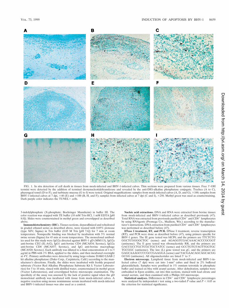
FIG. 1. In situ detection of cell death in tissues from mock-infected and BHV-1-infected calves. Thin sections were prepared from various tissues. Free 39-OHtermini were detected by the addition of terminal deoxynucleotidyltransferase and revealed by the anti-DIG-alkaline phosphatase conjugate. Trachea (A to C),pharyngeal tonsil (D to F), and turbinate mucosa (G to I) were tested. Original magnifications: samples from mock-infected calves (A, D, and G), 3100; samples fromBHV-1-infected calves at 7 dpi, 340 (E) and 3100 (B, H, and F); samples from infected calves at 7 dpi (C and I), 3250. Methyl green was used as counterstaining.Dark purple color indicates the TUNEL1 cells.
VOL. 73, 1999 INDUCTION OF APOPTOSIS BY BHV-1 8659
RESULTSBHV-1 infection induces apoptosis in lymphoid tissues. To
determine if apoptosis occurred in lymphoid tissue afterBHV-1 infection, calves were infected for 7 days and apoptosiswas examined in lymph nodes. Previous studies establishedthat cultured lymphocytes undergo apoptosis when infected (9,16–18), but it is not known if apoptosis occurs when calves are
infected. Pharyngeal tonsil contained many TUNEL-positive(TUNEL1) cells at 7 dpi (Fig. 1E and F) relative to mock-infected calves (Fig. 1D). TUNEL1 cells were localized in thecorticomedullary junction in germinal centers of secondarylymphoid follicles and subcapsular regions (Fig. 1E and F; Fig.2B and D). TUNEL1 cells were also detected in lymphoidareas located within the tracheal submucosa, which corre-
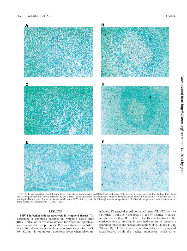
FIG. 2. In situ detection of cell death in lymph nodes from mock-infected and BHV-1-infected calves. Thin sections were prepared as described for Fig. 1 fromcervical lymph nodes from a mock-infected (A) and a BHV-1-infected calf (B), retropharyngeal lymph nodes from a mock-infected (C) and a BHV-1 infected calf (D),and inguinal lymph nodes from a mock-infected (E) and a BHV-1 infected calf (F). All samples are at a magnification of 3100. Methyl green was used to counterstain.Dark purple color indicates the TUNEL1 cells.
sponds to mucosa-associated lymphoid tissue (Fig. 1B and C),and turbinate membrane (Fig. 1H and I).
To determine if lymph nodes near the site of infection con-tained increased TUNEL1 cells, cervical and retropharyngeallymph nodes were examined at 7 dpi. Relative to mock-in-fected calves, cervical and retropharyngeal lymph nodes con-tained more TUNEL1 cells and staining was located in or nearthe follicle (Fig. 2B and D). Inguinal lymph node from infectedcalves also had TUNEL1 cells (Fig. 2F), but fewer than inpharyngeal tonsil. As expected, few or no TUNEL1 cells were
detected in the corresponding tissues of mock-infected animals(Fig. 1A, D, and G; Fig. 2A, C, and E). In summary, followinginfection, higher levels of TUNEL1 cells were detected inpharyngeal tonsil and other lymph nodes.
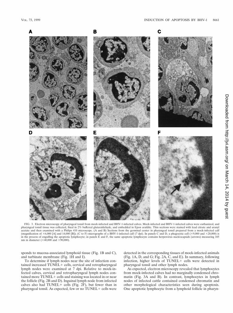
As expected, electron microscopy revealed that lymphocytesfrom mock-infected calves had no marginally condensed chro-matin (Fig. 3A and B). In contrast, lymphocytes in lymphnodes of infected cattle contained condensed chromatin andother morphological characteristics seen during apoptosis.One apoptotic lymphocyte from a lymphoid follicle in pharyn-
FIG. 3. Electron microscopy of pharyngeal tonsil from mock-infected and BHV-1-infected calves. Mock-infected and BHV-1-infected calves were euthanized, andpharyngeal tonsil tissue was collected, fixed in 2% buffered glutaraldehyde, and embedded in Epon araldite. Thin sections were stained with lead citrate and uranylacetate and then examined with a Philips 410 microscope. (A and B) Sections from the germinal center in pharyngeal tonsil prepared from a mock-infected calf(magnification of 36,000 [A] and 14,000 [B]); (C to F) micrographs of a BHV-1-infected calf (7 dpi). In panels C and D, a phagocytic cell (39,000 and 320,000) isin the process of engulfing the apoptotic lymphocyte; in panels E and F, the same apoptotic lymphocyte contains herpesvirus nucleocapsids (arrows) measuring 105nm in diameter (340,000 and 390,000).
VOL. 73, 1999 INDUCTION OF APOPTOSIS BY BHV-1 8661
geal tonsil of BHV-1-infected calves is shown in Fig. 3C to F.This lymphocyte has condensed chromatin shaped like a horse-shoe or half-moon (Fig. 3C to F). The cell adjacent to thelymphocyte appears to be a dendritic cell or macrophage. Theclose proximity of the lymphocyte to the large cell suggestedthat it was phagocytosed. However, this was unlikely becausewe found no evidence of two double membranes surroundingthe lymphocyte, which is indicative of phagocytosis. Herpesvi-rus nucleocapsid structures measuring 105 nm in diameterwere detected inside the apoptotic nucleus (Fig. 3E and F). Allapoptotic lymphocytes had morphological changes consistentto that shown in Fig. 3C, and many contained herpesvirusnucleocapsids within apoptotic nuclei.
To test whether infection led to decreased levels of CD41
and CD81 T-lymphocyte populations in pharyngeal tonsil andretropharyngeal lymph nodes, mononuclear cells were pre-pared by density gradient centrifugation. Adherent cells wereseparated from the other mononuclear cells by incubating on aplastic culture dish for 2 h at 37°C in a humidified CO2 incu-bator. T cells were not readily detected in the adherent pop-ulation based on cell morphology and hematoxylin-eosin stain-ing (57). At 7 dpi, threefold fewer CD41 T cells were detectedin the pharyngeal tonsil (n 5 2, P 5 0.013). CD81 T-cellnumbers were also decreased in the pharyngeal tonsil at 7 dpi(n 5 2, P 5 0.022) (Fig. 4). The retropharyngeal lymph nodealso had decreased levels of CD41 T cells (n 5 2, P 5 0.02)and CD81 T cells (n 5 2, P 5 0.028) at 7 dpi. In summary,these results demonstrated that lymph nodes located close tothe initial site of infection contained high levels of apoptoticlymphocytes, herpesvirus nucleocapsids were detected in theselymphocytes, and the numbers of T cells decreased.
Detection of gD in lymphoid tissue of infected calves. Theresults in Fig. 1 to 3 suggested that BHV-1 could infect lym-phocytes of calves, leading to a reduction in the number of Tcells in the tonsil and retropharyngeal lymph nodes. To test
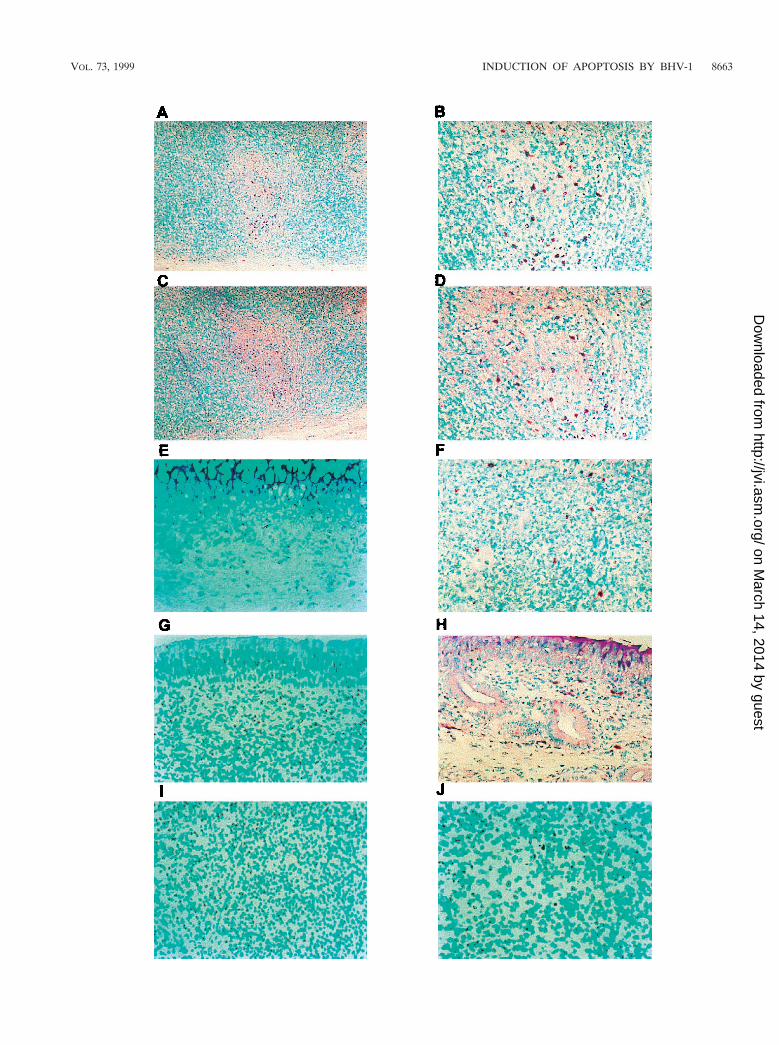
this hypothesis, we examined the relevant tissues for viral gly-coprotein (gD) by using a monoclonal antibody and IHC. gDwas consistently detected in lymphoid and nonlymphoid tissuesfrom two infected calves but not from two uninfected calves(Fig. 5 and data not shown). In lymphoid tissues, gD wasdetected in the germinal centers, the corticomedullary junctionof lymphoid follicles, the subcapsular region, and randomlythroughout individual lymphoid tissues (Fig. 5A and B). gDwas also detected in the submucosa of turbinate and trachea(Fig. 5F and H). Consecutive sections of pharyngeal tonsilrevealed that some of the infected cells also expressed thelymphocyte surface protein CD2 present in the germinal center(Fig. 5A to D). We observed overlapping of gD and CD21 cellsin 13 individual cells from a single germinal center. The loca-tion of these CD21 T lymphocytes suggested they are CD41 Tcells, which are normally present in germinal centers to acti-vate B lymphocytes. In other lymph nodes (cervical, retropha-ryngeal, and parotid), gD was also detected (data not shown).A few gD-positive cells were present in the inguinal lymphnode (Fig. 5J). Although this study suggested that gD wasexpressed in these tissues as a result of productive infection, itwas also possible that virus was delivered to lymph nodes viathe lymphatic system but infection did not occur.
Analysis of T lymphocytes in PBMC during acute infection.Figures 1 to 5 demonstrated that virus infection of lymphocytesoccurred in lymphoid tissue near the site of infection. How-ever, it was not clear whether circulating lymphocytes becomeinfected or undergo apoptosis as a consequence of infection.Six-month-old calves that were seronegative for BHV-1 werebled three times before BHV-1 infection, and the normal levelsof CD41 and CD81 T lymphocytes were compared to those ininfected calves. After BHV-1 infection, calves were bled atregular intervals (5, 7, 9, 12, and 16 dpi) and the distribution ofCD41 and CD81 T lymphocytes was determined. It was notnecessary to incubate PBMC with plastic to remove adherentcells and debris because T cells in PBMC were readily sepa-rated from other cell populations during FACS (57). BHV-1infection reduced the CD41 T-cell population in PBMC atdays 5 (n 5 4, P 5 0.014), 7 (n 5 4, P 5 0.002), 9 (n 5 3, P 50.009), and 12 (n 5 4, P 5 0.008) (Table 1). CD81 T-cellpopulations also decreased slightly at days 7 (n 5 4, P 5 0.001)and 9 (n 5 3, P 5 0.010).
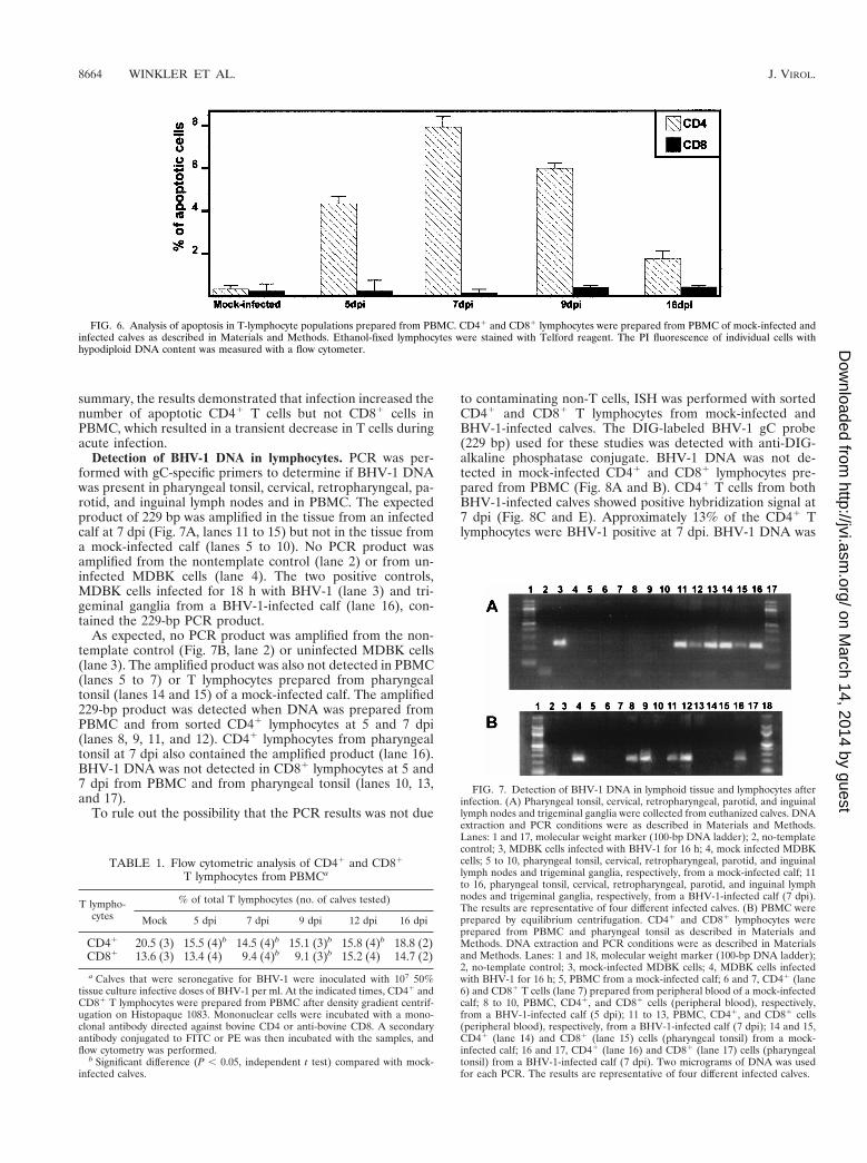
Two-color immunofluorescence of PBMC to measure apo-ptosis in CD41 and CD81 T lymphocytes was performed withmouse anti-bovine CD4 and CD8 antibodies, FITC-labeledsecondary antibodies, and PI. The percentage of apoptoticcells was determined by measuring the subgenomic DNA peakin mock-infected and infected calves at different times postin-fection. Figure 6 shows representative data from flow cytomet-ric analysis stained with PI. Increased apoptosis was detectedin CD41 T lymphocytes from PBMC at days 5 (n 5 2, P 50.006), 7 (n 5 2, P 5 0.002), 9 (n 5 2, P 5 0.002), and 16 (n 52, P 5 0.022) after infection. At 7 dpi, numbers of CD41 Tcells undergoing apoptosis were 30-fold higher than in thesame animal prior to infection. In contrast, the number ofapoptotic CD81 T lymphocytes was not significantly differentfrom the number in BHV-1-infected calves (P . 0.05). In
FIG. 4. Analysis of T-lymphocyte populations in pharyngeal tonsil and ret-ropharyngeal lymph nodes. CD41 and CD81 lymphocytes from mock-infectedand BHV-1-infected calves (7 dpi) were prepared from pharyngeal tonsil andretropharyngeal lymph node as described in Materials and Methods. Mononu-clear cells were incubated with monoclonal antibodies directed against bovineCD41 and CD81 T cells. FITC- or PE-conjugated IgG that was directed againstthe monoclonal antibody was used as a secondary antibody. FITC- or PE-positivecells were identified by flow cytometry.
FIG. 5. gD and CD2 detection by IHC on tissue sections of mock-infected and BHV-1-infected calves. Tissue sections were incubated with gD or CD2 monoclonalantibodies overnight at 4°C. After the slide was washed, the biotinylated secondary antibody was added. The alkaline phosphatase reaction was performed as describedin Materials and Methods. (A and B) Pharyngeal tonsil stained for BHV-1 gD in BHV-1-infected calves (7 dpi) (magnifications of 3100 and 3250, respectively); (Cand D) pharyngeal tonsil stained for the lymphocyte surface protein CD2 (magnification of 3100 and 3250, respectively) in consecutive sections from panels A andB; (E to I) gD staining of tissue from the turbinate membrane (F), tracheal submucosa (H), and inguinal lymph nodes (J) of calves infected for 7 days and gD stainingof turbinate mucosa (E), tracheal mucosa (G), and inguinal lymph node (I) of an infected calf (all at a magnification of 3250). The results are representative of dataobtained from three different calves.
summary, the results demonstrated that infection increased thenumber of apoptotic CD41 T cells but not CD81 cells inPBMC, which resulted in a transient decrease in T cells duringacute infection.
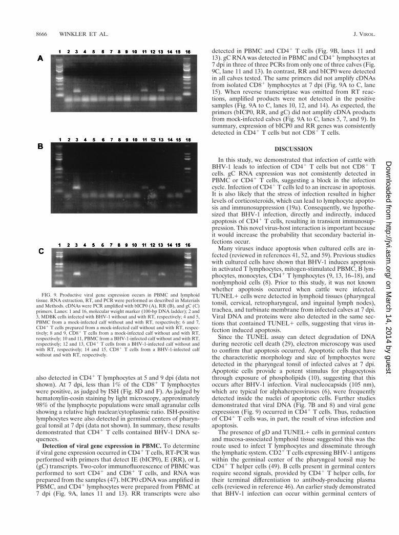
Detection of BHV-1 DNA in lymphocytes. PCR was per-formed with gC-specific primers to determine if BHV-1 DNAwas present in pharyngeal tonsil, cervical, retropharyngeal, pa-rotid, and inguinal lymph nodes and in PBMC. The expectedproduct of 229 bp was amplified in the tissue from an infectedcalf at 7 dpi (Fig. 7A, lanes 11 to 15) but not in the tissue froma mock-infected calf (lanes 5 to 10). No PCR product wasamplified from the nontemplate control (lane 2) or from un-infected MDBK cells (lane 4). The two positive controls,MDBK cells infected for 18 h with BHV-1 (lane 3) and tri-geminal ganglia from a BHV-1-infected calf (lane 16), con-tained the 229-bp PCR product.
As expected, no PCR product was amplified from the non-template control (Fig. 7B, lane 2) or uninfected MDBK cells(lane 3). The amplified product was also not detected in PBMC(lanes 5 to 7) or T lymphocytes prepared from pharyngealtonsil (lanes 14 and 15) of a mock-infected calf. The amplified229-bp product was detected when DNA was prepared fromPBMC and from sorted CD41 lymphocytes at 5 and 7 dpi(lanes 8, 9, 11, and 12). CD41 lymphocytes from pharyngealtonsil at 7 dpi also contained the amplified product (lane 16).BHV-1 DNA was not detected in CD81 lymphocytes at 5 and7 dpi from PBMC and from pharyngeal tonsil (lanes 10, 13,and 17).
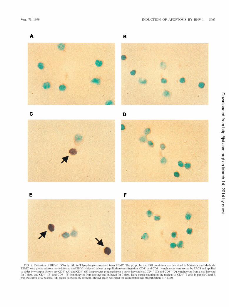
To rule out the possibility that the PCR results was not due
to contaminating non-T cells, ISH was performed with sortedCD41 and CD81 T lymphocytes from mock-infected andBHV-1-infected calves. The DIG-labeled BHV-1 gC probe(229 bp) used for these studies was detected with anti-DIG-alkaline phosphatase conjugate. BHV-1 DNA was not de-tected in mock-infected CD41 and CD81 lymphocytes pre-pared from PBMC (Fig. 8A and B). CD41 T cells from bothBHV-1-infected calves showed positive hybridization signal at7 dpi (Fig. 8C and E). Approximately 13% of the CD41 Tlymphocytes were BHV-1 positive at 7 dpi. BHV-1 DNA was
FIG. 6. Analysis of apoptosis in T-lymphocyte populations prepared from PBMC. CD41 and CD81 lymphocytes were prepared from PBMC of mock-infected andinfected calves as described in Materials and Methods. Ethanol-fixed lymphocytes were stained with Telford reagent. The PI fluorescence of individual cells withhypodiploid DNA content was measured with a flow cytometer.
FIG. 7. Detection of BHV-1 DNA in lymphoid tissue and lymphocytes afterinfection. (A) Pharyngeal tonsil, cervical, retropharyngeal, parotid, and inguinallymph nodes and trigeminal ganglia were collected from euthanized calves. DNAextraction and PCR conditions were as described in Materials and Methods.Lanes: 1 and 17, molecular weight marker (100-bp DNA ladder); 2, no-templatecontrol; 3, MDBK cells infected with BHV-1 for 16 h; 4, mock infected MDBKcells; 5 to 10, pharyngeal tonsil, cervical, retropharyngeal, parotid, and inguinallymph nodes and trigeminal ganglia, respectively, from a mock-infected calf; 11to 16, pharyngeal tonsil, cervical, retropharyngeal, parotid, and inguinal lymphnodes and trigeminal ganglia, respectively, from a BHV-1-infected calf (7 dpi).The results are representative of four different infected calves. (B) PBMC wereprepared by equilibrium centrifugation. CD41 and CD81 lymphocytes wereprepared from PBMC and pharyngeal tonsil as described in Materials andMethods. DNA extraction and PCR conditions were as described in Materialsand Methods. Lanes: 1 and 18, molecular weight marker (100-bp DNA ladder);2, no-template control; 3, mock-infected MDBK cells; 4, MDBK cells infectedwith BHV-1 for 16 h; 5, PBMC from a mock-infected calf; 6 and 7, CD41 (lane6) and CD81 T cells (lane 7) prepared from peripheral blood of a mock-infectedcalf; 8 to 10, PBMC, CD41, and CD81 cells (peripheral blood), respectively,from a BHV-1-infected calf (5 dpi); 11 to 13, PBMC, CD41, and CD81 cells(peripheral blood), respectively, from a BHV-1-infected calf (7 dpi); 14 and 15,CD41 (lane 14) and CD81 (lane 15) cells (pharyngeal tonsil) from a mock-infected calf; 16 and 17, CD41 (lane 16) and CD81 (lane 17) cells (pharyngealtonsil) from a BHV-1-infected calf (7 dpi). Two micrograms of DNA was usedfor each PCR. The results are representative of four different infected calves.
TABLE 1. Flow cytometric analysis of CD41 and CD81
a Calves that were seronegative for BHV-1 were inoculated with 107 50%tissue culture infective doses of BHV-1 per ml. At the indicated times, CD41 andCD81 T lymphocytes were prepared from PBMC after density gradient centrif-ugation on Histopaque 1083. Mononuclear cells were incubated with a mono-clonal antibody directed against bovine CD4 or anti-bovine CD8. A secondaryantibody conjugated to FITC or PE was then incubated with the samples, andflow cytometry was performed.
b Significant difference (P , 0.05, independent t test) compared with mock-infected calves.
FIG. 8. Detection of BHV-1 DNA by ISH in T lymphocytes prepared from PBMC. The gC probe and ISH conditions are described in Materials and Methods.PBMC were prepared from mock-infected and BHV-1-infected calves by equilibrium centrifugation. CD41 and CD81 lymphocytes were sorted by FACS and appliedto slides by cytospin. Shown are CD41 (A) and CD81 (B) lymphocytes prepared from a mock-infected calf, CD41 (C) and CD81 (D) lymphocytes from a calf infectedfor 7 days, and CD41 (E) and CD81 (F) lymphocytes from another calf infected for 7 days. Dark purple staining in the nucleus of CD41 T cells in panels C and Ewas indicative of a positive ISH signal (denoted by arrows). Methyl green was used for counterstaining; magnification is 31,000.
VOL. 73, 1999 INDUCTION OF APOPTOSIS BY BHV-1 8665
also detected in CD41 T lymphocytes at 5 and 9 dpi (data notshown). At 7 dpi, less than 1% of the CD81 T lymphocyteswere positive, as judged by ISH (Fig. 8D and F). As judged byhematoxylin-eosin staining by light microscopy, approximately98% of the lymphocyte populations were small agranular cellsshowing a relative high nuclear/cytoplasmic ratio. ISH-positivelymphocytes were also detected in germinal centers of pharyn-geal tonsil at 7 dpi (data not shown). In summary, these resultsdemonstrated that CD41 T cells contained BHV-1 DNA se-quences.
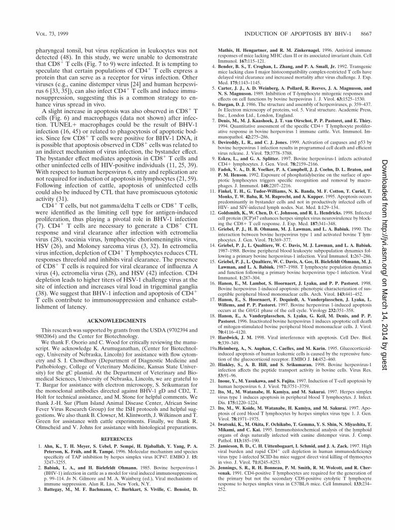
Detection of viral gene expression in PBMC. To determineif viral gene expression occurred in CD41 T cells, RT-PCR wasperformed with primers that detect IE (bICP0), E (RR), or L(gC) transcripts. Two-color immunofluorescence of PBMC wasperformed to sort CD41 and CD81 T cells, and RNA wasprepared from the samples (47). bICP0 cDNA was amplified inPBMC, and CD41 lymphocytes were prepared from PBMC at7 dpi (Fig. 9A, lanes 11 and 13). RR transcripts were also
detected in PBMC and CD41 T cells (Fig. 9B, lanes 11 and13). gC RNA was detected in PBMC and CD41 lymphocytes at7 dpi in three of three PCRs from only one of three calves (Fig.9C, lane 11 and 13). In contrast, RR and bICP0 were detectedin all calves tested. The same primers did not amplify cDNAsfrom isolated CD81 lymphocytes at 7 dpi (Fig. 9A to C, lane15). When reverse transcriptase was omitted from RT reac-tions, amplified products were not detected in the positivesamples (Fig. 9A to C, lanes 10, 12, and 14). As expected, theprimers (bICP0, RR, and gC) did not amplify cDNA productsfrom mock-infected calves (Fig. 9A to C, lanes 5, 7, and 9). Insummary, expression of bICP0 and RR genes was consistentlydetected in CD41 T cells but not CD81 T cells.
DISCUSSION
In this study, we demonstrated that infection of cattle withBHV-1 leads to infection of CD41 T cells but not CD81 Tcells. gC RNA expression was not consistently detected inPBMC or CD41 T cells, suggesting a block in the infectioncycle. Infection of CD41 T cells led to an increase in apoptosis.It is also likely that the stress of infection resulted in higherlevels of corticosteroids, which can lead to lymphocyte apopto-sis and immunosuppression (19a). Consequently, we hypothe-sized that BHV-1 infection, directly and indirectly, inducedapoptosis of CD41 T cells, resulting in transient immunosup-pression. This novel virus-host interaction is important becauseit would increase the probability that secondary bacterial in-fections occur.
Many viruses induce apoptosis when cultured cells are in-fected (reviewed in references 41, 52, and 59). Previous studieswith cultured cells have shown that BHV-1 induces apoptosisin activated T lymphocytes, mitogen-stimulated PBMC, B lym-phocytes, monocytes, CD41 T lymphocytes (9, 13, 16–18), andnonlymphoid cells (8). Prior to this study, it was not knownwhether apoptosis occurred when cattle were infected.TUNEL1 cells were detected in lymphoid tissues (pharyngealtonsil, cervical, retropharyngeal, and inguinal lymph nodes),trachea, and turbinate membrane from infected calves at 7 dpi.Viral DNA and proteins were also detected in the same sec-tions that contained TUNEL1 cells, suggesting that virus in-fection induced apoptosis.
Since the TUNEL assay can detect degradation of DNAduring necrotic cell death (29), electron microscopy was usedto confirm that apoptosis occurred. Apoptotic cells that havethe characteristic morphology and size of lymphocytes weredetected in the pharyngeal tonsil of infected calves at 7 dpi.Apoptotic cells provide a potent stimulus for phagocytosisthrough exposure of phospholipids (10), suggesting that thisoccurs after BHV-1 infection. Viral nucleocapsids (105 nm),which are typical for alphaherpesviruses (6), were frequentlydetected inside the nuclei of apoptotic cells. Further studiesdemonstrated that viral DNA (Fig. 7B and 8) and viral geneexpression (Fig. 9) occurred in CD41 T cells. Thus, reductionof CD41 T cells was, in part, the result of virus infection andapoptosis.
The presence of gD and TUNEL1 cells in germinal centersand mucosa-associated lymphoid tissue suggested this was theroute used to infect T lymphocytes and disseminate throughthe lymphatic system. CD21 T cells expressing BHV-1 antigenswithin the germinal center of the pharyngeal tonsil may beCD41 T helper cells (49). B cells present in germinal centersrequire second signals, provided by CD41 T helper cells, fortheir terminal differentiation to antibody-producing plasmacells (reviewed in reference 46). An earlier study demonstratedthat BHV-1 infection can occur within germinal centers of
FIG. 9. Productive viral gene expression occurs in PBMC and lymphoidtissue. RNA extraction, RT, and PCR were performed as described in Materialsand Methods. cDNAs were PCR amplified with bICP0 (A), RR (B), and gC (C)primers. Lanes: 1 and 16, molecular weight marker (100-bp DNA ladder); 2 and3, MDBK cells infected with BHV-1 without and with RT, respectively; 4 and 5,PBMC from a mock-infected calf without and with RT, respectively; 6 and 7,CD41 T cells prepared from a mock-infected calf without and with RT, respec-tively; 8 and 9, CD81 T cells from a mock-infected calf without and with RT,respectively; 10 and 11, PBMC from a BHV-1-infected calf without and with RT,respectively; 12 and 13, CD41 T cells from a BHV-1-infected calf without andwith RT, respectively; 14 and 15, CD81 T cells from a BHV-1-infected calfwithout and with RT, respectively.
pharyngeal tonsil, but virus replication in leukocytes was notdetected (48). In this study, we were unable to demonstratethat CD81 T cells (Fig. 7 to 9) were infected. It is tempting tospeculate that certain populations of CD41 T cells express aprotein that can serve as a receptor for virus infection. Otherviruses (e.g., canine distemper virus [24] and human herpesvi-rus 6 [33, 35]), can also infect CD41 T cells and induce immu-nosuppression, suggesting this is a common strategy to en-hance virus spread in vivo.
A slight increase in apoptosis was also observed in CD81 Tcells (Fig. 6) and macrophages (data not shown) after infec-tion. TUNEL1 macrophages could be the result of BHV-1infection (16, 45) or related to phagocytosis of apoptotic bod-ies. Since few CD81 T cells were positive for BHV-1 DNA, itis possible that apoptosis observed in CD81 cells was related toan indirect mechanism of virus infection, the bystander effect.The bystander effect mediates apoptosis in CD81 T cells andother uninfected cells of HIV-positive individuals (11, 25, 39).With respect to human herpesvirus 6, entry and replication arenot required for induction of apoptosis in lymphocytes (21, 59).Following infection of cattle, apoptosis of uninfected cellscould also be induced by CTL that have promiscuous cytotoxicactivity (31).
CD41 T cells, but not gamma/delta T cells or CD81 T cells,were identified as the limiting cell type for antigen-inducedproliferation, thus playing a pivotal role in BHV-1 infection(7). CD41 T cells are necessary to generate a CD81 CTLresponse and viral clearance after infection with ectromeliavirus (28), vaccinia virus, lymphocytic choriomeningitis virus,HSV (26), and Moloney sarcoma virus (3, 32). In ectromeliavirus infection, depletion of CD41 T lymphocytes reduces CTLresponses threefold and inhibits viral clearance. The presenceof CD81 T cells is required for viral clearance of influenza Avirus (4), ectromelia virus (28), and HSV (42) infection. CD4depletion leads to higher titers of HSV-1 challenge virus at thesite of infection and increases viral load in trigeminal ganglia(38). We suggest that BHV-1 infection and apoptosis of CD41
T cells contribute to immunosuppression and enhance estab-lishment of latency.
ACKNOWLEDGMENTS
This research was supported by grants from the USDA (9702394 and9802064) and the Center for Biotechnology.
We thank F. Osorio and C. Wood for critically reviewing the manu-script. We acknowledge K. Arumuganathan, (Center for Biotechnol-ogy, University of Nebraska, Lincoln) for assistance with flow cytom-etry and S. I. Chowdhury (Department of Diagnostic Medicine andPathobiology, College of Veterinary Medicine, Kansas State Univer-sity) for the gC plasmid. At the Department of Veterinary and Bio-medical Sciences, University of Nebraska, Lincoln, we are grateful toT. Bargar for assistance with electron microscopy, S. Srikumaran forthe monoclonal antibodies directed against BHV-1 gD and CD2, T.Holt for technical assistance, and M. Stone for helpful comments. Wethank J.-H. Sur (Plum Island Animal Disease Center, African SwineFever Virus Research Group) for the ISH protocols and helpful sug-gestions. We also thank B. Clowser, M. Klintworth, J. Wilkinson and T.Green for assistance with cattle experiments. Finally, we thank R.Olmscheid and V. Johns for assistance with histological preparations.
REFERENCES
1. Ahn, K., T. H. Meyer, S. Uebel, P. Sempe, H. Djaballah, Y. Yang, P. A.Peterson, K. Fruh, and R. Tampe. 1996. Molecular mechanism and speciesspecificity of TAP inhibition by herpes simplex virus ICP47. EMBO J. 15:3247–3255.
2. Babiuk, L. A., and H. Bielefeldt Ohmann. 1985. Bovine herpesvirus-1(BHV-1) infection in cattle as a model for viral induced immunosuppression,p. 99–114. In N. Gilmore and M. A. Wainberg (ed.), Viral mechanisms ofimmune suppression. Alan R. Liss, New York, N.Y.
3. Battegay, M., M. F. Bachmann, C. Burhkart, S. Viville, C. Benoist, D.
Mathis, H. Hengartner, and R. M. Zinkernagel. 1996. Antiviral immuneresponses of mice lacking MHC class II or its associated invariant chain. CellImmunol. 167:115–121.
4. Bender, B. S., T. Croghan, L. Zhang, and P. A. Small, Jr. 1992. Transgenicmice lacking class I major histocompatibility complex-restricted T cells havedelayed viral clearance and increased mortality after virus challenge. J. Exp.Med. 175:1143–1145.
5. Carter, J. J., A. D. Weinberg, A. Pollard, R. Reeves, J. A. Magnuson, andN. S. Magnuson. 1989. Inhibition of T-lymphocyte mitogenic responses andeffects on cell functions by bovine herpesvirus 1. J. Virol. 63:1525–1530.
6. Dargan, D. J. 1986. The structure and assembly of herpesviruses, p. 359–437.In Electron microscopy of proteins, vol. 5. Viral structure. Academic Press,Inc., London Ltd., London, England.
7. Denis, M., M. J. Kaashoek, J. T. van Oirschot, P. P. Pastoret, and E. Thiry.1994. Quantitative assessment of the specific CD41 T lymphocyte prolifer-ative response in bovine herpesvirus 1 immune cattle. Vet. Immunol. Im-munopathol. 42:275–286.
8. Devireddy, L. R., and C. J. Jones. 1999. Activation of caspases and p53 bybovine herpesvirus 1 infection results in programmed cell death and efficientvirus release. J. Virol. 73:3778–3788.
9. Eskra, L., and G. A. Splitter. 1997. Bovine herpesvirus-1 infects activatedCD41 lymphocytes. J. Gen. Virol. 78:2159–2166.
10. Fadok, V. A., D. R. Voelker, P. A. Campbell, J. J. Coehn, D. L. Braton, andP. M. Henson. 1992. Exposure of phosphatidylserine on the surface of apo-ptotic lymphocytes triggers specific recognition and removal by macro-phages. J. Immunol. 148:2207–2216.
11. Finkel, T. H., G. Tudor-Williams, N. K. Banda, M. F. Cotton, T. Curiel, T.Monks, T. W. Baba, R. M. Ruprecht, and A. Kupper. 1995. Apoptosis occurspredominantly in bystander cells and not in productively infected cells ofHIV- and SIV-infected lymph nodes. Nat. Med. 1:129–134.
12. Goldsmith, K., W. Chen, D. C. Johnson, and R. L. Hendricks. 1998. Infectedcell protein (ICP)47 enhances herpes simplex virus neurovirulence by block-ing the CD81 T cell response. J. Exp. Med. 187:341–348.
13. Griebel, P. J., H. B. Ohmann, M. J. Lawman, and L. A. Babiuk. 1990. Theinteraction between bovine herpesvirus type 1 and activated bovine T lym-phocytes. J. Gen. Virol. 71:369–377.
14. Griebel, P. J., L. Qualtiere, W. C. Davis, M. J. Lawman, and L. A. Babiuk.1987–1988. Bovine peripheral blood leukocyte subpopulation dynamics fol-lowing a primary bovine herpesvirus-1 infection. Viral Immunol. 1:267–286.
15. Griebel, P. J., L. Qualtiere, W. C. Davis, A. Gee, H. Bielefeldt Ohmann, M. J.Lawman, and L. A. Babiuk. 1987–1988. T lymphocyte population dynamicsand function following a primary bovine herpesvirus type-1 infection. ViralImmunol. 1:287–304.
16. Hanon, E., M. Lambot, S. Hoornaert, J. Lyaku, and P. P. Pastoret. 1998.Bovine herpesvirus 1-induced apoptosis: phenotypic characterization of sus-ceptible peripheral blood mononuclear cells. Arch. Virol. 143:441–452.
17. Hanon, E., S. Hoornaert, F. Dequiedt, A. Vanderplasschen, J. Lyaku, L.Willems, and P. P. Pastoret. 1997. Bovine herpesvirus 1-induced apoptosisoccurs at the G0/G1 phase of the cell cycle. Virology 232:351–358.
18. Hanon, E., A. Vanderplasschen, S. Lyaku, G. Keil, M. Denis, and P. P.Pastoret. 1996. Inactivated bovine herpesvirus 1 induces apoptotic cell deathof mitogen-stimulated bovine peripheral blood mononuclear cells. J. Virol.70:4116–4120.
19. Hardwick, J. M. 1998. Viral interference with apoptosis. Cell Dev. Biol.9:339–349.
19a.Heimberg, A., N. Auphan, C. Caelles, and M. Karin. 1995. Glucocorticoid-induced apoptosis of human leukemic cells is caused by the repressive func-tion of the glucocorticoid receptor. EMBO J. 14:452–460.
20. Hinkley, S., A. B. Hill, and S. Srikumaran. 1998. Bovine herpesvirus-1infection affects the peptide transport activity in bovine cells. Virus Res.53:91–96.
21. Inoue, Y., M. Yasukawa, and S. Fujita. 1997. Induction of T-cell apoptosis byhuman herpesvirus 6. J. Virol. 71:3751–3759.
22. Ito, M., M. Watanabe, H. Kamiya, and M. Sakurai. 1997. Herpes simplexvirus type 1 induces apoptosis in peripheral blood T lymphocytes. J. Infect.Dis. 175:1220–1224.
23. Ito, M., W. Koide, M. Watanabe, H. Kamiya, and M. Sakurai. 1997. Apo-ptosis of cord blood T lymphocytes by herpes simplex virus type 1. J. Gen.Virol. 78:1971–1975.
24. Iwatsuki, K., M. Okita, F. Ochikubo, T. Gemma, Y. S. Shin, N. Miyashita, T.Mikami, and C. Kai. 1995. Immunohistochemical analysis of the lymphoidorgans of dogs naturally infected with canine distemper virus. J. Comp.Pathol. 113:185–190.
25. Jamieson, B. D., C. H. Uittenbogaart, I. Schmid, and J. A. Zack. 1997. Highviral burden and rapid CD41 cell depletion in human immunodeficiencyvirus type 1-infected SCID-hu mice suggest direct viral killing of thymocytesin vivo. J. Virol. 71:8245–8253.
26. Jennings, S. R., R. H. Bonneau, P. M. Smith, R. M. Wolcott, and R. Cher-venak. 1991. CD4-positive T lymphocytes are required for the generation ofthe primary but not the secondary CD8-positive cytolytic T lymphocyteresponse to herpes simplex virus in C57BL/6 mice. Cell Immunol. 133:234–252.
VOL. 73, 1999 INDUCTION OF APOPTOSIS BY BHV-1 8667
27. Jones, C. 1998. Alphaherpesvirus latency: its role in disease and survival ofthe virus in nature. Adv. Virus Res. 51:47–99.
28. Karupiah, G., R. M. L. Buller, N. van Rooijen, C. J. Duarte, and J. Chen.1996. Different roles for CD41 and CD81 T lymphocytes and macrophagesubsets in the control of a generalized virus infection. J. Virol. 70:8301–8309.
29. Kressel, M., and P. Groscurth. 1994. Distinction of apoptotic and necroticcell death by in situ labelling of fragmented DNA. Cell Tissue Res. 278:549–556.
30. Lan, H. C., M. A. Chambers, J. A. Fergunson, K. K. Srivastava, and P. G.Reddy. 1996. Effect of bovine herpesvirus-1 on expression of interleukin-2receptors and effect of interleukin-12 on lymphocyte proliferation. Vet. Mi-crobiol. 49:59–66.
31. Lawman, M. J., P. Griebel, D. L. Hutchings, W. C. Davis, J. Heise, L.Qualtiere, and L. A. Babiuk. 1987–1988. Generation of IL-2 dependentbovine cytotoxic T lymphocyte clones reactive against BHV-1 infected targetcells: loss of genetic restriction and virus specificity. Viral Immunol. 1:163–176.
32. Leist, T. P., M. Kohler, and R. M. Zinkernagel. 1989. Impaired generationof anti-viral cytotoxicity against lymphocytic choriomeningitis and vacciniavirus in mice treated with CD41-specific monoclonal antibody. Scand. J. Im-munol. 30:679–686.
33. Lusso, P., A. De Maria, C. Balotta, S. E. DeRocco, P. D. Markham, and R. C.Gallo. 1991. Productive infection of human CD41 and CD81 mature hu-man T cell populations and clones by HHV-6. Transcriptional down-regu-lation of CD3. J. Immunol. 147:685–691.
34. Maccario, R., P. Comoli, E. Percivalle, D. Montagna, F. Locatelli, and G.Gerna. 1995. Herpes simplex virus-specific human cytotoxic T-cell coloniesexpressing either gamma delta or alpha beta T-cell receptor: role of acces-sory molecules on HLA-unrestricted killing of virus-infected targets. Immu-nology 85:49–56.
35. Manickan, E., R. J. Rouse, Z. Yu, W. S. Wire, and B. T. Rouse. 1995. Geneticimmunization against herpes simplex virus. Protection is mediated by CD4/1T lymphocytes. J. Immunol. 155:259–265.
36. Mikloska, Z., A. M. Kesson, M. E. Penfold, and A. L. Cunningham. 1996.Herpes simplex virus protein targets for CD4 and CD8 lymphocyte cytotox-icity in cultured epidermal keratinocytes treated with interferon-gamma.J. Infect. Dis. 173:7–17.
37. Milligan, G. N., and D. I. Bernstein. 1995. Analysis of herpes simplex virus-specific T cells in the murine female genital tract following genital infectionwith herpes simplex virus type 2. Virology 212:481–489.
38. Morrison, L. A., and D. M. Knipe. 1997. Contributions of antibody and T cellsubsets to protection elicited by immunization with a replication-defectivemutant of herpes simplex virus type 1. Virology 239:315–326.
39. Muro-Cacho, C. A., G. Pantaleo, and A. S. Fauci. 1995. Analysis of apoptosisin lymph nodes in HIV-infected persons. J. Immunol. 154:5556–5566.
40. Nataraj, S., S. Eidmann, M. J. Hariharan, J. H. Sur, G. A. Perry, and S.Srikumaran. 1997. Bovine herpesvirus 1 downregulates the expression ofbovine MHC class I molecules. Viral Immunol. 10:21–34.
41. Pignata, C., M. Fiore, S. de Filippo, M. Cavalcanti, L. Gaetaniello, and I.Scotese. 1998. Apoptosis as a mechanism of peripheral blood mononuclearcell death after measles and varicella-zoster virus infections in children.Pediatr. Res. 43:77–83.
42. Posavad, C. M., D. M. Koelle, and L. Corey. 1996. High frequency of CD81cytotoxic T-lymphocyte precursors specific for herpes simplex viruses inpersons with genital herpes. J. Virol. 70:8165–8168.
43. Reed, L. J., and H. Muench. 1938. A simple method of estimating fifty percent endpoints. Am. J. Hyg. 27:493–497.
44. Rinaldo, C. R., Jr., and D. J. Torpey. 1993. Cell-mediated immunity andimmunosuppression in herpes simplex virus infection. Immunodeficiency5:33–90.
45. Rossi, C. R., and G. K. Kiesel. 1977. Susceptibility of bovine macrophagesand tracheal ring cultures to bovine viruses. Am. J. Vet. Res. 38:1705–1708.
46. Scadding, G. K. Immunology of the tonsil: a review. J. R. Soc. Med. 83:104–107.
47. Schang, L. M., and C. Jones. 1997. Analysis of bovine herpesvirus 1 tran-scripts during a primary infection of trigeminal ganglia of cattle. J. Virol.71:6786–6795.
48. Schuh, J. C. L., H. Bielefeldt Ohmann, L. A. Babiuk, and C. E. Doige. 1992.Bovine herpesvirus-1-induced pharyngeal tonsil lesions in neonatal andweanling calves. J. Comp. Pathol. 106:243–253.
49. Schuh, J. C. L., and L. W. Oliphant. 1992. Development and immunophe-notyping of the pharyngeal tonsil (adenoid) in cattle. J. Comp. Pathol.106:229–241.
50. Sciammas, R., P. Kodukula, Q. Tang, R. L. Hendricks, and J. A. Bluestone.1997. T cell receptor-gamma/delta cells protect mice from herpes simplexvirus type 1-induced lethal encephalitis. J. Exp. Med. 185:1969–1975.
51. Sheffy, B. E., and S. Rodman. 1973. Activation of latent infectious bovinerhinotracheitis infection. J. Am. Vet. Med. Assoc. 163:850–851.
52. Shen, Y., and T. E. Shenk. 1995. Viruses and apoptosis. Curr. Opin. Genet.Dev. 5:105–111.
53. Simmons, A., and D. C. Tscharke. 1992. Anti-CD8 impairs clearance ofherpes simplex virus from the nervous system: implications for the fate ofvirally infected neurons. J. Exp. Med. 175:1337–1344.
54. Spear, A. 1993. Entry of alphaherpesviruses into cells. Semin. Virol. 4:167–180.
55. Takahashi, K., S. Sonoda, K. Higashi, T. Kondo, H. Takahashi, M. Taka-hashi, and K. Yamanishi. 1989. Predominant CD4 T-lymphocyte tropism ofhuman herpesvirus 6-related virus. J. Virol. 63:3161–3163.
56. Tigges, M. A., S. Leng, D. C. Johnson, and R. L. Burke. 1996. Human herpessimplex virus (HSV)-specific CD81 CTL clones recognize HSV-2-infectedfibroblasts after treatment with IFN-gamma or when virion host shutofffunctions are disabled. J. Immunol. 156:3901–3910.
57. Winkler, M. T., and C. Jones. Unpublished observation.58. Wyler, R., M. Engels, and M. Schwyzer. 1989. Infectious bovine rhinotra-
cheitis/vulvovaginitis (BHV-1), p. 172–183. In G. Witman (ed.), Herpesvi-ruses diseases of cattle, horses, and pigs, developments in veterinary medi-cine. Kluver Academic Publishers, Boston, Mass.
59. Yasukawa, M., Y. Inoue, H. Ohminami, K. Terrada, and S. Fujita. 1998.Apoptosis of CD41 T lymphocytes in human herpesvirus-6 infection. J. Gen.Virol. 79:143–147.
60. York, I. A., C. Roop, D. W. Andrews, S. R. Riddell, F. L. Graham, and D. C.Johnson. 1994. A cytosolic herpes simplex virus protein inhibits antigenpresentation to CD81 T lymphocytes. Cell 77:525–535.