76
Bronchiectasis Jeff Bowden Head of Respiratory and Sleep Services Southern Adelaide Local Health Network

Bronchiectasis
Jeff Bowden
Head of Respiratory and Sleep Services
Southern Adelaide Local Health Network

Disclosures
• Honoraria; Astra Zeneca, Glaxo Smith Kline, Novartis
• Educational Support; Eli Lilly, Astra Zeneca, Novartis
• Funding for Research Trials; Glaxo Smith Kline, Novartis, KaloBios, Astra Zeneca, Bayer
• Advisory Boards for Glaxo Smith Kline and Astra Zeneca

Outline
– Basic Anatomy and Physiology
– What is bronchiectasis
– Aetiology
– Assessment
– Treatment

Outline
– Basic Anatomy and Physiology
– What is bronchiectasis
– Aetiology
– Assessment
– Treatment

The Respiratory Tract
Upper Lower
http://www.nlm.nih.gov/medlineplus/ency/imagepages

Anatomy of the Lower Respiratory Tract
© Novartis 2000

Host defenses along the airways
© Current Medicine, Inc

Mucus
Basement membrane
Goblet (mucous) cell Nerve
Ciliated cells Basal
cell
Brush cell Basal
cell
Goblet cell (discharging) Nerve
Undifferentiated cell Kulchitsky
cell
Serous cell
Basement membrane
Clara cell
Basal cell
Ciliated cells
Nerves
Clara cell
Undifferentiated cell
Magnified detail of cilium
Cross section
© Novartis
Tracheobronchial Secretion and
Mucociliary Transport
© Boehringer, 1996

Interactions of respiratory epithelium and microbes
© Current Medicine, Inc

Mechanisms of microbial attachment and host resistance
© Current Medicine, Inc

Immunoglobulins; targeting proteins, critical for immune function

Outline
– Basic Anatomy and Physiology
– What is bronchiectasis
– Aetiology
– Assessment
– Treatment

Definition of Bronchiectasis
• “bronckos” (airway) and “ektasis” (widening)
• Bronchiectasis is characterised by abnormal, irreversible bronchial dilatation or a fixed increase in airway diameter
• Bronchiectasis is a common lung disease characterised by chronic infection in small airways
• Results in some parts of the lung becoming damaged, scarred and dilated, allowing infected mucus to build up in pockets
ALF Fact Sheet, Sept 2014

CT Scan of Chest

Bronchiectasis
• The diagnosis of bronchiectasis requires a CT scan of the lungs which demonstrates abnormal widening of the airways or bronchi
• Despite its low profile,
bronchiectasis is a common condition and patients will often have symptoms for many years before a diagnosis is made

Other Respiratory Infections
• Pneumonia – Infection of the lung parenchyma, (alveoli)
• Acute Bronchitis – Inflammatory Change in large airways and bronchi, may be
bacterial or viral
• Bronchiolitis – Inflammatory change in peripheral bronchioles, usually
viral
• Lower Respiratory Tract Infection – Any of the above, or combination thereof

Other Chronic Respiratory Conditions
• Chronic Obstructive Pulmonary Disease – Chronic lung injury, usually due to smoking, with narrowing of the
airways due to chronic bronchitis or emphysema, or a combination thereof
• Chronic Bronchitis – Chronic cough and sputum production due to chronic
inflammation of the airways
• Emphysema – Destruction of the airspaces within the lung, with loss of
radial traction on the airways

Outline
– Basic Anatomy and Physiology
– What is bronchiectasis
– Aetiology
– Assessment
– Treatment

•Aetiologies and factors associated with bronchiectasis
•Cystic fibrosis
•Post-infection (eg. tuberculosis, adenovirus, recurrent pneumonia)
•Primary or secondary immune deficiency (eg. hypogammaglobulinaemia, lung
and bone marrow transplantation, malignancy, HIV/AIDS, HTLV1)
•Mucociliary dysfunction (eg primary ciliary dyskinesia)
•Chronic obstructive pulmonary disease and smoking
•Congenital causes (eg. Mounier-Kuhn syndrome, Young syndrome)
•Postobstruction (eg. with a foreign body)
•Pulmonary fibrosis and pneumoconiosis (eg. silicosis)
•Recurrent small volume aspiration (eg. from upper airway secretions or gastric
contents)
•Allergic bronchopulmonary aspergillosis
•Systemic inflammatory diseases (eg. rheumatoid arthritis, sarcoidosis)
Chang AB, Bell SC, Byrnes CA, et al. Chronic suppurative lung disease and bronchiectasis
in children and adults in Australia and New Zealand. Med J Aust 2010;193:356–65.


Foreign Body
Peanut in Left main stem bronchus which had increased is size as it absorbs the secretions within the
bronchus.Courtesy Dr Trent Quinlan, ENT surgeon, Omaha Childrens Hospital(CHS).
https://www.mypacs.net/cases/FOREIGN-BODY-ASPIRATION-AT-LEAST-TWO-PEANUTS-5220614.html

Sputum retention
and occlusion of
right lower lobe
bronchi in an
elderly women with
depressed cough
Sputum retention

Cystic Fibrosis
• Autosomal recessive condition
• 1 in 2000 to 1 in 2500
• 1 in 25 people carry a mutation of the cystic fibrosis gene
• ethnic variation
– African American 1 in 15,300
– Hispanics 1 in 8000
– occurs in Asian populations
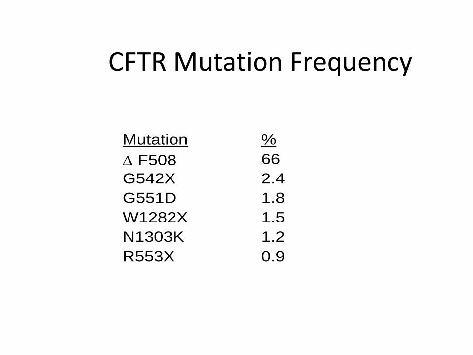
CFTR Mutation Frequency
Mutation %
F508 66
G542X 2.4
G551D 1.8
W1282X 1.5
N1303K 1.2
R553X 0.9

Downloaded from: Textbook of Respiratory Medicine (on 1 March 2007 09:29 PM)
© 2007 Elsevier
CysticFibrosisTransmembrane
conductanceReceptor(CFTR)

Downloaded from: Textbook of Respiratory Medicine (on 1 March 2007 09:29 PM)
© 2007 Elsevier
Normal Airways

Downloaded from: Textbook of Respiratory Medicine (on 1 March 2007 09:29 PM)
© 2007 Elsevier
Cystic Fibrosis Airways


Cystic Fibrosis
Outline
– Basic Anatomy and Physiology
– What is bronchiectasis
– Aetiology
– Assessment
– Treatment

Features that may suggest bronchiectasis in a patient presenting with chronic respiratory symptoms
•Chronic cough and sputum production
•Digital clubbing (this is rare in COPD and asthma)
•Lack of a significant smoking history (less than an average of 20 cigarettes per
day for 10 years) in a person with suspected COPD
•History of recurrent and/or severe pneumonia including tuberculosis
•Presence of 'unusual organisms' in sputum (eg. Aspergillus,
atypical/nontuberculous mycobacteria, Pseudomonas aeruginosa, Escherichia
coli, Klebsiella pneumoniae)
•Childhood associated with significant environmental and social disadvantage*
* This includes Aboriginal and Torres Strait Islander people, as well as people
who have immigrated from low income countries.52 In this group of people,
tuberculosis as the cause of chronic respiratory symptoms should also be
considered
Maguire, C. Bronchiectasis A guide for primary care
Australian Family Physician Volume 41, No.11, November 2012 Pages 842-85
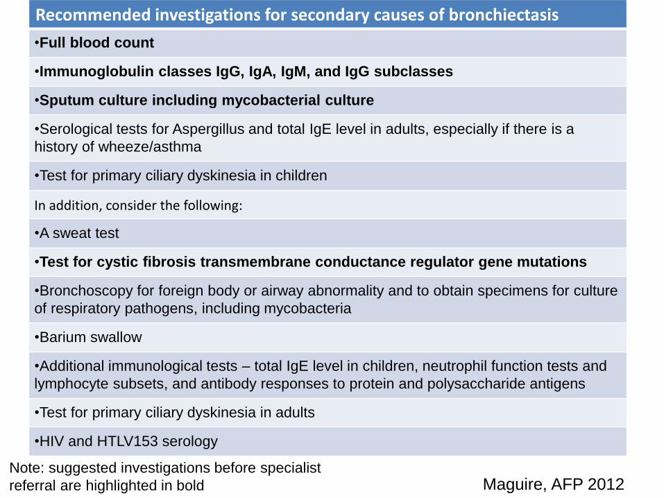
Recommended investigations for secondary causes of bronchiectasis
•Full blood count
•Immunoglobulin classes IgG, IgA, IgM, and IgG subclasses
•Sputum culture including mycobacterial culture
•Serological tests for Aspergillus and total IgE level in adults, especially if there is a
history of wheeze/asthma
•Test for primary ciliary dyskinesia in children
In addition, consider the following:
•A sweat test
•Test for cystic fibrosis transmembrane conductance regulator gene mutations
•Bronchoscopy for foreign body or airway abnormality and to obtain specimens for culture
of respiratory pathogens, including mycobacteria
•Barium swallow
•Additional immunological tests – total IgE level in children, neutrophil function tests and
lymphocyte subsets, and antibody responses to protein and polysaccharide antigens
•Test for primary ciliary dyskinesia in adults
•HIV and HTLV153 serology
Note: suggested investigations before specialist
referral are highlighted in bold Maguire, AFP 2012


Post-infectious bronchiectasis

Panbronchiolitis

Allergic Bronchopulmonary Aspergillosis (ABPA)

ABPA
www.aspergillus.man.ac.uk
Before bronchoscopy
After bronchoscopy

Mucus plugging with ABPA
Macroscopic view of sliced left upper lobe of lung showing obstruction Macroscopic view of sliced left upper lobe of lung showing obstruction of lower segmental bronchi
A
B
P
A
-
P
t
N
W
M
u
c
o
i
d
i
m
p
a
c
t
i
o
n
,
l
o
c
a
l
i
s
e
d
b
r
o
n
c
h
i
e
c
t
a
s
i
s
a
n
d
s
u
r
g
e
r
y
(
u
p
p
e
r
l
o
b
e
c
t
o
m
y
)
i
n
a
p
a
t
i
e
n
t
w
i
t
h
A
B
P
A
Macroscopic view of sliced left upper lobe of lung showing obstruction of lower segmental bronchi
Bronchoscopy of left upper lobe


CF-Bronchiectasis

Outline
– Basic Anatomy and Physiology
– What is bronchiectasis
– Aetiology
– Assessment
– Treatment

Outline
– Basic Anatomy and Physiology
– What is bronchiectasis
– Aetiology
– Assessment
– Treatment
• Specific Treatment
• General measures
• CF Therapy

Specific Treatment of bronchiectasis
Condition Treatment
Immune Deficiency Immunoglobulin infusions
Allergic Broncho-Pulmonary Aspergillosis Antifungal Therapy
Mycobacterial Infection Anti-mycobacterial therapy
Airway Obstruction Removal of foreign body
Inflammatory Bowel Disease Immuno-suppressive therapy
Rheumatoid Arthritis Anti-inflammatory
Aspiration Anti-reflux measures

Outline
– Basic Anatomy and Physiology
– What is bronchiectasis
– Aetiology
– Assessment
– Treatment
• Specific Treatment
• General measures
• CF Therapy

Rationale for Treatment of Bronchiectasis


Physiotherapy Options
• Postural drainage
• Huffing Techniques
• Flutter valve devices
– Flutter
– Acapella
– Aerobika
https://youtu.be/n7X3QP8HJKA
Thorax 2002;57:446–448


Risks of Antibiotic Resistance

Inhaled Antibiotics
• Reduce sputum bacterial load
• Increase likelihood of eradication of pseudomonas
• Reduce the risk of acute exacerbations,
• Do not reduce risk of hospitalisation
• Do not improve lung function
• Do not improve quality of life

Evidence for Antibiotics; reduced bacterial load

Mucolytic Therapy
• Tablets
– Bromhexine (also Syrup), Bisolvon
– N-acetyl cysteine
• Inhaled therapy
– Hypertonic Saline
– Mannitol
– DNAase

Bronchiectasis Clinic
R. Wilson (London, United Kingdom). How do I manage a NCFBE patient in my clinic every day? ERS School Course 2013 - Hot topics in the management of non-CF bronchiectasis
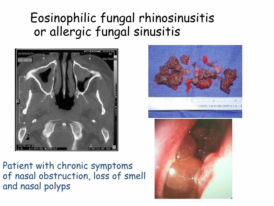
Eosinophilic fungal rhinosinusitis or allergic fungal sinusitis
Patient with chronic symptoms of nasal obstruction, loss of smell and nasal polyps
Ponikau et al, Mayo Clinic Proc 1999;74:877 & WWW.aspergillus.man.ac.uk

Outline
– Basic Anatomy and Physiology
– What is bronchiectasis
– Aetiology
– Assessment
– Treatment
• Specific Treatment
• General measures
• CF Therapy

•Gene Therapy
•Anti-inflammatory/Anti-infective
•Hypertonic Saline
•Ivacaftor
•Transplantation
Cystic Fibrosis:
Possible Therapies

Gene Therapy
• Still looking for the right vector
• Most give short term expression
• Use of stem cells?
• Still waiting for breakthrough

Proposed actions of Macrolides
• Modulation of inflammatory pathway – reduced neutrophil chemotactic activity
– reduced cytokines: IL-8, TNF-a, GM-CSF
• Neutrophils – chemotaxis, oxidation, apoptosis
• Pseudomonas – suppression of quorum sensing factors
– virulence factors: adherence, alginate, mobility
• Alteration of biofilm properties

X
Inate lactoferrin N Engl J Med, Vol. 347, No. 14 · October 3, 2002

Effect of long term treatment with azithromycin on disease
parameters in cystic fibrosis: a randomised trial J Wolter, S Seeney, S Bell, S Bowler, P Masel, J McCormack
Thorax 2002;57:212–216

A Controlled Trial of Long-Term Inhaled Hypertonic Saline in
Patients with Cystic Fibrosis Mark R. Elkins, M.H.Sc., et al for the National Hypertonic Saline in Cystic Fibrosis (NHSCF) Study Group
Volume 354(3):229-240 2006
Ivacaftor
• Selective CFTR potentiator
• Improves function of the CFTR protein
• Only benefits those with G551D mutation
• Improves lung function by 10%

Bilateral Lung Transplant Aust & NZ 1992 -2005
Cystic Fibrosis 40%
Emphysema 19% CFA 5%
Bronchiectasis 11%
Misc 9%
AAT 9%
Other PH 1% PPH 4%
ANZCOTR 2005
Eisenmengers 1%

ADULT LUNG TRANSPLANTATION Kaplan-Meier Survival By Diagnosis (Transplants: January 1994 – June 2003)
0
25
50
75
100
0 1 2 3 4 5 6 7 8 9 10
Years
Su
rviv
al (%
)
ALPHA-1 (N=1,127) CF (N= 1,934)
COPD (N= 4,888) IPF (N= 2,058)
PPH (N= 533) SARCOIDOSIS (N = 314)
HALF-LIFE Alpha-1: 5.1 Years; CF: 5.8 Years; COPD: 4.8
Years; IPF: 3.7 Years; PPH: 4.3 Years; Sarcoidosis: 4.0 Years
Survival comparisons
COPD vs. IPF: p < 0.0001
Alpha-1 vs. CF: p = 0.0248
Alpha-1 vs. IPF: p < 0.0001
Alpha-1 vs. PPH: p = 0.0021
CF vs. COPD: p = 0.0006
CF vs. IPF: p < 0.0001
CF vs. PPH: p < 0.0001
CF vs. Sarcoidosis: p = 0.0007
ISHLT 2005 J Heart Lung Transplant 2005;24: 945-982

CF described
Sweat
test
Antibiotics anti pseudomonas
rhDNAse
Abnormal
Nasal PD
CF gene
Inflammation targeted
Tests of
basic defect
Centres, Comprehensive
care, CF Foundation
Life Expectancy with CF


