1
Burkholderia pseudomallei triggers altered inflammatory profiles in a whole blood model of 1
type 2 diabetes-melioidosis co-morbidity 2
Running title: A model of diabetes melioidosis co-morbidity 3
Jodie Morris*1, Natasha Williams1, Catherine Rush1, Brenda Govan1, Kunwarjit Sangla2, Robert
Norton3, Natkunam Ketheesan1
1. Infectious Diseases and Immunopathogenesis Research Group, Discipline of Microbiology
and Immunology, James Cook University, Townsville, Queensland, Australia 4811
2. Department of Endocrinology, Townsville Hospital, Townsville, Queensland, Australia 4811
3. Pathology Queensland, Townsville Hospital, Townsville, Queensland, Australia 4811
*Corresponding author:
Jodie L Morris
Building 87, Discipline of Microbiology and Immunology
James Cook University
Townsville, Queensland, Australia 4811
Tel.: +61 7 4781 6646; Fax: +61 7 47 791526; E-mail: [email protected]
Abstract Word Count: 250
Text Word Count: 6,160 4
5
Copyright © 2012, American Society for Microbiology. All Rights Reserved.Infect. Immun. doi:10.1128/IAI.00212-12 IAI Accepts, published online ahead of print on 2 April 2012
on February 10, 2018 by guest
http://iai.asm.org/
Dow
nloaded from
2
Key words: type 2 diabetes, melioidosis, Burkholderia pseudomallei, inflammation 6 7
on February 10, 2018 by guest
http://iai.asm.org/
Dow
nloaded from
3
Abstract
Melioidosis is a potentially fatal disease caused by the bacterium, Burkholderia 8
pseudomallei. Type 2 diabetes (T2D) is the most common co-morbidity associated with 9
melioidosis. B. pseudomallei isolates from melioidosis patients with T2D are less virulent in 10
animal models than those from patients with melioidosis and no identifiable risk factors. We 11
developed an ex vivo whole blood assay as a tool for comparison of early inflammatory profiles 12
generated by T2D and non-diabetic (ND) individuals in response to a B. pseudomallei strain of 13
low virulence. Peripheral blood from individuals with T2D, with either poor (PC-T2D; n=6) or 14
well-controlled glycemia (WC-T2D; n=8), and healthy ND (n=13) individuals was stimulated 15
with B. pseudomallei. Oxidative burst, myeloperoxidase (MPO) release, expression of pathogen 16
recognition receptors (TLR2, TLR4, CD14) and activation markers (CD11b, HLA-DR) were 17
measured on polymorphonuclear (PMN) leukocytes and monocytes. Plasma inflammatory 18
cytokine (IL-6, IL-12p70, TNF-α, MCP-1, IL-8, IL-1β, IL-10) concentrations were also 19
determined. Following stimulation, oxidative burst and MPO levels were significantly elevated in 20
blood from PC-T2D compared to controls. Differences were also observed in expression of 21
TLR2, CD14 and CD11b on phagocytes from T2D and ND individuals. Levels of IL-12p70, 22
MCP-1 and IL-8 were significantly elevated in blood from PC-T2D compared to ND individuals. 23
Notably, differential inflammatory responses of PC-T2D, WC-T2D and ND individuals to 24
B. pseudomallei occur independently of bacterial load and confirm the efficacy of this model of 25
T2D-melioidosis co-morbidity as a tool for investigation of dysregulated PMN and monocyte 26
responses to B. pseudomallei underlying susceptibility of T2D individuals to melioidosis. 27
on February 10, 2018 by guest
http://iai.asm.org/
Dow
nloaded from
4
Introduction
Melioidosis, caused by Burkholderia pseudomallei is an increasingly important disease in 28
the tropics (17, 25). Melioidosis is the third most common cause of death from infectious disease 29
in northeast Thailand (37). In northern Australia, melioidosis pneumonia is an increasingly 30
recognized cause of community-acquired pneumonia (19). Rapid progression to septic shock and 31
death are common complications (19). Type 2 diabetes (T2D) is the most significant risk factor 32
associated with susceptibility to infection with B. pseudomallei. Up to 42% of patients with 33
melioidosis in Australia (17, 19), 60% of patients in Thailand (37) and 76% of patients in India 34
(48) have pre-existing T2D. A considerable proportion (43%) of non-diabetic patients with 35
melioidosis have other underlying risk factors associated with progression to T2D including 36
increased body mass index (BMI), hypertension, hypertriglyceridemia and renal problems 37
(Townsville Hospital melioidosis database, R. Norton, unpublished data). There are 38
inconsistencies in reports on the impact of T2D on severity of B. pseudomallei infection (19, 30, 39
55). Given T2D is the most common risk factor for melioidosis and that individuals with pre-40
existing diabetes are more likely to present with acute rather than chronic infection (18, 19), this 41
co-morbid condition undoubtedly impacts on the ability of the host immune response to control 42
B. pseudomallei infection. 43
44
The inflammatory nature of insulin resistance and T2D are well documented (52). Defects 45
in cellular immune responses, which tend to be more pronounced in diabetic individuals with 46
poor glycaemic control (40, 41, 53, 57), have been described for other infections (33). However, 47
the impact of T2D on immunopathogenesis of B. pseudomallei infection remains an under 48
researched area. We have previously demonstrated variation in virulence levels amongst clinical 49
on February 10, 2018 by guest
http://iai.asm.org/
Dow
nloaded from
5
isolates of B. pseudomallei in animal models (58). In BALB/c mice, the virulence of 50
B. pseudomallei isolates from patients with pre-existing T2D is significantly lower than isolates 51
recovered from patients with no identifiable risk factors (58), suggesting immunopathological 52
changes associated with diabetes increase susceptibility to otherwise innocuous strains of 53
B. pseudomallei. 54
55
Recent in vitro studies have demonstrated defects in the response of individual leukocyte 56
types from diabetic hosts toward B. pseudomallei (11, 45, 65). Compared to healthy controls, 57
mononuclear leukocytes isolated from peripheral blood of diabetic individuals express lower 58
levels of IL-17 following exposure to B. pseudomallei (45). Recently, we demonstrated that bone 59
marrow-derived DC (BMDC) and peritoneal elicited macrophages (PEM) isolated from 60
streptozotocin-induced diabetic mice are impaired in their ability to internalize and kill 61
B. pseudomallei, in vitro (65). In addition, polymorphonuclear leukocytes (PMN) from patients 62
with melioidosis and co-morbid T2D have been shown to have impaired phagocytosis and 63
migration in response to IL-8, and a reduced ability to delay apoptosis following exposure to 64
B. pseudomallei (11). Defects in PMN function in T2D, excess alcohol consumption and renal 65
disease are well described and were the basis for trialing therapy with granulocyte colony 66
stimulating factor (G-CSF) in melioidosis (12, 14, 31, 46, 67). However, a disadvantage of 67
functional assays using a single leukocyte type in isolation is the inability to consider the 68
modulating effects of other cell types and plasma constituents on inflammatory responses 69
important in controlling B. pseudomallei infection. 70
71
on February 10, 2018 by guest
http://iai.asm.org/
Dow
nloaded from
6
Therefore, in the current study we developed an ex vivo whole-blood assay and 72
characterized its efficacy as a tool for assessing early inflammatory responses toward 73
B. pseudomallei infection in susceptible hosts. We compared bacterial loads and inflammatory 74
profiles in peripheral blood of individuals with T2D and in healthy individuals without T2D 75
following exposure to a B. pseudomallei strain with low virulence in an animal model of 76
melioidosis (5). In addition, we sought to determine the influence of glycemic control on 77
inflammatory responses to B. pseudomallei, by comparing profiles in blood from individuals with 78
either poorly-controlled or well-controlled T2D. Comparable bacterial numbers were 79
demonstrated for all groups throughout the experimental period. Importantly, however we 80
demonstrate contrasting expression of pathogen recognition receptors (PRRs; TLR2, TLR4, 81
CD14) and activation markers (CD11b, HLA-DR) on PMN and monocytes from T2D 82
individuals, particularly those with poorly-controlled glycemia, and healthy controls in response 83
to stimulation with B. pseudomallei. Similarly, exposure to B. pseudomallei led to differences in 84
the secretion of inflammatory cytokines between controls and individuals with T2D. Parallels 85
between the findings of the current study with that described in patients (62) and animal models 86
(6, 32, 59) support a role for our whole blood model of diabetes-melioidosis co-morbidity as a 87
valuable tool in investigating impaired early host cell interactions that underlie susceptibility to 88
melioidosis. 89
Methods
Participants
A total of 14 individuals with diabetes were recruited through the outpatient Endocrinology 90
Clinic of the Townsville Hospital, Queensland, Australia. T2D was diagnosed according to the 91
on February 10, 2018 by guest
http://iai.asm.org/
Dow
nloaded from
7
World Health Organization criteria (66). Participants with T2D consisted of 6 males and 8 92
females, aged 31-78 years (mean: 54.4 years, SD: 12.6 years). The mean duration from the first 93
diagnosis of diabetes was 11.8 years (SD: 8.9, range: 6 months-33 years). Participants with T2D 94
were being administered the following drugs: sulphonyl urea (8 patients), α-glucosidase (2 95
patients), biguanide (9 patients), and no medication, dietary control only (3). Individuals with 96
diabetes were subdivided based on their history of glycemic control, as indicated by the 97
percentage glycosylated hemoglobin (HbA1c) on monthly clinic visits over a period of at least 6 98
months: poor (PC-T2D; HbA1c >8.5%; n=6) or well-controlled (WC-T2D; HbA1c 5.5-7.5%; 99
n=8). Healthy, non-diabetic (ND) controls were age- and gender-matched (mean: 56 years, SD: 100
11.7 years; Range: 29-76 years). Participants, those with diabetes or healthy controls, had no 101
previous history of melioidosis and were seronegative for antibodies against B. pseudomallei 102
according to an indirect hemagglutination assay (3). 103
Venous blood (40mL) was obtained from participants in sodium heparin Vacutainer™
tubes (Becton Dickinson). Experiments were performed over a series of runs, where each run
consisted of at least one diabetic individuals, age- and gender-matched to a healthy control
individual. This study was approved by the human ethics review committees of the Townsville
Hospital (71/04) and James Cook University (H3483). All participants gave informed consent.
104
Biochemical and hematological measurements 105
Blood cell counts including numbers and proportions of leukocytes, erythrocyte 106
sedimentation rate (ESR), HbA1c and C-reactive protein (CRP) were determined in unstimulated 107
peripheral blood by the routine diagnostic laboratory at the Townsville Hospital. 108
on February 10, 2018 by guest
http://iai.asm.org/
Dow
nloaded from
8
Whole blood stimulation assay
A previously characterized (5) B. pseudomallei clinical strain of low virulence
(NCTC13179) was used for in vitro stimulation in whole blood assays. The isolate was grown to
logarithmic phase, washed twice and resuspended in phosphate buffered saline (PBS, pH 7.2) to 4
x 108 cfu/mL, as described previously (5). B. pseudomallei was added to whole blood for the
assays described below at a multiplicity of infection (MOI) of 1(leukocyte): 5 (bacteria).
Unstimulated, control samples received an equivalent volume of PBS only. Stimulated and
unstimulated whole blood was incubated at 37oC in 5% CO2 with gentle mixing for up to 3.5 hrs.
B. pseudomallei persistence
Persistence of B. pseudomallei throughout the culture period was monitored in stimulated
blood samples by measuring total and intracellular bacterial loads at 1 and 4 hrs of incubation.
Total B. pseudomallei concentration (intracellular and extracellular) was determined by culturing
serial dilutions of whole blood onto Ashdown agar at the time points indicated. Intracellular
bacterial loads were determined following lysis of erythrocytes (Erythrocyte Lysis Buffer,
Qiagen) and washing of remaining leukocytes. Leukocytes were resuspended to the original
volume and serial dilutions were plated onto AA. Colonies were enumerated after 48hrs of
culture and B. pseudomallei load determined. Assays were performed in duplicate at each time
point for all samples. Data for total bacterial load is expressed as log10 cfu/ml of blood.
Intracellular bacterial loads are expressed as log10 cfu/106 leukocytes.
Flow cytometric analysis of PMN and monocytes cell surface activation markers 109
on February 10, 2018 by guest
http://iai.asm.org/
Dow
nloaded from
9
We compared the activation of blood phagocytes from PC-T2D, WC-T2D and ND 110
individuals following exposure to B. pseudomallei using flow cytometry. The PRRs, TLR2, 111
TLR4 and CD14 have previously been demonstrated to play a role in the pathogenesis of 112
melioidosis (61, 63, 64). We therefore assessed their expression on PMN and monocytes prior to, 113
and following stimulation with B. pseudomallei. In addition, blood phagocyte activation was 114
compared for PC-T2D, WC-T2D and ND individuals by measuring changes in the expression of 115
cell surface activation markers on PMN (CD11b) and monocytes (CD11b, HLA-DR) following 116
stimulation with B. pseudomallei. Monoclonal antibodies were purchased from BD Biosciences 117
(CD45, CD3, CD16, CD14, HLA-DR and isotype controls) and Jomar Biosciences (CD11b and 118
isotype control) conjugated to fluorescein isothiocyanate (FITC), phycoerythrin (PE) or 119
peridinin-chlorophyll protein (PerCP). Toll-like receptor (TLR)-2-Alexa Fluor® 488 and TLR-4-120
biotinylated (detected with streptavidin-conjugated PE) antibodies were also purchased from BD 121
Biosciences. All monoclonal antibodies, except TLR-4, were employed in direct 122
immunofluorescence tests using whole blood according to the manufacturers’ instructions. 123
Briefly, appropriate monoclonal antibodies were added to 100μl of whole blood and incubated on 124
ice for 30 min. Erythrocyte lysing solution (1 x FACSlyse, BD Biosciences) was added for 10 125
min at room temperature. Samples were washed twice then resuspended in 2% 126
paraformaldehyde. Immunofluorescence-positive cells were determined by flow cytometry (BD 127
FACScan, BD Biosciences). PMN activation was assessed by CD11b, TLR2 and TLR4 128
expression after gating on CD16+ granulocytes. Monocyte activation was assessed by CD11b, 129
CD14, HLA-DR, TLR2 and TLR4 expression after gating on CD45+CD3- mononuclear 130
leukocytes. Sample analysis was performed with CellQuestPro software (BD, Biosciences). Data 131
on February 10, 2018 by guest
http://iai.asm.org/
Dow
nloaded from
10
is expressed as mean fluorescence intensity in unstimulated (unstim) and B. pseudomallei-132
stimulated (stim) samples. 133
Myeloperoxidase (MPO) Activity
The release of MPO from activated PMN was measured in plasma from stimulated and
unstimulated samples at 1 and 3.5 hrs of culture using a sandwich ELISA for human MPO
according to the manufacturer’s protocol (Hycult® Biotech). Samples were analyzed in duplicate
in the same run.
Oxidative Burst Activity
Intracellular oxidative burst activity of PMN and monocytes was determined by flow
cytometry using the fluorogenic substrate dihydrorhodamine 123 (DHR; Invitrogen). One
hundred microlitres of whole blood was mixed with B. pseudomallei at a MOI 1:5 (or PBS only
for unstimulated control samples) for 30 min. DHR (100μM) was added to the samples at 37oC
and incubation was continued for a further 10 min in order to allow nonfluorescent DHR to
convert to fluorescent rhodamine 123 upon the production of reactive oxygen species. Lysing
solution (1xFACSlyse) was added for 10 min at room temperature. After washing, CD16-PE was
added to samples and incubated for 20 min on ice. Samples were washed twice and resuspended
in 2% paraformaldehyde for flow cytometric analaysis. Granulocyte populations were gated on
forward versus side-scatter plots, followed by gating on CD16+ granulocytes. Monocytes were
gated on forward versus side scatter plots. Oxidative burst was monitored by determining the
proportion of cells positive for rhodamine 123 and the relative fluorescence intensities of the
gated PMN or monocytes.
on February 10, 2018 by guest
http://iai.asm.org/
Dow
nloaded from
11
Inflammatory cytokine analysis
At 1 and 3.5 hrs of incubation, one millilitre aliquots of B. pseudomallei-stimulated and
unstimulated blood were centrifuged (500 x g, 5 min). Plasma was removed and stored at -70oC
until further analysis. Inflammatory cytokines were measured with the BD Biosciences CBA™
kit: IL-8, IL-1β, IL-6, IL-10, TNF-α and IL-12p70 (Human Inflammatory Cytokines) using a BD
FACScan flow cytometer (BD Biosciences) and a BD Biosciences CBA™ MCP-1 Flex kit using
a BD FACSCalibur flow cytometer (BD Biosciences), according to the manufacturers’
instructions. Assays were performed in duplicate at each time point for all samples. Cytokine
concentration (pg/mL) was determined with calibration curves separately established using the
CBA analysis software (BD Biosciences).
Statistical analysis
Differences in inflammatory responses of unstimulated and B. pseudomallei stimulated 134
samples were examined using Wilcoxon signed rank test. Differences in inflammatory responses 135
between PC-T2D, WC-T2D and ND groups were calculated using Kruskal-Wallis test. Simple 136
linear correlations between HbA1c, as a marker of glycaemic control, and immunological 137
measures at 3.5 hrs of culture were determined by Spearman’s rank correlation coefficient. A p-138
value less than 0.05 was considered significant. All statistical analyses were conducted using 139
Graphpad Prism v5 software. Results are expressed as median ± interquartile range (IQR) unless 140
otherwise specified. 141
on February 10, 2018 by guest
http://iai.asm.org/
Dow
nloaded from
12
Results
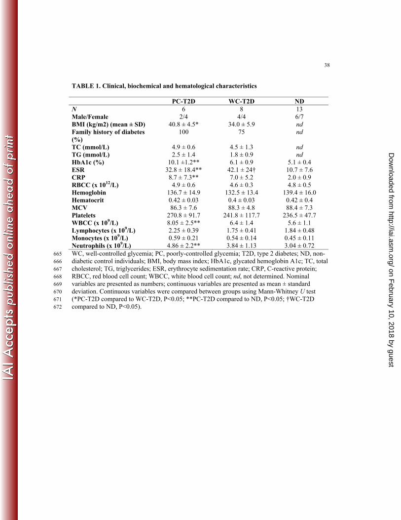
Clinical, biochemical and hematological characteristics
Compared to ND individuals, ESR, CRP and leukocyte numbers were elevated in 142
unstimulated blood from PC-T2D individuals (Table 1). Increased leukocyte levels corresponded 143
to neutrophilia in PC-T2D individuals. 144
145
B. pseudomallei persistence 146
Growth kinetics of B. pseudomallei in whole blood was comparable for PC-T2D, WC-T2D 147
and ND individuals throughout the experimental period (Fig. S1). Following stimulation, there 148
were no significant differences in total or intracellular bacterial loads between the three groups at 149
1 or 4 hrs of culture. 150
151
PMN activation 152
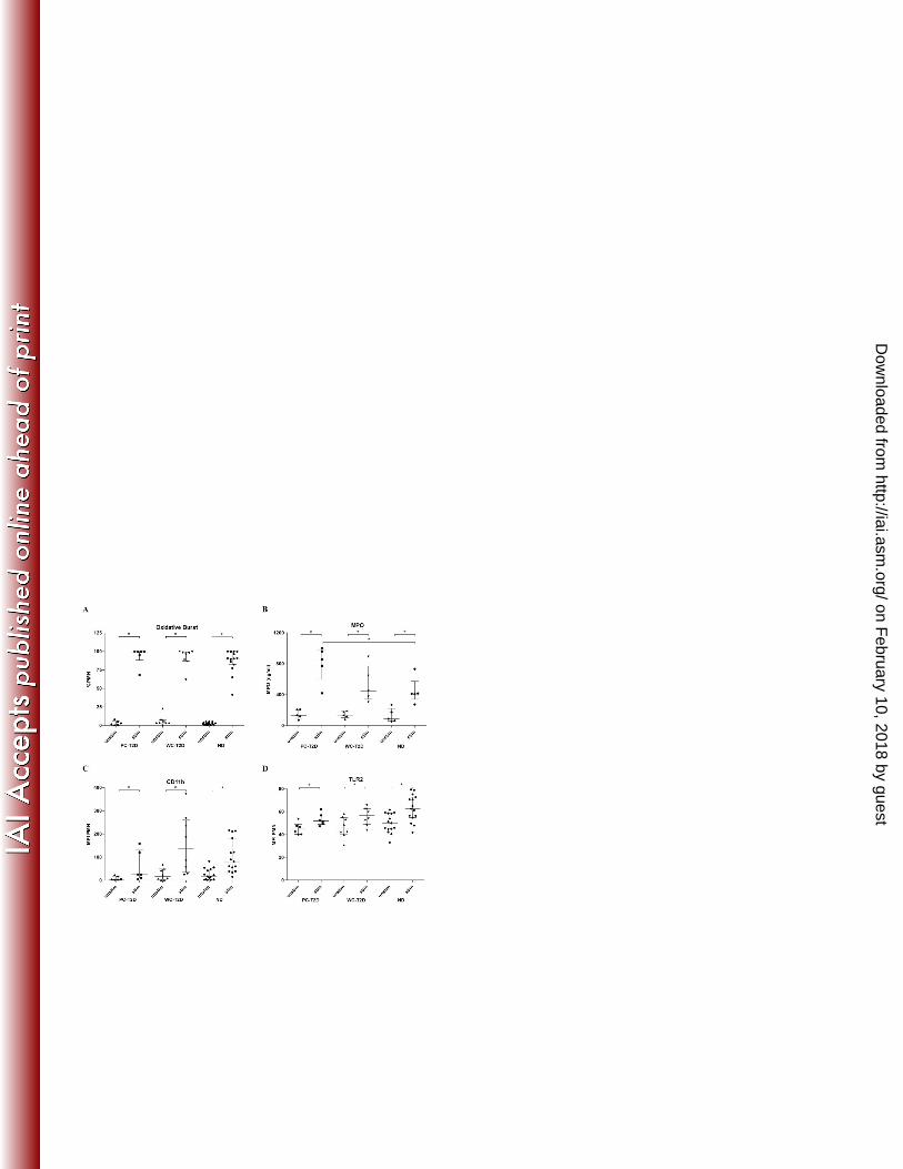
Following B. pseudomallei stimulation, the percentage of PMN undergoing oxidative burst 153
(Fig. 1a) increased significantly for all groups (42-fold for PC-T2D, 25-fold for WC-T2D and 36-154
fold for ND individuals). While there was a trend for greater levels of oxidative burst activity for 155
PMN from PC-T2D compared to ND and WC-T2D individuals, this did not reach statistical 156
significance. Levels of MPO, an enzyme that is liberated from activated PMN, were similarly 157
increased in B. pseudomallei-stimulated blood samples (Fig. 1b) and were significantly higher in 158
blood from PC-T2D compared to ND individuals (P=0.05; 6.5-fold v 4.7 fold increase for PC-159
T2D and ND individuals, respectively). 160
161
on February 10, 2018 by guest
http://iai.asm.org/
Dow
nloaded from
13
We also measured surface expression of the integrin, CD11b, as a marker of PMN 162
activation. CD11b levels increased on PMN from all groups following exposure to 163
B. pseudomallei (Fig. 1c). There was a trend for lower CD11b expression on PMN from PC-T2D 164
individuals following stimulation, compared to WC-T2D (P=0.08) and ND (P=0.094) 165
individuals. 166
167
TLR2 expression was comparable on PMN from PC-T2D, WC-T2D and ND at baseline 168
(Fig. 1d). After exposure to B. pseudomallei, TLR2 expression increased significantly for all 169
groups, with the highest levels observed on PMN from ND individuals (Fig. 1d). Minimal TLR4 170
expression was detected on PMN at baseline among the three groups and these levels remained 171
relatively unchanged following exposure to B. pseudomallei (data not shown). 172
173
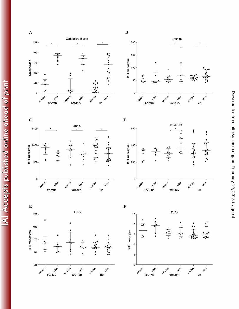
Monocyte Activation 174
Following stimulation, the percentage of DHR+ monocytes (Fig. 2a) and the intensity of the 175
oxidative burst (data not shown) increased significantly for all groups, with the highest fold-176
change observed for WC-T2D individuals (12.8-fold) compared to PC-T2D (4.3 fold) and ND 177
individuals (8.7-fold). There was a trend for higher levels of oxidative burst activity in monocytes 178
from PC-T2D compared to monocytes from ND individuals (P=0.067) following stimulation. 179
Monocyte oxidative responses to B. pseudomallei reflected the same trends observed in PMN, 180
whereby both the number of cells undergoing oxidative burst activity, and the intensity of the 181
oxidative burst response was increased in individuals with T2D, particularly those with poorly-182
controlled glycemia. 183
184
on February 10, 2018 by guest
http://iai.asm.org/
Dow
nloaded from
14
Compared to PMN, there was only a slight increase in CD11b expression on monocytes 185
following exposure to B. pseudomallei (Fig. 2b), with significant increases observed only on 186
monocytes from WC-T2D (P=0.023) and ND (P=0.048) individuals. In contrast, CD14 187
expression decreased significantly on monocytes from PC-T2D (P=0.031), WC-T2D (P=0.039) 188
and ND (P=0.013) individuals following stimulation with B. pseudomallei (Fig. 2c). While 189
minimal changes were observed in HLA-DR expression on monocytes from PC-T2D and ND 190
individuals after exposure to B. pseudomallei, a significant increase in HLA-DR levels occurred 191
on monocytes from WC-T2D individuals following stimulation (P = 0.003, Fig. 2d). 192
193
Compared to ND individuals, there was a trend for increased TLR2 expression on resting, 194
unstimulated monocytes from T2D individuals, PC-T2D and WC-T2D (Fig. 2e). Following 195
exposure to B. pseudomallei, expression of TLR2 decreased on monocytes from PC-T2D (1.1-196
fold) and WC-T2D (1.1-fold) and remained relatively unchanged on monocytes from ND 197
individuals, though these changes were not significant. No significant differences were observed 198
in baseline TLR4 expression on monocytes from T2D and ND individuals (Fig. 2f). However, 199
compared to monocytes from ND individuals, TLR4 expression was increased on monocytes 200
from PC-T2D (P= 0.039) in response to B. pseudomallei exposure. 201
202
Inflammatory cytokines 203
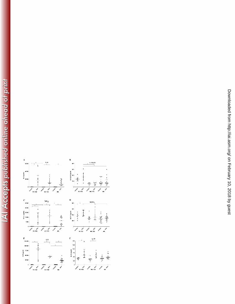
There were no statistically significant differences in baseline plasma concentrations of IL-204
1β, IL-8, IL-10 or IL-12p70 between PC-T2D, WC-T2D and ND individuals. However, 205
compared to WC-T2D, IL-6 (P=0.031) and TNF-α ((P=0.001) concentrations were elevated in 206
baseline plasma from PC-T2D individuals (Fig. 3a, c). There was also a trend for increased MCP-207
on February 10, 2018 by guest
http://iai.asm.org/
Dow
nloaded from
15
1 levels in baseline plasma from PC-T2D individuals, compared to plasma from ND individuals 208
(Fig. 3d, P=0.083). 209
210
Following stimulation of whole blood with B. pseudomallei for 1hr, plasma concentrations 211
of IL-1β, IL-8, IL-10, IL-12p70, IL-6 and TNF-α increased significantly for all groups, though 212
levels were similar between PC-T2D, WC-T2D and ND individuals (Fig. S2). MCP-1 levels 213
remained unchanged at 1hr post-stimulation (data not shown). After 3.5 hrs of exposure to 214
B. pseudomallei, significant increases in levels of IL-6 (Fig. 3a; 237-fold, 218-fold and 181-fold 215
for PC-T2D, WC-T2D and ND individuals, respectively), TNF-α (Fig. 3c; 141-fold, 447-fold and 216
86-fold for PC-T2D, WC-T2D and ND individuals, respectively) and IL-1β (92-fold, 116-fold 217
and 78-fold for PC-T2D, WC-T2D and ND individuals, respectively) were observed for all 218
groups. IL-12p70 concentrations were significantly higher in plasma from PC-T2D compared to 219
WC-T2D (P=0.005) and ND individuals (P=0.039) following stimulation for 3.5 hrs. Similarly, 220
exposure to B. pseudomallei led to elevated levels of IL-8 in plasma from PC-T2D, compared to 221
ND individuals (P=0.032; 182-fold v 43-fold, respectively) at 3.5 hrs post-stimulation. IL-10 222
concentrations also increased in B. pseudomallei-stimulated blood, though differences only 223
reached significance for PC-T2D (1.5 fold) and WC-T2D (1.6 fold) individuals (Fig. 3f). 224
225
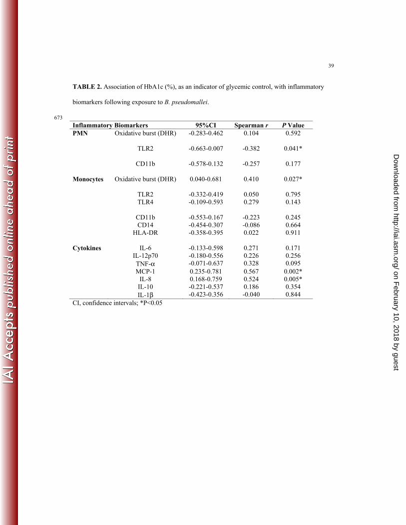
Effect of HbA1c on inflammatory profiles 226
We analysed the effect of glycaemic control, indicated by HbA1c levels, on inflammatory 227
biomarkers following B. pseudomallei exposure (Table 2). TLR2 expression on PMN correlated 228
negatively with HbA1c levels (P=0.041). In contrast, following exposure to B. pseudomallei the 229
percentage of monocytes undergoing respiratory burst (P=0.027) and the concentrations of 230
on February 10, 2018 by guest
http://iai.asm.org/
Dow
nloaded from
16
MCP-1 (P=0.002) and IL-8 (P=0.005) correlated positively with HbA1c levels. No significant 231
correlation was observed between age, gender or time since diagnosis of T2D and inflammatory 232
markers following exposure to B. pseudomallei (data not shown). 233
234
Discussion
235 It is widely recognized that T2D is associated with increased rates of infection and 236
infection-related mortalities (33, 50). However, there is a paucity of literature concerning 237
diabetes-induced immune alterations and their effect on subsequent host-pathogen interactions. 238
Despite being the most significant risk factor associated with susceptibility to melioidosis, the 239
impact of pre-existing diabetes on the immunopathogenesis of B. pseudomallei infection is 240
unclear. Bacteremia and sepsis are common clinical findings of melioidosis (18). We had 241
previously demonstrated that compared to individuals with no identifiable risk factors, 242
individuals with T2D are more susceptible to infection with less virulent B. pseudomallei isolates 243
(58). Therefore, using a whole-blood stimulation assay, the current study was the first to 244
demonstrate differences in inflammatory profiles of leukocytes from whole blood of diabetic and 245
non-diabetic individuals in response to a B. pseudomallei strain of low virulence, despite 246
comparable bacterial loads. While strong inflammatory responses were induced in blood from 247
both diabetic and healthy controls, exposure to B. pseudomallei led to increased levels of IL-248
12p70, MCP-1, IL-8 and IL-10 in blood from individuals with T2D, particularly those with 249
poorly controlled glycemia. Interestingly, differences were also observed in PRR and cell surface 250
activation marker expression on phagocytes from diabetic and non-diabetic individuals. It is 251
noteworthy that the contrasting inflammatory responses were not a reflection of B. pseudomallei 252
numbers, since total and intracellular bacterial loads were comparable for diabetics and non-253
on February 10, 2018 by guest
http://iai.asm.org/
Dow
nloaded from
17
diabetic individuals throughout the experimental period. Given the importance of PRR signaling 254
in determining the subsequent activation of diverse immune response pathways, the contrasting 255
expression identified on phagocytes in the current study may underpin the susceptibility of T2D 256
individuals to melioidosis. 257
258
Chronic low-grade inflammation and oxidative stress is well documented in T2D and plays 259
an important role in the development of many of the metabolic complications associated with 260
insulin resistance (27, 44). In addition to many metabolic abnormalities, a variety of 261
immunological abnormalities continue to be identified that conceivably contribute to the 262
increased susceptibility of T2D to infections such as melioidosis (35, 44). Prolonged 263
hyperglycemia, oxidative stress and production of advanced glycated end products contribute to 264
local production of inflammation and tissue damage and delayed wound repair in diabetics (68). 265
In the current study, unstimulated blood from individuals with T2D, particularly those with 266
poorly controlled glycemia, had increased erythrocyte sedimentation rates, C-reactive protein, 267
TNF-α and IL-6 levels, all of which are consistent with an underlying inflammatory state which 268
has been shown to occur in association with T2D (35, 43, 44). 269
270
Recruitment, migration and activation of leukocyte subsets are crucial events during an 271
immune response that are regulated by chemokines and inflammatory cytokines. However, while 272
these mediators are indispensable for the effective clearance of a pathogen, an excessive 273
inflammatory response is potentially destructive and may contribute to disease progression (60). 274
The pro-inflammatory cytokine profiles observed in the current study are consistent with those 275
described in melioidosis sepsis (29, 62) and both IL-6 and IL-10 have been shown to be 276
on February 10, 2018 by guest
http://iai.asm.org/
Dow
nloaded from
18
independent predictors of mortality (51). Interestingly, levels of MCP-1 and IL-8 correlated with 277
glycaemic control, with highest concentrations measured in plasma from PC-T2D individuals 278
(high % HbA1c). In contrast to WC-T2D and ND individuals, IL-12p70 was also elevated in 279
plasma from PC-T2D individuals following stimulation with B. pseudomallei. It is noteworthy 280
that the anti-inflammatory cytokine, IL-10 increased significantly in blood from T2D individuals. 281
To our knowledge there have been no previous published comparisons of inflammatory cytokine 282
production between diabetic and non-diabetic melioidosis patients. Wiersinga et al. (62) did 283
however report the increased expression of mRNA for genes encoding IL-1β, IL-6, ΤΝF−α, IFN-284
γ and IL-10 in peripheral blood leukocytes from patients with melioidosis sepsis. In contrast to 285
the current study, MCP-1 and IL-8 levels were lower in monocytes of patients with melioidosis 286
sepsis compared to controls (62). However, comparisons of cytokine responses between the ex 287
vivo model used in the current study and that of Wiersinga et al. (62) are not possible given 288
peripheral blood from the melioidosis sepsis study was collected from melioidosis patients after 289
the commencement of antimicrobial therapy. Nevertheless, despite differences in the focus of the 290
study by Wiersinga et al. (62) and our current study, both have demonstrated the induction of 291
both pro-inflammatory and anti-inflammatory cytokines in leukocytes from patients with 292
melioidosis sepsis and from susceptible T2D individuals after stimulation with B. pseudomallei, 293
respectively. 294
295
We have previously demonstrated differential inflammatory cytokine responses in our 296
animal models of melioidosis, in which BALB/c mice are highly susceptible to B. pseudomallei 297
infection and C57BL/6 mice, relatively resistant (6, 59). Differences have been described in the 298
kinetics and magnitude of KC, a homologue of IL-8 in humans, MCP-1, IL-12 and IL-10 mRNA 299
on February 10, 2018 by guest
http://iai.asm.org/
Dow
nloaded from
19
induction between the two mouse strains (6, 59). Compared to C57BL/6 mice, susceptible 300
BALB/c mice had elevated inflammatory chemokine and cytokine responses in liver and spleen 301
within 48 hrs of infection with B. pseudomallei, corresponding to a more extensive inflammatory 302
cell infiltrate that is rich in PMN (6, 59). Paradoxically, the increased inflammatory response to 303
B. pseudomallei in BALB/c mice is associated with increased bacterial dissemination and 304
mortality (6, 59). This suggests that the exaggerated inflammatory cytokine production and PMN 305
infiltration to sites of B. pseudomallei infection, apparent by day two of infection, may be a final, 306
desperate response of the susceptible host following failed earlier attempts at controlling 307
pathogen replication. Recently we characterized for the first time an animal model of T2D 308
melioidosis co-morbidity (32) and describe a disease pattern in diabetic mice that is similar to 309
that observed in the susceptible BALB/c mice. Compared to non-diabetic mice, hyperproduction 310
of pro-inflammatory cytokines including ΤΝF−α, IL-1β and IL-6 and extensive PMN infiltration 311
and tissue damage at sites of infection, preceded mortality in the first few days following 312
B. pseudomallei infection in diabetic mice (32). Data from the current study, demonstrates that 313
control of B. pseudomallei persistence within the first few hours of exposure is similar for PC-314
T2D, WC-T2D and ND individuals. Importantly, despite comparable bacterial loads, the 315
inflammatory pathways triggered in leukocytes in response to this bacterium differ significantly 316
for T2D and ND individuals. 317
318
In the current study there was evidence of lower pro-inflammatory cytokines in blood from 319
WC-T2D compared to PC-T2D. Given the inflammatory nature of diabetes, there has been 320
increasing interest in the identification of compounds with anti-oxidative and anti-inflammatory 321
properties in the treatment of infection in individuals with diabetes (10). This is supported by 322
on February 10, 2018 by guest
http://iai.asm.org/
Dow
nloaded from
20
recent evidence that for patients with melioidosis sepsis, those with pre-existing T2D that were 323
taking glyburide, a sulphonylurea with anti-inflammatory properties, were more likely to survive 324
than non-diabetics or T2D individuals receiving other anti-diabetic treatments prior to diagnosis 325
with melioidosis (34). T2D individuals recruited to the current study were receiving one or more 326
of a variety of anti-diabetic medication classes including sulphonylureas, α-glucosidases and 327
biguanides. Unfortunately, this meant that group sizes were inadequate for analysis of potential 328
effects of anti-diabetic medication on inflammatory profiles generated in response to 329
B. pseudomallei exposure. Nevertheless, the findings in the current study have clearly 330
demonstrated that glycemic control does influence the host inflammatory response to 331
B. pseudomallei. The hyperinflammatory response observed between 24 and 48 hrs in susceptible 332
BALB/c (6, 59), in diabetic mice (32), and in patients with melioidosis sepsis (62), reflect the 333
inflammatory profiles observed in the whole blood assay used in the current study. Based on our 334
findings, we propose that very early interactions between B. pseudomallei and PMN and 335
monocytes are altered in the diabetic host, such that a compensatory dysregulated 336
hyperinflammatory response is triggered independently of bacterial load, resulting in increased 337
local tissue damage and bacterial dissemination. 338
339
Both PMN and monocytes are vulnerable to functional disturbances due to hyperglycemia-340
induced stress (33, 35, 68). Oxidative stress is well described in diabetes, contributing to 341
endothelial dysfunction and vascular inflammation (20). Trends for increased oxidative burst 342
activity observed in PMN and monocytes from PC-T2D individuals both in unstimulated and 343
stimulated blood compared to healthy controls are consistent with the findings of others (4, 23). 344
on February 10, 2018 by guest
http://iai.asm.org/
Dow
nloaded from
21
MPO, a principle enzyme released by activated PMN, was significantly elevated in plasma from 345
PC-T2D individuals compared to ND individuals following exposure to B. pseudomallei. 346
PMN migration from the bloodstream into sites of infection within tissues occurs rapidly through 347
a process that is tightly regulated. Activated PMN display an upregulated expression of CD11b 348
and other integrins and chemokine receptors, which facilitate their transmigration through into 349
sites of infection within tissue (15). Reduced PMN function has been demonstrated in patients 350
with melioidosis and co-morbid T2D (11). Based on the association of PMN functional defects 351
with the most common underlying risk factors for melioidosis, G-CSF, which stimulates the 352
production and function of PMN, was trialed as an adjunctive therapy for the treatment of septic 353
shock from melioidosis (12, 14, 31, 46, 67). Although G-CSF had no effect on mortality for 354
patients with severe melioidosis sepsis (12), its efficacy in treatment of patients with less severe 355
forms of melioidosis has not been investigated. In the current study we examined CD11b 356
expression, which mediates inflammation by regulating leukocyte adhesion and migration, as 357
additional measures of PMN and monocyte activation in response to B. pseudomallei exposure. 358
CD11b was upregulated on PMN from all groups after stimulation with B. pseudomallei, similar 359
to responses described in sepsis patients (38). However, there was a trend for reduced levels of 360
CD11b on PMN from PC-T2D at baseline and following stimulation, supporting decreased 361
migratory capacity previously described for PMN from T2D individuals (11, 28). In contrast, 362
CD11b expression was comparable for monocytes from T2D and ND individuals prior to and 363
following stimulation with B. pseudomallei. In addition to CD11b, we measured changes in 364
expression of HLA-DR as cell surface markers of monocytes activation. HLA-DR, or the MHC 365
class II surface receptor, has a central role in antigen specific interactions between monocytes and 366
lymphocytes. Similar to CD11b, HLA-DR levels remained relatively unchanged in PC-T2D and 367
on February 10, 2018 by guest
http://iai.asm.org/
Dow
nloaded from
22
ND individuals. However, it remains to be determined whether other integrins or chemokine 368
receptors involved in monocyte activation are altered in diabetic individuals following exposure 369
to B. pseudomallei. 370
371
PRRs play an important role in the activation and regulation of the innate immune 372
response. TLRs are transmembrane proteins that mediate host cell signaling in response to a 373
range of extracellular and endosomal pathogen-associated molecules. TLR4 permits signaling in 374
response to lipopolysaccharide (LPS) and noninfective inflammatory stimuli such as heat shock 375
protein 60 (HSP60) (reviewed in 42). TLR2, which can heterodimerise with other TLRs, 376
responds to a more diverse group of ligands including lipoproteins, peptidoglycans and atypical 377
LPS (reviewed in 41). B. pseudomallei has been shown to interact with CD14, TLR2 and TLR4 378
(61, 63, 64) and our group recently identified differences in TLR2 and TLR4 expression on 379
monocytes following stimulation with B. pseudomallei isolates of low and high virulence (M. 380
Feterl and N. Ketheesan, unpublished data). TLRs have also been implicated in the pathogenesis 381
of insulin resistance and its complications, with observations that TLR2 and TLR4 expression is 382
increased on monocytes from diabetic individuals (21, 22). Endogenous ligands such as fatty 383
acids, heat shock proteins, hyaluronan and high mobility group B1 protein induce the production 384
of pro-inflammatory cytokines through the stimulation of TLR2 and TLR4 in individuals with 385
diabetes (21). Given the importance of TLR2, TLR4 and CD14 in melioidosis, we therefore 386
investigated changes in the expression of these pathogen recognition receptors on phagocytes 387
from T2D and ND individuals in response to B. pseudomallei stimulation. CD14, one of the first 388
described PRRs, exists in a membrane bound form on monocytes where it serves as a ligand-389
binding component of the LPS-receptor complex (7). Membrane-bound CD14 can also be shed 390
on February 10, 2018 by guest
http://iai.asm.org/
Dow
nloaded from
23
into a soluble form upon monocyte activation (7). CD14 expression decreased on monocytes 391
exposed to B. pseudomallei, with greater decreases observed for monocytes from T2D 392
individuals compared to ND individuals. Changes in CD14 expression may reflect internalisation 393
of membrane-bound CD14 (39) or increased shedding of surface CD14 (48). Decreased 394
membrane CD14 on monocytes, described as monocytes hyporesponsiveness, has been shown to 395
correlate with sepsis severity (9, 49). Our findings of decreased surface expression of CD14 on 396
monocytes from T2D individuals suggest alterations in monocyte functional capacity following 397
exposure to B. pseudomallei may contribute to more severe disease progression in T2D 398
individuals. This is consistent with the clinical observation that individuals with diabetes are 399
more likely to present with melioidosis sepsis than individuals with no identifiable risk factors 400
(19). 401
402
TLR4 expression on PMN was low, as previously reported (36). In contrast, TLR2 403
expression increased on PMN from both T2D and ND individuals after stimulation, with levels 404
tending to be greatest on PMN from healthy controls. On monocytes, TLR4 levels increased 405
marginally following B. pseudomallei exposure, with no significant differences observed between 406
groups. Whilst TLR2 expression remained relatively unchanged on monocytes from ND 407
individuals in response to B. pseudomallei stimulation, there was a trend for decreased TLR2 408
expression on monocytes from both PC-T2D and WC-T2D individuals. The pattern of decreased 409
TLR2 and CD14 levels on monocytes from diabetic individuals in the current study is consistent 410
with the profile described for patients with severe sepsis, though no comparisons have been made 411
between diabetic and non-diabetic patients (9, 49, 62). In addition, peripheral blood samples from 412
the sepsis studies were collected from patients following commencement of antimicrobial therapy 413
on February 10, 2018 by guest
http://iai.asm.org/
Dow
nloaded from
24
after hospital admission, making it difficult to draw parallels to the early changes in TLR 414
expression measured in our whole blood stimulation assay. 415
416
The MyD88-dependent pathway is activated by TLR2 and TLR4, resulting in the 417
production of pro-inflammatory cytokines such as TNF-α, IL-6 and IL-8 (reviewed in 42). TLR4 418
also activates the TIR domain-containing adaptor-inducing interferon β (IFN-β) (TRIF)-419
dependent pathway leading to increased production of IL-12, IL-10 and IFN-β (reviewed in 42). 420
Compared to ND individuals, plasma from PC-T2D individuals had elevated levels of IL-12 and 421
IL-10, in addition to higher concentrations of TNF-α, IL-6 and IL-8, suggesting that both 422
MyD88- and TRIF-dependent pathways had been activated. This is supported by the observation 423
of slight increases in TLR4 expression and decreased CD14 activation on monocytes from PC-424
T2D individuals after exposure to B. pseudomallei. Given the contrasting TLR and CD14 425
activation and inflammatory cytokine response generated in leukocytes from PC-T2D and ND 426
individuals in the current study, it is likely that future kinetic studies focusing on PRR activation 427
in response to B. pseudomallei will demonstrate differences within these groups that may be 428
important in directing the generation of subsequent inflammatory responses. 429
430
TLR activation not only influences PMN survival, but also has a regulatory effect on 431
chemokine receptor expression on leukocytes, thereby influencing their migratory capacity (1, 432
26, 47). Downregulation of chemokine receptors aids retention of PMN at sites of infection and 433
bacterial phagocytosis has been shown to promote this downregulation (24). A reduced migratory 434
response of diabetic PMN toward IL-8 has been demonstrated in vitro (11) and is supported by 435
the differences observed in CD11b expression on PMN from PC-T2D and ND PMN individuals 436
on February 10, 2018 by guest
http://iai.asm.org/
Dow
nloaded from
25
in the current study. A role for altered PMN migratory response to B. pseudomallei in the 437
susceptibility of diabetic individuals to melioidosis is also supported by findings that 438
upregulation of ICAM-1, involved in CD11b/CD18-mediated neutrophil adhesion and migration 439
through endothelium, is attenuated in endotoxemia in diabetic individuals compared to healthy 440
controls (2). Murine studies have highlighted the importance of CD11b/CD18 upregulation on 441
PMN in the early control of bacterial replication following infection (8). Within the first hours of 442
Gram negative bacterial infection, a rapid PMN influx is evident at sites of inflammation in 443
immune-competent mice (69). However, a short delay in very early PMN migration provides an 444
opportunity for the bacterium to replicate and express virulence factors that facilitate its 445
persistence in the host, thereby influencing disease progression (69). We propose a similar 446
mechanism is associated with susceptibility of T2D individuals to melioidosis, whereby an initial 447
delay in PMN migration and/or activation allows B. pseudomallei to multiply, establish an 448
intracellular niche and therefore rendering subsequent inflammatory responses insufficient to 449
contain infection. Our data is supported by a recent transcriptome analysis of innate immune 450
responses triggered after infection B. pseudomallei in a chemically-induced diabetic mouse 451
model. Despite comparable bacterial loads in the first 24hrs of infection, differential expression 452
profiles for genes involved in PRR signalling pathways were demonstrated for diabetic and non-453
diabetic mice (13). Whilst 16 hrs post-infection was the earliest time point assessed in the 454
microarray studies of diabetic mice (13), they provide further support to our data that very early 455
defects (< 3.5 hrs) in the ability of phagocytes from diabetic individuals to detect and respond 456
appropriately to B. pseudomallei underlies contrasting disease progression in diabetic and non-457
diabetic hosts. Further studies to investigate interactions between B. pseudomallei, PRR 458
on February 10, 2018 by guest
http://iai.asm.org/
Dow
nloaded from
26
signalling and the effects on leukocyte migration to sites of infection in individuals at risk of 459
melioidosis are certainly warranted. 460
461
Our study has clear limitations. Specifically the sample size was small and our 462
investigations measured selected markers of the inflammatory response within 4 hrs of exposure 463
to B. pseudomallei. Nonetheless, we believe that the data presented here identifies novel 464
differences in early cellular responses to B. pseudomallei between diabetic and non-diabetic 465
individuals which are not attributed to differences in bacterial persistence. In contrast to studies 466
examining functional responses of single leukocyte subsets, the use of a whole blood approach 467
enabled us to examine the responses of relevant leukocytes simultaneously, therefore more 468
closely modeling in vivo inflammatory responses generated in the early stages of B. pseudomallei 469
in diabetic and non-diabetic individuals. In addition, utilization of a whole blood assay avoids 470
potential bias from cellular stimulation associated with leukocyte subset isolation techniques 471
(54). Data from this proof of principle study provide an immunological basis for the clinical 472
observations observed in patients with T2D and melioidosis co-morbidity (19, 62), supporting the 473
utility of ex vivo whole blood assays as a tool for evaluation of the mechanisms underlying 474
susceptibility to B. pseudomallei in this at risk population. Such studies are not possible utilizing 475
blood from patients with diabetes and melioidosis, given by the time they are hospitalized the 476
infection has already been established. In addition, in most instances, patients recruited for such 477
studies would have commenced antimicrobial therapy. Use of a whole blood model will enable 478
investigation of the very early interactions between human leukocytes and B. pseudomallei that 479
are important in determining the outcome of infection. Given the complexity of leukocyte-480
leukocyte crosstalk in innate immunity, use of a whole blood model also permits measurement of 481
on February 10, 2018 by guest
http://iai.asm.org/
Dow
nloaded from
27
inflammatory changes following B. pseudomallei exposure that more closely reflect in vivo 482
interactions than would be possible using specific leukocyte subsets in isolation, in vitro. 483
484
In summary, this is the first study to describe and characterize an ex vivo whole blood
model of co-morbidity between diabetes and melioidosis. We have shown for the first time that
contrasting inflammatory profiles for PC-T2D, WC-T2D and ND individuals are evident as early
as 3.5 hrs following exposure to a B. pseudomallei strain of low virulence, despite comparable
bacterial loads. Data from our study suggest very early interactions between B. pseudomallei and
PMN and monocytes are altered in hosts with diabetes contributing to impaired leukocyte
activation and the development of an exaggerated inflammatory response. The availability of a
model that reflects clinical parameters observed in patients with T2D and melioidosis is
invaluable since it will facilitate further studies of the dysregulated interactions between
phagocytes and B. pseudomallei in the initial stages of infection in susceptible hosts and the
effects of therapeutics on these interactions. This is significant, given the continuing increase in
the prevalence of T2D which could potentially place more people at risk of severe melioidosis in
endemic regions.
5. Acknowledgements
The authors are grateful to the volunteers who participated in the current study and wish to
thank Kelly Hodgson and Marshall Feterl for their laboratory assistance and Lauren Kromoloff
for assistance with collation of clinical data. This project was supported by a grant from the
Townsville Hospital Private Practice Research Fund.
on February 10, 2018 by guest
http://iai.asm.org/
Dow
nloaded from
28
References
1. Alves-Filho, J. C., et al. 2009. Regulation of chemokine receptor by toll-like receptor 2 is 485
critical to neutrophil migration and resistance to polymicrobial sepsis. Proc. Natl. Acad. 486
Sci. U.S.A 106:4018-4023. 487
2. Andreasen, A. S., et al. 2010. Type 2 diabetes mellitus is associated with impaired 488
cytokine response and adhesion molecule expression in human endotoxemia. Intensive 489
Care Med. 36:1548-1555. 490
3. Ashdown, L. R. 1987. Indirect hemagglutination test for melioidosis. Med. J. Aust. 491
147:364-365. 492
4. Ayilavarapu, S., et al. 2010. Diabetes-induced oxidative stress is mediated by Ca2+-493
independent phospholipase A2 in neutrophils. J. Immunol. 184:1507-1515. 494
5. Barnes, J., and Ketheesan, N. 2005. Melioidosis: routes of infection. Emerg. Infect. Dis. 495
4:638-639. 496
6. Barnes, J. L., Ulett, G. C., Ketheesan, N., Clair, T., Summers, P. M., and Hirst, R. G. 497
2001. Induction of multiple chemokine and colony-stimulating factor genes in experimental 498
Burkholderia pseudomallei infection. Immunol. Cell. Biol. 79:490-501. 499
7. Bazil, V., and Strominger, J. L. 1991. Shedding as a mechanism of down-modulation of 500
CD14 on stimulated human monocytes. J. Immunol. 147:1567-1574. 501
8. Borjesson, D. L., Simon, S. I., Hodzic, E., Ballantyne, C. M., and Barthold, S. W. 2002. 502
Kinetics of CD11b/CD18 upregulation during infection with the agent of human 503
granulocytic ehrlichiosis in mice. Lab. Invest. 82:303-311. 504
9. Brunialti, M. K. C., Martins, P. S., Barbosa de Carvalho, H., Machado, F. R., 505
Barbosa, L. M., and Salomao, R. 2006. TLR2, TLR4, CD14, CD11b, and CD11c 506
on February 10, 2018 by guest
http://iai.asm.org/
Dow
nloaded from
29
expressions on monocytes surface and cytokine production in patients with sepsis, severe 507
sepsis, and septic shock. Shock 25:351-357. 508
10. Ceriello, A., and Testa, R. 2009. Antioxidant anti-inflammatory treatment in type 2 509
diabetes. Diabetes Care 32:S232-236. 510
11. Chanchamroen, S., Kewcharoenwong, C., Susaengrat W., Ato, M., and 511
Lertmemongkolchai, G. 2009. Human polymorphonuclear responses to Burkholderia 512
pseudomallei in healthy and diabetic subjects. Infect. Immun. 77:456-463. 513
12. Cheng, A., et al. 2007. A randomized controlled trial of granulocyte colony stimulating 514
factor for the treatment of septic shock due to melioidosis in Thailand. Clin. Infect. Dis. 515
45:308-314. 516
13. Chin, C.-Y., Monack, D. M., and Nathan, S. 2011. Delayed activation of host innate 517
immune pathways in streptozotocin-induced diabetic hosts leads to more severe disease 518
during infection with Burkholderia pseudomallei. Immunol. Accepted Article, 519
DOI: 10.1111/j.1365-2567.2011.03544. 520
14. Chonchol, M. 2006. Neutrophil dysfunction and infection risk in end-stage renal disease. 521
Semin. Dial. 19:291-296. 522
15. Cowburn, A. S., Condliffe, A. M., Farahi, N., Summers, C., and Chilvers, E. R. 2008. 523
Advances in neutrophil biology: clinical implications. Chest 134:606-612. 524
16. Currie, B., et al. 2000. Endemic melioidosis in tropical Northern Australia: a 10-year 525
prospective study and review of the literature. Clin. Infect. Dis. 31:981-986. 526
17. Currie, B., et al. 2000. The epidemiology of melioidosis in Australia and Papua New 527
Guinea. Acta. Trop. 74:121-127. 528
on February 10, 2018 by guest
http://iai.asm.org/
Dow
nloaded from
30
18. Currie, B., et al. 2004. Melioidosis epidemiology and risk factors from a prospective 529
whole population study in northern Australia. Trop. Med. Int. Health 9:1167-1174. 530
19. Currie, B., Ward, L., and Cheng, A. 2010. The epidemiology and clinical spectrum of 531
melioidosis: 540 cases from the 20 year Darwin prospective study. PLoS Negl. Trop. Dis. 532
4:e900. 533
20. Dandona, P., Aljada, A., Chaudhuri, A., and Mohanty, P. 2004. Endothelial 534
dysfunction, inflammation and diabetes. Rev. Endocr. Metab. Disord. 5:189-197. 535
21. Dasu, M. R., Devaraj, S., Park, S., and Jialal, I. 2010. Increased toll-like receptor (TLR) 536
activation and TLR ligands in recently diagnosed type 2 diabetic subjects. Diabetes Care 537
33:861-868. 538
22. Devaraj, S., Jialal, I., Yun, J.-M., and Bremer, A. 2011. Demonstration of increased toll-539
like receptor 2 and toll-like receptor 4 expression in monocytes of type 1 diabetes mellitus 540
patients with microvascular complications. Metabolism 60:256-259. 541
23. Ding, Y., Kantarci, A., Hasturk, H., Trackman, P.C., Malabanan, A., and Van Dyke, 542
T.E. 2007. Activation of RAGE induces elevated O2- generation by mononuclear 543
phagocytes in diabetes. J. Leuk. Biol. 81:520-527. 544
24. Doroshenko, T., et al. 2002. Phagocytosing neutrophils down-regulate the expression of 545
chemokine receptors CXCR1 and CXCR2. Blood 100:2668-2671. 546
25. Douglas, M., Lum, G., Roy, J., Fisher, D. A., Anstey N. M., and Currie, B. J. 2004. 547
Epidemiology of community-acquired and nosocomial bloodstream infections in tropical 548
Australia: a 12 month prospective study. Trop. Med. Int. Health 9:795-804. 549
on February 10, 2018 by guest
http://iai.asm.org/
Dow
nloaded from
31
26. Dragomir, E., and Simionescu, M. 2006. Monocyte chemoattractant protein-1 – a major 550
contributor to the inflammatory process associated with diabetes. Archives Physiol. 551
Biochem. 112:239-244. 552
27. Fernandez-Real, J. M., and Pickup, J. C. 2008. Innate immunity, insulin resistance and 553
type 2 diabetes. Trends Endocrinol. Metab. 19:10-16. 554
28. Fortes, Z. B., Farsky, S. P., Oliveira, M. A., and Garcia-Leme, J. 1991. Direct vital 555
microscopic study of defective leukocyte-endothelial interactions in diabetes mellitus. 556
40:1267-1273. 557
29. Friedland, J. S., Suputtamongkol, Y., and Remck, D. G. 1992. Prolonged elevation of 558
interleukin-8 and interleukin-6 concentrations in plasma and of leukocyte interleukin-8 559
mRNA levels during septicemic and localized Pseudomonas pseudomallei infection. Infect. 560
Immun. 60:2402-2408. 561
30. Hassan, M., et al. 2010 Incidence, risk factors and clinical epidemiology of melioidosis: a 562
complex socio-ecological emerging infectious disease in the Alor Setar region of Kedah, 563
Malaysia. BMC Infect. Dis. 10:302. 564
31. Heinzelmann, M., Mercer-Jones, M. A, and Passmore, J. C. 1999. Neutrophils and renal 565
failure. Am. J. Kidney Dis. 34:384-399. 566
32. Hodgson, K., Morris, J., Williams, N., Govan, B., and Ketheesan, N. 2010 567
Immunological defects associated with type 2 diabetes contribute to susceptibility to 568
bacterial infection, PP-060-23. Intern. Immunol. 22 (Suppl 1, Pt 3): iii97-iii110. 569
33. Jacob, A., Steinberg, M.L., Yang, J., Dong, W, Ji, Y., and Wang, P. 2008. Sepsis-570
induced inflammation is exacerbated in an animal model of type 2 diabetes. Int. J. Clin 571
Exp. Med. 1:22-31. 572
on February 10, 2018 by guest
http://iai.asm.org/
Dow
nloaded from
32
34. Koh, G. C. K. W., et al. 2011. Glyburide is anti-inflammatory and associated with reduced 573
mortality in melioidosis. Clin. Infect. Dis. 52:717-725. 574
35. Kolb, H., and Mandrup-Poulsen, T. 2005. An immune origin of type 2 diabetes? 575
Diabetologia 48:1038-1050. 576
36. Kurt-Jones, E. A., et al. 2002. Role of Toll-like receptor 2 (TLR2) in neutrophil 577
activation: GM-CSF enhances TLR2 expression and TLR2-mediated interleukin responses 578
in neutrophils. Blood 100:1860-1868. 579
37. Limmathurotsakul, D., et al. 2010. Increasing incidence of human melioidosis in 580
Northeast Thailand. Am. J. Trop. Med. Hyg. 82: 1113-1117. 581
38. Lin, R. Y., Astiz, M. E., Saxon, J. C., and Rackow, E. C. 1993 Altered leukocyte 582
immunophenotypes in septic shock. Studies of HLA-DR, CD11b, CD14 and IL-2R 583
expression. Chest 104:847-853. 584
39. Lin, S. M., et al. 2004. Differential regulation of membrane CD14 expression and 585
endotoxin-tolerance in alveolar macrophages. Am. J. Respir. Cell Mol. Biol. 31:162-170. 586
40. Mustaffa A. S., El-Shamy, A. M., Madi, N. M., Amoudy, H. A., and Al-Attiyah, R. 587
2007. Cell-mediated immune responses to complex and single mycobacterial antigens in 588
tuberculosis patients with diabetes. Med. Princ. Pract. 17:325-330. 589
41. Naguib, G., Al-Mashat, H., Desta, T., and Graves, E. T. 2004. Diabetes prolongs the 590
inflammatory response to bacterial stimulus through cytokine dysregulation. J. Invest. 591
Dermatol. 123:87-92. 592
42. Netea, M. G., van der Graaf, C., Van der Meer, J. W., and Kullberg, B. J. 2004. Toll-593
like receptors and the host defense against microbial pathogens: bringing specificity to the 594
innate-immune system. J. Leukoc. Biol. 75:749-755. 595
on February 10, 2018 by guest
http://iai.asm.org/
Dow
nloaded from
33
43. Packiavathy, A. S. C., Ramalingam, M. 2010 Evaluation of haematological and 596
immunogical status in type 2 diabetes. Chemistry 7:1029-1032. 597
44. Pickup, J. C., and Crook, M. A. 1998. Is type II diabetes mellitus a disease of the innate 598
immune system? Diabetologia 41:1241–1248. 599
45. Pongcharoen, S., et al. 2008. Reduced interleukin-17 expression of Burkholderia 600
pseudomallei-infected peripheral blood mononuclear cells of diabetic patients. Asian Pac. J. 601
Allergy Immunol. 26: 63-69. 602
46. Powell, K., Ulett, G., Hirst, R., and Norton, R. 2003. G-CSF immunotherapy for 603
treatment of acute disseminated murine melioidosis. FEMS Microbiol, Lett, 224:315-318. 604
47. Sabroe, I., Dower, S. K., and Whyte, M. K. B. 2005. The role of toll-like receptors in the 605
regulation of neutrophil migration, activation, and apoptosis. Clin. Infect. Dis. 41(S7): 606
S421-426. 607
48. Saravu, K., et al. 2010. Melioidosis in southern India: epidemiological and clinical profile. 608
Southeast Asian J. Trop. Med. Pub. Health 41:401-409. 609
49. Schaaf, B., et al. 2009. Mortality in human sepsis is associated with down regulation of 610
Toll-like receptor 2 and CD14 expression on blood monocytes. Diagn. Pathol. 4:12. 611
50. Shah, B., and Hux, J. 2003. Quantifying the risk of infectious diseases for people with 612
diabetes. Diabetes Care 26:510-513. 613
51. Simpson, A. J., et al. 2000. Prognostic value of cytokine concentrations (tumor necrosis 614
factor-alpha, interleukin-6, and interleukin-10) and clinical parameters in severe 615
melioidosis. J. Infect. Dis. 181:621-625. 616
on February 10, 2018 by guest
http://iai.asm.org/
Dow
nloaded from
34
52. Spranger, J., et al. 2003. Inflammatory cytokines and the risk to develop type 2 diabetes: 617
results of the prospective population-based European Prospective Investigation into Cancer 618
and Nutrition (EPIC)-Postdam Study. Diabetes 52:812-817. 619
53. Stentz, F, B., and Kitabchi, A. E. 2003. Activated T lymphocytes in Type 2 diabetes: 620
implications from in vitro studies. Curr. Drug Targets 4:493-503. 621
54. Stibenz, D., and Buhrer C. 1994. Down-regulation of L-selectin Surface Expression by 622
Various Leukocyte Isolation Procedures. Scandanavian J. Immunol. 39:59-63. 623
55. Suputtamongkol, Y., et al. 1999. Risk factors for melioidosis and bacteremic melioidosis. 624
Clin. Infect. Dis. 29:408-413. 625
56. Takahashi, K., et al. 2003. Adiposity elevates plasma MCP-1 levels leading to the 626
increased CD11b-positive monocytes in mice. J. Biolog. Chem. 278:46654-46660. 627
57. Tennenberg, S., Finkenauer, R., and Dwivedi, A. 1999. Absence of lipopolysaccharide-628
induced inhibition of neutrophil apoptosis in patients with diabetes. Arch. Surg. 134:1229-629
1233. 630
58. Ulett, G., et al. 2001. Burkholderia pseudomallei virulence: definition, stability and 631
association with clonality. Microb. Infect. 3:621-63.1 632
59. Ulett, G., Ketheesan, N., and Hirst, R. 2000. Cytokine gene expression in innately 633
susceptible BALB/c mice and relatively resistant C57BL/6 mice during infection with 634
virulent Burkholderia pseudomallei infection. Infect. Immun. 68:2034-2042. 635
60. Walley, K. R., Lukacs, N. W., Standiford, T. J., Strieter, R. M., and Kunkel, S. L. 636
1996. Balance of inflammatory cytokines related to severity and mortality of murine sepsis. 637
Infect. Immun. 64:4733-4738. 638
on February 10, 2018 by guest
http://iai.asm.org/
Dow
nloaded from
35
61. West, T. E., Ernst, R. K., Jansson-Hutson, M. J., and Skerrett, S. J. 2008. Activation of 639
toll-like receptors by Burkholderia pseudomallei. BMC Immunol. 9:46. 640
62. Wiersinga, W. J., et al. 2007. High-throughput mRNA profiling characterizes the 641
expression of inflammatory molecules in sepsis caused by Burkholderia pseudomallei. 642
Infect. Immun.75:3074-3079. 643
63. Wiersinga, W. J., de Vos, A. F., Wieland, C. W., Leendertse, M., Roelofs, J. J., and 644
van der Poll, T. 2008, CD14 impairs host defense against Gram negative sepsis caused by 645
Burkholderia pseudomallei in mice. J. Infect. Dis. 198:1388-1397. 646
64. Wiersinga, W. J., et al. 2007. Toll-like receptor 2 impairs host defense in Gram-negative 647
sepsis caused by Burkholderia pseudomallei (melioidosis). PLoS Medicine 4:e248 648
65. Williams, N. L., Morris, J. L., Rush, C., Govan, B. L., and Ketheesan, N. 2011. Impact 649
of streptozotocin-induced diabetes on functional responses of dendritic cells and 650
macrophages toward Burkholderia pseudomallei. FEMS Immunol. Med. Microbiol. 651
61:218-227. 652
66. World Health Organization Expert Committee on the Diagnosis and Classification of 653
Diabetes Mellitus. 2003. Report of the Expert Committee on the Diagnosis and 654
Classification of Diabetes Mellitus. Diabetes Care 26:S5-S20. 655
67. Wu, D., and Cederbaum, A. I. 2003. Alcohol, oxidative stress, and free radical damage. 656
Alcohol Res. Health 27:277-284. 657
68. Yan, S. F., Ramasamy, R., Naka, Y., and Schmidt, A. M. 2003. Glycation, 658
inflammation, and RAGE: A scaffold for the macrovascular complications of diabetes and 659
beyond. Circ. Res. 93:1159-1163. 660
on February 10, 2018 by guest
http://iai.asm.org/
Dow
nloaded from
36
69. Yang K. K., et al. 2002. Neutrophil influx in response to a peritoneal infection with 661
Salmonella is delayed in lipopolysaccharide-binding protein or CD14-deficient mice. J. 662
Immunol. 169: 4475-4480. 663
664
on February 10, 2018 by guest
http://iai.asm.org/
Dow
nloaded from
37
FIG. 1 Biomarker levels on PMN from PC-T2D, WC-T2D and ND individuals prior to (unstim)
and following exposure (stim) to B. pseudomallei [(MOI) 1(leukocyte): 5 (bacteria)]. (A) The
percentage of PMN undergoing oxidative burst and the (B) plasma concentration of MPO
released from PMN was determined. In addition, expression of (C) CD11b and (D) TLR2 was
determined as measures of PMN activation in response to B. pseudomallei stimulation. Results
are expressed as median + IQR, *P<0.05
FIG. 2 Biomarker levels on monocytes from PC-T2D, WC-T2D and ND individuals prior to
(unstim) and following exposure (stim) to B. pseudomallei [(MOI) 1(leukocyte): 5 (bacteria)].
(A) The percentage of monocytes undergoing oxidative burst was determined. Monocyte
activation was also compared among groups by determining changes in expression of (B) CD11b,
(C) CD14, (D) HLA-DR, (E) TLR2 and (F) TLR4 prior to and following exposure to
B. pseudomallei. Results are expressed as median + IQR, *P<0.05
FIG. 3 Inflammatory cytokine production in whole blood from PC-T2D, WC-T2D and ND
individuals prior to (unstim) and following 3.5 hr exposure (stim) to B. pseudomallei [(MOI)
1(leukocyte): 5 (bacteria)]. Plasma concentrations of (A) IL-6, (B) IL-12p70, (C) TNF-α, (D)
MCP-1, (E) IL-8 and (F) IL-10 were measured in duplicate. Data represent median cytokine
concentration (pg/ml) in unstimulated and stimulated blood within each participant group (ND,
WCD, PCD) + IQR. *P<0.05
on February 10, 2018 by guest
http://iai.asm.org/
Dow
nloaded from
38
TABLE 1. Clinical, biochemical and hematological characteristics
PC-T2D WC-T2D ND N 6 8 13 Male/Female 2/4 4/4 6/7 BMI (kg/m2) (mean ± SD) 40.8 ± 4.5* 34.0 ± 5.9 nd Family history of diabetes (%)
100 75 nd
TC (mmol/L) 4.9 ± 0.6 4.5 ± 1.3 nd TG (mmol/L) 2.5 ± 1.4 1.8 ± 0.9 nd HbA1c (%) 10.1 ±1.2** 6.1 ± 0.9 5.1 ± 0.4 ESR 32.8 ± 18.4** 42.1 ± 24† 10.7 ± 7.6 CRP 8.7 ± 7.3** 7.0 ± 5.2 2.0 ± 0.9 RBCC (x 1012/L) 4.9 ± 0.6 4.6 ± 0.3 4.8 ± 0.5 Hemoglobin 136.7 ± 14.9 132.5 ± 13.4 139.4 ± 16.0 Hematocrit 0.42 ± 0.03 0.4 ± 0.03 0.42 ± 0.4 MCV 86.3 ± 7.6 88.3 ± 4.8 88.4 ± 7.3 Platelets 270.8 ± 91.7 241.8 ± 117.7 236.5 ± 47.7 WBCC (x 109/L) 8.05 ± 2.5** 6.4 ± 1.4 5.6 ± 1.1 Lymphocytes (x 109/L) 2.25 ± 0.39 1.75 ± 0.41 1.84 ± 0.48 Monocytes (x 109/L) 0.59 ± 0.21 0.54 ± 0.14 0.45 ± 0.11 Neutrophils (x 109/L) 4.86 ± 2.2** 3.84 ± 1.13 3.04 ± 0.72 WC, well-controlled glycemia; PC, poorly-controlled glycemia; T2D, type 2 diabetes; ND, non-665 diabetic control individuals; BMI, body mass index; HbA1c, glycated hemoglobin A1c; TC, total 666 cholesterol; TG, triglycerides; ESR, erythrocyte sedimentation rate; CRP, C-reactive protein; 667 RBCC, red blood cell count; WBCC, white blood cell count; nd, not determined. Nominal 668 variables are presented as numbers; continuous variables are presented as mean ± standard 669 deviation. Continuous variables were compared between groups using Mann-Whitney U test 670 (*PC-T2D compared to WC-T2D, P<0.05; **PC-T2D compared to ND, P<0.05; †WC-T2D 671 compared to ND, P<0.05). 672
on February 10, 2018 by guest
http://iai.asm.org/
Dow
nloaded from
39
TABLE 2. Association of HbA1c (%), as an indicator of glycemic control, with inflammatory
biomarkers following exposure to B. pseudomallei.
673
Inflammatory Biomarkers 95%CI Spearman r P Value PMN Oxidative burst (DHR) -0.283-0.462 0.104 0.592 TLR2 -0.663-0.007 -0.382 0.041* CD11b -0.578-0.132 -0.257 0.177 Monocytes Oxidative burst (DHR) 0.040-0.681 0.410 0.027* TLR2 -0.332-0.419 0.050 0.795 TLR4 -0.109-0.593 0.279 0.143 CD11b -0.553-0.167 -0.223 0.245 CD14 -0.454-0.307 -0.086 0.664 HLA-DR -0.358-0.395 0.022 0.911 Cytokines IL-6 -0.133-0.598 0.271 0.171 IL-12p70 -0.180-0.556 0.226 0.256 TNF-α -0.071-0.637 0.328 0.095 MCP-1 0.235-0.781 0.567 0.002* IL-8 0.168-0.759 0.524 0.005* IL-10 -0.221-0.537 0.186 0.354 IL-1β -0.423-0.356 -0.040 0.844 CI, confidence intervals; *P<0.05
on February 10, 2018 by guest
http://iai.asm.org/
Dow
nloaded from
on February 10, 2018 by guest
http://iai.asm.org/
Dow
nloaded from
on February 10, 2018 by guest
http://iai.asm.org/
Dow
nloaded from
on February 10, 2018 by guest
http://iai.asm.org/
Dow
nloaded from