120
By: Diana Blum Msn NURS 2150 Metropolitan Community College Neuro/ musculoskeletal
| Date post: | 16-Dec-2015 |
| Category: |
Documents |
| Upload: | amanda-norman |
| View: | 219 times |
| Download: | 2 times |

By: Diana Blum Msn
NURS 2150
Metropolitan Community College
Neuro/musculoskeletal

Selective Anatomy 12 cranial nerves 31 spinal nerves
Neuron transmits impulses to facilitate movement or sensation
Meninges serve as protection of the brain and spinal cord
Bronca’s area in frontal lobe forms speech
Hypothalamus regulates water, appetite, temp
CSF: surrounds and cushions brain and cord
Physical assessment Orientation
LOC
Memory◦ LTM (DOB)
◦ STM (mode of transportation to hospital)
◦ Immediate memory (repeat 3 words after 5 minutes)
Attention◦ Serial 7 test
Language/copying◦ Follows simple commands
Cognition ◦ Current events
functional Assessment Appearance
Speech
Motor function
Family history
Ethnicity
Diet
ADLs
Right handed or left handed◦ Brain injury is more pronounced in dominant hemisphere

Sensory assessment Pain and temp
◦ Cotton ball vs paper clip
◦ Cold vs warm
Touch◦ Pt closes eyes and you touch hand etc and then have them touch
where you touched
ABNORMAL FINDINGS Propioception-position sense below injury Contralateral- loss of sensation in opposite side of body
affected

Motor assessment Hand grasps
Foot strength
Arm drift
Coordination
Gait
Balance
Reflexes
ABNORMAL FINDINGS
tremors, weakness, paralysis, jerking muscles

Rapid assessment Glascow coma scale: eye opening, motor response, and
verbal response◦ painful stimuli
Supraorbital pressure Sternal rub Mandibular pressure Trapezius squeeze
◦ LOC Decortication-hands/arms turned in Decerebration- hands/ arms turned out
◦ Pupil assess Response to light


The GCS is scored between 3 and 15, 3 being the worst score, and 15 the best.
It is composed of three parts: Best Eye Response, Best Verbal Response, Best Motor Response
When doing a neuro assessment it is important to watch for trends indicating a decreasing LOC.
Keep in mind that when patients have ingested alcohol,
mind altering drugs, have hypoglycemia or shock with a systolic BP <80, the GCS may be invalid.
9 to 12 is a moderate injury
8 or less is a severe brain injury.
7 or less = Coma

A client has a 5 on the Glasgow Coma Scale. When assessing this client, the nurse would expect what level of consciousness?
Sleepy or drowsy
Stuporous
Fully alert and oriented
Comatose

This is testable material.. So read CHAPTER 20
That was review from nurs 2520 and A&P

Seizures/EpilepsySeizure: abnormal sudden, excessive, uncontrollable electrical d/c of neurons w/in the brain that may result in altered LOC, motor/sensory ability, and/or behavior.• No known cause but may be from tumors


Types of Seizures
Tonic-Clonic: lasts 2-5 minutes• Rigidity/stiffening arms/legs and Loss of Consciousness cyanosis excess
drooling
Tonic: loss of consciousness, muscle contraction and relaxation
Clonic: rhythmic jerking, may bite tongue, incontinence• Post seizure lethargy
Absence: more common in kids, runs in families, blank staring, loss of consciousness (resembles daydreaming)
Myoclonic: brief jerking or stiffening, symmetric or assymetric movement
Atonic (akinetic): sudden loss of muscle tone, lasts for few seconds confusion after seizure.
Partial: begin in one part of cerebral hemisphere, most often in adults and are less responsive to medical treatment
Complex Partial: blacks out for 1-3 minutes and automatisms present (lip smacking, picking), amnesia after seizure,temporal lobe most affected
Simple partial: remains conscious, senses unusual sensation, smell, or pain before (déjà vu). Unilateral movement during seizure, and may have tachycardia, flushing, or psychic symptoms
Idopathic: account for ½ of seizures, no known cause
Causes Metabolic disorders
ETOH withdrawl
Electrolyte disturbances
Heart disease
Altered gene function• Defective genes for
channels that regulate ions in/out of cell
• Myoclonus clients are missing cystain B protein
• Etc.
Triggers• Physical activity• Stress• Fatigue• Alcohol or caffeine• Certain foods

Epilepsy Def: chronic disorder characterized by recurrent unprovoked seizure activity.• May be caused from abnormality in electrical neuronal activity,
abnormal transmitters, or both.
Approximately 2 million people in the USA with epilepsy

can be defined as abnormal, uncontrolled electrical activity in brain cells.
Nerve cells transmit signals to and from the brain in two ways by • (1) altering the concentrations of salts (sodium, potassium, calcium)
within the cell
• (2) releasing chemicals called neurotransmitters (gamma aminobutyric acid). The change in salt concentration conducts the impulse from one end of the nerve cell to the other.

Types of Epilepsy
Primary or idopathic• Not associated with identifiable brain lesion
Secondary • Most common cause is brain lesion, tumor or trauma
Status epilepticus• Prolonged seizures that last greater than 5 minutes or repeated seizures over the course of thirty minutes.• Causes:
• Med withdrawl• Infection• Acute alcohol withdrawl• Head trauma• Cerebral edema• Metabolic disturbances

CONVULSIVE STATUS EPIEPTICUS IS A NEUROLOGICAL EMERGENCY AND MUST BE TREATED PROMPTLY AND AGGRESSIVELY.• Call 911or staff emergency• Get airway established if needed by RT, Anesthesia • O2 as needed• Establish large bore IV access• Start NS• Get ABGs• Transfer to ICU
Education of seizure/epilepsy patient
Teach importance of taking meds as prescribed
Promote balanced diet, rest, and stress reduction techniques
Instruct pt. to keep a seizure diary to identify causative factors

Phases of seizures
Preicteral phase: aura present.. The first phase involves alterations in smell, taste, visual perception, hearing, and emotional state. This is known as an aura, which is actually a small partial seizure that is often followed by a larger event.
Ictus: The seizure.. There are two major types of seizure: partial and generalized. What happens to the person during the seizure depends on where in the brain the disruption of neural activity occurs.
Postictal state: The period in which the brain recovers from the insult
it has experienced. Drowsiness and confusion are commonly experienced during this phase. the period in which the brain recovers from the insult it has experienced

TREATMENTNonsurgical• Antiepileptic drugs• Seizure precautions
• During: • Protect the client from injury• Do not force anything into
mouth• Turn client to side• Loosen restrictive clothes• Do not restrain
• After• Take vitals• Perform neuro checks• Keep on side• Allow rest• document
Teach family• Info about disease• Info about medication• Support groups available• Teach about alcohol
avoidance• To investigate state laws
pertaining to driving and working with machinery
• Care of seizure client
Surgical treatmentVagal nerve stimulation• For simple or complex partial seizures• Stimulating device is surgically placed in the left chest wall with a lead wire on the vagus nerve
• Activates with hand held magnet
Corpuscalostomy• Used for tonic-clonic seizures• For those not candidates for other surgical procedures• Sections of the anterior and 2/3 of the corpus collosum are created to prevent neural discharges
Nursing diagnosisRisk for falls
Ineffective coping
Risk for ineffective breathing
HUNTINGTON’S DISEASEFormerly huntington’s chorea
Hereditary
Transmitted as an autosomal dominant trait at time of conception
25000 people in usa have
2 main symptoms are progressive mental status changes and choreiform movements (rapid, jerky) in the limbs trunk and face

No known cause
No known treatment
Only prevention is to not have children
Antipsychotics and monoamine depleting agents used to manage movement
TX: PT, OT, speech therapy, meal planning by dietician, HHC, social work to line up community resources
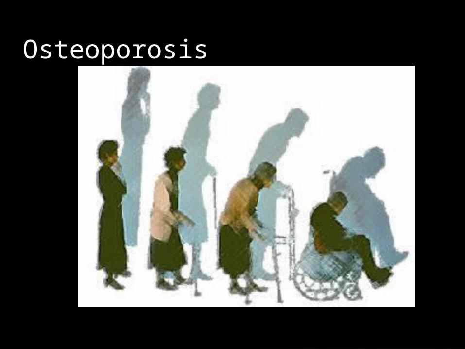
Osteoporosis

Metabolic condition
Bone demineralizes
Easy to fracture
Wrist, hip, and vertebrae are most affected

Osteopenia: low bone mass
Osteoclasic: bone resorption
Decreased bone mineral density
40-45% loss in women throughout lifespan
Trebecular (Spongy bone) is lost first
Then Cortical (compact bone) lost 2nd
Pathophysiology is unknown

classesGeneralized:involves many structures• Primary: more common
• Post menopausal women
• Men in 60s-70s
• seconday
Regional: limb involved• r/t fx, injury, paralysis, joint inflammation
• Immobilization greater than 8-12 weeks
• Weightless environment (astronauts)
Health preventionTeach about exercise
Teach about diet rich in calcium
Teach about bone health
Teach about safety

Assessment Risk for falls
Head to toe assessment• Inspect and palpate vertebrae
Assess pain
Assess for fallophobia
No definitive lab tests
Bone scan to check density

Nursing diagnosisRisk for falls
Impaired physical mobility
Acute or chronic pain

InterventionsClient education is #1
Hormone replacements
Calcium supplements
Multivitamins
Diet
Fall prevention
Exercise
Pain management
Braces

Osteomalacia Softening of the bone tissue
Inadequate mineralization of osteoid (mature compact and spongy bone)
Vitamin D deficiency is a key player
Similar characteristics with osteoporosis
Rare in USA
Prevent with vitamin D, sun exposure, and diet

s/s: early stages : nonspecific• Muscle weakness
• Bone pain
• Hypophosphatemia
• Hypocalcemia
• Generalized bone tenderness

Paget’s Disease

Metabolic disorder of bone remodeling
Bone deposits that are weak, enlarged, and disorganized
Phases:• Active increased osteoclasts cause massive bone destruction• Osteoclasts are multinuclear
• Mixed• Inactive 2nd phase
• New bone becomes sclerotic and very hard
• Osteoclasts return to normal amount
2nd most common bone disease
Most common sites are vertebrae, femur, skull, sternum, and pelvis
Unknown cause
Assessment 80% asymptomatic
Assess past history of fractures, skin color and temp, gout, hyperparathyroidism, lethargy, hyperuricemia
Pain that is aching, deep, poor description
Pain worsens with weight bearing and pressure
Pain most noticeable at nite or at rest
Arthritis at infected joints
Assess posture, gait, and balance
Assess vision, speech, and swallowing, hydrocephalus,
Neoplasm is the dreaded complication

Diagnostics Serum alk phosphate• Those treated for paget’s need ALP drawn 3-4 times/year
Urine hydroxyproline• Shows bone collagen turnover and degree of severity
Calcium levels are normal or elevated
Increase noted in uric acid• May initially be thought to be gout
X-rays, CT, MRI, bone biopsy

Treatment
Drugs for pain relief
Drugs to decrease bone resorption
Calcitonin (thyroid hormone)
Mithramycin (antineoplastic)
Biphosphanates
Heat therapy
Gentle massage
Exercise
PT
Diet
Osteotomy or joint replacement

osteomylelitis

Inflammatory process
Increase in vascularity and edema
Vessel becomes thrombosed once inflamed
Ischemia is next
Then necrosis
Sequestrium forms and retards bone healing
Categories Exogenous: infection enters from outside
Endogenous: infection enters from inside
Contiguous: results from skin infection
The most common offending organism is pseudomonas aeruginosa
Staph, salmonella are aslo culprits

s/s and assessmentPain
Fever
Erythema
Heat
Swelling
Assess circulation
Assess for septic shock

Treatment Contact precautions
IV antibx therapy
PICC line
Use sterile techniques
Pain meds
Hyperbaric oxygen therapy
Bone grafts
Muscle flaps
Amputations

Chondrogenic Osteochondroma: most common, benign, tumor…onsets in childhood, grows until skeletal maturity..has a bony stalk like appearance..may become malignant
Chondroma: lesion of mature hyaline cartilage of the hand and feet. Ribs, sternum, spine, and long bones can also be affected…can get at any age or gender

Osteogenic Osteoid osteoma: pinkish granular appearance..any bone affected..femur and tibia most affected
Osteoblastoma: affects vertebrae and long bones..large in size and lies in spongy bone..reddish granular appearance
Giant cell tumor: origin unknown..aggressive and extensive..affects women 20s-30s

Assessment/ tx Assess pain
Palpate involved area
CT scan and MRI done for diagnosis
Interventions• Meds and surgery combination
• Pain meds
• Meds taken with meals or milk
•

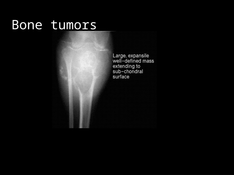
Malignant bone tumorsPrimary: originate in bone / 2nd ary: mets to bone
• Primary
Osteosarcoma: most common
• Large lesion, pain and swelling of short duration, warm site, central portion is sclerotic, usually mets to lung in 2 yrs then death
Ewing’s sarcoma: most malignant
• Pain and swelling, fever, anemia, leukocytosis, pelvis and lower extremities most affected, any age..but kids and young adults age 20s more
• Pelvic yields poor prognosis
Chondrosarcoma: dull pain, swelling for long period..• pelvis and femur fore affected
• Destroys bone and often calcifies
• Affect middle age to elders and more in men
Fibrosarcoma: from fibrous tissue; most common in long bones of legs and mets to lungs• Histiocytoma is most malignant type
• Local tenderness, with or w/o mass palpated

Bone Mets Primary tumors are in prostate, breast, kidney, thyroid, and lung
Fractures are major problem with management• Femur and acetabulum
Primarily affects those under 40
Assess/ diagnostics
Assess pain, swelling, palpate for masses
Monitor vs
Assess ADLs
Assess support structures
Assess coping skills
Check ALP levels for elevation
CT scan
Stage tumor
Nursing diagnosis/tx Pain
Anticipatory grieving
Disturbed body image
Fear
Anxiety
Tx• Pain management, chemo, radiation, surgery, dressing changes, be
active listener, establish goals, safety precautions, HHC

Carpal Tunnel

Education Use ergonomic work stations
Teach client to take regular breaks
s/s• Parathesia in hands
• Weak pinch, clumsiness, weakness
• Hand activity worsens symtoms
• Swelling may occur
Tx: nsaids, surgery

Dupuytren’s contracturesSlow progressive contracture
Common problem
Affects 4th or 5th digit of the hand
Trigger finger release surgery performed to fix
Disorders of the foot
Hammertoe: fix with surgery
Tarsal tunnel syndrome: ankle version of carpal tunnel
Plantar fasciitis: inflammation of the plantar fascia located in the arch of the foot• s/s: pain in arch, pain worsens w/ wt bearing
• Tx: ice, rest, stretches, strapping, nsaids, surgery
Hallux valgus: aka bunion
Associated with 8th cranial nerve or cellebellum
Menieres disease is an example of a disorder of vertigo.
most common 30-60
Ultimately just means dizzy
Vertigo
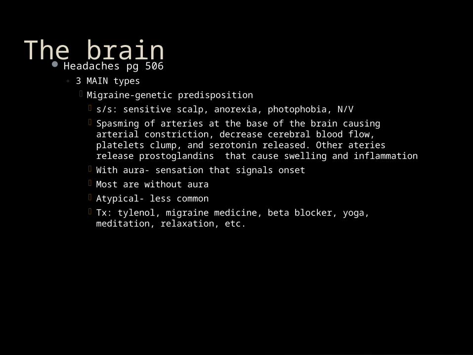
The brain Headaches pg 506
◦ 3 MAIN types Migraine-genetic predisposition
s/s: sensitive scalp, anorexia, photophobia, N/V Spasming of arteries at the base of the brain causing arterial
constriction, decrease cerebral blood flow, platelets clump, and serotonin released. Other ateries release prostoglandins that cause swelling and inflammation
With aura- sensation that signals onset Most are without aura Atypical- less common Tx: tylenol, migraine medicine, beta blocker, yoga, meditation,
relaxation, etc.
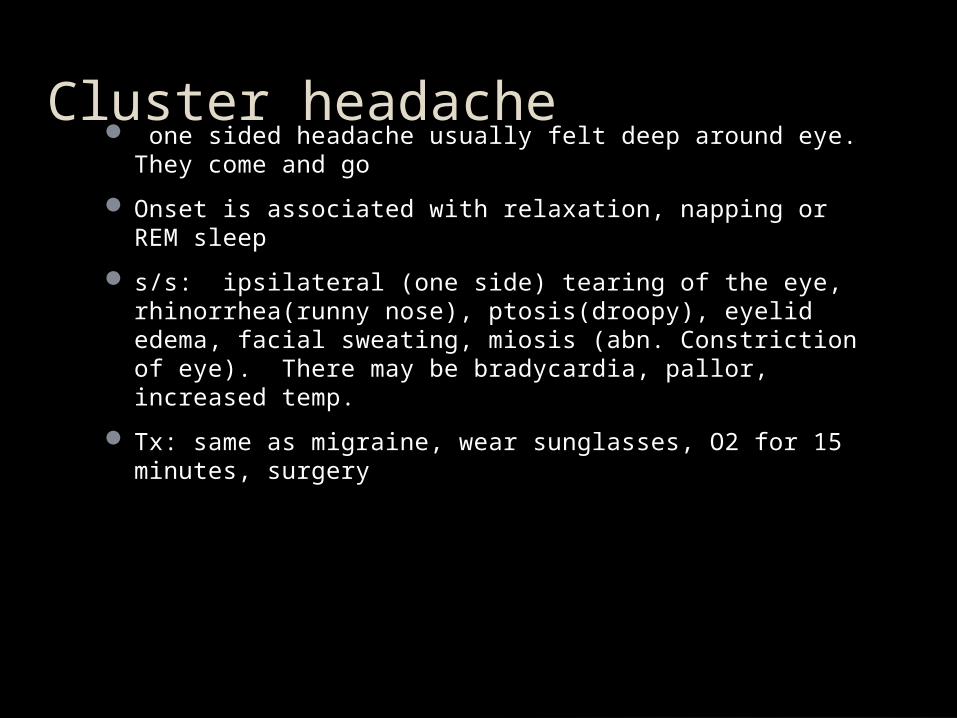
Cluster headache one sided headache usually felt deep around eye. They
come and go
Onset is associated with relaxation, napping or REM sleep
s/s: ipsilateral (one side) tearing of the eye, rhinorrhea(runny nose), ptosis(droopy), eyelid edema, facial sweating, miosis (abn. Constriction of eye). There may be bradycardia, pallor, increased temp.
Tx: same as migraine, wear sunglasses, O2 for 15 minutes, surgery
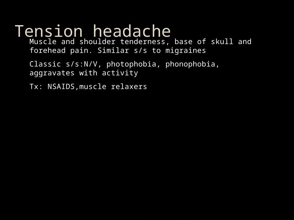
Tension headacheMuscle and shoulder tenderness, base of skull and forehead pain. Similar s/s to migraines
Classic s/s:N/V, photophobia, phonophobia, aggravates with activity
Tx: NSAIDS,muscle relaxers


Alzheimer’s Diseasehttp://www.youtube.com/watch?v=Z6lA1P2tF0o&feature=related

Stages Early• mild
Middle• moderate
Late• severe

s/sAggressive
Rapid mood swings
Increased confusion at nite (sundowner’s)
Decrease interest in personal appearance
Inappropriate clothing selection
Loss of bowel/bladder
Decreased appetite
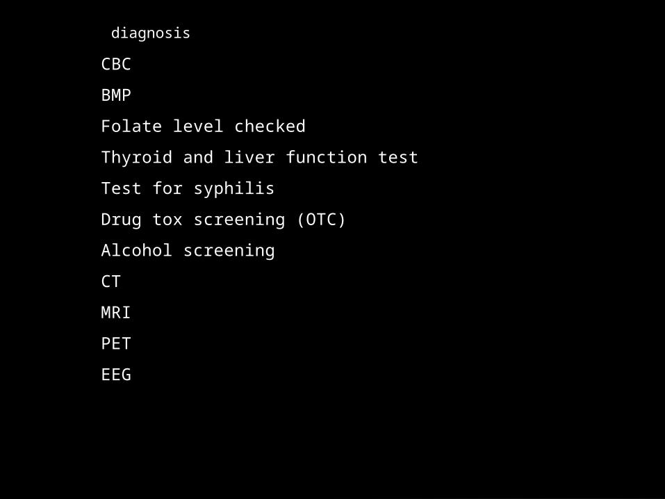
diagnosis
CBC
BMP
Folate level checked
Thyroid and liver function test
Test for syphilis
Drug tox screening (OTC)
Alcohol screening
CT
MRI
PET
EEG

Nursing diagnosisChronic confusion
Risk for injury
Disturbed sleep pattern

Tx
Meds
Prevent overstimulation
Be consistent
Reorient
Promote independence
Bowel/bladder training
Promote facial recognition
Speech therapy
Safety precautions
Minimize agitations

http://www.youtube.com/watch?v=Xv1tMioGgXI

Monitor electrolytes especially sodium levels and protein levels
Chart 8-2 talks about labs to monitor– albumin transferrin---prealbumin total lymphocytes.
Diets chart 8-4
Nutrition
glaucoma
2 types• Primary open angle: most common
• Angle closure: less common..emergency

s/sOpen angle: small cresent shaped defect
Angle closure: visual fields quickly decrease, severe pain around eye, headache, n/v, halos, blurred vision

Macular degenerationCentral vision declines
Mild blurring or distortion
More rapid to produce in smokers

Spinal Cord Injury(SCI)
Chapter 24

Causes of SCIPrimary
• Hyperflexion (moved forward excessively)• Hyperextension (MVA)• Axial loading (blow at top of head causes shattering)• Excessive rotation (turning beyond normal range)• Penetrating (knife, bullet)
Secondary
• Neurogenic shock• Vascular insult • Hemorrhage• Ischemia• Electrolyte imbalance

Types
Complete: spinal cord severed and no nerve impulses below level of injury• Cervical/Thoracic
Incomplete: allow some function and movement below level of injury• Includes:• Central cord syndrome • Anterior cord syndrome• Brown-Séquard syndrome

CompleteTetraplegia (quadriplegia): paralysis from neck down• Loss of bowel and bladder control
• Loss of motor function
• Loss of reflex activity
• Loss of sensation
• Coping issues
*Christopher Reeve is example of this injury*
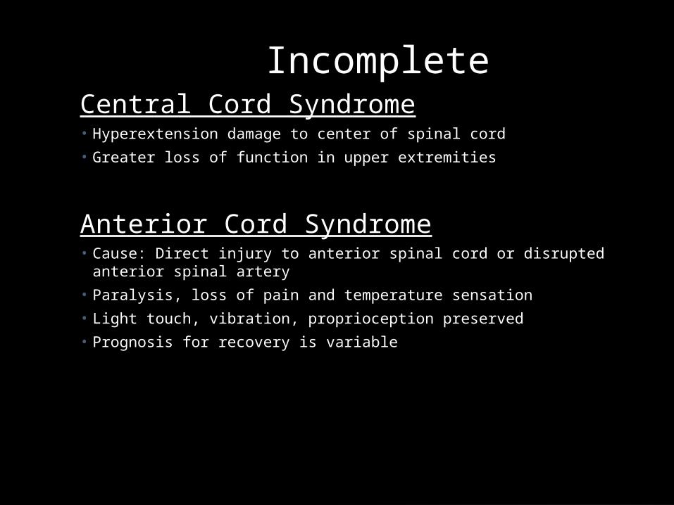
IncompleteCentral Cord Syndrome• Hyperextension damage to center of spinal cord
• Greater loss of function in upper extremities
Anterior Cord Syndrome• Cause: Direct injury to anterior spinal cord or disrupted anterior spinal
artery
• Paralysis, loss of pain and temperature sensation
• Light touch, vibration, proprioception preserved
• Prognosis for recovery is variable

IncompletePosterior cord lesion• Damage to posterior white and gray matter• Motor function intact, but loss of vibratory sense, crude touch, and
position sensation
Brown Sequard syndrome• Result of penetrating injury that causes hemisection of spinal cord.• Motor function , proprioception, vibration, and deep touch are lost on
the same side as injury (ipsilateral)• On the other side (contralateral) the sensation of pain, temperature
and light touch are affected

Assessment1st -respiratory status
2nd - intra-abdominal hemorrhage (hypotension, tachycardia, weak and thready pulse)
3rd assess motor function
• C4-5 apply downward pressure while the client shrugs• C5-6 apply resistance while client pulls up arms• C7 apply resistance while pt straightens flexed arms• C8 check hand grasp• L2-4 apply resistance while the client lifts legs from
bed• L5 apply resistance while client dorsiflexes feet• S1 apply resistance while client plantar flexes feet

ComplicationsCerebral ischemia
DVT/PE
Pneumonia/Atelectasis
Vomiting and Aspiration
GI stress ulcers
Constipation
UTI
Pressure Ulcers

Autonomic DysreflexiaSevere HTN, bradycardia, sever headache, nasal stuffiness, and flushing
• Caused by noxious stimuli like distended bladder or constipation
Immediate interventions
• Place in sitting position• Call doctor • Loosen tight clothes• Check foley tubing if present• Check for impaction• Check room temp• Monitor BP q10-15 minutes• Give nitrates or hydralazine per md order

TreatmentImmobilize fx- C-collarProper body alignment• Traction is possible
Monitor VS q4 hr and prnNeuro checks q4 hr and prnMonitor for neurogenic shock (hypotension and bradycardia)Prepare for possible surgeryTeach skin care, ADLs, wound prevention techniques, bowel and bladder training, medications, and sexuality

NRSG DX for SCI
Ineffective tissue perfusion r/t interruption of arterial flow
Ineffective airway clearance r/t SCI
Ineffective breathing pattern r/t SCI
Impaired gas exchange r/t SCI

Traumatic Brain Injury(TBI)

Head Injury Classification:
Severe Head Injury----GCS score of 8 or less
Moderate Head Injury----GCS score of 9 to 12
Mild Head Injury----GCS score of 13 to 15
(Adapted from: Advanced Trauma Life Support: Course for Physicians, American College of Surgeons, 1993).

Superficial InjuriesCommon
Abrasions
“Goose Eggs”
Lacerations• Scalp is very vascular
Xray if suspect skull fracture

Skull FracturesCategorized according to type and severity
Frequently seen in conjunction with brain injuries
Linear skull fractures
Comminuted skull fractures
Basal skull fractures
Possible associated cranial nerve deficits

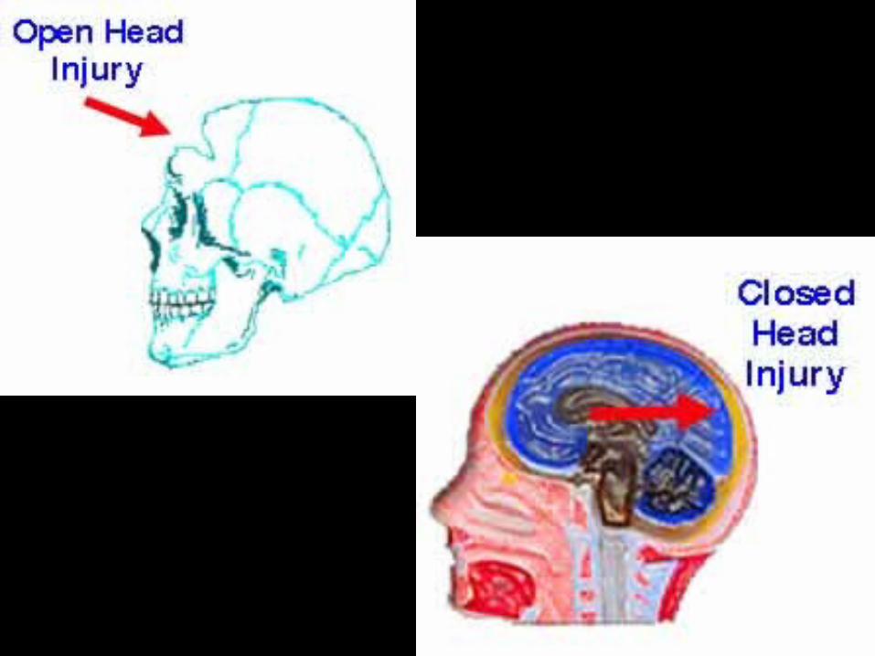
Open Skull FracturesLinear- simple clean break
Depressed - bone pressed in towards tissue
Open -lacerated scalp that creates opening to brain tissue
Comminuted - bone fragments and depresses into brain tissue
Basilar- unique fx at base of skull with CSF leaking though the ear or nose• Racoon eyes/Battles sign

http://www.pearlau.com.au/jpg/raccoon%2520eyes%

http://www.pearlau.com.au/jpg/raccoon%2520eyes

Closed Skull FracturesClosed- blunt trauma • Mild concussion-brief LOC• Diffuse axonal injury- usually from MVA • May go into coma
• Contusion-bruising of brain• Site of impact (coupe)• Opposite side of impact (contrecoupe)

Intracranial HematomasEpidural- bleed b/w skull and dura• Laceration of artery or vien
Subdural-bleed below dura and arachoid layers • Acute, subacute, chronic
Intracerebral-accumulation of blood in brain tissue• Blunt trauma
• Penetrating wounds
• Acceleration/deceleration injuries


Increased Intracranial Pressure(ICP)
Pg 568

Increase is caused by an increase in the volume of any of the intracranial components
Drivers of increased ICP• Hypoxia – triggers the vasodilatory cascade
• Ischemia in acute brain injury
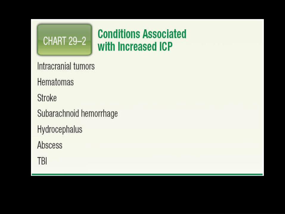

Increased ICPNormal ICP 10-15mmHg
Normal increases occur with coughing, sneezing, defecation
Leading cause of death for head trauma
As ICP increases cerebral perfusion decreases causing tissue hypoxia, decrease serum pH, and increase in CO2
ICP continued3 types of edema• Vasogenic: increase in brain tissue volume
• Cytotoxic: result of hypoxia
• Interstitial: occurs with brain swelling

Assessment
Hydrocephalus
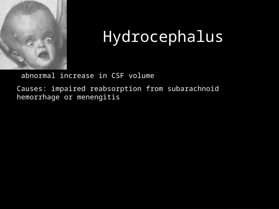
abnormal increase in CSF volume
Causes: impaired reabsorption from subarachnoid hemorrhage or menengitis

Brain HerniationIncreased ICP will shift and move brain tissue downward
Central Herniation• Downward shift to brainstem
• S/S• Cheyne stokes , pinpoint pupils, hemodynamic instability
The most life threatening is Uncal because it causes pressure on the 3rd cranial nerve• S/S
• Dilated, nonreactive pupils, ptosis, rapidly decreased LOC

Herniation syndromes.

Movement/musculoskeletal
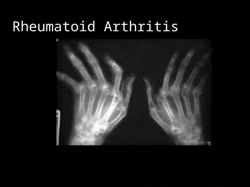
Rheumatoid Arthritis
Most common connective tissue disorders
Most destructive to joints
RA factors looked for in lab
Assess sedrate
Assess immunoglobins
MRIs performed
EMGs are performed to measure function

Assessment/ S/S continued
Joint stiffness
Swelling
Pain
Fatigue
Weight loss
Reddened joints
Deformity of joints
Baker’s cysts may occur and cause pain
Dry eyes, dry mouth, dry vagina
Assess ADLs, coping, pain
interventions
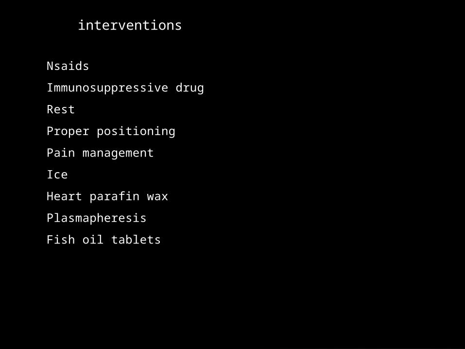
Nsaids
Immunosuppressive drug
Rest
Proper positioning
Pain management
Ice
Heart parafin wax
Plasmapheresis
Fish oil tablets

GoutType of arthritis
Urate crystals deposit in joints
Primary gout is most common
Inflammation is key sign
2nd ary is when too much uric acid in blood
Can affect kidneys
Meds to treat
Pain management

Fibromyalgia
Chronic pain syndrome
Pain is burning or gnawing
Headache and jaw pain are also common
Chest pain is common
Pain control is the key• Muscle relaxers, nsaids, antidepressants

Muscular distrophies9 types
Progression is slow or fast
Most common is severe X linked recessive
Diagnosis is difficult
Comfort is key
Treat symptoms
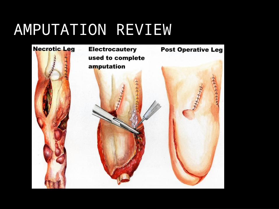
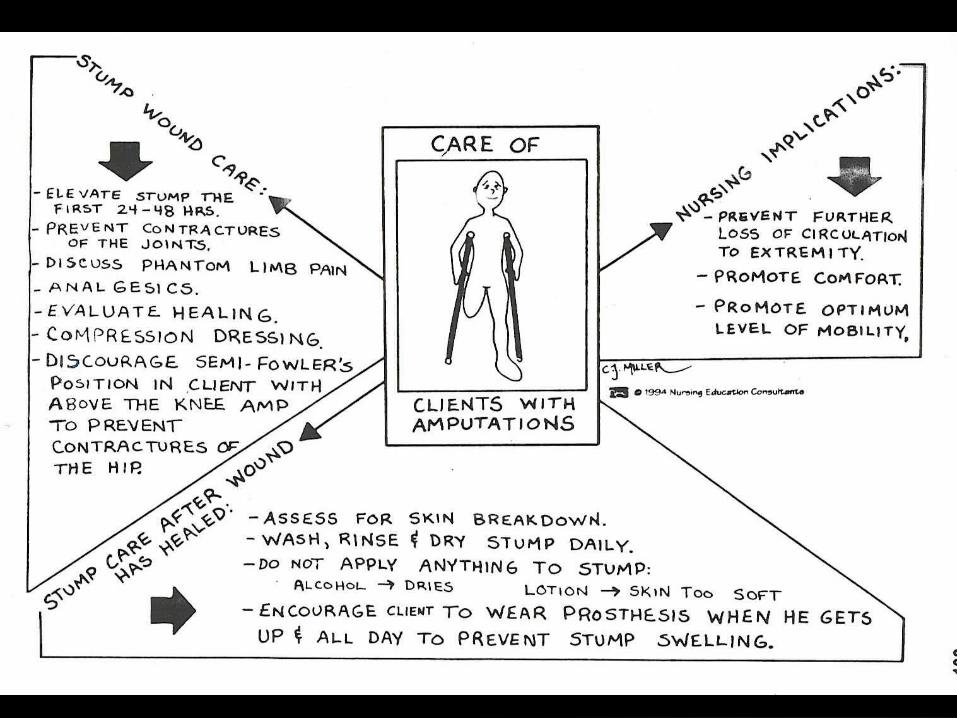
AMPUTATION REVIEW


amputations
Removal of part of the body
Types• Surgical-example digit
• Traumatic- example digit
Levels• Lower extremity: digits, bka, aka,
midfoot
• Upper extremity: hands, fingers, arms
Complications• Hemorrhage
• Infection
• Phantom limb pain: perceive pain in the amputated limb
• Immobility
• Neuroma: sensitive tumor consisting of nerve cells found at several nerve endings
• Contractures
Review Meds on 599-604
Review cranial nerves
TIPS!!