ASSOCIATION BETWEEN MYOPIA AND AMPLITUDE OF ACCOMMODATION IN YOUNG ADULTS by DR. NG SOK LIN MD(USM) Dissertation Submitted In Partial Fulfillment Of The Requirements F.or The Degree Of Master Of Medicine (Ophthalmology) UNIVERSITI SA INS MALAYSIA 2001
Transcript
ASSOCIATION BETWEEN MYOPIA AND AMPLITUDE OF ACCOMMODATION
IN YOUNG ADULTS
by
DR. NG SOK LIN
MD(USM)
Dissertation Submitted In Partial Fulfillment Of The
Requirements F.or The Degree Of Master Of Medicine (Ophthalmology)
UNIVERSITI SA INS MALAYSIA
2001
11
ACKNOWLEDGEMENT
My sincere thanks and deepest appreciation to my supervisor, Dr. Elias Hussein, lecturer
of Department of Ophthalmology, Hospital Universiti Sains Malaysia, for his guidance
and support throughout the planning and duration of this study and for his invaluable
advice and constructive criticism during the preparation of this dissertation.
I wish to express my greatest appreciation to Dr Mohtar Ibrahim, Head Department of
Ophthalmology, and all the lecturers in the Department of Ophthalmology, Hospital
Universiti Sains Malaysia (HUSM). Thank for their outstanding teaching, guidance and
encouragement throughout my course. My warmest appreciation to all my fellow
colleagues for their friendship, cooperation, assistance and encouragement.
A special thanks goes to our statistician, Dr. Syed Hatim and my colleagues from
Department of Community Medicine, HUSM, Dr. Maizun Binti Mohd Zain, Dr. Wan
Mohamad Zahirnuddin Bin Wan Mohamad, Dr. Suhaiza Binti Sulaiman and Dr. Nor
Sa' adah Binti Bachok, for their endless assistance in statistics.
My thanks to Encik Amir for his excellent photography.
My warmest appreciation to my husband, Dr. Yew Cheng Hoe, my children, Wei Chee
and Wei Ni and my parents, without whose support and encouragement I could never
complete this course.
TABLE OF CONTENTS
TITLE
ACKNOWLEDGE1\4ENT
TABLE OF CONTENTS
LIST OF TABLES
LIST OF FIGURES
ABSTRAK
ABSTRACT
TEXT
I. INTRODUCTION
I .1. Objectives
I .1. I. General Objective
1.1.2. Specific Objectives
2. LITERATURE REVIEW
3. MATERIALS AND :METHODS
3 .1. Research design
3.2. Population, setting and time
3.3. Sampling and sample size
3.3 .1. Sampling procedure
3.3.2. Sample size
3.3 .3. Plans for minimizing error
3.4. Selection criteria
PAGE
ii
111-VI
vii-viii
IX-X
Xl-Xll
Xlll-XIV
I
8
9
9
10
25
26
26
26
26
28
28
29
m
3. 4 .1. Inclusion criteria
3.4.2. Exclusion criteria
3. 5. Defmition of terms
3.5 .1. Amplitude of accommodation (A)
3.5.2. Accommodation
3.5.3. The Far Point (FP)
3.5.4. The Near Point (NP)
3.5.5. The dioptric power of the resting eye
3.5.6. The dioptric power of the accommodated eye
3.5.7. Myopia
3.5.8. Low myopia.
3.5.9. Moderate to high myopia.
3. 5.1 0. Emmetropia
3. 5 .11. Early onset myopia
3. 5.12. Late onset myopia
3.6. Units of observation
3. 6 .1. Refractive Error
3.6.2. Near Point Distance (NP)
3.6.3. Far Point Distance (FP)
3.6.4. Dioptric value of the near point distance (P)
3. 6. 5. Dioptric value of the far point distance (R)
3.6.6. Amplitude of Accommodation (A)
29
29
30
30
30
30
30
30
31
31
31
31
31
31
32
32
32
32
32
32
32
32
lV
3. 7. Instruments
3.7.1. Retinoscope.
3.7.2. Trial lens set.
3.7.3. Trial frame.
3.7.4. Snellen acuity chart.
3.7.5. R.A.F. rule.
3.7.6. Automated refractometer
3.7.7. Focimeter
3.8. Methods
3 .8.1. Methods of data collection
3.8.2. Formula for calculating the amplitude of accommodation.
3.8.3. Statistical Methods.
4. RESULTS
4.1. Demographic Characteristics
4.2. Risk Factors associated with Myopia.
4.2.1. Association between Myopia and amplitude of accommodation.
4.2.2. Myopia versus sex.
4.2.3. Myopia versus race.
4.2.4. Myopia and family history of myopia.
4.2.5. Myopia versus place of residence.
4.2.6. Multivariate Logistic Regression Analysis.
v
33
33
33
33
34
34
34
34
41
41
43
43
44
45
47
47
48
49
50
52
53
VI
4.3. Risk factors associated with Degree of Myopia. 54
4.3.1. Degree of myopia versus amplitude of accommodation. 54
4.3 .2. Degree of myopia versus sex. 55
4.3.3. Degree of myopia versus race. 56
4.3.4. Degree of myopia versus family history of myopia and 58 place of residence.
4.3.5. Relationship between degree of myopia and onset of 59 myopia.
Table 2.1. Prevalence of visual impairment due to refractive error based on 13 sex, ethnicity, age group, urban-rural residence and state in Malaysia.
Table 2.2. Dander's table showing amplitude of accommodation in 24
relation to age.
Table 3.1. Nwnber of students based on age group and refractive error. 27
Table 4.1. Baseline characteristics of medical and nursing students, USM in 46 three refractive groups (emmetropia, low myopia and moderate to high myopia).
Table 4.2. Mean refraction and Mean Amplitude of Accommodation between 47 emmetropic and myopic students.
Table 4.3. Nwnber of subjects in relation to sex and refractive status. 48
Table 4.4. Nwnber of subjects in relation to race and refractive status. 50
Table 4.5. Number of subjects in relation to family history of myopia and 51 refractive status.
Table 4.6. Nwnber of subjects in relation to place of residence and refractive 52
status.
Table 4.7. Logistic Regression Modeling. The risk of Myopia (Refractive 53 error~ -0.75 Diopter) and Various Risk Factors.
Table 4.8. Mean Amplitude of accommodation and Mean refraction between 54 students with low myopia and students with moderate to high
myopta.
Table 4.9. Number of subjects in relation to sex and degree of myopia. 55
Table 4.10. Number of subjects in relation to race and degree of myopia. 57
Table 4.11. Number of subjects in relation to family history of myopia and 58 degree of myopia.
Table 4.12. Number of subjects in relation to place of residence and degree of 59 myopta.
Table 4.13. Number of subjects in relation to age of onset of myopia and 60 degree of myopia.
Table 4.14. Logistic Regression Modeling. The risk ofhigher degree of 61 myopia (Refractive error~ -3.00 Diopter) and Various Risk Factors.
Vlll
IX
LIST OF FIGURES.
Figure 3.1.
Figure 3.2.
Figure 3.3.
Figure 3.4.
Figure 3.5.
Figure 3.6.
Figure 3.7.
Figure 3.8.
Figure 3.9.
Figure 3.10.
Figure 3.11.
Figure 3.12.
Figure 3.13.
Figure 4.1.
Figure 4.2.
Figure 4.3.
Figure 4.4.
Figure 4.5.
Figure 4.6.
Page
Retinoscope. 35
Trial lens set. 35
Trial frame. 36
Visual acuity tested with Snellen acuity chart. 36
R.A.F. rule. 37
Automated refractometer. 37
Focimeter. 38
Occluder. 38
+2.50 Diopter lens. 38
Retinoscopy examination. 39
Subjective refraction. 39
Measurement of far point distance with R.A.F. rule. 40
Measurement of near point distance with R.A.F. rule. 40
Mean amplitude of accommodation in medical I nursing students with or without tnyopia.
48
Number of subjects according to sex and refractive status. 49
Number of subjects according to race and refractive status. 50
Number of subjects based on refractive status and family history 51 of myopia.
Number of subjects according to place of residence and 52 refractive status.
Mean amplitude of accommodation in medical/ nursing students with low tnyopia and moderate to high tnyopia.
55
X
Figure 4.7. Nwnber of subjects according to sex and degree of tnyopia. 56
Figure 4.8. Number of subjects according to race and degree of myopia. 57
Figure 4.9. Number of subjects according to family history of myopia and 58 degree of myopia.
Figure 4.10. Number of subjects according to place of residence and degree 59 of myopia.
Figure 4.11. Number of subjects according to degree and onset of myopia. 60
X1
ABSTRAK.
Objektif: Tujuan penyelidikan ini adalah untuk mengkaji hubungan diantara amplitud
akomodasi (amplitude of accommodation) dan masalah refraksi (refractive error) di
kalangan penuntut·penuntut perubatan dan kejururawatan daripada Pusat Pengajian Sains
Perubatan (PPSP), Universiti Sains Malaysia. Disamping itu, faktor-faktor lain yang
berkaitan dengan rabunjauh (myopia) turut disiasat.
Tatacara: Sejumlah 110 penuntut-penuntut perubatan dan kejururawatan daripada PPSP
Universiti Sains Malaysia, Kubang Kerian, Kelantan terlibat dalam penyelidikan ini.
Sample dipilih daripada populasi dengan kaedah 'stratified random sampling' (stratified
random sampling method). Masalah rabun masing-masing ditentukan, jarak 'near point'
(near point distance) dan jarak 'far point'' (far point distance) diukur pada kedua-dua
mata pada masa yang berasingan. Amplitud akomodasi dikira secara penolakan 'far
point'(unit diopter) daripada 'near point' (unit diopter). Data analisis dilakukan dengan
program SPSS.
Keputusan: Rabun jauh ( ditakritkan sebagai mereka yang mempunyai masalah refraksi
sebanyak -0.7 5 diopter dan keatas) tnempunyai amplitud akomodasi yang rendah
(p=0.012). Faktor-faktor lain yang berkaitan dengan masalah rabun jauh tennasuk faktor
keturunan (p=0.002) dan faktor etnik (ethnic) (p=0.021). Penuntut-penuntut yang
mempunyai latarbelakang keluarga yang berabun jauh merupakan kumpulan yang lebih
berisiko ('odds ratio' 4.4, p=0.002). Analisis pelbagai logistik regrasi (multivariate
X11
logistic regression analysis) menunjukkan bahawa amplitud akomodasi yang rendah
(p=0.009) serta faktor keturunan (p=0.002) mempunyai huhungan rapat dengan rabun
jauh. Mereka yang mengidap rabun jauh pada peringkat umur muda (early onset myopia)
berisiko tinggi untuk mendapat rabun jauh yang teruk (p=0.005). Dari analisis pelbagai
logistik regrasi, rabun jauh pada peringkat umur muda didapati mempunyai risiko
sebanyak lima perpuluhan satu kali ganda menjadi rabun jauh yang teruk ('odds ratio'=
5.1, p=0.005).
Kesimpulan: Penyelidikan ini mendapati amplitud akomodasi yang rendah dan faktor
keturunan merupakan faktor-faktor penting berkaitan dengan rabtm jauh. Disamping itu;
rabun jauh pada peringkat umur muda didapati berisiko tinggi untuk menjadi rabun jauh
yang lebih teruk.
Xlll
ABSTRACT.
Objectives: To describe the association between amplitude of accommodation and
refractive error as well as to evaluate the risk factors associated with myopia among
medical and nursing students from School of Medical Sciences, Universiti Sains
Malaysia.
Methods: A total of 110 medical and nursing students with both eyes classified into the
same refractive state namely emmetropia, low myopia and moderate to high myopia were
enrolled in this study. The sample was randomly selected from the population with the
stratified random sampling method. Refractive error, near point distance and far point
distance were measured. Each eye of each subject was tested separately. The amplitude
of accommodation was calculated by subtracting the far point (in diopters) from the near
point (in diopter). Data analyzes was performed using SPSS software.
Results: Myopes were defined as those with refractive error of -0.75 diopter or more,
have lower accommodative amplitudes (p=O.Ol2). Family history of myopia (p=0.002)
and race (p=0.021) were other risk factors associated with myopia. Those with family
history of myopia are at higher risk of getting myopia (odds ratio 4.4, p=0.002). After
multivariate logistic regression analysis adjusting for various relevant variables, lower
amplitude of accommodation (p=0.009) and family history of myopia (p=0.002)
remained associated with myopia. Myopes with early onset myopia were related to higher
XIV
degree of myopia (p=0.005). Early onset myopic subjects are at 5.1 times greater risk of
getting higher degree of myopia (odds ratio= 5.1, p=0.005).
Conclusions: This study demonstrates that lower amplitude of accommodation along
with family history of myopia is important risk factors associated with myopia. On the
other hand, the early onset of myopia is the risk factor for higher degree of myopia.
1
1. INTRODUCTION
2
1. INTRODUCTION
Myopia, or nearsightedness, has been undergoing a major re-evaluation in recent years
both by ophthalmologists and basic scientists, though for different reasons. For
ophthalmologists the rise of refractive surgery in the past decade has seen myopia
changing from a condition requiring optical correction to one that can be managed
surgically with the aid of the excimer laser and other techniques. For basic scientists
interested in the control of eye growth, the past decade has been equally revolutionary
with a huge increase in the understanding of mechanism by which eye growth is
regulated by the quality of the retinal image. This research offers insights into why
myopia develops in humans and offers clinicians a novel perspective from which to
approach the management of myopia. Rather than attempting to alter corneal curvature to
"treat" myopia, it may be possible to prevent or "cure' myopia by directly manipulating
the growth mechanisms of the eye.
Myopia is defined clinically as a mismatch between the power of the optical elements of
the eye and the axial length that causes images to focus in front of the retina and results in
blurry images on the retina. The corrective lenses or other refractive treatment is required
to produce a clear image (Wensor et al., 1999; Zadnik et al., 1994).
The public health and economic impact of myopia, the most common eye condition in the
world, is enormous. In the United States, the cost of correcting refractive errors with
spectacles or contact lenses is estimated to be 2 million dollars per year (Saw et al.,
1996).
3
Many investigations have been carried out during the last 150 years to detect factors '
which cause myopia. In the ophthalmic literature, there has been extensive discussion
whether myopia is caused by hereditary or environmental factors. The causes of myopia
are unclear, although evidence supports both genetic and environmental components.
Hereditary factors are implicated in myopia and one widely accepted explanation for the
role of education and intelligence is that the accommodation involved in near work ,
particularly reading, can provoke elongation of the eyeball in genetically susceptible
individuals (Teasdale et al., 1988). Interest has been focused on accommodation because
of the established association between myopia and the amount of close work, or working
at a close distance, years of education and intelligence. It remains unclear, however, to
what extent greater levels of education and intelligence are associated with a greater
degree of myopia.
The earliest suggestion of a link between accommodation and myopia appears to be that
of Kepler ( 1611 ). Whilst numerous subsequent authors have also suggested that the
development of myopia is related to the action of accommodation (Stansbury, 1948;
Goldschmidt, 1968; Duke-Elder and Abrams, 1970; Curtin, 1970, 1985), to date no clear
mechanism has been elucidated.
The Early Treatment Diabetic Retinopathy Study, a randomized clinical trial, was
designed to study the timing of photocoagulation and aspirin therapy for diabetic
retinopathy. A test of accommodation was perfonned at baseline in patients who were
younger than 46 years and had best-corrected visual acuity of 20/40 or better (Bratm et
4
al., 1995). The study shows that eyes with myopia have lower accommodative amplitude.
Eyes with lower amplitudes of accommodation must use more of their accommodative
reserve for near work. So myopia may be an adaptation that develops in eyes with
reduced accommodative amplitudes to reduce the demands of near work.
One limitation of this study is that the study population was composed entirely of persons
with diabetic retinopathy and lower amplitudes of accommodation (Fong, 1997). Another
study demonstrated that diabetes and duration of diabetes, along with age, were important
risk factors for reduced accommodative amplitude (Braun et al., 1995). This fact has
initiated an idea of performing a study to find out the association between amplitudes of
accommodation and refractive error in the normal healthy individual. The aim of the
study was to find out whether low amplitude of accommodation also occurs among the
normal healthy myopic individual.
It is unclear at the present time whether myopia develops as a result of an abnormal
accommodative response, or alternatively that the accommodative stimulus is reduced as
a consequence of myopic development. Prospective studies measuring accommodative
amplitudes at baseline and monitoring the development of refractive error are needed to
determine whether lower amplitudes of accommodation lead to myopia.
The amplitude of accommodation reflects the maximum accommodative response, and
has been defined as the dioptric distance between the far point (point conjugate with the
retina when accommodation is fully relaxed) and the near-point (point conjugate with the
5
retina when accommodation is fully exerted) of accommodation (Rosenfield, 1997). A
number of studies have reported significant variations in this parameter with refractive
error. Maddock et al. (1981) and McBrien and Millodot (1986a) subdivided their myopic
population into either low (<3D) and high (>3D) myopes, or early- (myopia onset at 13
years of age or earlier) and late- (myopia onset at 15 years of age or later) onset myopes.
Since the late-onset myopes are also typically low myopes, both studies reported similar
findings, with low myopes having higher amplitudes than high myopes. However, both
myopic subgroups had higher amplitudes than either emmetropes or hyperopes. In
contrast, Fisher et al. (1987) did not obsetve any significant variation in the nearpoint of
accommodation with refractive error.
Myopia has become a problem of public health concern in the world. The prevalence of
myopia in Singapore is one of the highest worldwide. It is interesting to conduct study on
myopic population in Malaysia to find out whether myopia is also a problem of public
health concern in our country. However the nationwide myopic research was impossible
for me at this stage due to limitation of time and lack of manpower. In order to conduct a
myopic research within a short period of time and alone, the medical and nursing students
from School of Medical Sciences, Universiti Sains Malaysia (USM) were selected as my
study population. The primary objective of the study was to elicit the association between
amplitude of accommodation and myopia among healthy normal medical and nursing
students in USM. The aim of the study was to find out whether low amplitude of
accommodation also occurs among the normal healthy young myopic individual beside
diabetic patients discussed earlier. The second objective was to find out the possible risk
6
factors associated with myopia and degree of myopia among medical and nursing
students in USM.
The study was set out to answer three questions:
1. Is there any association between myopia and amplitude of accommodation in the
medical and nursing students in USM?
2. Does degree of myopia affects the amplitude of accommodation?
3. What are the risk factors for myopia and degree of myopia among medical and
nursing students in USM?
THE NULL HYPOTHESIS (Ho).
1. There was no association between amplitude of accommodation and myopia
in the medical and nursing students in USM.
2. There was no association between amplitude of accommodation and degree of
myopia.
3. There was no sosiodemographic factors (example: sex, race, family history of
myopia, place of residence) affecting myopia among the medical and nursing
students in USM.
1n the future, we may try to establish whether low amplitude of accommodation is the
cause for myopia or the other way round by conducting a prospective study measuring
7
accommodative amplitude at baseline and follow up to monitor the development of
refractive error.
8
1.1 OBJECTIVES
9
1.1. OBJECTIVES
1.1.1 GENERAL OBJECTIVE
To describe the association between amplitude of accommodation and refractive error
among medical and nursing students from School of Medical Sciences, Universiti Sains
Malaysia.
1.1.2 SPECIFIC OBJECTIVES
1. To measure the amplitude of accommodation in myopic and emmetropic
individuals.
u. To compare the amplitude of accommodation in relation to the refractive status
and degree of myopia.
111. To evaluate the risk factors for myopia and degree of myopia.
10
2. LITERATURE REVIEW
11
2. LITERATURE REVIEW.
Myopia is the state of refractive error in which parallel rays of light come to a focus in
front of the sentient layer of the retina when the eye is at rest (Duke-Elder and Abrams '
1970). Helmholtz defined myopia in terms of the position of the far point plane (objects
situated in this plane are focused on the retina), this being in front of the eye in myopia,
and pointed out that light entering the eye had to be divergent in order to be focused on
the retina of the myopic eye.
Myopia is measured by the spherical power in diopters of the diverging lens needed to
focus light onto the retina, which can be expressed as the spherical equivalent or
refraction in the least myopic meridian (Saw et al., 1996). The clinical correlates of
myopia include blurred distance vision, eye rubbing, and squinting.
During the past several years an increasing amount of data has become available
concerning the prevalence of myopia at various stages of life, particularly during the
early years. The prevalence of myopia is approximately 20% in the United States
population. This frequently varies with age, sex, race, ethnicity, occupation, environment,
and other factors in various sampled populations (Curtin & Whitmore, 1995).
Many studies show that by the age of 5 or 6 years, only about 2 percent of children have
myopia of 0.50 D or more (Kemph et al., 1928; Blum et al., 1959; Hirsch, 1964;
Laatikainen and Erkkila, 1980; Mantyjarvi, 1983). It is known that many children who
12
are emmetropic when entering school become myopic during the school years. The
prevalence of myopia of 0.50 D or more increases in a relatively linear manner from
about 2 percent at age 6 to about 20 percent at age 20. Between the ages of 20 and 40
years, the prevalence of myopia reaches a peak of about 30 percent. After which it begins
to decrease because of the tendency for some of the low myopes to lose their myopia,
rejoining the emmetropic group. But the prevalence of myopia (of 0.50 D or more)
increases somewhat in the later years of life due to the presence of nuclear lens changes,
as suggested by Hirsch (1958).
The prevalence of refractive anomalies varies widely from one geographical, racial, or
occupational group to another. Baldwin (1967) has reviewed much of the literature
concerning the prevalence of myopia in various racial and occupational groups. One of
the most interesting studies cited by Baldwin was that of Crawford and Hammar (1949),
who screened 50,000 school children of various racial groups living in Hawaii. They
found that the percentage of children having myopia ranged from about 3 percent for
Hawaiian children to 12 percent for Caucasian children and 17 percent for Chinese
children.
Even within a single racial or ethnic group, the prevalence of myopia has been found to
vary greatly with occupation. Baldwin (1967) reviewed the results of six studies in which
the prevalence of myopia was compared for near workers and those not engaged in near
work. The mean prevalence of myopia for near workers was approximately 33 percent as
compared to 15 percent for non-near workers.
13
In Malaysia, the National Eye Survey was carried out between 1996 and 1997 with the
primary objective to provide accurate and statistically representative infonnation
regarding the actual visual status and the prevalence of eye diseases in the Malaysia
population. Prevalence of visual impairment due to refractive error in Malaysia was
reported based on sex, ethnicity, age group, urban-rural residence and state (Table 2.1 ).
The overall prevalence of visual impainnent due to refractive error in Malaysia was
1.18% (Mohamad et. al., 1996).
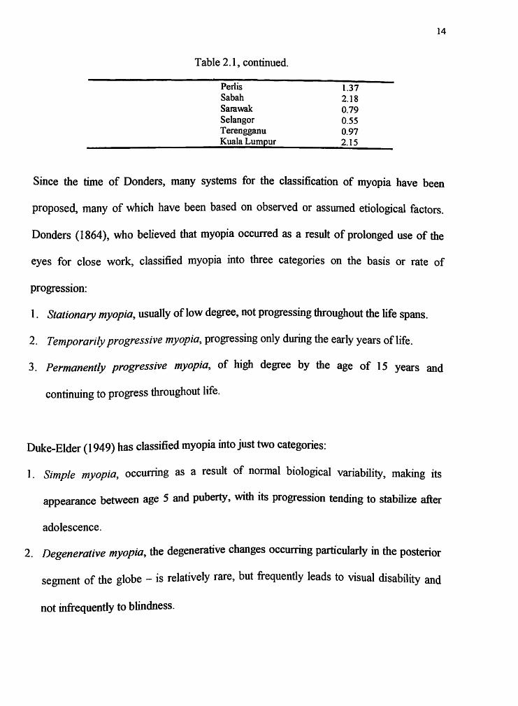
Table 2.1. Prevalence of visual impainnent due to refractive error based on sex, ethnicity, age group, urban-rural residence and state in Malaysia.
Overall Sex
Race
Age group
Urban or rural residence
State
Men Women
Malay Chinese Indian Others
0-9 10-19 20-29 30-39 40-49 S0-59 60-69 >=70
Urban Rural
Johore Kedah Kelantan Melaka Negeri Sembilan Pahang Penang Perak
Prevalence (%) 1.18
0.89 1.48
1.20 0.90 1.27 1.74
0.31 1.38 1.24 0.63 1.71 2.78 2.90 2.46
1.24 1.13
1.69 1.14 0.60 1.34 0.26 0.42 1.15 1.60
Table 2.1, continued.
Perl is Sa bah Sarawak Selangor Terengganu Kuala Lumpur
1.37 2.18 0.79 0.55 0.97 2.15
14
Since the time of Donders, many systems for the classification of myopia have been
proposed, many of which have been based on observed or assumed etiological factors.
Donders (1864), who believed that myopia occurred as a result of prolonged use of the
eyes for close work, classified myopia into three categories on the basis or rate of
progression:
1. Stationary myopia, usually of low degree, not progressing throughout the life spans.
2. Temporarily progressive myopia, progressing only during the early years of life.
3. Permanently progressive myopia, of high degree by the age of 15 years and
continuing to progress throughout life.
Duke-Elder (1949) has classified myopia into just two categories:
1. Simple myopia, occurring as a result of normal biological variability, making its
appearance between age 5 and puberty' with its progression tending to stabilize after
adolescence.
2. Degenerative myopia, the degenerative changes occurring particularly in the posterior
segment of the globe -is relatively rare, but frequently leads to visual disability and
not infrequently to blindness.
15
It is a widespread practice to distinguish two forms of myopia, a rare pathological form
and a less severe simple form. The pathological form can show several complications,
including chorioretinal degeneration, and it almost always appears in association with a
high refractive error, usually to some degree above -8.0 diopter (D). The much more
common myopia of degrees below -8.0 Dare almost always of the less severe form. It is
sometimes called "school myopia", because it typically develops in children of school
age and because epidemiological studies have persistently shown it to be associated with
high educational attainment (Teasdale et al., 1988).
A simple classification of myopia, though not a particularly informative one, is by
degree. Hine (1949) classified myopia of less than 3 D as low, of 3 D to 6 D as moderate,
and of more than 6 D as high. Hirschberg proposed a classification similar to Hine's
except with the additional category of very high myopia for refractive errors greater than
15 D. Severe myopia may be associated with myopic macular degeneration, cataract,
glaucoma, peripheral retinal changes (such as lattice degeneration), and retinal holes and
tears as well as retinal detachment. '
As a result of an epidemiological study of myopia in Denmark, Goldschmidt ( 1968)
proposed the existence of three types of myopia, classified on the basis of both the degree
of myopia and the age of onset:
1. Low myopia, the most frequent type of myopia, principally genetically determined,
developing during the first 20 years of life, progressing steadily and rarely exceeding
6.00 to 9.00 D.
16
2. Late myopia, developing after the cessation of bodily growth, seldom reaching higher
degrees, and seemingly related to excessive close work.
3. High myopia, either genetically or environmentally determined, frequently having an
early onset and capable of reaching excessive degrees, causing severely reduced
vision and degenerative changes in the eye over a period of years.
Defining myopia as "expansion glaucoma," brought about by an increase in intraocular
pressure, Kelly (1981) described three types of myopia, classified on the basis of
etiology:
1. Self-inflicted vitreous glaucoma (simple myopia) due to blockage at the zonular level,
accounting for 90 percent of myopia, occurring because the ciliary body, during
accommodation, pulls forward on the thick anterior vitreous, concentrating the zonule
and closing the zonular gap.
2. Active anterior chamber glaucoma (malignant glaucoma) due to the presence of a
retinoschisis like membrane blocking the trabecular area, accounting for 5 percent of
myopia.
3. Inactive glaucoma (congenital glaucoma) due to an intraocular pressure rises in utero.
In The Myopia, Basic Sciences and Clinical Management, Curtin (1985) introduced a
system of classification based on etiology, degree of myopia, and time of onset:
1. Physiologic myopia (simple or refractive myopia), developing postnatally because of
a correlation failure between the total refracting power of the eye and a normal axial
length.
17
2. Intermediate myopia (axial myopia), medium or moderate myopia, due to an
expansion of the posterior segment of the globe in excess of normal ocular growth,
subdivided into congenital, childhood, and late myopia.
3. Pathologic myopia, a special form of axial myopia, defined as the ocular disease in
which a number of serious complications are associated with abnonnal lengthening of
the eyeball. It is often associated with thinning of the scleral wall and posterior
staphyloma ..
Grosvenor (1987) has proposed a myopia classification base on the basis of age of onset.
Grosvenor's classification system includes four categories:
I. Congenital myopia. Myopia is present at birth and persists through infancy, with
high myopia being the general rule.
2. Youth-onset myopia/ school myopia. Becomes manifest during the early childhood
from about age 6 years through the teenage years and stabilizes by the late teens or
early twenties.
3. Early adult-onset myopia. This form of myopia has its onset during the period from
age 20 to about age 40. Many of those will have only a small amount of myopia and
will become emmetropic or even hyperopic in their later years.
4. Late adult-onset myopia. This form of myopia has its onset beyond the age of 40.
Both school and adult- onset myopia are mainly the result of idiopathic causes, while
congenital myopia is often associated with other abnormalities.
18
Unlike hyperopia or astigmatism, once myopia is found to exist, it tends to progress. For
example, data from Hirsch's (1964) longitudinal study of refraction between the ages of 5
to 6 and 13 to 14 years indicated that hyperopia tends to decrease by a small fraction of
1.00 D per year. Myopia occurring during the adult years, on the other hand, tends to
progress at a slower rate. In most cases the myopia tended to increase in a linear manner
into the middle or late teen years, then level off.
Both the Goss and Winkler (1983) data and the Grosvenor et al. (1987) data tend to
confirm the observation that the earlier a child becomes myopic, the more rapidly the
condition tends to progress. Consequently, a child who becomes myopic at an early age
(by age 6 or 7) will not only have more years to progress prior to cessation (at age 15 or
16 ± 2 years) but will be likely to progress at a significantly faster rate than if the myopia
had presented itself at a later age. Myopia tends to progress slowly in the adult years.
Myopic progression is connected with much use of the eyes in reading and close work
and with short reading distance (Parssinen et al., 1989).
Different studies have adopted different definitions of myopia. The most common
definitions are a refractive error greater than 0.25 diopter and a refractive error greater
than 0.50 diopter. The lack of uniform criteria has led to difficulties in comparing
prevalence rates in different studies. All studies should specify the definition of myopia
used and the range of refractive error of the subjects in the study.
19
Both environmental and genetic factors have been associated with the onset and
progression of myopia. The use-abuse theory states that close up work causes myopia, as
seen in the higher prevalence of myopia among persons who are more highly educated
and are in white collar occupations. The mechanisms underlying the environmental and
genetic factors, and the nature of the interaction between the two factors is not certain.
Educational level, intelligence, certain personality traits, and socioeconomic status have
all been associated with myopia (Saw et al., 1996). Premature and low birth weight
infants have a higher risk of developing myopia later in life (Quinn et al., 1992).
Family studies by Sorsby et al. and Keller demonstrated significant parent-child
correlations. There is a greater prevalence of myopia in children of myopic parents than
in children of nonmyopic parents. It is unknown, however, to what extent these familial
patterns are due to genetic or environmental factors. Zadnik et al found that children with
two myopic parents have longer eyes than do children with nonmyopic parents, even
though the children were still hyperopic at the time of measurement. However, these
children were less hyperopic than the children with nonmyopic parents. There is evidence
that the ocular components and refractive errors of monozygotic twins are more closely
aligned than they are for dizygotic twins, suggesting a genetic component (Zadnik et al.,
1994). The role of heredity is postulated to be more significant in persons with higher
degrees of myopia.
The exact mode of inheritance and possible genetic markers for myopia have not been
identified. Not all observations, such as the increase in myopia prevalence in Taiwan,
20
Singapore, and Hong Kong, can be explained solely by genetic causes. There may be an
interaction between genetic and environmental factors wherein some individuals have a
genetic predisposition such that they are more susceptible to environmental influences
causing myopia.
Acquired myopia, on the other hand, is a much greater problem because almost one-third
of the population in an industrialized society (and as many as two-thirds in some
population groups) will become myopic after several years of schooling or during the
adult years. A large amount of attention has been given to discovering the cause of
acquired myopia. In On the Anomalies of Accommodation and Refraction of the Eye
(1864), Donders proposed that myopia occurs as a result of prolonged tension on the eyes
during close work and elongation of the visual axes.
Near work has been linked to myopia for more than a century (Ware, 1813; Cobn, 1886;
Angle and Wissmann, 1980; Richler and Bear, 1980; Rosner and Belkin, 1987). Many
subsequent studies have demonstrated that higher prevalence of myopia are associated
with tasks involving significant amounts of near work require high accommodative
demand such as reading, writing, computer work, and close television viewing. The ,
incidence of myopia increases at the time children start attending school, and this
suggests that closeup work may be a cause of the development of myopia. An increased
prevalence of myopia is observed in certain occupations, such as microscopy, sewing,
and carpet weaving that require a large amount of time spent in closeup work. Further
evidence for the close-work hypothesis is the higher prevalence of myopia among college
21
graduates, with a higher number of new cases in the college years, compared with other
adults in the same age group. Although much has been made of the potential causative
role for accommodation in the development of myopia, studies of myopia and near work
all report associations, not necessarily causation. There are associations of near work with
both the prevalence and degree of myopia. Many factors appear to influence these
associations, including geographic considerations, occupation, age, gender, education,
intelligence and degree urbanization of place of residence (Au Eong et al., 1993).
Teasdale et al. (1988) observed that the prevalence of myopia increased with intelligence
test score. However, increasing intelligence did not correlate with the degree of refractive
error for myopia greater than 2.0 D. They concluded factors associated with intelligence
and education seem to be important in triggering the onset of myopia, they seem to be
much less important in determining the degree to which myopia progresses. (Teasdale et
al., 1988).
The dramatic increase in computer utilization in recent years, both in the workplace and
domestic environment, has led many practitioners to suggest that video display terminal
(VDT) use may be associated with the development or progression of myopia. However,
a review of the literature by Mutti and Zadnik (1996) noted a high prevalence of
asthenopia amongst computer users but no clear evidence of any association with myopia
progression (Mutti and Zadnik, 1996).
22
Myopia was found to be significantly higher in people with higher education levels, in
clerks and professionals (Paritsis et al., 1983; Wensor et al., 1999). The higher prevalence
of myopia associated with increased educational demands also suggests that near work
produces myopia.
A number of studies have reported markedly lower prevalence of myopia among more
rural populations (Gamer et al., I 988, 1990). These studies have been cited as evidence
of the environmental etiology of myopia. Lithander ( 1999) reported significant less
myopia in remote areas and high myopia was seen in one of Oman's major cities.
Accommodation is probably present from birth, but is initially inaccurate and principally
operative over a short range until the age of about 3 months. It is thought that the main
constraints on accommodative function in infants are attention and detection of the blur
signal. There is considerable evidence suggesting that, under ideal conditions of attention,
infants' accommodation is good enough to give them the acuity that their sensory system
can resolve (Evans, I 997).
The amplitude of accommodation is a measure of the closest point at which the eyes can
focus; it is the range from the far point to the near point in dioptres. Because it is
measured from the far point, the measurement needs to be taken with the distance
correction in place. It is therefore assessed after the refractive part of the routine
examination (Evans, 1997). The amplitude of accommodation decreases with age.
23
Accommodation can be stimulated either by moving a test object closer to the eyes or by
placing minus lenses in front of the eyes. Either of these procedures can be used to
determine the amplitude of accommodation. The first method called the push-up or
Donders method. The second method is called the minus lens method.
Mathematically, the amplitude of accommodation can be calculated from the reciprocals
of the near and far point distances measured in meter. These are the dioptric values of the
near and far point distances. The amplitude of accommodation is given by the formula
A =P -R
Where A is the amplitudes of accommodation in diopters.
p is the dioptric value of the near point distance.
R is the dioptric value of the far point distance.
The amplitude of accommodation declines with advancing age, giving rise to the
condition of presbyopia - the inability to focus near objects. This is due mainly to
sclerosis of the fibers of the crystalline lens and changes in its capsule, which reduce the
spontaneous steepening of its surfaces when the ciliary muscle contracts. Also it may be
that the ciliary muscle itself becomes less efficient with advancing age (after 40 years
old).
24
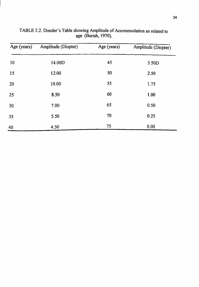
TABLE 2.2. Dander's Table showing Amplitude of Accommodation as related to age (Borish, 1970).
Age (years) Amplitude (Diopter) Age (years) Amplitude (Diopter)