Calcium Nephrolithiasis and Bone Health Noah S. Schenkman, MD Associate Professor of Urology and Residency Program Director, University of Virginia Health System; Charlottesville, Virginia Objectives: • Diagram the roles of vitamin D and Parathyroid hormone in calcium homeostasis • Describe the contribution of androgens and estrogens on osteoporosis and fractures in stone formers. • List 3 dietary interventions for stone formers with osteoporosis • Identify key pharmaceutical management of bone loss in stone formers including roles of thiazide diuretics and bisphosphonates
Transcript
Calcium Nephrolithiasis and Bone Health
Noah S. Schenkman, MD
Associate Professor of Urology and Residency Program Director, University of Virginia Health System;
Charlottesville, Virginia
Objectives: • Diagram the roles of vitamin D and Parathyroid hormone in
calcium homeostasis • Describe the contribution of androgens and estrogens on
osteoporosis and fractures in stone formers. • List 3 dietary interventions for stone formers with osteoporosis • Identify key pharmaceutical management of bone loss in stone
formers including roles of thiazide diuretics and bisphosphonates
Kidney Stones and Bone Health
Noah S. Schenkman, MD University of Virginia School of Medicine
1
1
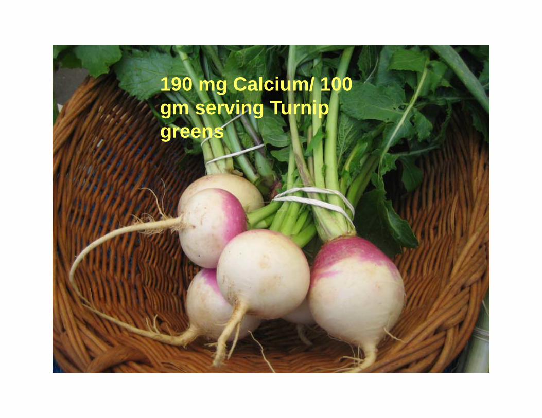
190 mg Calcium/ 100 gm serving Turnip greens
Are kidney stones an annoying episodic problem or manifestation of chronic metabolic disease? Are kidney stones deadly ? How do hormones play into all of this? What is the role of diet? Does giving dietary calcium to patients
with calcium stones make sense?
Osteoporosis
Most common bone disorder affecting humans compromised bone strength
increased risk of fracture
Osteoporosis
Peak bone mass: age 30 y/o in women Slow decline until menopause, then decline hastens. At age 80 , women have lost 30% of their bone mass 1st indication of disease is usually a fall with non-
vertebral fracture Marked height loss over the years may be sign of
underlying vertebral compression fracture.
7
Osteoporosis
White American women age 50: risk of osteoporotic fracture is 40% 2/3 of fx occur after age 75.
Hip fx: average age - 82 y/o : 25% increase in mortality in following year 25 % of women require long term care, 50%
have long term loss of mobility Avg. 3.8 yr. follow up RR for mortality was 6.7 for hip fx. 8.64 for vertebral fx.
Osteoporosis
Goal: reduce fracture risk Slow or stop bone loss or improve bone architecture and
strength 13-18 % of American women >50 y/o have osteoporosis of
the hip (less than 2.5 SD below the mean BMD of healthy,young, white women)
4% in age 50-59, 52% in age 80 or older Osteoporosis responsible for 90% of hip and spine fx in
white women age 65-84
Risk factors for osteoporosis
Idiopathic Hypercalciuria !
Bone Mineral Density
BMD: Factor of peak bone mineralization (age 30) and subsequent mineral loss T score most useful for postmenopausal
women: compare current BMD to mean BMD of
normal, young adult population of same gender
Management of osteoporosis in postmenopausal women: 2010 position statement of The North American Menopause Society.Menopause. 17(1):25-54, January 2010.
BMD-based definitions of bone density
Osteoporosis
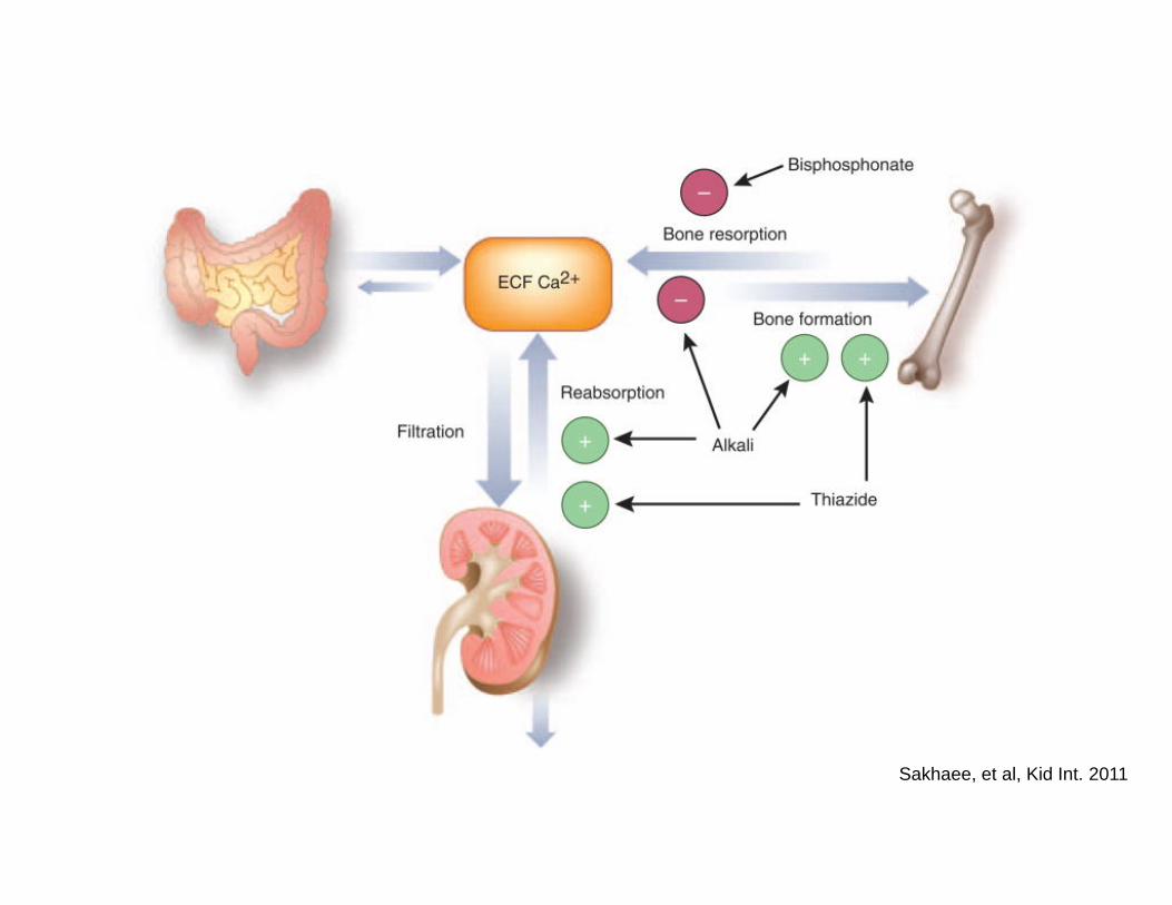
Calcium Homeostasis
Calcium Homeostasis
Parathyroid Hormone Regulates: kidney, bone , intestinal mucosa (indirect) Stimulated by decreases in serum calcium Inhibited by high serum calcium, elevated vit. D3
Effects in kidney: stimulates activation of vitamin D3 promotes calcium reabsorption suppresses tubular reabsorption of phosphate
Effect on bone: stimulates osteoclasts to breakdown apatite,
releasing calcium and phosphorus
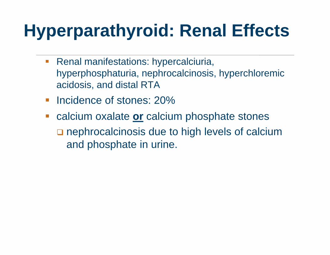
Hyperparathyroid May be responsible for stones in 2-8% of calcium stone
formers Hypercalcemia is hallmark of the disease Increased mobilization of calcium from bone leads to
osteoporosis Increased absorption from gut (PTH stimulated 1,25
OH D3 production) Increased tubular resorption of calcium in kidney


![Recurrent Nephrolithiasis in Adults: Comparative ......0.32 [CI, 0.14 to 0.74]). Strength of evidence for all these interventions was low. In patients with multiple past calcium stones,](https://static.documents.pub/doc/80x56/5f4064af5da64408a44e9dda/recurrent-nephrolithiasis-in-adults-comparative-032-ci-014-to-074.jpg)


