HOUNSFIELD REVIEW SERIES Cancer risks from diagnostic radiology E J HALL, DPhil, DSc, FACR, FRCR and D J BRENNER, PhD, DSc Center for Radiological Research, Columbia University Medical Center, New York, NY 10032, USA ABSTRACT. In recent years, there has been a rapid increase in the number of CT scans performed, both in the US and the UK, which has fuelled concern about the long-term consequences of these exposures, particularly in terms of cancer induction. Statistics from the US and the UK indicate a 20-fold and 12-fold increase, respectively, in CT usage over the past two decades, with per caput CT usage in the US being about five times that in the UK. In both countries, most of the collective dose from diagnostic radiology comes from high-dose (in the radiological context) procedures such as CT, interventional radiology and barium enemas; for these procedures, the relevant organ doses are in the range for which there is now direct credible epidemiological evidence of an excess risk of cancer, without the need to extrapolate risks from higher doses. Even for high-dose radiological procedures, the risk to the individual patient is small, so that the benefit/risk balance is generally in the patients’ favour. Concerns arise when CT examinations are used without a proven clinical rationale, when alternative modalities could be used with equal efficacy, or when CT scans are repeated unnecessarily. It has been estimated, at least in the US, that these scenarios account for up to one-third of all CT scans. A further issue is the increasing use of CT scans as a screening procedure in asymptomatic patients; at this time, the benefit/risk balance for any of the commonly suggested CT screening techniques has yet to be established. Received 15 November 2007 Revised 29 January 2008 Accepted 7 February 2008 DOI: 10.1259/bjr/01948454 ’ 2008 The British Institute of Radiology The use of X-rays as a diagnostic tool is so well established that it is hard to imagine contemporary medicine without them. At the same time, X-rays are a known and proven human carcinogen. It is the purpose of this review to address the benefit/risk balance associated with these two observations. Two findings have recently combined to spark concern over the long-term effects of diagnostic X-rays, particu- larly the induction of cancer. Firstly, as illustrated in Figure 1, CT usage over the past quarter of a century has risen ,12-fold in the UK and .20-fold in the US [1–3]. Current annual usage is estimated to be more than 3 million scans per year in the UK and more than 60 million per year in the US. Overall, the mean effective dose in the US from all medical X-rays has increased ,seven-fold over this period [4], with the result that medical exposures now represent, for the first time, the majority of the effective dose to which individuals in the US are exposed. These increases, driven in large part by the increases in CT usage, are a reflection of the fact that CT is such a rapid, simple and accurate diagnostic tool. Concerns arise because a CT scan results in organ radiation doses that are, typically, 100 times larger than those from conven- tional radiological procedures such as chest X-rays. The second recent development, as we shall discuss, is that there is now direct credible epidemiological evi- dence for a small risk of radiation-associated cancer at doses comparable to a few CT scans, or from other high- dose radiological procedures [5–8]. Indeed, as early as 2002, the International Commission on Radiological Protection (ICRP) commented that: ‘‘The absorbed dose to tissue from CT can often approach or exceed the levels known to increase the probability of cancer’’ [9]. Radiation exposure should always operate under the ‘‘As Low As Reasonably Achievable’’ (ALARA) principle and, as we discuss, opportunities do exist in the CT field for collective dose reduction, both by reducing the numbers of CT scans and by reducing the doses per scan. It is hoped that this review will promote ongoing dialogue [10] among radiologists, emergency room (ER) staff and other physicians, and indeed the public, as to practical ways to slow the increase in CT usage and CT doses, without compromising patient care. CT and its usage From its inception in the 1970s, the use of CT has increased rapidly in all developed countries, although usage rates vary greatly from country to country. In a survey from the mid-1990s, illustrated in Figure 2 [11], the number of CT scanners per million population was 64 in Japan, 26 in the US and 6 in the UK, the country where CT was invented. Figure 1 quantifies the increase in CT usage in the UK and the US over the past quarter of a century [3]. It is estimated that close to 3 million CT scans per year were Address correspondence to: E J Hall, Center for Radiological Research, Columbia University Medical Center, New York, NY 10032, USA. E-mail: [email protected]The British Journal of Radiology, 81 (2008), 362–378 362 The British Journal of Radiology, May 2008
Transcript
HOUNSFIELD REVIEW SERIES
Cancer risks from diagnostic radiology
E J HALL, DPhil, DSc, FACR, FRCR and D J BRENNER, PhD, DSc
Center for Radiological Research, Columbia University Medical Center, New York, NY 10032, USA
ABSTRACT. In recent years, there has been a rapid increase in the number of CT scansperformed, both in the US and the UK, which has fuelled concern about the long-termconsequences of these exposures, particularly in terms of cancer induction. Statisticsfrom the US and the UK indicate a 20-fold and 12-fold increase, respectively, in CTusage over the past two decades, with per caput CT usage in the US being about fivetimes that in the UK. In both countries, most of the collective dose from diagnosticradiology comes from high-dose (in the radiological context) procedures such as CT,interventional radiology and barium enemas; for these procedures, the relevant organdoses are in the range for which there is now direct credible epidemiological evidenceof an excess risk of cancer, without the need to extrapolate risks from higher doses.Even for high-dose radiological procedures, the risk to the individual patient is small, sothat the benefit/risk balance is generally in the patients’ favour. Concerns arise when CTexaminations are used without a proven clinical rationale, when alternative modalitiescould be used with equal efficacy, or when CT scans are repeated unnecessarily. It hasbeen estimated, at least in the US, that these scenarios account for up to one-third of allCT scans. A further issue is the increasing use of CT scans as a screening procedure inasymptomatic patients; at this time, the benefit/risk balance for any of the commonlysuggested CT screening techniques has yet to be established.
Received 15 November2007Revised 29 January 2008Accepted 7 February 2008
DOI: 10.1259/bjr/01948454
’ 2008 The British Institute of
Radiology
The use of X-rays as a diagnostic tool is so wellestablished that it is hard to imagine contemporarymedicine without them. At the same time, X-rays are aknown and proven human carcinogen. It is the purposeof this review to address the benefit/risk balanceassociated with these two observations.
Two findings have recently combined to spark concernover the long-term effects of diagnostic X-rays, particu-larly the induction of cancer. Firstly, as illustrated inFigure 1, CT usage over the past quarter of a century hasrisen ,12-fold in the UK and .20-fold in the US [1–3].Current annual usage is estimated to be more than3 million scans per year in the UK and more than60 million per year in the US. Overall, the mean effectivedose in the US from all medical X-rays has increased,seven-fold over this period [4], with the result thatmedical exposures now represent, for the first time, themajority of the effective dose to which individuals in theUS are exposed.
These increases, driven in large part by the increases inCT usage, are a reflection of the fact that CT is such a rapid,simple and accurate diagnostic tool. Concerns arisebecause a CT scan results in organ radiation doses thatare, typically, 100 times larger than those from conven-tional radiological procedures such as chest X-rays.
The second recent development, as we shall discuss, isthat there is now direct credible epidemiological evi-
dence for a small risk of radiation-associated cancer atdoses comparable to a few CT scans, or from other high-dose radiological procedures [5–8]. Indeed, as early as2002, the International Commission on RadiologicalProtection (ICRP) commented that: ‘‘The absorbed dose totissue from CT can often approach or exceed the levels knownto increase the probability of cancer’’ [9].
Radiation exposure should always operate under the‘‘As Low As Reasonably Achievable’’ (ALARA) principleand, as we discuss, opportunities do exist in the CT fieldfor collective dose reduction, both by reducing thenumbers of CT scans and by reducing the doses perscan. It is hoped that this review will promote ongoingdialogue [10] among radiologists, emergency room (ER)staff and other physicians, and indeed the public, as topractical ways to slow the increase in CT usage and CTdoses, without compromising patient care.
CT and its usage
From its inception in the 1970s, the use of CT hasincreased rapidly in all developed countries, althoughusage rates vary greatly from country to country. In asurvey from the mid-1990s, illustrated in Figure 2 [11],the number of CT scanners per million population was64 in Japan, 26 in the US and 6 in the UK, the countrywhere CT was invented.
Figure 1 quantifies the increase in CT usage in the UKand the US over the past quarter of a century [3]. It isestimated that close to 3 million CT scans per year were
Address correspondence to: E J Hall, Center for RadiologicalResearch, Columbia University Medical Center, New York, NY10032, USA. E-mail: [email protected]
The British Journal of Radiology, 81 (2008), 362–378
362 The British Journal of Radiology, May 2008
performed in the UK in 2005–2006, compared with0.25 million in 1980 [2, 7]. The corresponding figures forthe US are 69 million scans in 2007, compared with,2 million in 1980 [1, 7]. Taking into account the relativepopulations, the data indicate that the number of CTscans per person is five times greater in the US than inthe UK. There is perhaps a suggestion from the datain Figure 1 that the rate of increase in scans is slowing inthe US, but continuing to rise sharply in the UK.
A significant part of the UK increase is probably forpre-surgical diagnosis of acute appendicitis. For exam-ple, Dixon and Goldstone [12] report that UK radiologydepartments are currently experiencing a massiveincrease in requests for CT of the acute abdomen. Aparticular concern here, as discussed below, is thatappendicitis is largely a disease of young people [13], forwhom the radiation risks are correspondingly higher.
In 1997, the European Union issued a Directive on‘‘Health protection of individuals against the dangers of
ionizing radiation in relation to medical exposure’’ [14],from which followed corresponding UK regulations [15]and a detailed set of referral criteria guidelines [16]. Nocorresponding regulatory framework exists in the US,although the American College of Radiology has recentlypublished a valuable white paper on radiation dose inmedicine [17], which contains a series of recommenda-tions designed to slow the increase in US populationexposure from diagnostic radiology.
Organ doses produced by CT scans
Organ doses from CT examinations are considerablylarger than those from the corresponding conventionalradiograph (Table 1). For example, typical doses to thelung from a conventional chest X-ray range from about0.01 mGy to 0.15 mGy, whereas a typical dose to anorgan examined with CT, as discussed below, is around
Figure 1. Graphs illustrating therapid increase in the number of CTscans per year in (a) the UK and in(b) the US, as well as the number ofCT scans per person per year [1–3].Note that the number of scans perperson per year is about five timeslower in the UK than in the US.
Hounsfield Review Series: Cancer risks from diagnostic radiology
The British Journal of Radiology, May 2008 363
10 mGy to 20 mGy, and can be as high as 80 mGy for 64-slice CT coronary angiography.
The radiation doses to particular organs from anygiven CT scan depend upon a number of factors: themost important are the number of scans, the product oftube current and scan time (the ‘‘mAs’’), the patient size,the axial scan range, the scan pitch (the degree of overlapbetween adjacent CT ‘‘slices’’), the maximum tubevoltage (the kVp) and the particular scanner design[18]. Many of these parameters are under the control ofthe radiologist or radiographer, and ideally should betailored to the individual examination type and theindividual patient size, a practice that is increasing but isby no means universal [19]. It is always the case that therelative noise in CT images will increase as the radiationdose decreases, and so there will always be a trade-offbetween the need for low-noise images and the desir-ability to use low radiation doses [20].
Representative calculated organ doses from single CTscans are shown in Figure 3 for commonly used machinesettings [22] for either a single head scan or a singleabdominal scan, the two most common CT scans. Thenumber of CT scans in a given study is, of course, animportant factor in determining the dose. For example,Mettler et al [23] reported that almost all patients havingCT scans of the abdomen or pelvis had more than one CTscan on the same day; for all patients having CT scans,they reported that 30% had at least three scans, 7% hadmore than five scans, and 4% had nine or more CT scans.
It should also be borne in mind that doses associatedwith a given CT scan may vary considerably betweendifferent machines and institutions. For example, the USFood and Drug Administration conducted a survey of CThead scans in 203 facilities and found, as illustrated inFigure 4, that the institution-to-institution multiple-scanaverage dose varied by as much as a factor of 10 [24].
Radiation carcinogenesis at low doses
Data from atomic bomb survivors represent the ‘‘goldstandard’’ in the quantitative assessment of radiationcarcinogenesis risks at low doses. There are severalreasons for this:
1. The study involves a large non-selected population(,100 000), including all ages and both genders.
2. Approximately 30 000 of the survivors were exposedto low doses — specifically in the range of 5–100 mSv— which is roughly the relevant dose range for singleand multiple CT examinations.
3. Both cancer incidence and mortality data are avail-able.
4. Mortality follow-up is close to complete amongindividuals exposed as adults, and is more than 50%complete for exposed children.
5. The study has continued for 60 years (and isongoing), and has cost over 0.5 billion dollars. It is(hopefully) highly unlikely that any comparablestudy will ever be performed.
Two major conclusions from the A-bomb studies are,firstly, that the risk of all solid cancers is consistent witha linear increase in radiation dose, from low doses up to,2.5 Sv (Figure 5). The second major conclusion is thatchildren are much more radiosensitive than adults;indeed, there is a continuous decline in radiosensitivitywith age for most cancers (Figure 6). Lung cancer is anotable exception, with the radiation-associated relativerisk for lung cancer apparently increasing with age, up tomiddle age [6], implying that the absolute radiation-associated risk of lung cancer may not decreasesignificantly with age. The significance of this observa-tion for a variety of adult CT scans will be discussedbelow.
Radiation risks are reviewed at regular intervals by avariety of national and international organizations. Atthe international level, there are the ICRP and the UnitedNations Scientific Committee on the Effects of AtomicRadiation (UNSCEAR). At national levels, there are the
Table 1. Typical organ doses from various radiological examinations
Examination Relevant organ Relevant organ dose (mGy)
Figure 2. Number of CT scanners per million population inselected countries in the 1990s. Data from a 1991–1996survey reported by the United Nations Scientific Committeeon the Effects of Atomic Radiation (UNSCEAR) [11].
E J Hall and D J Brenner
364 The British Journal of Radiology, May 2008
UK Radiation Protection Division of the HealthProtection Agency and the US National Council onRadiological Protection and Measurements (NCRP), aswell as the US National Academy of Sciences BiologicalEffects of Ionizing Radiation (BEIR) committees. Thecurrent unanimous consensus of all of these bodies isthat, for doses ,100 mSv, the most appropriate riskmodel for radiation protection purposes is one in whichthe risk of radiation-induced stochastic effects, isparticular cancer induction, is assumed to decreaselinearly with decreasing dose with no threshold (theso-called ‘‘linear no-threshold’’ (LNT) model) [11, 25, 26].
This LNT hypothesis is often described as prudent andpossibly conservative, but it is certainly not proven.What can be said is that the measured cancer risks areconsistent with linearity. Not surprisingly, this hypoth-esis has been assailed on both sides — by those whobelieve that low radiation doses are more damaging thanlinearity (specifically, a linear extrapolation of risks fromhigher doses) predicts [27, 28], as well as by those who
believe that low radiation doses are less damaging thanlinearity would predict [29], or even that they arebeneficial [30].
In the present context, it is not necessary to take aposition on this LNT controversy. This is because the dosesinvolved in CT scans, interventional radiology and bariumenemas, which account for the majority of the collective
Figure 3. Estimated organ doses in mGy from typical singleCT scans of the (a) head and the (b) abdomen [3]. Asexpected, the main exposed organs are the brain for headCT, and the digestive organs for abdominal CT. As describedin the text, these doses depend on a variety of factors,including the number of scans (data here are for a singlescan) and the mAs setting. The data here refer to the medianmAs settings reported in the 2000 NEXT survey of CT usage[22, 24]. Note that, for a given mAs setting, paediatric dosesare much larger than adult doses, as there is less self-shielding, but mAs settings can be (but only sometimes are[19]) reduced for children, which proportionately reducesthe paediatric dose and the risk. The dose estimationmethodology used for these calculations has been describedelsewhere [38], but software to estimate organ doses forspecific ages and CT settings is now generally available [21].
Figure 4. During 2000–2001, the US Food and DrugAdministration conducted measurements in 203 institutions[24] to estimate the institution-to-institution variation inmultiple scan average doses (MSADs) involved in a no-contrast axial CT head scan. The frequency distribution ofresults, shown here, exhibits a 10-fold variation in the MSAD.
Figure 5. Estimated relative risks for cancer incidence in A-bomb survivors during the 195821994 follow-up periodrelative to controls [5]. The dotted curves represent ¡1standard error for the smoothed curve. The inset shows dataover the whole dose range of 0–2 Sv, to which a straight lineis fitted, i.e. the relative risk is proportional to the dose, withno threshold. The main figure is an expanded version of thelow-dose region up to 0.5 Sv. The straight line is taken fromthe inset data for the whole dose range. Because of anapparent distinction between distal and proximal zero-dosecancer rates, the unity baseline corresponds to zero-dosesurvivors within 3 km of the bombs. The dashed linerepresents the alternative baseline if the distal survivorswere not omitted.
Hounsfield Review Series: Cancer risks from diagnostic radiology
The British Journal of Radiology, May 2008 365
dose from diagnostic radiology, are just within the rangewhere we have credible and direct epidemiological evi-dence for an increased cancer risk in human populations [5–8]. By contrast, for example, a single-plane chestX-ray results in a maximum organ dose of less than0.2 mGy (see Table 1), which is much lower than thesmallest dose for which significant epidemiological data areavailable. Estimating the risk for very low dose proceduresdoes indeed involve a significant extrapolation, over asmuch as two orders of magnitude of dose, and is the subjectof much controversy; however, it is not directly relevant tothe higher radiological doses of interest in this review.
Limitations of epidemiological radiation-attributable cancer risk data
Report 126 from the NCRP [8] addressed the questionof uncertainties in the total fatal cancer risk estimatesused in radiation protection. The report consideredepidemiological uncertainties, dosimetric uncertainties,transfer of risk between populations, projections to alifetime risk, and extrapolation to low dose and/or lowdose-rate. The results suggest an overall uncertainty ofapproximately a factor of 3 below and above theestimated value. By far the biggest source of uncertaintyinvolves the extrapolation to low doses and the applica-tion of a dose-rate effectiveness factor. This uncertaintywas estimated to be a factor of 2–2.5, but much of thiscomponent of the uncertainty may not apply to the dosesinvolved in CT, in that we do not need to significantlyextrapolate risks to lower doses or dose rates.Epidemiology-based uncertainties were estimated at¡25%, dosimetric uncertainties at 0230%, transferbetween populations at 230% to +65%, and projectionsto a lifetime risk at 250% to +10%.
A limitation of the Japanese A-bomb data that mustalways be kept in mind is that the cohort size is large
(,100 000 individuals), but not infinitely large. Thus,stratification of the results, e.g. by age, results in a markeddecrease in statistical power. As such, when investigatingthe variation of radiosensitivity with age, all doses must beused, and when investigating the lowest dose for which asignificant excess cancer risk is evident, all ages must beused. Thus, it is probably not possible to answer somedetailed questions, such as ‘‘what is the lowest dose atwhich an excess cancer incidence is evident in children lessthan 10 years of age?’’.
What is the lowest dose for which cancer excess hasbeen demonstrated?
A-bomb survivorsSeveral analyses have addressed the question of the
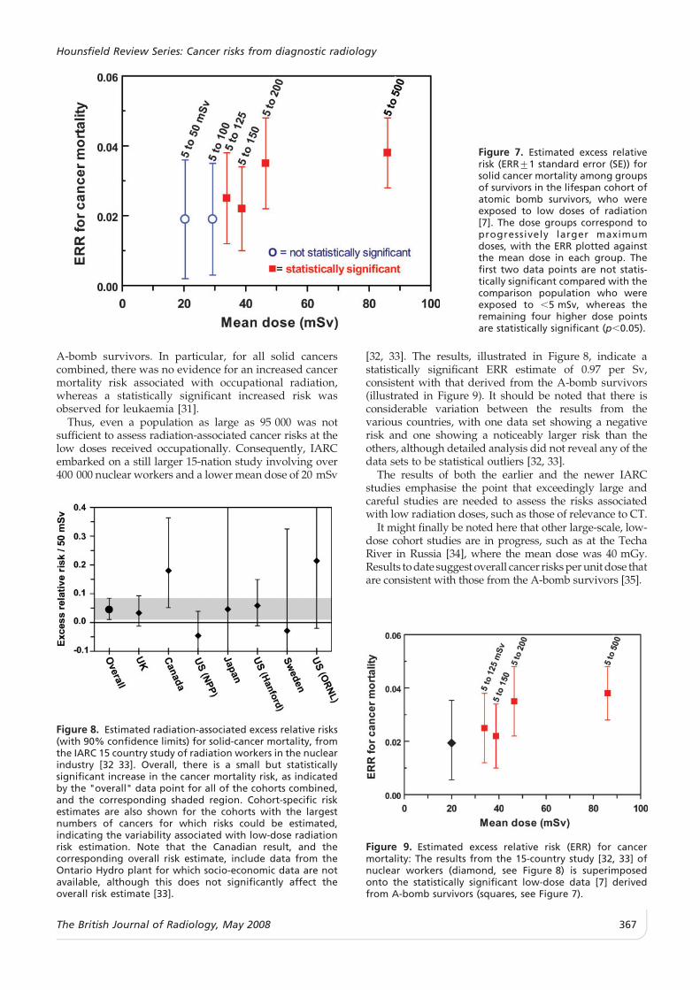
lowest dose for which a statistically significant increasein cancer risk is apparent [6, 7], with the caveat, asdiscussed above, that age-averaged data must be usedfor the analysis. A summary of the conclusions [7] isshown in Figure 7. The survivors are stratified intoprogressively larger dose bins, with the lowest being 5–50 mSv; the excess relative risk (ERR) is then plotted as afunction of the mean dose. The mean dose in the lowestdose bin at which the ERR is statistically significant is,35 mSv, which corresponds to the typical maximumorgan dose from two or three CT scans.
Nuclear workersIn 1995, the International Agency for Research on
Cancer (IARC) published the results of a large studyinvolving over 95 000 nuclear industry workers in theUS, UK and Canada, who received an average occupa-tional dose of ,40 mSv [31]. Because the confidenceintervals were wide, the results were consistent bothwith zero risk and with the risk based on analysis of the
Figure 6. Estimated attributablelifetime risk from a single small doseof radiation as a function of age atexposure [74]. Note the dramaticdecrease in radiosensitivity withage. The higher risk for the youngerage groups is not expressed untillate in life.
E J Hall and D J Brenner
366 The British Journal of Radiology, May 2008
A-bomb survivors. In particular, for all solid cancerscombined, there was no evidence for an increased cancermortality risk associated with occupational radiation,whereas a statistically significant increased risk wasobserved for leukaemia [31].
Thus, even a population as large as 95 000 was notsufficient to assess radiation-associated cancer risks at thelow doses received occupationally. Consequently, IARCembarked on a still larger 15-nation study involving over400 000 nuclear workers and a lower mean dose of 20 mSv
[32, 33]. The results, illustrated in Figure 8, indicate astatistically significant ERR estimate of 0.97 per Sv,consistent with that derived from the A-bomb survivors(illustrated in Figure 9). It should be noted that there isconsiderable variation between the results from thevarious countries, with one data set showing a negativerisk and one showing a noticeably larger risk than theothers, although detailed analysis did not reveal any of thedata sets to be statistical outliers [32, 33].
The results of both the earlier and the newer IARCstudies emphasise the point that exceedingly large andcareful studies are needed to assess the risks associatedwith low radiation doses, such as those of relevance to CT.
It might finally be noted here that other large-scale, low-dose cohort studies are in progress, such as at the TechaRiver in Russia [34], where the mean dose was 40 mGy.Results to date suggest overall cancer risks per unit dose thatare consistent with those from the A-bomb survivors [35].
Figure 7. Estimated excess relativerisk (ERR¡1 standard error (SE)) forsolid cancer mortality among groupsof survivors in the lifespan cohort ofatomic bomb survivors, who wereexposed to low doses of radiation[7]. The dose groups correspond toprogressively larger maximumdoses, with the ERR plotted againstthe mean dose in each group. Thefirst two data points are not statis-tically significant compared with thecomparison population who wereexposed to ,5 mSv, whereas theremaining four higher dose pointsare statistically significant (p,0.05).
Figure 8. Estimated radiation-associated excess relative risks(with 90% confidence limits) for solid-cancer mortality, fromthe IARC 15 country study of radiation workers in the nuclearindustry [32 33]. Overall, there is a small but statisticallysignificant increase in the cancer mortality risk, as indicatedby the "overall" data point for all of the cohorts combined,and the corresponding shaded region. Cohort-specific riskestimates are also shown for the cohorts with the largestnumbers of cancers for which risks could be estimated,indicating the variability associated with low-dose radiationrisk estimation. Note that the Canadian result, and thecorresponding overall risk estimate, include data from theOntario Hydro plant for which socio-economic data are notavailable, although this does not significantly affect theoverall risk estimate [33].
Figure 9. Estimated excess relative risk (ERR) for cancermortality: The results from the 15-country study [32, 33] ofnuclear workers (diamond, see Figure 8) is superimposedonto the statistically significant low-dose data [7] derivedfrom A-bomb survivors (squares, see Figure 7).
Hounsfield Review Series: Cancer risks from diagnostic radiology
The British Journal of Radiology, May 2008 367
Methodologies for assessing the potentialrisks associated with high-dose radiologicalexaminations
The potential health risk associated with a high dose (ina radiological context) examination can be quantitativelyassessed in several different ways. One simple approachuses the effective dose concept [36], which represents anattempt to provide a single number that is proportional tothe radiobiological ‘‘detriment’’ from a particular radiationexposure — with detriment representing a balancebetween carcinogenesis, life shortening and hereditaryeffects. Specifically, it is the sum of the equivalent doses toa number of radiosensitive organ/tissues, with each organbeing weighted by a tissue-specific committee-determinedweighting factor. The effective dose is commonly used inradiology to allow comparisons of the risks associatedwith different spatial dose distributions produced bydifferent imaging techniques. If the effective doses to all ofthe involved individuals are summed, the result is thecollective dose [37]. If the collective dose is then multipliedby a generic fatal cancer risk estimate for whole-bodyexposure (e.g. 5% Sv21 from ICRP Publication 60 [25]), theresult is a very crude estimate of the number of fatalcancers resulting from the procedure.
Such risk estimates based on effective dose andcollective dose are crude for two reasons:
1. Collective dose from radiological procedures includescontributions from the many low-dose procedures,such as routine chest X-rays and mammograms, whichinvolve doses far below those for which we have directevidence of cancer induction, i.e. it assumes validity ofthe LNT hypothesis down to the lowest doses.However, as we discuss below, most of the collectivedose is actually from high-dose procedures (e.g. CT,interventional radiology and barium enema).
2. Risk estimates based on effective dose are highlygeneric and include, for example, hereditary effectsthat are unlikely to be significant at doses relevant todiagnostic radiology. In addition, the weightingfactors used in the calculation of effective dose donot take into account the strong variations of radio-sensitivity with age and gender.
In the next section, we use the collective dose concept tomake some generic estimates of cancer risks fromdiagnostic radiology in the UK. Although we make arough correction by excluding low-dose procedures, therisk estimates for which would be highly speculative, theresults are still highly generic and, in the followingsections, more reliable risk estimates are described. Inparticular, rather than using effective dose and collectivedose, with all their inherent assumptions, a potentiallymore satisfactory method to assess the risk associated witha high-dose (in the radiological context) examination isfirst to measure (in an anthropomorphic phantom) orcalculate (using Monte Carlo techniques) individual organdoses. Given these organ doses, risks can be applied(ultimately derived from A-bomb survivors) that are dosespecific, organ specific, age specific, gender specific andcountry specific; finally, the resulting organ-specific riskscan be summed. Such an approach to CT risk estimationhas been used by several groups [3, 38–40].
Radiology in the UK, 2005–2006: generic riskestimates
Figure 10 shows the increase with time of the UKcollective dose from radiology, restricted to the threemajor radiological procedures (CT, interventional radi-ology and barium enema) where organ doses aresufficiently high that there is plausible direct evidencefrom epidemiological studies (see above) of an increasedcancer risk. For the time period 2005–2006, this restrictedcollective dose is ,21 600 man Sv per year, based on thenumber of procedures [1] and the average effective doseper procedure [41]. If all radiological procedures wereincluded, the corresponding collective dose would be,28 000 man Sv per year (thus, approximately three-quarters of the total radiology collective dose is fromhigh-dose procedures).
CT contributes the bulk of the collective dose, butinterventional radiology is also very much a growth area,both in the US and the UK. In 2006, as illustrated inFigure 10, interventional radiology in the UK accountedfor ,11% of the high-dose collective dose from radiologyprocedures [1, 41]. Although it represents a muchsmaller contribution to the collective dose than CT, it isgrowing just as rapidly. In fact, some patients under-going interventional radiology receive doses sufficientlyhigh as to cause deterministic effects in the skin, fromerythema to desquamation and, very occasionally, evennecrosis [42–45]. A mitigating factor is that most patientsreceiving interventional radiology are older and suffer-ing from life-threatening illnesses, so that the radiationrisks must be viewed in a broader context.
Barium enemas involve doses, and therefore risks, thatare comparable to CT. The number of barium enemasperformed is not increasing as rapidly as CT andinterventional radiology, so that it represents a decliningproportion of the collective dose.
With all the caveats discussed above, by applying therisk factor for fatal cancer suggested by the ICRP of5% Sv21 [37], a collective dose of 28 000 man Sv impliesthat the practice of diagnostic radiology in the UK wouldbe predicted to result in 5/100 6 28 000 or 1400 fatalcancers per year. If only high-dose (in the radiologicalcontext) procedures are included, the rationale for whichis described above, the corresponding predicted number
Figure 10. Diagram illustrating the recent increase in the UKcollective dose from high-dose radiological procedures. Onlythe main high-dose diagnostic procedures are included, i.e.CT, interventional radiology (INT) and barium enemas. Thenumber of procedures in each category was obtained fromthe UK Department of Health KH12 returns [2], and theaverage effective doses per procedure from Hart and Wall[41].
E J Hall and D J Brenner
368 The British Journal of Radiology, May 2008
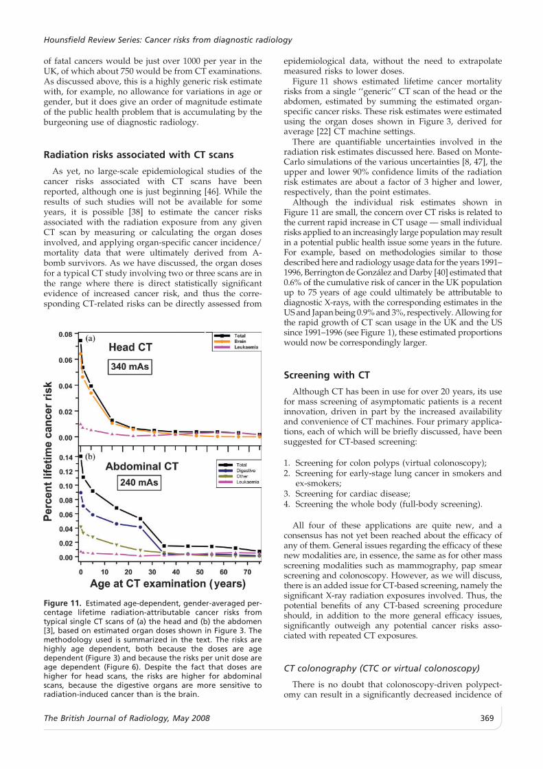
of fatal cancers would be just over 1000 per year in theUK, of which about 750 would be from CT examinations.As discussed above, this is a highly generic risk estimatewith, for example, no allowance for variations in age orgender, but it does give an order of magnitude estimateof the public health problem that is accumulating by theburgeoning use of diagnostic radiology.
Radiation risks associated with CT scans
As yet, no large-scale epidemiological studies of thecancer risks associated with CT scans have beenreported, although one is just beginning [46]. While theresults of such studies will not be available for someyears, it is possible [38] to estimate the cancer risksassociated with the radiation exposure from any givenCT scan by measuring or calculating the organ dosesinvolved, and applying organ-specific cancer incidence/mortality data that were ultimately derived from A-bomb survivors. As we have discussed, the organ dosesfor a typical CT study involving two or three scans are inthe range where there is direct statistically significantevidence of increased cancer risk, and thus the corre-sponding CT-related risks can be directly assessed from
epidemiological data, without the need to extrapolatemeasured risks to lower doses.
Figure 11 shows estimated lifetime cancer mortalityrisks from a single ‘‘generic’’ CT scan of the head or theabdomen, estimated by summing the estimated organ-specific cancer risks. These risk estimates were estimatedusing the organ doses shown in Figure 3, derived foraverage [22] CT machine settings.
There are quantifiable uncertainties involved in theradiation risk estimates discussed here. Based on Monte-Carlo simulations of the various uncertainties [8, 47], theupper and lower 90% confidence limits of the radiationrisk estimates are about a factor of 3 higher and lower,respectively, than the point estimates.
Although the individual risk estimates shown inFigure 11 are small, the concern over CT risks is related tothe current rapid increase in CT usage — small individualrisks applied to an increasingly large population may resultin a potential public health issue some years in the future.For example, based on methodologies similar to thosedescribed here and radiology usage data for the years 1991–1996, Berrington de Gonzalez and Darby [40] estimated that0.6% of the cumulative risk of cancer in the UK populationup to 75 years of age could ultimately be attributable todiagnostic X-rays, with the corresponding estimates in theUS and Japan being 0.9% and 3%, respectively. Allowing forthe rapid growth of CT scan usage in the UK and the USsince 1991–1996 (see Figure 1), these estimated proportionswould now be correspondingly larger.
Screening with CT
Although CT has been in use for over 20 years, its usefor mass screening of asymptomatic patients is a recentinnovation, driven in part by the increased availabilityand convenience of CT machines. Four primary applica-tions, each of which will be briefly discussed, have beensuggested for CT-based screening:
1. Screening for colon polyps (virtual colonoscopy);2. Screening for early-stage lung cancer in smokers and
ex-smokers;3. Screening for cardiac disease;4. Screening the whole body (full-body screening).
All four of these applications are quite new, and aconsensus has not yet been reached about the efficacy ofany of them. General issues regarding the efficacy of thesenew modalities are, in essence, the same as for other massscreening modalities such as mammography, pap smearscreening and colonoscopy. However, as we will discuss,there is an added issue for CT-based screening, namely thesignificant X-ray radiation exposures involved. Thus, thepotential benefits of any CT-based screening procedureshould, in addition to the more general efficacy issues,significantly outweigh any potential cancer risks asso-ciated with repeated CT exposures.
CT colonography (CTC or virtual colonoscopy)
There is no doubt that colonoscopy-driven polypect-omy can result in a significantly decreased incidence of
Figure 11. Estimated age-dependent, gender-averaged per-centage lifetime radiation-attributable cancer risks fromtypical single CT scans of (a) the head and (b) the abdomen[3], based on estimated organ doses shown in Figure 3. Themethodology used is summarized in the text. The risks arehighly age dependent, both because the doses are agedependent (Figure 3) and because the risks per unit dose areage dependent (Figure 6). Despite the fact that doses arehigher for head scans, the risks are higher for abdominalscans, because the digestive organs are more sensitive toradiation-induced cancer than is the brain.
Hounsfield Review Series: Cancer risks from diagnostic radiology
The British Journal of Radiology, May 2008 369
colorectal cancer [48], and that there is suboptimalcompliance with current guidelines for colorectal cancerscreening [49]. Screening using CT colonography, oftenreferred to as CTC or ‘‘virtual colonoscopy’’, wassuggested as early as 1983 [50], but has only recentlybecome a potential option for mass screening [51, 52].
Several recent large-scale studies have shown that CTCis at least as sensitive and specific as conventional opticalcolonoscopy in detecting adenomas of diameter >10 mm[53, 54] — a result confirmed by preliminary results of theNational US CT Colonography Trial [55]. CTC may wellhave the potential to increase colorectal cancer screeningcompliance, in part because of the possibility that it can beperformed with reduced laxative [56, 57] or non-cathartic[58, 59] pre-examination bowel preparation.
It is clear that CTC, at least in the US, is reasonablyclose to being used for mass screening, although it is notyet approved for most US third-party reimbursements.An issue that confronts CTC is its reduced sensitivityand specificity for detecting lesions ,10 mm, althoughlesions smaller than this typically have no more than a1% chance of containing a frank malignancy. Anotherissue is the relatively early developmental stage of non-cathartic or minimally cathartic protocols, with standar-dized approaches still to be established.
If CTC were to become a standard screening tool forthe over-50s, the potential ‘‘market’’ would be ,100 mil-lion people in the US and 20 million in the UK. Even ifthe recommended CTC frequency were to be thatcurrently recommended for optical colonoscopy (everydecade), this would imply that millions of CTC scansmight be performed each year. It is pertinent, therefore,to consider the radiation exposure and any potentialradiation risk to the population from such a massscreening programme. Because of the advantageousgeometry of a CTC scan, the dose/noise trade-off canbe very much weighted towards low-dose higher-noiseimages, while still maintaining sensitivity and specificity,at least for polyps .10 mm in diameter [60–64].
Table 2 [65] shows estimated CTC organ doses for oneof the more common CT scanners, with typical scannerparameters. To provide an estimate of scanner-to-scanner dose variations, Table 3 [65] shows the radiationdose to the colon estimated for five of the more commonCT scanners in current use, using identical scanner
parameters in each case; the coefficient of variation of thedose to the colon is ,20%.
Table 2 also shows the estimated absolute lifetimecancers risks associated with the radiation exposure frompaired CTC scans in a 50-year-old individual [65]. Asexpected, the main organs at risk are the colon, stomachand bladder, as well as the leukaemic cancers. All of theestimated absolute radiation risks are relatively small,with the largest being ,0.05% (1 in 2000). Summed overall of the organs at risk, the estimated absolute lifetimerisk of cancer induction from a pair of CTC scans (withthe scanner parameters from Table 2) in a 50-year old is,0.14%, approximately 1 in 700. Estimated risks forcancer mortality would, of course, be less.
The estimated risks are, of course, dependent upon thescanner settings used, particularly the mAs and the pitch.There is good evidence [62, 66] to suggest that the mAs andthus the dose for CTC could be decreased considerablyfurther. In addition, automatic tube current modulation,discussed elsewhere in this review, has been shown toreduce CTC doses by a further 35% [67]. Thus, it seemsclear that, in terms of the radiation exposure, the benefit/risk ratio is potentially large for virtual colonoscopy.
Low-dose CT screening for early-stage lung cancerin smokers and ex-smokers
Lung cancer is the leading cause of cancer-relatedmortality and is, of course, strongly associated with pastsmoking history. Thus, there is much interest in using low-dose CT scans for the regular screening of smokers andformer smokers for early-stage lung cancer. This is alogical next step after the failure of earlier attempts toscreen this population with conventional chest X-rays [68];low-dose lung CT clearly has a much greater sensitivity fordetecting small pulmonary lesions than does conventionalradiography [69]. A National Lung Cancer Screening Trialis currently underway in the US [70].
As with virtual colonoscopy, the geometry for lung CTis quite advantageous, and this allows the use of relativelylow-dose (i.e. noisier) images, while still maintaining goodsensitivity for detecting small lesions [71].
The potential mortality benefits of lung cancer screen-ing have been much debated [72, 73], and it is fair to saythat, at the very earliest, the issue will not be resolveduntil the completion of the National Lung Cancer
Table 2. Typical organ doses and estimated additionalabsolute gender-averaged lifetime cancer risks associatedwith a paired CTC screening examination of a healthy 50 yearold [65].
Organ dose frompaired CTC scansa
(mGy)
Additional absolute lifetimecancer risk from paired CTCscans at age 50 years (%)
Screening Trial in 2009. Less attention has been paid tothe potential radiation risks, specifically radiation-induced lung cancer, associated with the radiation fromthese CT scans. In part, this is because the screeningtechnique involves ‘‘low dose’’, rather than standard, CTlung scans, and partly because ERRs of radiation-induced cancer generally decrease markedly withincreasing age [74].
There are, however, indications that the radiation riskto the lung associated with this screening technique maybe significant. Firstly, cancer risks from radiation aregenerally multiplicative of the background cancer risk[75], which is of course high for lung cancer in smokers;this general observation has been borne out in terms ofthe interaction between radiation and smoking, whichmost authors have suggested is near-multiplicative [76–79], although an intermediate interaction betweenadditive and multiplicative has also been suggested forradon exposure [80], and there is one suggestion of anadditive interaction [81] in A-bomb survivors. Secondly,although ERRs for cancer generally decrease markedlywith increasing age at exposure, radiation-induced lungcancer does not show this decrease in ERR withincreasing age [6].
These considerations suggest that the risk of radiation-induced lung cancer associated with the radiation fromrepeated low-dose CT scans of the lung in smokers maynot be negligible. A recent estimate [82], based on theorgan-specific risk estimation techniques describedabove, suggests that a 50-year-old smoker planning anannual lung screening CT would incur an estimatedradiation-related lifetime lung cancer risk of 0.5%, inaddition to his/her otherwise expected lung cancer riskof ,14% (the radiation-associated cancer risk to anyother organ is far lower). The estimated radiation risk seta baseline of benefit that annual CT screening mustsubstantially exceed. This risk/benefit analysis suggeststhat a reduction in mortality from annual CT screening ofmore than 3% would be necessary to outweigh thepotential radiation risks [82].
CT-based cardiac screening for heart disease
Since the introduction of Agatston’s scoring system[83] for quantifying artery calcium levels, there has beenincreasing interest in using CT as a screening test forcardiovascular risk [84–86]. A variety of studies hassuggested that coronary artery calcium might indeed bea good predictor of cardiovascular events such as acutemyocardial infarction, coronary revascularization andsudden death [87–90]. These results have contributed tothe SHAPE (Screening for Heart Attack Prevention andEducation) task force call for non-invasive screening,either with CT or ultrasound, of all asymptomatic men45–75 years of age and asymptomatic women 55–75 years of age (except those defined as very low risk)to detect individuals with sub-clinical atherosclerosis[91]. In the US, this amounts to 61 million people, and inthe UK to approximately 12 million people.
Neither the sensitivity nor the specificity of CT-basedcalcium screening has yet been well established [92, 93].In particular, many dangerous patches of arterial diseaseare not yet calcified, and so would be missed, leading to
decreased sensitivity; furthermore, many calcifiedarteries will have normal blood flow, leading todecreased specificity.
Because of its rapid motion, CT screening of the heartpresents special problems. In particular, information canonly be obtained when the heart is relatively still, i.e. indiastole. Typically, this is done using retrospective gatedtechniques, so that the dose delivered in other parts ofthe heart’s cycle is effectively wasted, leading to highorgan doses, particularly to the lung and breast [39, 94].For adults aged over 45 years, it would be expected thatthe lung risks would considerably outweigh any risks tothe breast [74]. Assuming the SHAPE recommendationsfor screening all asymptomatic men and women aged45–75 years and 55–75 years, respectively, Table 4 showsestimates of the predicted radiation-associated lungcancer mortality if all of these 61 million people in theUS were screened with multi-detector row CT once,involving a lung dose of 10 mGy [94]. The total predictedmortality is ,7000, or about 1 in 8000.
As discussed elsewhere in this review, the use ofprospective electrocardiogram-triggered coronary CT,where the machine is off during other parts of thecardiac cycle, has the potential to reduce the dose andtherefore the risk significantly, perhaps by a factor of 4[95]. Thus, the radiation concerns will be significantlyreduced if CT were to become a realistic option.
Full-body CT screening
There has been a recent wave of interest in the use offull-body CT screening of non-symptomatic adults [96–99]. The technique is intended to be an early detectiondevice for a variety of diseases including lung cancer,coronary artery disease and colon cancer. At present, theevidence for the utility of this technique is anecdotal, andthere is considerable controversy [100] regarding itsefficacy; to date, no studies have reported a life-prolonging benefit. Because of the nature of the scan,the false-positive rate is expected to be high, and a studyon full-body CT screening [101] found that 37% of thosescreened were recommended for further evaluation,whereas the overall evaluable disease prevalence isprobably ,2% [102].
Another aspect that is important in assessing full-bodyscreening is the potential risk from the radiationexposure associated with full-body CT scans. Typicalorgan doses from a single full body scan are ,9 mGy tothe lung, 8 mGy to the digestive organs and 6 mGy tothe bone marrow [103]. The effective dose is ,7 mSv,and therefore if, for example, five such scans wereundertaken in a lifetime, the effective dose would be,35 mSv. To put these doses into perspective, a typicalscreening mammogram produces a dose of ,2.6 mGy tothe breast [104], with a corresponding effective dose of,0.13 mSv. Based on the risk estimation methodologiesdescribed above, the estimated lifetime cancer mortalityrisks from a single full-body scan are ,4.5 6 1024
(about 1 in 2200) for a 45 year old and ,3.3 6 1024
(about 1 in 3000) for a 65 year old [103]. The riskestimates for multiple scans, which would be necessary iffull-body CT screening was to become a useful screeningtool, are correspondingly larger. For example, a 45 year
Hounsfield Review Series: Cancer risks from diagnostic radiology
The British Journal of Radiology, May 2008 371
old who plans on undergoing 10 three-yearly full-bodyscans would potentially accrue an estimated lifetimecancer mortality risk of 0.33% (about 1 in 300) [103].
The issue of whole-body screening has recently beenaddressed by the UK Committee on Medical Aspects ofRadiation in the Environment (COMARE) [105]. Theyconcluded that ‘‘there is little evidence that demonstrates, forwhole body CT scanning, the benefit outweighs the detriment.We recommend therefore that services offering whole body CTscanning of asymptomatic individuals should stop doing soimmediately’’.
Can CT doses be reduced?
The short answer is yes. There are a variety of CTparameters that can be optimized in order to deliver aminimum dose while obtaining the desired information,and there is much published research in this area [106,107]. In particular, mAS, filtration, collimation and peaktube voltage can all be optimized.
Much interest has focused on automated exposurecontrol. In general, exposure control is based on thenotion that lower CT image noise will typically beachieved at the cost of higher doses, so the image noiselevel should be no better than sufficient for thediagnostic task at hand. Given a desired noise leveland the geometry of the patient, either manual [108] orautomated [106, 107, 109, 110] exposure control techni-ques can be used to generate a CT setting that willminimize the patient dose.
All of the major CT scanner manufacturers now offersome type of automated exposure control, in which theuser defines the desired image quality, resulting inmachine-recommended settings [106]. The CT controlsystem can then adjust the tube current according to thepatient’s size, and can also optionally adjust the tubecurrent continuously during a given rotation and/orduring movement along the z-axis, according to thepatient’s size and body habitus, to produce an imageconsistent with the image quality requirements.
Patient size is a particularly important issue. It hasbeen known for many years that, for the same imagequality requirement, smaller (e.g. paediatric) patientsrequire lower mAs settings [111]; however, for many
years, paediatric CT was often performed with the samesettings as adult CT [19]. Automated and semi-auto-mated exposure-control systems, as well as increasedphysician awareness, have resulted in significantimprovements in this regard.
Finally, one area in which much technologicalimprovement has recently occurred is CT coronaryangiography. Because of cardiac motion, cardiac CThas generally been retrospectively gated, obtaininguseful information only during diastole and resultingin unnecessary exposure throughout the rest of the heartcycle [39]. Prospective electrocardiogram-triggered 64-slice helical CT, in which CT is only ‘‘on’’ duringdiastole, can result in a sharp decrease in radiation dose[95].
Can CT usage be reduced?
Irrespective of the absolute levels of CT-associatedrisk, it is clearly desirable to reduce CT usage, as long aspatient care is not compromised. However, this will notbe an easy task. Physicians are often subject to significantpressures (some country specific) from the medicalsystem, the medico-legal system and from the public toprescribe CT. As we have discussed, in most (non-screening) scenarios, CT is the appropriate choice, butthere is undoubtedly a significant proportion of potentialsituations where CT is not medically justifiable or whereequally effective alternatives exist.
Tellingly, a straw poll [112] of paediatric radiologistssuggested that perhaps one-third of CT exams could bereplaced by alternative approaches, or not performed at all[113]. Examples include the use of CT, or the use ofmultiple CT scans, for the management of blunt trauma[114–118], seizures [119, 120] and chronic headaches [121].
There is also a variety of scenarios in which CT usagecould be replaced by other imaging modalities, withoutsignificant loss of efficacy. For example, patients with ahistory of nephrolithiasis and flank pain, or with knownchronic kidney stones, are at increased risk for multipleCT exams, resulting in potentially high cumulativedoses. In such cases, combinations of sonography andunenhanced abdominal radiography (kidneys, uretersand bladder) would be an appropriate alternative to
Table 4. Estimated radiation-associated mortality risks for CT cardiac screening
Estimates are for lung cancer mortality, which is expected to dominate the risk. The risk estimates assume the Screening forHeart Attack Prevention and Education (SHAPE) [91] guidelines, calling for screening of all asymptomatic women 55–75 yearsof age (left four columns) and asymptomatic men 45–75 years of age (right four columns). It was assumed that each of the61 million individuals in this age group in the US receives one multidetector row CT for calcium scoring, with a typical lungdose of 10 mGy [94]. The total predicted lung cancer deaths is ,7000 out of the 61 million population.
E J Hall and D J Brenner
372 The British Journal of Radiology, May 2008
multiple CT scans [122–124]. Another example is the useof CT in screening for abdominal aortic aneurysm inpatients at risk; although CT is an excellent solution,several ultrasound-based devices have been shown to beequally effective and practical to use in an ER situation[125, 126].
A third area is the use of CT as a primary tool for pre-surgical diagnosis of acute appendicitis [127]. CT islargely replacing ultrasound for this purpose [128], andhas a very high sensitivity and specificity for diagnosingappendicitis. A particular issue here is that appendicitisis predominantly a young person’s disease [13], and sothe radiation risks per unit dose are higher than foradults. Several recent reports [129, 130] have highlightedthe utility and practicality of clinical practice guidelinesfor diagnosing paediatric appendicitis, using selectiveCT and ultrasound scans. Specifically, the guidelinesrecommend immediate surgery or further evaluationwith either CT or ultrasound depending on the patient’sspecific clinical presentation. Selective imaging guide-lines for paediatric appendicitis have been shown todecrease markedly the number of CT scans performed(by a reported 40% [129]) with minimal diminution indiagnostic accuracy.
Beyond these clinical issues, however, a problem ariseswhen CT scans are requested in the practice of defensivemedicine, or when a CT scan, justified in itself, isrepeated as the patient passes through the medicalsystem, often simply because of a lack of communication.It is possible that the wider use of electronic radiologyinformation systems and patient records will reduce thisproblem in the future.
Part of the issue is that physicians often view CTexams in the same light as other radiological procedures,despite the fact that CT-related doses are typically muchhigher. In a recent survey of radiologists and ERphysicians [131], about three-quarters of physicianssignificantly underestimated the radiation dose from aCT scan, whereas 53% of radiologists and 91% of ERphysicians did not believe that CT scans increased cancerrisks.
This concern is encapsulated by an Editorial commentregarding CT angiography [132], but which appliesequally well to many CT applications: ‘‘due to its easieravailability, CT of the pulmonary arteries may, however, beused more liberally in patients with low clinical suspicion’’.This trend towards a somewhat less selective use ofdiagnostic CT, for better or worse, has occurred in manydifferent applications of CT, and is in considerable partresponsible for the rapid increases in CT use.
Understanding, using and communicating CTrisk estimates
In 1983, the Royal Society introduced a usefulstratification of risks [133]. They proposed that a risk ofone in a million is acceptable as part of everyday lifeactivities such as commercial air travel. Conversely, anannual risk of 1 in 100, e.g. that associated with coalmining in the 19th century, was considered unaccepta-ble. Between these extremes, a risk of 1 in 1000 (whichcorresponds approximately to an abdominal CT in achild) was considered acceptable, provided:
1. the individual receives a potential benefit that out-weighs the potential risk;
2. everything possible has been done to reduce orminimize the risk;
3. the individual or parent is aware of the risk.
We discuss the first two points elsewhere in thisreview. The risk/benefit balance, which is well estab-lished as being highly favourable in the majority,although not all, of diagnostic CT examinations, iscurrently far less well established for CT-based screeningexams. With regard to the second point, we discusselsewhere the new technologies being introduced tolower CT doses and the issue of paediatric CT dosereduction.
Regarding the third point — risk communication — arecent US survey concluded that, although most aca-demic medical centres currently have guidelines forinformed consent regarding CT, only a minority ofinstitutions inform patients about the possible radiationrisks and alternatives to CT [134]. There may well besome concern here that a patient who needs a CT scanmight refuse it because of anxiety over received cancerrisk information, but the evidence does not support thisconcern; for example, in a recently published US study[135], when parents were informed about CT risks, theirwillingness to have their child undergo a CT did notsignificantly change, although they became more willingto consider other imaging options if equally effective. NoCTs were cancelled or deferred after receiving riskinformation. It appears that, given the appropriateinformation, patients can make a balanced judgment asto the risk/benefit balance for CT [135–138].
In the UK, the Royal College of Radiologists (RCR) hasrecommended [139], with regard to high-dose proce-dures such as CT, that ‘‘all examinations having a knownpotential risk of complications of the order of >1:2000 shouldbe brought to the attention of the patient when seekingconsent’’. The RCR suggests that ‘‘the clinical radiologistwill already have reviewed the clinical indication for theexamination in order to ensure that risk/benefit has beenproperly evaluated. However, the patient may wish to discussfurther the necessity for or the desirability of the radiationexposure involved. Additional information may be needed. Thetime and effort of the radiological team in discussing theseaspects of radiological care require special workload andtimetabling arrangements within the imaging department’’.This is a highly desirable, although possibly somewhatidealized, scenario. For example, in a recent UK survey[140] of 500 outpatient non-emergency first-time atten-dees for ultrasound (300 patients), CT (150 patients) orMRI (50 patients), less than half of the patients indicatedthey even knew the type of investigation for which theyhad been referred.
Finally, when assessing risk, it is important todistinguish between individual risk and collective publichealth risk. Although the risk to the individual is smalland acceptable for the symptomatic patient, the exposedpopulation is large and increasing. Even a smallindividual radiation risk, when multiplied by such ahuge number, adds up to a significant long-term publichealth problem that will not become evident for manyyears. One is reminded of examples from the past, suchas the use of multiple fluoroscopies in the management
Hounsfield Review Series: Cancer risks from diagnostic radiology
The British Journal of Radiology, May 2008 373
of artificial pneumothorax in TB patients. This wasconsidered an acceptable use of radiation from about1930 to 1950, and only in the mid 1960s was there asuggestion of an increased breast cancer risk [141], whichhas since been well established and quantified insubsequent decades [142, 143]. The fluoroscopic doseswere an order of magnitude larger than the doses ofrelevance to CT, but the number of individuals exposedto CT in the modern era is undoubtedly several orders ofmagnitude larger than the number of TB patients whoreceived multiple fluoroscopies.
Conclusions
N There has been a major increase in the collective dosefrom medical radiation within the past two decades,fuelled mostly by the rapid increase in the use of CTscans.
N About three-quarters of the collective dose fromradiology is the result of high-dose procedures, inparticular CT, interventional radiology and bariumenemas. For these procedures, the organ dosesinvolved are sufficiently large that there is directstatistically significant evidence of small increasedcancer risks, based on epidemiological data.
N Lower-dose procedures, such as mammography orconventional radiography, require models to estimateany associated cancer risk.
N The ‘‘gold standard’’ for risk estimates of radiation-induced cancer at doses relevant to CT is the study ofthe A-bomb survivors: ,30 000 survivors wereexposed to doses corresponding to one or a few CTscans. The low-dose A-bomb cancer risk data areconsistent with the results from large-scale epidemio-logical studies in nuclear workers.
N Risk to an individual from a high-dose radiologicalprocedure, such as a specific CT scan, is optimallyestimated by measuring or calculating organ doses,and then applying organ-specific, age-specific, gen-der-specific and country-specific cancer risk esti-mates. For CT doses, such risk estimates areprobably good to within a factor of approximately 3in either direction.
N The majority of diagnostic radiological procedures,including CT, in symptomatic patients involve anextremely small individual risk, which is justified bythe medical need.
N In contrast, the various proposed applications of CT-based health screening of asymptomatic populationsare not yet in a position where the potential benefitscan be quantitatively balanced against the potentialradiation risks.
N There is considerable potential, using ongoing tech-nological developments, to reduce CT doses, andtherefore the associated risks.
N Even in symptomatic patients, there is a significantnumber of situations where a CT scan either need not bedone or could be reasonably replaced with anotherimaging modality — perhaps one-third of all diagnosticCT scans fall into this category. Common examplesinclude (i) pre-operative diagnosis for appendicitis,particularly in children, where selective guidelines
could reduce CT usage considerably, and (ii) patientswith a history of flank pain or kidney stones, wheresonography plus radiography is an alternative tomultiple CT scans.
N Regardless of individual risk, the burgeoning collectivedose from CT must signal the possibility that we arecreating a future public health problem. Ongoingdialogue is important among radiologists, ER physiciansand other physicians, and indeed the public, to establishpractical ways to slow the increase in CT usage and CTdoses, without compromising patient care.
References
1. International Marketing Ventures. 2006 CT MarketSummary Report. Rockville, MD; 2006. Available from:http://www.imvinfo.com [Accessed 12 February 2008].
2. UK Department of Health. Department of Health HospitalActivity Statistics: Imaging and Radiodiagnostics Data files.Available from: http://www.performance.doh.gov.uk/hospitalactivity/data_requests/imaging_and_radiodiag-nostics.htm [Accessed 12 February 2008].
3. Brenner DJ, Hall EJ. Computed tomography--an increasingsource of radiation exposure. N Engl J Med2007;357:2277–84.
4. Mettler FA. Magnitude of radiation uses and doses in theUnited States. Health Phys 2008; in press.
5. Pierce DA, Preston DL. Radiation-related cancer risks atlow doses among atomic bomb survivors. Radiat Res2000;154:178–86.
6. Preston DL, Ron E, Tokuoka S, Funamoto S, Nishi N, SodaM, et al. Solid cancer incidence in atomic bomb survivors:1958–1998. Radiat Res 2007;168:1–64.
7. Brenner DJ, Doll R, Goodhead DT, Hall EJ, Land CE, LittleJB, et al. Cancer risks attributable to low doses of ionizingradiation: assessing what we really know. Proc Natl AcadSci USA 2003;100:13761–6.
8. NCRP. Uncertainties in fatal cancer risk estimates used inradiation protection. Report 126. Bethesda, MD: NationalCouncil on Radiation Protection and Measurements; 1997.Report No 126.
10. Goske MJ, Applegate KE, Boylan J, Butler PF, Callahan MJ,Coley BD, et al. The Image Gently campaign: workingtogether to change practice. AJR Am J Roentgenol2008;190:273–4.
11. UNSCEAR. Sources and effects of ionizing radiation:United Nations Scientific Committee on the Effects ofAtomic Radiation: UNSCEAR 2000 report to the GeneralAssembly. New York, NY: United Nations; 2000.
12. Dixon AK, Goldstone KE. Abdominal CT and the EuratomDirective. Eur Radiol 2002;12:1567–70.
13. Al-Omran M, Mamdani M, McLeod RS. Epidemiologicfeatures of acute appendicitis in Ontario, Canada. Can JSurg 2003;46:263–8.
14. Euratom. Health protection against individuals against thedangers of ionizing radiation in relation to medicalexposure. EU Directive 1997/43/Euratom. Brussels,Belgium; 1997.
15. HMSO. The ionising radiation (medical exposure) regula-tions: Statutory Instrument 2000. No 1059. London, UK;2000. Available from http://www.opsi.gov.uk/si/si2000/20001059.htm [Accessed 12 February 2008].
16. Royal College of Radiologists. Making the best use of aDepartment of Radiology: Guidelines for doctors, 5th edn.London, UK: Royal College of Radiologists; 2003.
E J Hall and D J Brenner
374 The British Journal of Radiology, May 2008
17. Amis ES Jr., Butler PF, Applegate KE, Birnbaum SB,Brateman LF, Hevezi JM, et al. American College ofRadiology white paper on radiation dose in medicine. JAm Coll Radiol 2007;4:272–84.
18. McNitt-Gray MF. AAPM/RSNA Physics Tutorial forResidents: topics in CT. Radiation dose in CT.Radiographics 2002;22:1541–53.
19. Paterson A, Frush DP, Donnelly LF. Helical CT of the body:are settings adjusted for pediatric patients? AJR Am JRoentgenol 2001;176:297–301.
21. Stamm G, Nagel HD. CT-EXPO--a novel program for doseevaluation in CT. Fortschr Geb Rontgenstr Nuklearmed2002;174:1570–6.
22. What’s NEXT? Nationwide evaluation of x-ray trends: 2000Computed Tomography. CRCPD Publication NEXT_2000CT-T: Conference of Radiation Control Directors andUS Food and Drug Administration; 2006: Report No CRCPDNEXT_2000CT-T. Available from: http://www.crcpd.org/Pubs/NexTrifolds/NEXT2000CT_T.pdf [Accessed 14 March2008]
23. Mettler FA Jr, Wiest PW, Locken JA, Kelsey CA. CTscanning: patterns of use and dose. J Radiol Prot2000;20:353–9.
24. Stern S, Kaczmarek R, Spelic D, Suleiman O. NationwideEvaluation of X-ray Trends (NEXT) 2000–2001 survey ofpatient radiation exposure from computed tomographic(CT) examinations in the United States (see also www.fda.gov/cdrh/ct/ct-next.ppt). Radiology 2001;221:161.
25. ICRP. ICRP Publication 103: Recommendations of the ICRP.Annals of the ICRP 2007:37.
26. NCRP. Evaluation of the linear nonthreshold dose-responsemodel for ionizing radiation. Bethesda, MD: NationalCouncil on Radiation Protection and Measurements; 2001:Report No 136.
27. Gofman JW, Tamplin AR. Fluoroscopic radiation and riskof primary lung cancer following pneumothorax therapy oftuberculosis. Nature 1970;227:295–6.
28. Mangano JJ. A short latency between radiation exposurefrom nuclear plants and cancer in young children. Int JHealth Serv 2006;36:113–35.
29. Tubiana M. Dose-effect relationship and estimation of thecarcinogenic effects of low doses of ionizing radiation: Thejoint report of the Academie des Sciences (Paris) and ofthe Academie Nationale de Medecine. Int J Radiat OncolBiol Phys 2005;63:317–9.
30. Feinendegen LE. Evidence for beneficial low level radiationeffects and radiation hormesis. Br J Radiol 2005;78:3–7.
31. Cardis E, Gilbert ES, Carpenter L, Howe G, Kato I,Armstrong BK, et al. Effects of low doses and low doserates of external ionizing radiation: cancer mortality amongnuclear industry workers in three countries. Radiat Res1995;142:117–32.
32. Cardis E, Vrijheid M, Blettner M, Gilbert E, Hakama M, HillC, et al. Risk of cancer after low doses of ionising radiation:retrospective cohort study in 15 countries. Br Med J2005;331:77.
33. Cardis E, Vrijheid M, Blettner M, Gilbert E, Hakama M, HillC, et al. The 15-country collaborative study of cancer riskamong radiation workers in the nuclear industry: estimates ofradiation-related cancer risks. Radiat Res 2007;167:396–416.
34. Degteva MO, Vorobiova MI, Tolstykh EI, Shagina NB,Shishkina EA, Anspaugh LR, et al. Development of animproved dose reconstruction system for the Techa Riverpopulation affected by the operation of the MayakProduction Association. Radiat Res 2006;166:255–70.
35. Krestinina LY, Davis F, Ostroumova E, Epifanova S,Degteva M, Preston D, et al. Solid cancer incidence and
low-dose-rate radiation exposures in the Techa Rivercohort: 1956 2002. Int J Epidemiol 2007;36:1038–46.
36. ICRP. Recomendations of the International Commission onRadiological Protection, ICRP Publication 26. Oxford, UK:Pergamon Press; 1977.
37. ICRP. 1990 Recommendations of the InternationalCommission on Radiological Protection: Report 60.Oxford, UK: Pergamon Press; 1991.
38. Brenner DJ, Elliston CD, Hall EJ, Berdon WE. Estimatedrisks of radiation-induced fatal cancer from pediatric CT.AJR Am J Roentgenol 2001;176:289–96.
39. Einstein AJ, Henzlova MJ, Rajagopalan S. Estimating risk ofcancer associated with radiation exposure from 64-slicecomputed tomography coronary angiography. JAMA2007;298:317–23.
40. Berrington de Gonzalez A, Darby S. Risk of cancer fromdiagnostic X-rays: estimates for the UK and 14 othercountries. Lancet 2004;263:345–51.
41. Hart D, Wall BF. UK population dose from medical X-rayexaminations. Eur J Radiol 2004;50:285–91.
42. Shope TB. Radiation-induced skin injuries from fluoro-scopy. Radiographics 1996;16:1195–9.
43. Stone MS, Robson KJ, LeBoit PE. Subacute radiationdermatitis from fluoroscopy during coronary artery stent-ing: evidence for cytotoxic lymphocyte mediated apoptosis.J Am Acad Dermatol 1998;38:333–6.
44. Vano E, Arranz L, Sastre JM, Moro C, Ledo A, Garate MT, etal. Dosimetric and radiation protection considerationsbased on some cases of patient skin injuries in interven-tional cardiology. Br J Radiol 1998;71:510–6.
45. Nahass GT, Cornelius L. Fluoroscopy-induced radioderma-titis after transjugular intrahepatic portosystemic shunt.Am J Gastroenterol 1998;93:1546–9.
46. Pearce M. Long-term sequelae of radiation exposure due toCT in childhood — a study in progress. In: 2008 UKRadiological Congress; 2008; Birmingham, UK.
47. Land CE, Gilbert E, Smith JM. Report of the NCI-CDCWorking Group to Revise the 1985 NIHRadioepidemiological Tables. NIH Publication 03-5387.See also www.irep.nci.nih.gov. Bethesda: NIH; 2003.
48. Citarda F, Tomaselli G, Capocaccia R, Barcherini S, CrespiM. Efficacy in standard clinical practice of colonoscopicpolypectomy in reducing colorectal cancer incidence. Gut2001;48:812–5.
49. Subramanian S, Amonkar MM, Hunt TL. Use of colono-scopy for colorectal cancer screening: evidence from the2000 national health interview survey. Cancer EpidemiolBiomarkers Prev 2005;14:409–16.
50. Coin CG, Wollett FC, Coin JT, Rowland M, DeRamos RK,Dandrea R. Computerized radiology of the colon: apotential screening technique. Comput Radiol1983;7:215–21.
51. Pickhardt PJ, Kim DH. CT colonography (virtual colono-scopy): a practical approach for population screening.Radiol Clin North Am 2007;45:361–75.
52. van Dam J, Cotton P, Johnson CD, McFarland BG, PineauBC, Provenzale D, et al. AGA future trends report: CTcolonography. Gastroenterology 2004;127:970–84.
53. Macari M, Bini EJ, Xue X, Milano A, Katz SS, Resnick D, etal. Colorectal neoplasms: prospective comparison of thin-section low-dose multi-detector row CT colonography andconventional colonoscopy for detection. Radiology2002;224:383–92.
54. Kim DH, Pickhardt PJ, Taylor AJ, Leung WK, Winter TC,Hinshaw JL, et al. CT colonography versus colonoscopy forthe detection of advanced neoplasia. N Engl J Med2007;357:1403–12.
55. Johnson CD. For the ACRIN study investigators. Theprimary results of the National CT Colonography Trial.In: Fall 2007 Meeting of the American College of Radiology
Hounsfield Review Series: Cancer risks from diagnostic radiology
The British Journal of Radiology, May 2008 375
Imaging Network (ACRIN); 2007 September 19; Arlington,VA.
56. Taylor SA, Slater A, Burling DN, Tam E, Greenhalgh R,Gartner L, et al. CT colonography: optimisation, diagnosticperformance and patient acceptability of reduced-laxativeregimens using barium-based faecal tagging. Eur Radiol2008;18:32–42.
58. Iannaccone R, Laghi A, Catalano C, Mangiapane F,Lamazza A, Schillaci A, et al. Computed tomographiccolonography without cathartic preparation for the detec-tion of colorectal polyps. Gastroenterology2004;127:1300–11.
59. Johnson CD, Manduca A, Fletcher JG, MacCarty RL,Carston MJ, Harmsen WS, et al. Noncathartic CT colono-graphy with stool tagging: performance with and withoutelectronic stool subtraction. AJR Am J Roentgenol2008;190:361–6.
60. Hara AK, Johnson CD, Reed JE, Ahlquist DA, Nelson H,Ehman RL, et al. Reducing data size and radiation dose forCT colonography. AJR Am J Roentgenol 1997;168:1181–4.
61. van Gelder RE, Venema HW, Serlie IW, Nio CY, DetermannRM, Tipker CA, et al. CT colonography at differentradiation dose levels: feasibility of dose reduction.Radiology 2002;224:25–33.
62. Iannaccone R, Laghi A, Catalano C, Brink JA, MangiapaneF, Trenna S, et al. Detection of colorectal lesions: lower-dosemulti-detector row helical CT colonography compared withconventional colonoscopy. Radiology 2003;229:775–81.
63. Wessling J, Fischbach R, Meier N, Allkemper T, KlusmeierJ, Ludwig K, et al. CT colonography: protocol optimizationwith multi-detector row CT--study in an anthropomorphiccolon phantom. Radiology 2003;228:753–9.
64. Luz O, Buchgeister M, Klabunde M, Trabold T, Kopp AF,Claussen CD, et al. Evaluation of dose exposure in 64-sliceCT colonography. Eur Radiol 2007;17:2616–21.
65. Brenner D, Georgsson MA. Mass screening with CTcolonography: should the radiation exposure be of concern?Gastroenterology 2005;129:328–37.
66. Johnson KT, Johnson CD, Anderson SM, Bruesewitz MR,McCollough CH. CT colonography: determination ofoptimal CT technique using a novel colon phantom.Abdom Imaging 2004;29:173–6.
67. Graser A, Wintersperger BJ, Suess C, Reiser MF, Becker CR.Dose reduction and image quality in MDCT colonographyusing tube current modulation. AJR Am J Roentgenol2006;187:695–701.
68. Fontana RS. The Mayo Lung Project: a perspective. Cancer2000;89:2352–5.
69. Henschke CI, McCauley DI, Yankelevitz DF, Naidich DP,McGuinness G, Miettinen OS, et al. Early lung cancer actionproject: overall design and findings from baseline screen-ing. Lancet 1999;354:99–105.
70. Vastag B. Lung screening study to test popular CT scans.JAMA 2002;288:1705–6.
72. Bach PB, Jett JR, Pastorino U, Tockman MS, Swensen SJ,Begg CB. Computed tomography screening and lung canceroutcomes. JAMA 2007;297:953–61.
73. Henschke CI, Yankelevitz DF, Libby DM, Pasmantier MW,Smith JP, Miettinen OS. Survival of patients with stage Ilung cancer detected on CT screening. N Engl J Med2006;355:1763–71.
74. National Research Council of the National Academies.Health Risks from Exposure to Low Levels of IonizingRadiation - BEIR VII. Washington, DC: The NationalAcademies Press; 2006.
75. NRC. Health effects of exposure to low levels of ionizingradiation: BEIR V. Washington, DC: National AcademyPress; 1990.
76. Gilbert ES, Stovall M, Gospodarowicz M, Van Leeuwen FE,Andersson M, Glimelius B, et al. Lung cancer aftertreatment for Hodgkin’s disease: focus on radiation effects.Radiat Res 2003;159:161–73.
77. Alcobia I, Dilao R, Parreira L. Spatial associations of centro-meres in the nuclei of hematopoietic cells: Evidence for cell-type-specific organizational patterns. Blood 2000;95:1608–15.
78. Samet JM, Pathak DR, Morgan MV, Key CR, Valdivia AA,Lubin JH. Lung cancer mortality and exposure to radonprogeny in a cohort of New Mexico underground uraniumminers. Health Phys 1991;61:745–52.
79. Kaufman EL, Jacobson JS, Hershman DL, Desai M, NeugutAI. Effect of breast cancer radiotherapy and cigarettesmoking on risk of second primary lung cancer. J ClinOncol 2008;26:392–8.
80. NRC. Health effects of exposure to radon: BEIR VI.Washington, DC: National Academy Press; 1999.
81. Pierce DA, Sharp GB, Mabuchi K. Joint effects of radiationand smoking on lung cancer risk among atomic bombsurvivors. Radiat Res 2003;159:511–20.
82. Brenner DJ. Radiation risks potentially associated with low-dose CT screening of adult smokers for lung cancer.Radiology 2004;231:440–5.
83. Agatston AS, Janowitz WR, Hildner FJ, Zusmer NR,Viamonte M Jr, Detrano R. Quantification of coronaryartery calcium using ultrafast computed tomography. J AmColl Cardiol 1990;15:827–32.
84. Selvester RH, Ahmed J, Tolan GD. Asymptomatic coronaryartery disease detection: update 1996. A screening protocolusing 16-lead high-resolution ECG, ultrafast CT, exercisetesting, and radionuclear imaging. J Electrocardiol1996;29(Suppl):135–44.
85. Thompson BH, Stanford W. Update on using coronarycalcium screening by computed tomography to measurerisk for coronary heart disease. Int J Cardiovasc Imaging2005;21:39–53.
86. Lepor NE. Screening CT-coronary angiography: ready forprime time? Rev Cardiovasc Med 2006;7:198–204.
87. Arad Y, Spadaro LA, Goodman K, Newstein D, Guerci AD.Prediction of coronary events with electron beam computedtomography. J Am Coll Cardiol 2000;36:1253–60.
88. Greenland P, LaBree L, Azen SP, Doherty TM, Detrano RC.Coronary artery calcium score combined with Framinghamscore for risk prediction in asymptomatic individuals.JAMA 2004;291:210–5.
89. Kondos GT, Hoff JA, Sevrukov A, Daviglus ML, GarsideDB, Devries SS, et al. Electron-beam tomography coronaryartery calcium and cardiac events: a 37-month follow-up of5635 initially asymptomatic low- to intermediate-riskadults. Circulation 2003;107:2571–6.
90. Shaw LJ, Raggi P, Schisterman E, Berman DS, Callister TQ.Prognostic value of cardiac risk factors and coronary arterycalcium screening for all-cause mortality. Radiology2003;228:826–33.
91. Naghavi M, Falk E, Hecht HS, Jamieson MJ, Kaul S, BermanD, et al. From vulnerable plaque to vulnerable patient--PartIII: Executive summary of the Screening for Heart AttackPrevention and Education (SHAPE) Task Force report. Am JCardiol 2006;98:2H–15H.
92. Leontiev O, Dubinsky TJ. CT-based calcium scoring toscreen for coronary artery disease: why aren’t we there yet?AJR Am J Roentgenol 2007;189:1061–3.
E J Hall and D J Brenner
376 The British Journal of Radiology, May 2008
93. Waugh N, Black C, Walker S, McIntyre L, Cummins E,Hillis G. The effectiveness and cost-effectiveness ofcomputed tomography screening for coronary arterydisease: systematic review. Health Technol Assess2006;10:iii–ivix–x1–41.
94. Hunold P, Vogt FM, Schmermund A, Debatin JF, KerkhoffG, Budde T, et al. Radiation exposure during cardiac CT:effective doses at multi-detector row CT and electron-beam CT. Radiology 2003;226:145–52.
95. Horiguchi J, Kiguchi M, Fujioka C, Shen Y, Arie R,Sunasaka K, et al. Radiation dose, image quality, stenosismeasurement, and CT densitometry using ECG-triggeredcoronary 64-MDCT angiography: a phantom study. AJRAm J Roentgenol 2008;190:315–20.
96. Illes J, Fan E, Koenig BA, Raffin TA, Kann D, Atlas SW.Self-referred whole-body CT imaging: current implicationsfor health care consumers. Radiology 2003;228:346–51.
104. Kruger RL, Schueler BA. A survey of clinical factors andpatient dose in mammography. Med Phys 2001;28:1449–54.
105. COMARE. Committee on Medical Aspects of Radiation inthe Environment (COMARE). 12th report: The impact ofpersonally initiated x-ray computed tomography scanningfor the health assessment of asymptomatic individuals.Didcot, UK: The Health Protection Agency; 2007.Available from: http//www.comare.org.uk/documents/COMPARE12thReport.pdf [Accessed 12 February 2008].
106. McCollough CH, Bruesewitz MR, Kofler JM Jr. CT dosereduction and dose management tools: overview ofavailable options. Radiographics 2006;26:503–12.
107. Kalra MK, Maher MM, Toth TL, Schmidt B, WestermanBL, Morgan HT, et al. Techniques and applications ofautomatic tube current modulation for CT. Radiology2004;233:649–57.
108. Aldrich JE, Chang SD, Bilawich AM, Mayo JR. Radiationdose in abdominal computed tomography: the role ofpatient size and the selection of tube current. Can AssocRadiol J 2006;57:152–8.
109. Lehmann KJ, Wild J, Georgi M. Clinical use of software-controlled X-ray tube modulation with ‘‘Smart-Scan’’ inspiral CT. Aktuelle Radiol 1997;7:156–8.
110. Keat N. CT scanner automatic exposure control systems.London, UK: Medicines and Healthcare ProductsRegulatory Agency; 2005: Report 05016.
111. Robinson AE, Hill EP, Harpen MD. Radiation dosereduction in pediatric CT. Pediatr Radiol 1986;16:53–4.
113. Donnelly LF. Reducing radiation dose associated withpediatric CT by decreasing unnecessary examinations.AJR Am J Roentgenol 2005;184:655–7.
114. Ruess L, Sivit CJ, Eichelberger MR, Gotschall CS, TaylorGA. Blunt abdominal trauma in children: impact of CT on
operative and nonoperative management. AJR Am JRoentgenol 1997;169:1011–4.
115. Navarro O, Babyn PS, Pearl RH. The value of routinefollow-up imaging in pediatric blunt liver trauma. PediatrRadiol 2000;30:546–50.
116. Renton J, Kincaid S, Ehrlich PF. Should helical CTscanning of the thoracic cavity replace the conventionalchest X-ray as a primary assessment tool in pediatrictrauma? An efficacy and cost analysis. J Pediatr Surg2003;38:793–7.
117. Kaups KL, Davis JW, Parks SN. Routinely repeatedcomputed tomography after blunt head trauma: does itbenefit patients? J Trauma 2004;56:475–80.
118. Durham SR, Liu KC, Selden NR. Utility of serial computedtomography imaging in pediatric patients with headtrauma. J Neurosurg 2006;105:365–9.
119. Maytal J, Krauss JM, Novak G, Nagelberg J, Patel M. Therole of brain computed tomography in evaluating childrenwith new onset of seizures in the emergency department.Epilepsia 2000;41:950–4.
120. Dzienis W, Tarasow E, Kochanowicz J, Szulc A, Walecki J,Kubas B. Utility of computed tomography and selectedMR sequences in the diagnostics of patients with partialepileptic attacks. Med Sci Monit 2007;13(Suppl 1):49–54.
121. Lewis DW, Dorbad D. The utility of neuroimaging in theevaluation of children with migraine or chronic dailyheadache who have normal neurological examinations.Headache 2000;40:629–32.
122. Katz SI, Saluja S, Brink JA, Forman HP. Radiation doseassociated with unenhanced CT for suspected renal colic:impact of repetitive studies. AJR Am J Roentgenol2006;186:1120–4.
123. Ripolles T, Agramunt M, Errando J, Martinez MJ,Coronel B, Morales M. Suspected ureteral colic: plain filmand sonography vs unenhanced helical CT. A prospectivestudy in 66 patients. Eur Radiol 2004;14:129–36.
124. Catalano O, Nunziata A, Altei F, Siani A. Suspectedureteral colic: primary helical CT versus selective helicalCT after unenhanced radiography and sonography. AJRAm J Roentgenol 2002;178:379–87.
125. Costantino TG, Bruno EC, Handly N, Dean AJ. Accuracyof emergency medicine ultrasound in the evaluation ofabdominal aortic aneurysm. J Emerg Med 2005;29:455–60.
126. Vidakovic R, Feringa HH, Kuiper RJ, Karagiannis SE,Schouten O, Dunkelgrun M, et al. Comparison withcomputed tomography of two ultrasound devices fordiagnosis of abdominal aortic aneurysm. Am J Cardiol2007;100:1786–91.
127. Stephen AE, Segev DL, Ryan DP, Mullins ME, Kim SH,Schnitzer JJ, et al. The diagnosis of acute appendicitis in apediatric population: to CT or not to CT. J Pediatr Surg2003;38:367–71.
128. Partrick DA, Janik JE, Janik JS, Bensard DD, Karrer FM.Increased CT scan utilization does not improve thediagnostic accuracy of appendicitis in children. J PediatrSurg 2003;38:659–62.
129. Garcia Pena BM, Cook EF, Mandl KD. Selective imagingstrategies for the diagnosis of appendicitis in children.Pediatrics 2004;113:24–8.
130. Smink DS, Finkelstein JA, Garcia Pena BM, Shannon MW,Taylor GA, Fishman SJ. Diagnosis of acute appendicitis inchildren using a clinical practice guideline. J Pediatr Surg2004;39:458–63, Discussion.
131. Lee CI, Haims AH, Monico EP, Brink JA, Forman HP.Diagnostic CT scans: assessment of patient, physician, andradiologist awareness of radiation dose and possible risks.Radiology 2004;231:393–8.
132. Diederich S. Radiation dose in helical CT for detection ofpulmonary embolism. Eur Radiol 2003;13:1491–3.
Hounsfield Review Series: Cancer risks from diagnostic radiology
The British Journal of Radiology, May 2008 377
133. The Royal Society of London. Risk Assessment: a StudyGroup Report. London, UK: The Royal Society of London;1983.
134. Lee CI, Flaster HV, Haims AH, Monico EP, Forman HP.Diagnostic CT scans: institutional informed consent guide-lines and practices at academic medical centers. AJR Am JRoentgenol 2006;187:282–7.
135. Larson DB, Rader SB, Forman HP, Fenton LZ. Informingparents about CT radiation exposure in children: it’s OK totell them. AJR Am J Roentgenol 2007;189:271–5.
136. Ludwig RL, Turner LW. Effective patient education inmedical imaging: public perceptions of radiation exposurerisk. J Allied Health 2002;31:159–64.
138. Hendee WR. Personal and public perceptions of radiationrisks. Radiographics 1991;11:1109–19.
139. Royal College of Radiologists. Standards for patientconsent particular to radiology. London, UK: The Boardof the Faculty of Clinical Radiology. The Royal College ofRadiologists; 2005.
140. Chesson RA, McKenzie GA, Mathers SA. What do patientsknow about ultrasound, CT and MRI? Clin Radiol2002;57:477–82.
141. Mackenzie I. Breast cancer following multiple fluorosco-pies. Br J Cancer 1965;19:1–8.
142. Miller AB, Howe GR, Sherman GJ, Lindsay JP, Yaffe MJ,Dinner PJ, et al. Mortality from breast cancer after irradiationduring fluoroscopic examinations in patients being treatedfor tuberculosis. N Engl J Med 1989;321:1285–9.
143. Boice JD Jr, Preston D, Davis FG, Monson RR. Frequentchest X-ray fluoroscopy and breast cancer incidenceamong tuberculosis patients in Massachusetts. RadiatRes 1991;125:214–22.