Cardiología Nuclear: su aplicación en el estudio de la sincronía ventricular Ernesto V. García, PhD* Emory University School of Medicine Curso Regional de Técnicas Avanzadas, Cardiología Nuclear IAEA y la Fundación CardioInfantil Bogotá, Colombia 5 al 9 de Noviembre, 2012 *receives royalties from the sale of the following software programs: Emory Cardiac Toolbox, PERFEX, Heartfusion, Synctool and ExSPECT II. * Consultant for Lantheus *receives research funding from GE, and has an equity position with Syntermed inc.
Transcript
Cardiología Nuclear: su aplicación en el
estudio de la sincronía ventricular
Ernesto V. García, PhD*
Emory University School of Medicine Curso Regional de Técnicas Avanzadas, Cardiología Nuclear
IAEA y la Fundación CardioInfantil
Bogotá, Colombia
5 al 9 de Noviembre, 2012
*receives royalties from the sale of the following software programs: Emory Cardiac Toolbox, PERFEX, Heartfusion, Synctool and ExSPECT II.
* Consultant for Lantheus
*receives research funding from GE, and has an equity position with Syntermed inc.
• Como medirlo con IPM
• Que exactitud tiene
• Cuales son las características clínicas
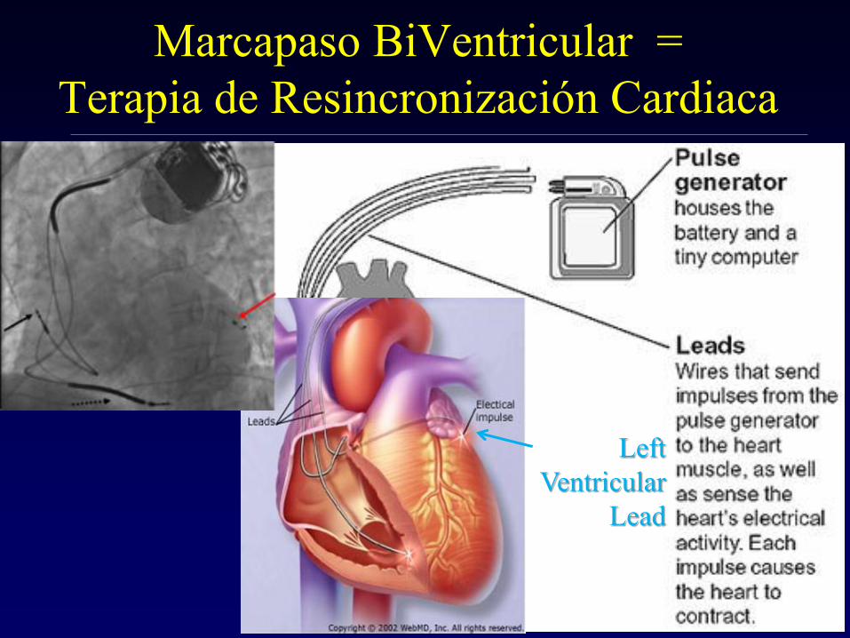
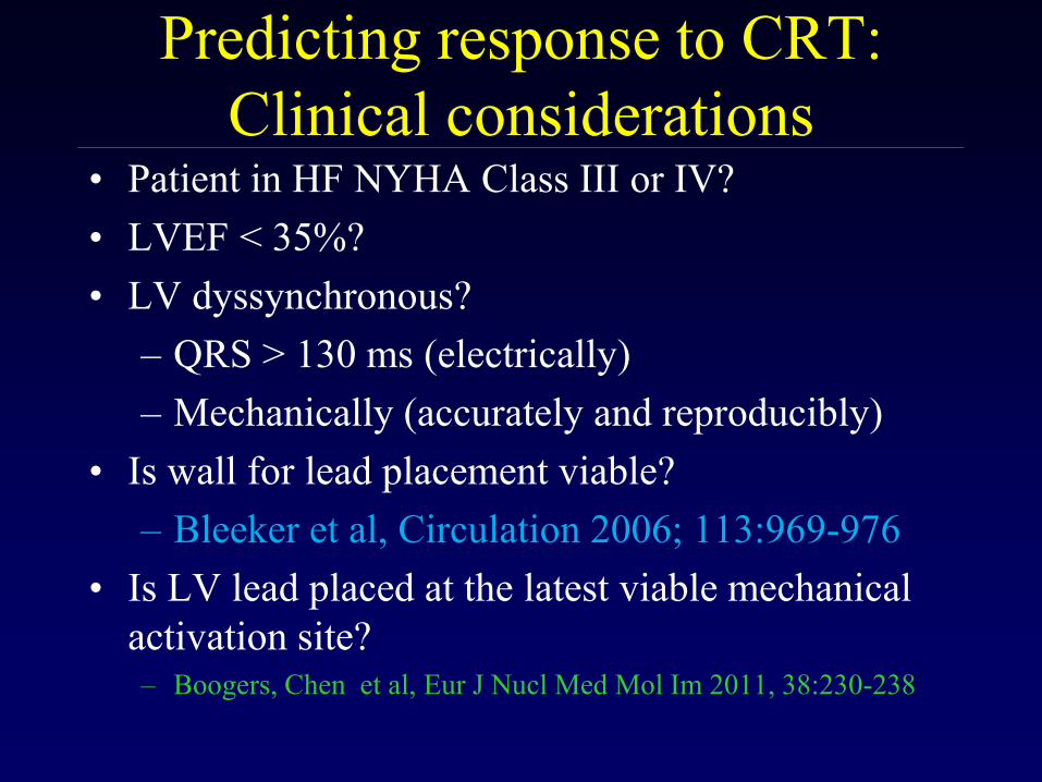
• Como usarlo para predecir la
respuesta al TRC y para guiar donde
colocar el electrodo del TRC
SPECT usado en
Disincronía sistólica intraventricular
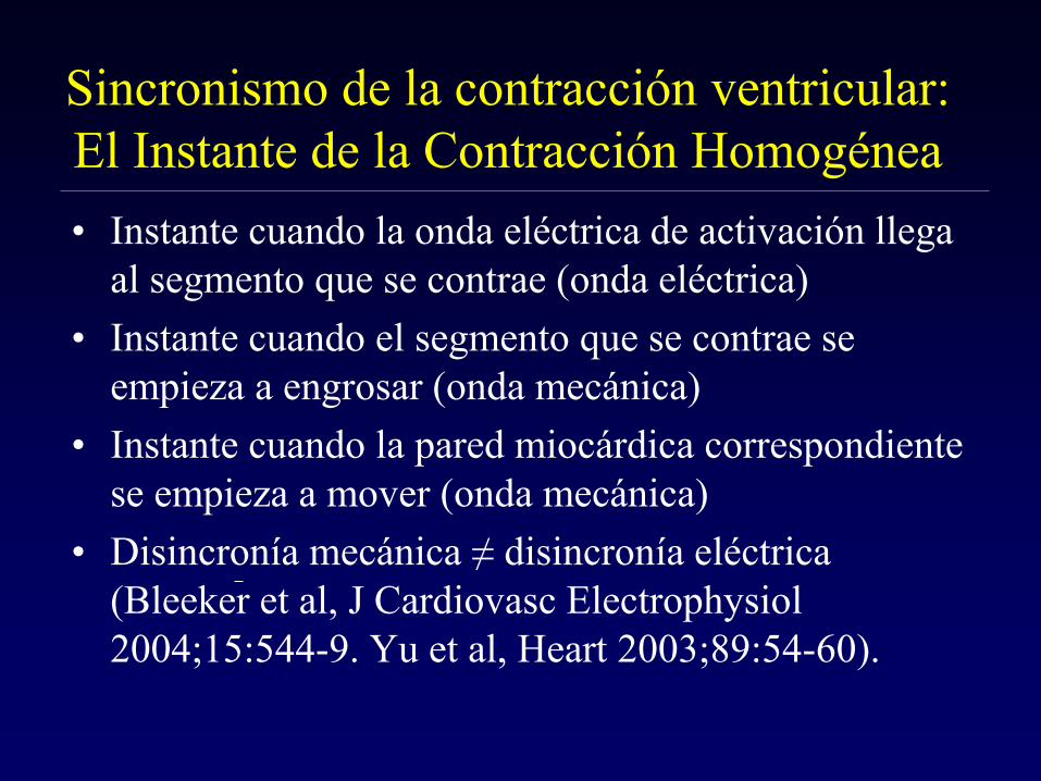
Sincronismo de la contracción ventricular:
El Instante de la Contracción Homogénea
• Instante cuando la onda eléctrica de activación llega
al segmento que se contrae (onda eléctrica)
• Instante cuando el segmento que se contrae se
empieza a engrosar (onda mecánica)
• Instante cuando la pared miocárdica correspondiente
se empieza a mover (onda mecánica)
• Disincronía mecánica ≠ disincronía eléctrica
(Bleeker et al, J Cardiovasc Electrophysiol
2004;15:544-9. Yu et al, Heart 2003;89:54-60).
MUGA: Analisis de Fase
phase
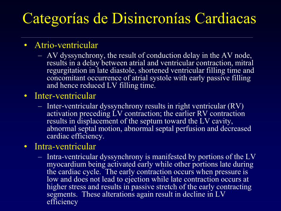
Categorías de Disincronías Cardiacas
• Atrio-ventricular – AV dyssynchrony, the result of conduction delay in the AV node,
results in a delay between atrial and ventricular contraction, mitral regurgitation in late diastole, shortened ventricular filling time and concomitant occurrence of atrial systole with early passive filling and hence reduced LV filling time.
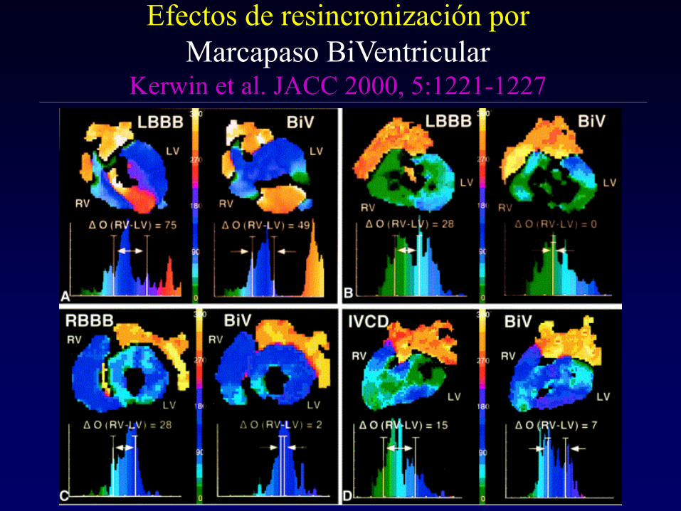
• Inter-ventricular – Inter-ventricular dyssynchrony results in right ventricular (RV)
activation preceding LV contraction; the earlier RV contraction results in displacement of the septum toward the LV cavity, abnormal septal motion, abnormal septal perfusion and decreased cardiac efficiency.
• Intra-ventricular – Intra-ventricular dyssynchrony is manifested by portions of the LV
myocardium being activated early while other portions late during the cardiac cycle. The early contraction occurs when pressure is low and does not lead to ejection while late contraction occurs at higher stress and results in passive stretch of the early contracting segments. These alterations again result in decline in LV efficiency
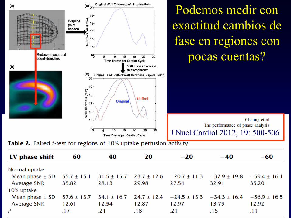
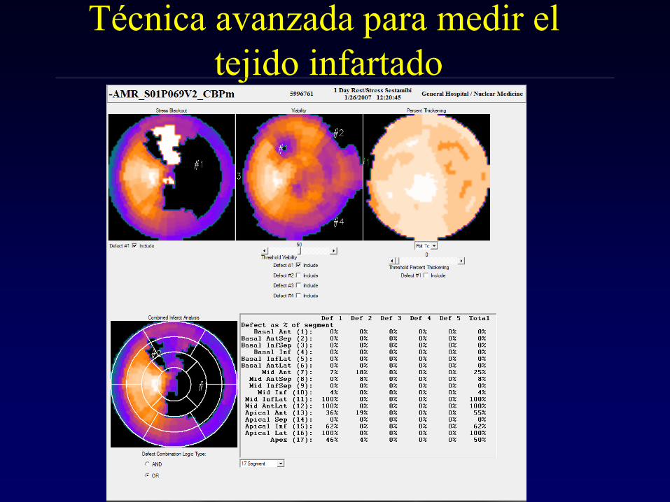
Con que exactitud podemos medir
el engrosamiento? • Using partial volume effect: Δ cts ~ Δ thickness
Galt et al. IEEE Trans Med Imag
1990;9:144-50
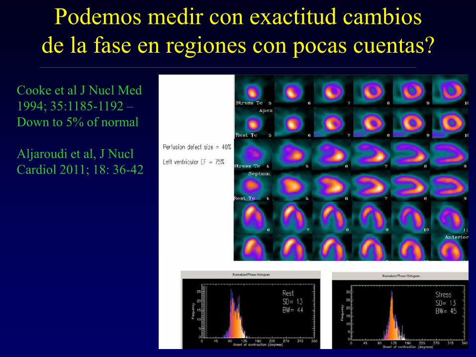
Cooke et al. J Nucl Med 1994;
35:1185-1192
Nichols et al:
Galt et al. IEEE Trans
Med Imag 1990;9:144-
50
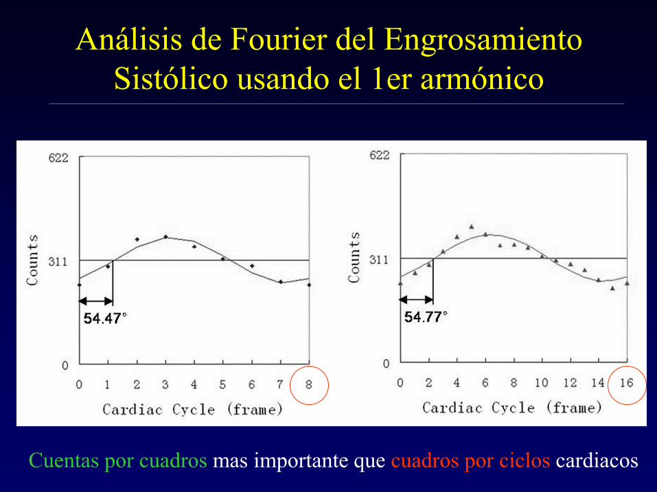
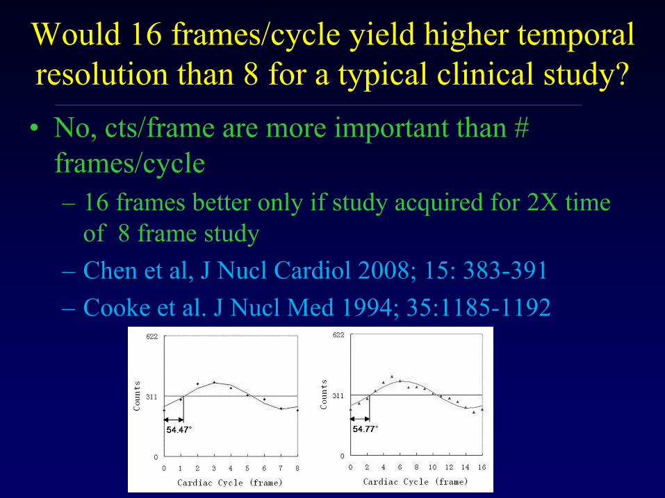
Temporal Sampling
Frame 1
2
8
Cooke et al. J Nucl
Med 1994; 35:1185-
1192
Phase
Thickening C
ounts
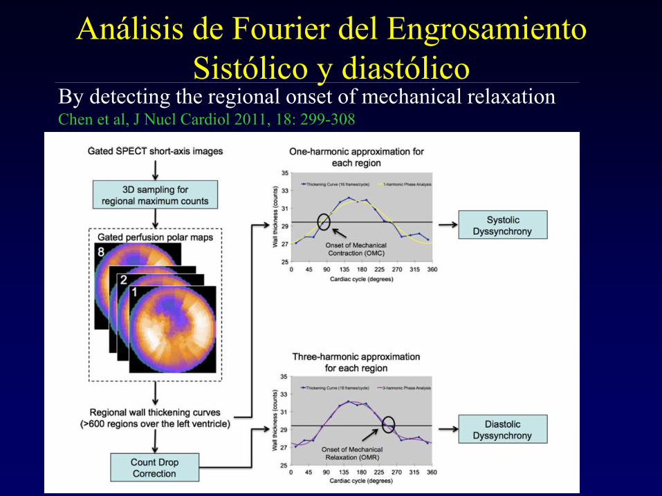
Gated-SPECT de Perfusión Miocárdica : Análisis
del Engrosamiento de la Pared durante la Sístole
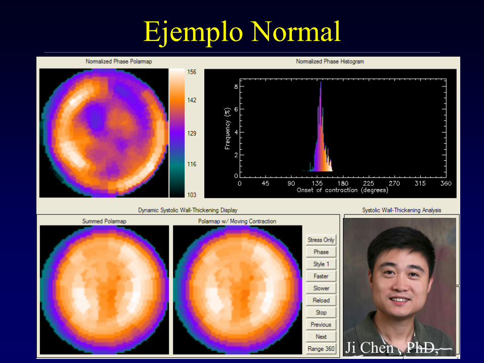
Ejemplo Normal
Ji Chen , PhD,
Patrones Normales
Chen et al, JNC 2005;12:687-95
Henneman MM, Chen J, et al. JNM 2007;
48:1104-1111
Ancho de Banda
135°
43°
Cutoff
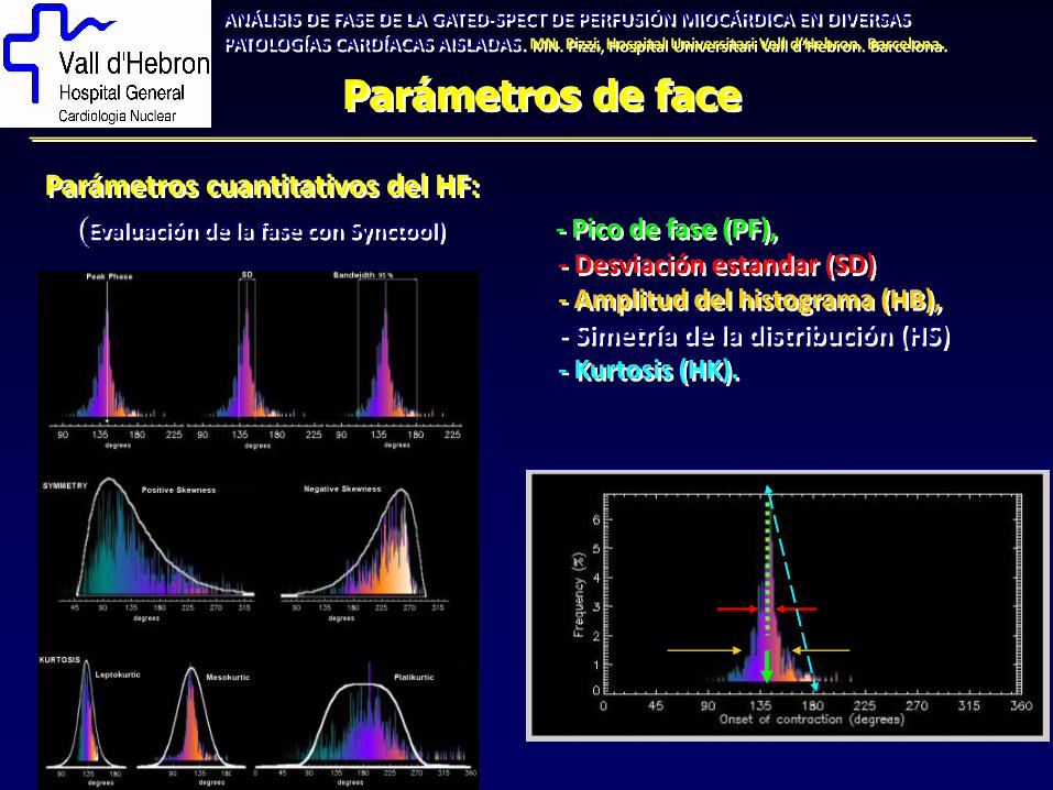
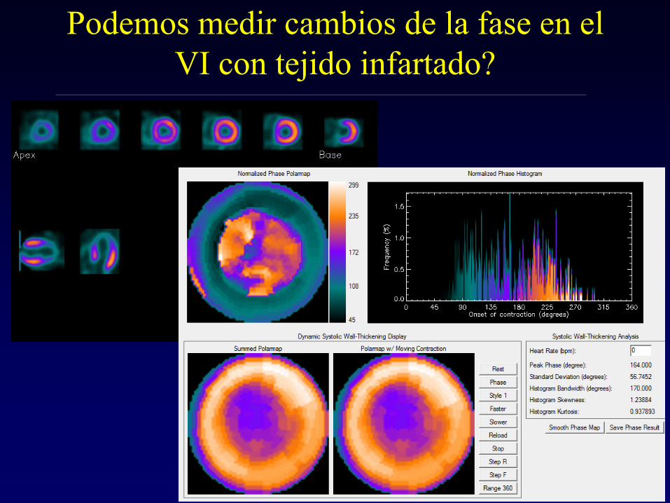
Parámetros de face
ANÁLISIS DE FASE DE LA GATED-SPECT DE PERFUSIÓN MIOCÁRDICA EN DIVERSAS PATOLOGÍAS CARDÍACAS AISLADAS. MN. Pizzi, Hospital Universitari Vall d’Hebron. Barcelona.
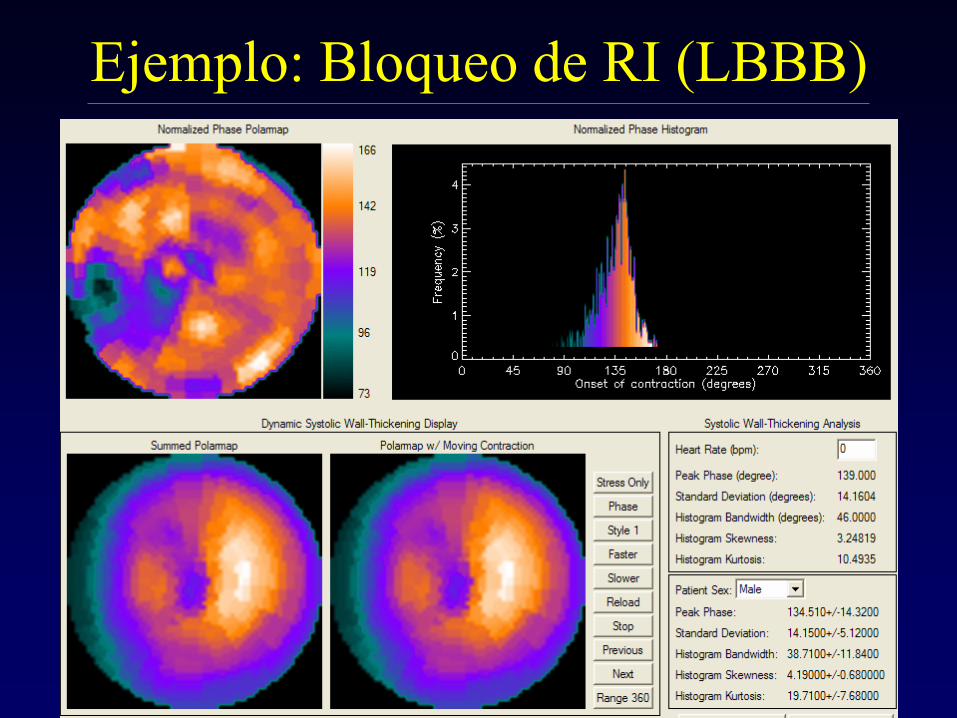
Parámetros cuantitativos del HF:
(Evaluación de la fase con Synctool) - Pico de fase (PF), - Desviación estandar (SD) - Amplitud del histograma (HB), - Simetría de la distribución (HS) - Kurtosis (HK).
Análisis de la Fase del Gated-SPECT
de Perfusión Miocárdica
• Phase analysis of ECG-
gated myocardial perfusion
imaging (MPI) had been
developed to measure left-
ventricular dyssynchrony
• Preliminary normal limits
had been generated
Chen et al, J Nucl Cardiol 2005;12:687-95
Ejemplo: Bloqueo de RI (LBBB)
Ejemplos clínicos
ANÁLISIS DE FASE DE LA GATED-SPECT DE PERFUSIÓN MIOCÁRDICA EN DIVERSAS PATOLOGÍAS CARDÍACAS AISLADAS. MN. Pizzi, Hospital Universitari Vall d’Hebron. Barcelona.
2-Hemibloqueos (HBR): 20 3-Bloqueo de rama derecha (BRD): 40 4-Bloqueo de rama izquierda (BRI): 37 5-Marcapasos (MCP): 26 6-Infarto del miocardio (IM): 71 7-Miocardiopatía dilatada (MD): 17