43
Dr. Ahmad Al-Shafei, MBChB, PhD, MHPE Associate Professor in Physiology KSU Cardiovascular Block Cardiac Electric Activity
| Date post: | 02-Jan-2016 |
| Category: |
Documents |
| Upload: | pamela-barrett |
| View: | 214 times |
| Download: | 0 times |

Dr. Ahmad Al-Shafei, MBChB, PhD, MHPE
Associate Professor in Physiology
KSU
Dr. Ahmad Al-Shafei, MBChB, PhD, MHPE
Associate Professor in Physiology
KSU
Cardiovascular Block
Cardiac Electric Activity

Learning outcomes
State and define the main electrical and mechanical properties of the heart: Autorhythmicity. Excitability. Conductivity. Contractility.
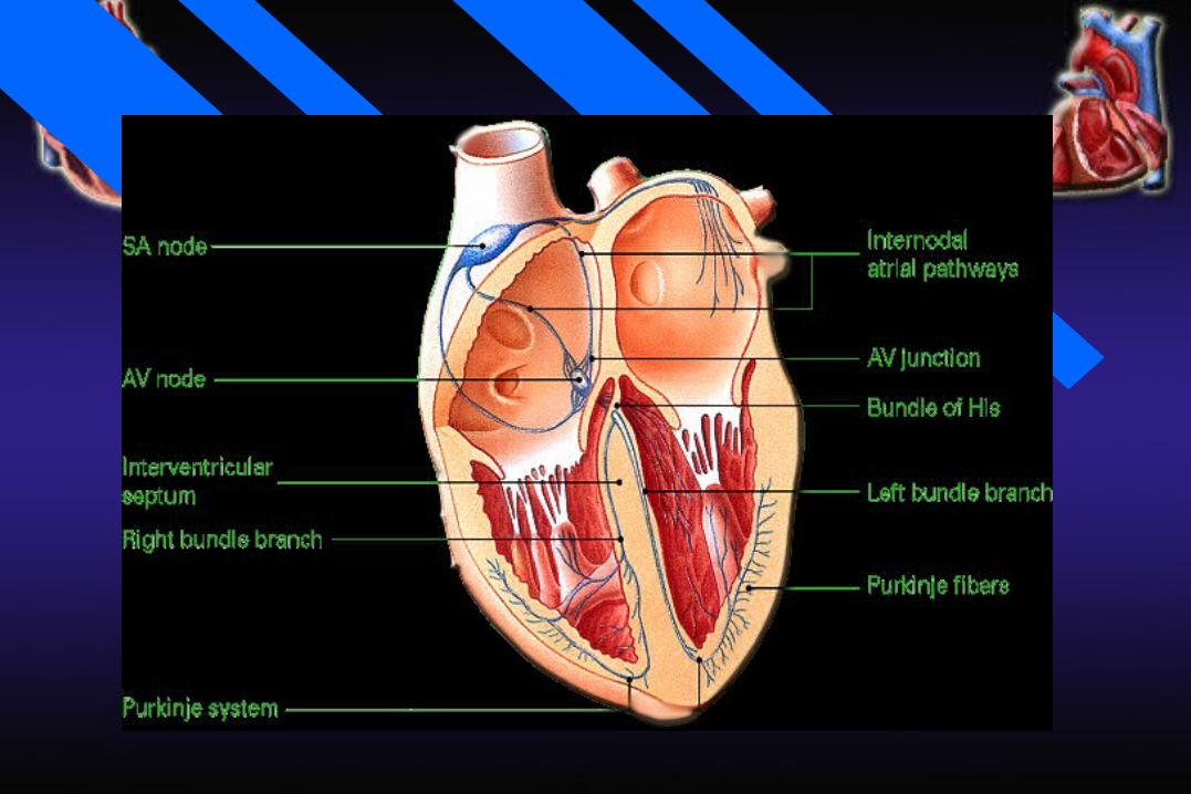
Describe the structure and function of the different parts of the conducting system of the heart.Discuss the genesis of the resting membrane potential in the heart.Compare and contrast the ionic currents during the different phases of the action potential in myocytes.Compare and contrast fast-response and slow-response action potentials in the heart.Describe the physiological significance of the plateau phase and refractory period of a ventricular working muscle cell.Discuss the electrical activity of the pacemaker.Describe the sequence of normal conduction in the heart.Define intrinsic heart rate.Discuss regulation of heart rate under different physiological conditions.
After reviewing the PowerPoint presentation, lecture notes and associated material, the student should be able to:

Learning Resources
Textbooks :
Guyton and Hall, Textbook of Medical Physiology; 12th Edition.Mohrman and Heller, Cardiovascular Physiology; 7th Edition.Ganong’s Review of Medical Physiology; 24th Edition.
Websites:
http://accessmedicine.mhmedical.com/

How the heart performs its function?
The heart has four basic properties which are essential for its functioning as the central pump of the CVS. These are:
1. Autorhythmicity 2. Conductivity 3. Excitability 4. Contractility

How the heart performs its function?
Contractions in both skeletal & cardiac muscles are triggered by a rapid change in voltage called “action potential”. However, action potentials in cardiac muscle are different from skeletal muscle in that:
They are self-generatingThey are conducted directly from cell to cellThey have longer duration
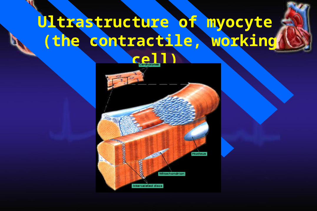
Ultrastructure of myocyte (the contractile, working cell)

Cardiac Cell
AMPVoltmeter
To oscilloscope
The resting membrane potential
(RMP)
The intracellular potential of the resting myocyte is found to be –80 mV to –90 mV (i.e., 80 – 90 mV lower than the extra cellular potential). This is called the resting membrane potential (RMP).
In atrial and ventricular cells, this RMP is stable until external stimulation (excitation) is applied.
In the sino-atrial node cells in particular, and many conduction fibers, the RMP is not stable, drifting towards zero with time.
Electrical potentials arise from:
Differences in the concentrations of ions across the membrane.
The presence of selective ion-conducting channels spanning the membrane, namely K+, Na+, and Ca2+.

Ion pumps and exchangers establish differences in ions concentration across the cell membrane.
Characteristics ofa resting ventricular muscle
Na+
Extracellular
140 mM/L
Ca++ 1.2 mM/L
K+ 4 mM/L
0.0001 mM/L
10 mM/L
140 mM/L
Intracellular

K+
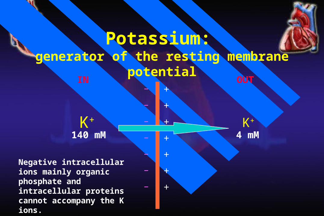
140 mMK+
4 mM
IN OUT
Potassium: generator of the resting membrane potential
Negative intracellular ions mainly organic phosphates and intracellular proteins cannot accompany the K+ ions.
+
+
+
+
+
+
+
_
_
_
_
_
_
_
K+
140 mMK+
4 mM
IN OUT
Potassium: generator of the resting membrane potential
Negative intracellular ions mainly organic phosphate and intracellular proteins cannot accompany the K ions.
_
_
_
_
_
_
_
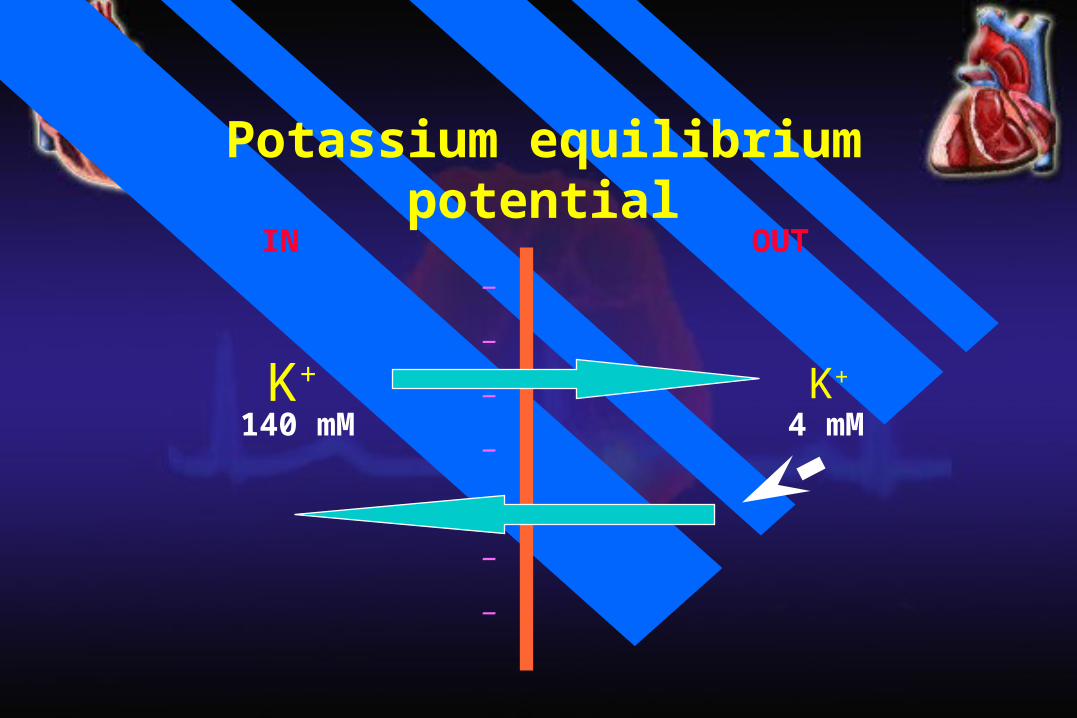
Potassium equilibrium potential
K+
140 mMK+
4 mM
IN OUT
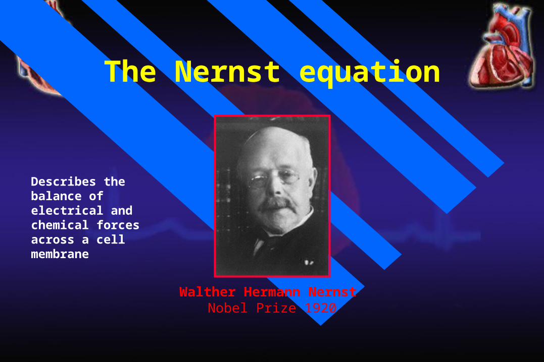
Walther Hermann Nernst Nobel Prize 1920
The Nernst equation
Describes the balance of electrical and chemical forces across a cell membrane
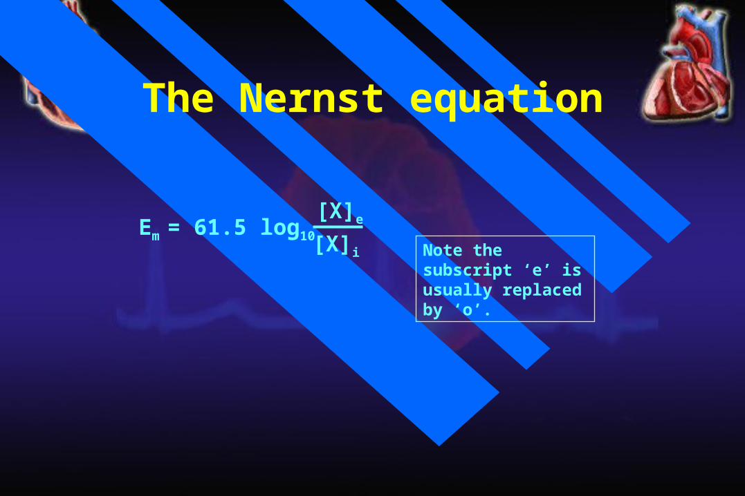
The Nernst equation
Em = 61.5 log10
[X]e
[X]i Note the subscript ‘e’ is usually replaced by ‘o’.
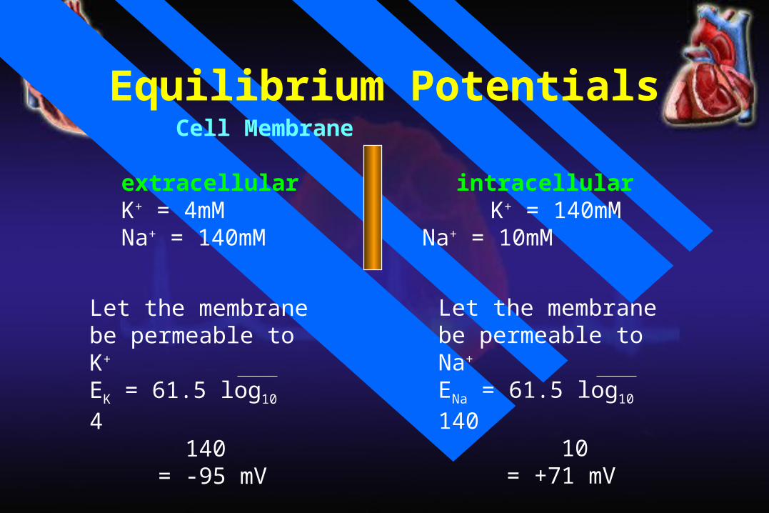
Equilibrium Potentials Cell Membrane
extracellular intracellular K+ = 4mM K+ = 140mM Na+ = 140mM Na+ = 10mM
Let the membrane be permeable to K+
EK = 61.5 log10 4 140
= -95 mV
Let the membrane be permeable to Na+
ENa = 61.5 log10 140 10
= +71 mV
+
+
+
+
+
+
+
_
_
_
_
_
_
_
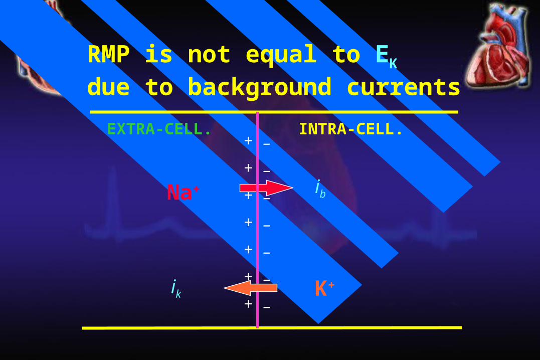
Na+
EXTRA-CELL. INTRA-CELL.
ik
RMP is not equal to EK due to background currents
ib
K+

Action potential and ionic currents in cardiac cells
Cardiac action potentials can be broadly classified into tow types, termed fast-response and slow-response potentials.
Atrial, ventricular and His-Purkinje cells have fast-response action potentials, and sinus node and atrioventricular (AV) node have slow-response action potentials.
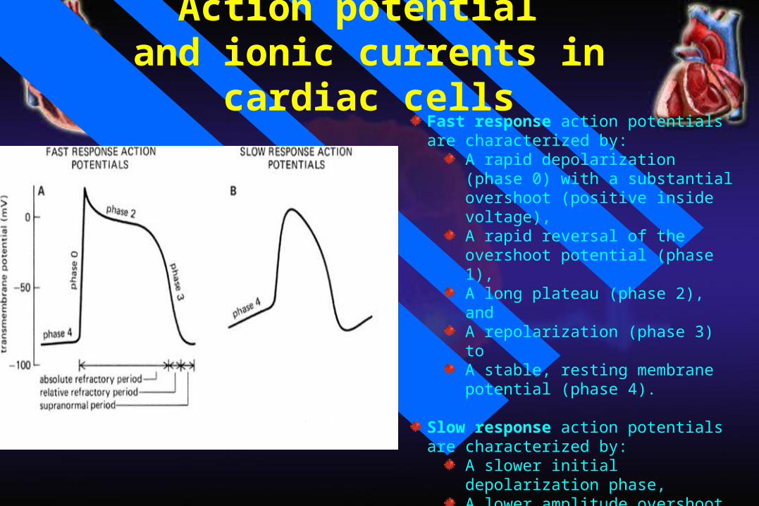
Action potential and ionic currents in cardiac cells
Fast response action potentials are characterized by:
A rapid depolarization (phase 0) with a substantial overshoot (positive inside voltage),A rapid reversal of the overshoot potential (phase 1),A long plateau (phase 2), andA repolarization (phase 3) toA stable, resting membrane potential (phase 4).
Slow response action potentials are characterized by:
A slower initial depolarization phase, A lower amplitude overshoot,A shorter and less stable plateau phase, andA repolarization to an unstable, slowly depolarizing "resting" potential
MEM
BRAN
E P
OTE
NTI
AL (m
V)
0 0
-50 -50
-100 -100
SANVENTRICULULARCELL
0
12
3
4
4
0 3
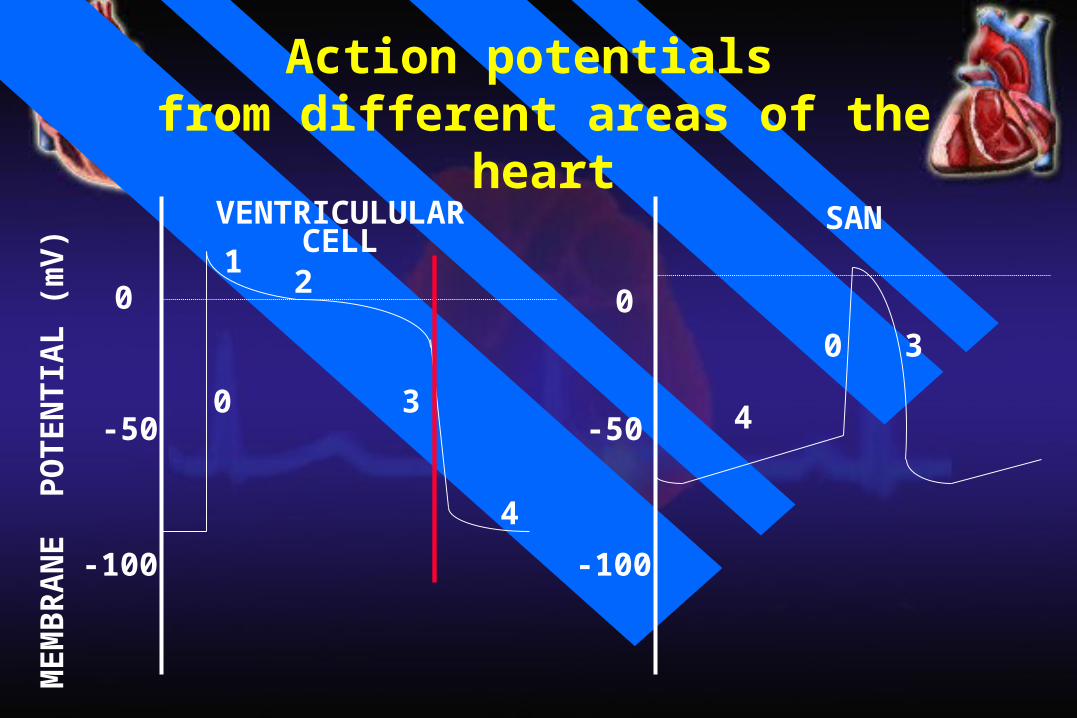
Action potentials from different areas of the heart
MEM
BRAN
E P
OTE
NTI
AL (m
V)
-90
0
0
12
3
4
TIME
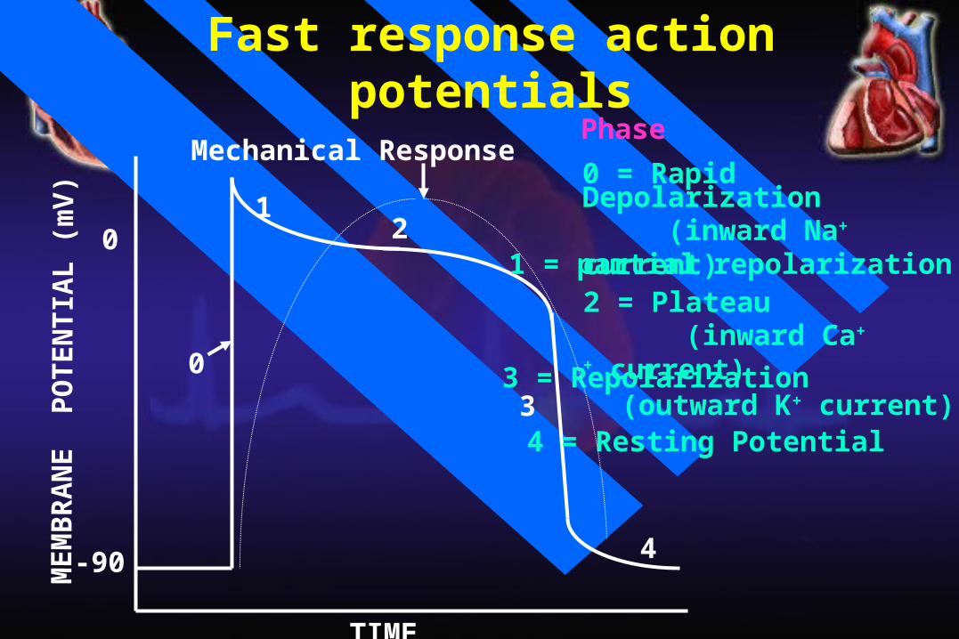
Phase
0 = Rapid Depolarization (inward Na+ current)
1 = partial repolarization2 = Plateau (inward Ca++ current)3 = Repolarization (outward K+ current)4 = Resting Potential
Mechanical Response
Fast response action potentials


Refractory periodsImportance of the long plateau
Cardiac muscle cannot be tetanized because the duration of the effective refractory period is approximately equal to the duration of the mechanical event.
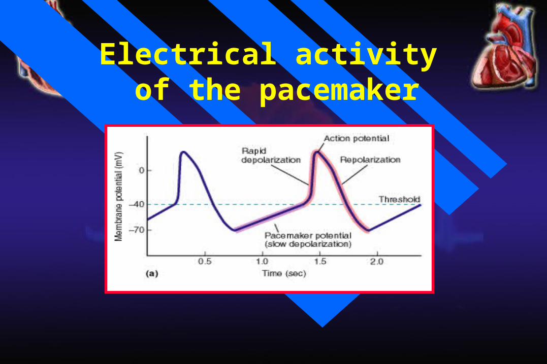
Electrical activity of the pacemaker
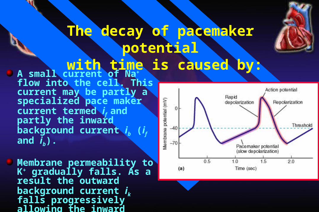
The decay of pacemaker potential with time is caused by:
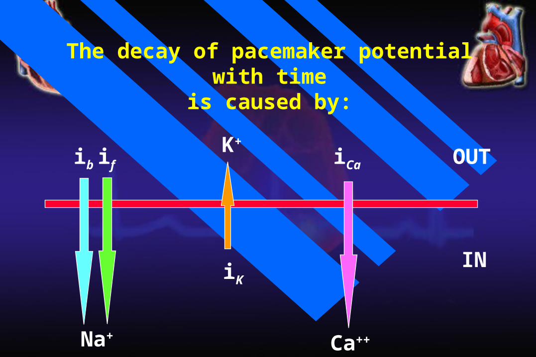
A small current of Na+ flow into the cell. This current may be partly a specialized pace maker current termed if and partly the inward background current ib (if and ib).
Membrane permeability to K+ gradually falls. As a result the outward background current ik falls progressively allowing the inward currents (if and ib) to dominate increasingly.
Ca2+ current in the later part.
OUT
IN
Na+
if
Ca++
iCaK+
iK
ib
The decay of pacemaker potential with time
is caused by:

[Na+]e high[K+]e low[Ca2+]e high
[Na+]i low[K+]i high[Ca2+]i low
extracellularfluid
intracellular
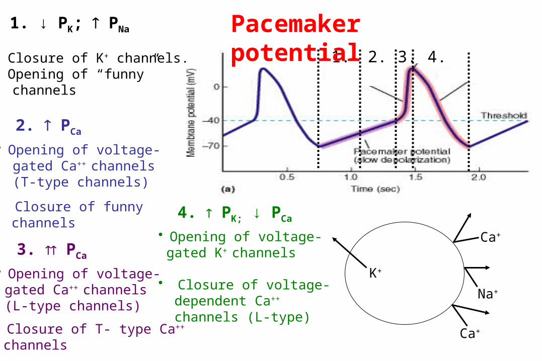
Pacemaker potential

1. ↓ PK; PNa
• Closure of K+ channels.• Opening of “funny” channels (Na+)
[Na+]e high[K+]e low[Ca2+]e high
[Na+]i low[K+]i high[Ca2+]i low
1.
extracellularfluid
intracellular
K+Na+
Pacemaker potential

1. ↓ PK; PNa
• Closure of K+ channels.• Opening of “funny” channels
2. PCa
• Opening of voltage- gated Ca++ channels (T-type channels)
1. 2.
K+
Na+
Ca+
• Closure of funny channelsT channel
Pacemaker potential

1. ↓ PK; PNa
• Closure of K+ channels.• Opening of “funny” channels
2. PCa
• Opening of voltage- gated Ca++ channels (T-type channels)
3. PCa
• Opening of voltage- gated Ca++ channels (L-type channels)
1. 2. 3.
K+
Ca+
Ca+
• Closure of funny channels
Na+
T channel
L channel• Closure of T- type Ca++ channels
Pacemaker potential
1. ↓ PK; PNa
• Closure of K+ channels.• Opening of “funny” channels
2. PCa
• Opening of voltage- gated Ca++ channels (T-type channels)
1. 2. 3. 4.
4. PK; ↓ PCa
• Opening of voltage- gated K+ channels
• Closure of voltage- dependent Ca++ channels (L-type)
K+
Ca+
Ca+
• Closure of funny channels
Na+
3. PCa
• Opening of voltage- gated Ca++ channels (L-type channels)
• Closure of T- type Ca++
channels
Pacemaker potential

Pacemakers(in order of their inherent rhythm)
Sino-atrial (SA) nodeAtrio-ventricular (AV) nodeBundle of HisBundle branchesPurkinje fibers

Cardiac pacemakers
The sinoatrial has the fastest intrinsic rate (~90-100 beats/min) and is the normal pacemaker
The atrioventricular node is the next fastest (~40-60 beats/min) followed by cells in the bundle of His (15-30).
The fastest pacemaker normally drives the heart and suppresses other pasemakers (overdrive suppression).
A beat generated outside the normal pacemaker is an ectopic beat.
The site that generates an ectopic beat is known as an ectopic focus (foci pl.) or ectopic pacemaker.

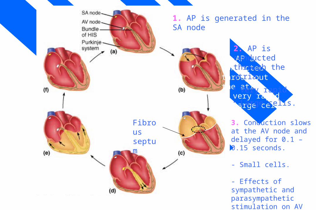
a. AP is generated in the SA node
Conduction of the action potential
in the heart

Conduction of the action potential in the heart(Cell to cell conduction of action potential)

1. AP is initiated in the SA node
2. AP is conducted through the atria.
- Very rapid.
- Large cells
SA node versus AV node(frequency and refractory period)
b. AP are conductedthroughoutthe atria• very rapid• large cells
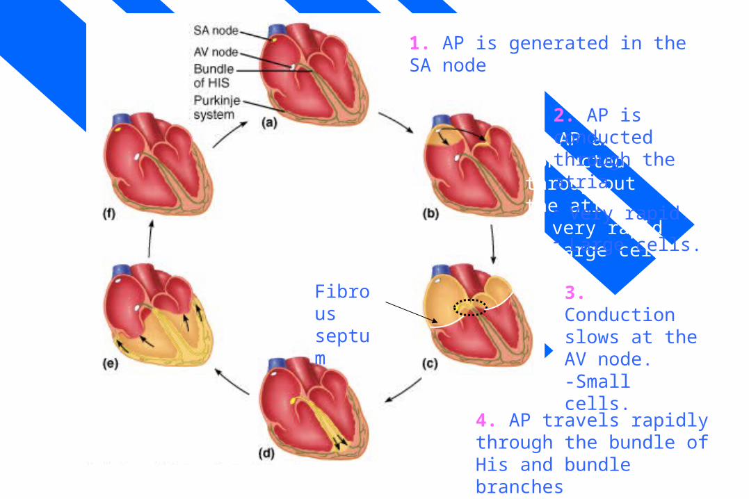
1. AP is generated in the SA node
2. AP is conducted through the atria.
- Very rapid.
- Large cells.
3. Conduction slows at the AV node and delayed for 0.1 – 0.15 seconds.
- Small cells.
- Effects of sympathetic and parasympathetic stimulation on AV nodal conductivity.
Fibrous septum
SA node versus AV node(frequency and refractory period)
b. AP are conductedthroughoutthe atria• very rapid• large cells
1. AP is generated in the SA node
2. AP is conducted through the atria.
- Very rapid.
- Large cells.
3. Conduction slows at the AV node.-Small cells.
Fibrous septum
4. AP travels rapidly through the bundle of His and bundle branches
SA node versus AV node(frequency and refractory period)
b. AP are conductedthroughoutthe atria• very rapid• large cells
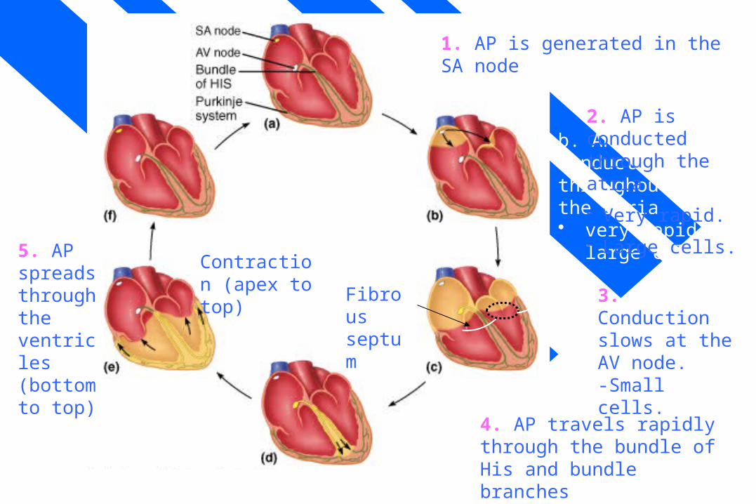
1. AP is generated in the SA node
2. AP is conducted through the atria.
- Very rapid.
- Large cells.
3. Conduction slows at the AV node.-Small cells.
Fibrous septum
4. AP travels rapidly through the bundle of His and bundle branches
5. AP spreads through the ventricles (bottom to top)
Contraction (apex to top)

SA node versus AV node(frequency and refractory period)
b. AP are conductedthroughoutthe atria• very rapid• large cells
1. AP is generated in the SA node
2. AP is conducted through the atria.
- Very rapid.
- Large cells.
3. Conduction slows at the AV node.-Small cells.
Fibrous septum
4. AP travels rapidly through the bundle of His and bundle branches
5. AP spreads through the ventricles (bottom to top)
Contraction (apex to top)
6. Rest
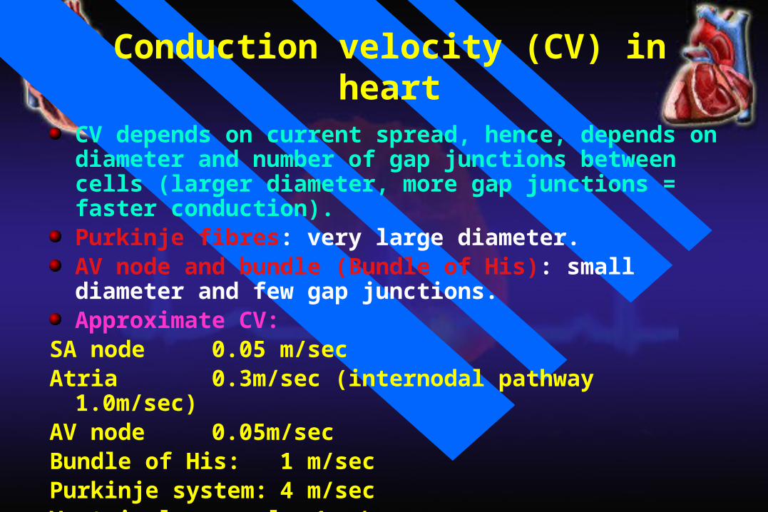
Conduction velocity (CV) in heart
CV depends on current spread, hence, depends on diameter and number of gap junctions between cells (larger diameter, more gap junctions = faster conduction).Purkinje fibres: very large diameter.AV node and bundle (Bundle of His): small diameter and few gap junctions.Approximate CV:
SA node 0.05 m/secAtria 0.3m/sec (internodal pathway 1.0m/sec)AV node 0.05m/secBundle of His: 1 m/secPurkinje system: 4 m/secVentricular muscle: 1 m/sec

Cardiac rhythm
The term rhythm refers to the regularity of: Initiation of cardiac impulses. Sequence of excitation of the heart.
i.e., regularity of the electrical activity of the heart.
The normal cardiac rhythm is called sinus rhythm.
Any variation from the normal rhythm (sinus rhythm) is termed: a cardiac arrhythmia or dysrhythmia.


