212
The road to optimized nerve reconstruction Caroline A. Hundepool

The road to optimized nerve reconstruction
Caroline A. Hundepool

Design, cover & graphics RidderprintPrinted by Ridderprint
© Caroline A. Hundepool, 2016
Publication of this thesis was financially supported by: Nederlands Vereniging voor Plastische Chirurgie, de Esser Stichting, Maatschap Plastische Chirurgie Erasmus MC, Stichting Kortjakje, Van Wijngaarden Medical, Annafonds, Rabobank Rotterdam, Schouten Zekerheid, Astellas Pharma, Chiesi Pharma.

The Road to Optimized Nerve Reconstruction
De weg naar optimaal zenuwherstel
Proefschrift
Ter verkrijging van de graad van doctor aande Erasmus Universiteit Rotterdam
op gezag van de rector magnificus
Prof. dr. H.A.P. Pols
en volgens besluit van het College voor Promoties.
De openbare verdediging zal plaatsvinden opvrijdag 11 november 2016 om 13:30
door
Caroline Anna Hundepool
geboren op 13 april 1989te Leidschendam

PROmOTiecOmmissie
Promotor Prof. dr. S.E.R. Hovius
Overige Leden Prof. dr. M.J.A. Malessy Prof. dr. P.A.E. Sillevis Smitt
Dr. T.J.H. Ruigrok
copromotor dr. T.H.J. Nijhuis
Paranimfen Dr. R.R. Dijkman L.F. Bulstra, MSc

Aan mijn ouders


cONTeNTs
chapter 1 General introduction 9
PART i clinical problem 25chapter 2 Prognostic factors for outcome after median, ulnar and combined
median-ulnar nerve injuries: a prospective study27
chapter 3 Early posttraumatic psychological stress following peripheral nerve injury: a prospective study
43
PART ii evaluation 55chapter 4 Ultrasonographic quantification of intrinsic hand muscle cross-
sectional area; reliability and validity for predicting muscle strength57
chapter 5 Motor nerve recovery in a rabbit model: Description and validation of a noninvasive ultrasound technique
75
chapter 6 Non-invasive ultrasound of the tibial muscle for longitudinal analysis of nerve regeneration in rats
89
PART iii engineering a nerve allograft 103chapter 7 Optimizing decellularization techniques to create a new nerve
allograft: an in-vitro study using rodent nerve segments105
chapter 8 Application of Elastase in the decellularization of human sensory and motor nerves
119
chapter 9 Return of motor function with decellularized nerve allografts using elastase in a rat sciatic nerve model
133
PART iV improving the nerve allograft 149chapter 10 The effect of stem cells in bridging peripheral nerve defects: a
meta-analysis151
chapter 11 General Discussion 177chapter 12 Summary 191
Nederlandse Samenvatting 197
Appendices List of publications 203PhD Portfolio 207Curriculum Vitae 209Dankwoord 211


Chapter 1General introduction


11
General Introduction
11. iNTROducTiON
Peripheral nerve injuries are devastating injuries, which can lead to severe disability. Nerve in-juries are relatively common. It occurs with up to 3% of all patients admitted to Level I trauma centers. Most of the injuries to peripheral nerves occur in the upper extremities. Nerve injury will lead to significant impairment in motor function and causes sensory loss. Depending on the level of nerve injury the consequences can be devastating and have great impact on a patient’s life and ability to perform daily activities such as work and hobbies. Nerve injury not only causes physical disability. There is evidence it also has great consequences psycho-logically. Cognitive, emotional and behavioral aspects influence recovery. It is important these factors are recognized so that the quality of patient care can be improved[1]. The last decades both experimental and clinical research has been focused on optimizing the reconstruction of nerve injuries. The studies in this thesis are focused on the optimization of nerve reconstruc-tion.
2. ANATOmy
When nerve injury occurs and the nerve is transected the electrical signals are disrupted. Peripheral nerves start in the myelum and end in the designated end-point (either sensory or motory). The nerves consist of an outer layer, the epineurium, which protects the nerve fascicles. The vasa nervorum provide the vascularization of the nerves and are interconnected with the epineural collagen and fibrotic fibers. In small nerves (i.e. nerves with a small diam-eter) these vasa nervorum will solely supply the nerve. In nerves with a larger diameter the
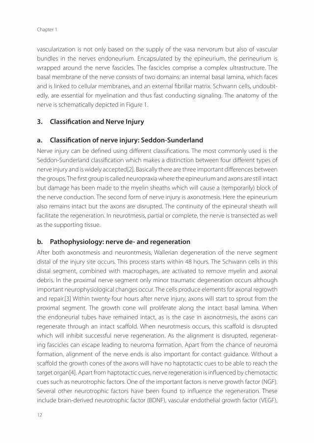
Figure 1. The anatomy of a peripheral nerve with the different components. The epineurium, perineu-rium and endoneutrium are depicted. (Published with permission of the Mayo Foundation for Medical Education and Research. All rights reserved, copyright © 2015)
Chapter 1
12
vascularization is not only based on the supply of the vasa nervorum but also of vascular bundles in the nerves endoneurium. Encapsulated by the epineurium, the perineurium is wrapped around the nerve fascicles. The fascicles comprise a complex ultrastructure. The basal membrane of the nerve consists of two domains: an internal basal lamina, which faces and is linked to cellular membranes, and an external fibrillar matrix. Schwann cells, undoubt-edly, are essential for myelination and thus fast conducting signaling. The anatomy of the nerve is schematically depicted in Figure 1.
3. classification and Nerve injury
a. classification of nerve injury: seddon-sunderlandNerve injury can be defined using different classifications. The most commonly used is the Seddon-Sunderland classification which makes a distinction between four different types of nerve injury and is widely accepted[2]. Basically there are three important differences between the groups. The first group is called neuropraxia where the epineurium and axons are still intact but damage has been made to the myelin sheaths which will cause a (temporarily) block of the nerve conduction. The second form of nerve injury is axonotmesis. Here the epineurium also remains intact but the axons are disrupted. The continuity of the epineural sheath will facilitate the regeneration. In neurotmesis, partial or complete, the nerve is transected as well as the supporting tissue.
b. Pathophysiology: nerve de- and regenerationAfter both axonotmesis and neurontmesis, Wallerian degeneration of the nerve segment distal of the injury site occurs. This process starts within 48 hours. The Schwann cells in this distal segment, combined with macrophages, are activated to remove myelin and axonal debris. In the proximal nerve segment only minor traumatic degeneration occurs although important neurophysiological changes occur. The cells produce elements for axonal regrowth and repair.[3] Within twenty-four hours after nerve injury, axons will start to sprout from the proximal segment. The growth cone will proliferate along the intact basal lamina. When the endoneurial tubes have remained intact, as is the case in axonotmesis, the axons can regenerate through an intact scaffold. When neurotmesis occurs, this scaffold is disrupted which will inhibit successful nerve regeneration. As the alignment is disrupted, regenerat-ing fascicles can escape leading to neuroma formation. Apart from the chance of neuroma formation, alignment of the nerve ends is also important for contact guidance. Without a scaffold the growth cones of the axons will have no haptotactic cues to be able to reach the target organ[4]. Apart from haptotactic cues, nerve regeneration is influenced by chemotactic cues such as neurotrophic factors. One of the important factors is nerve growth factor (NGF). Several other neurotrophic factors have been found to influence the regeneration. These include brain-derived neurotrophic factor (BDNF), vascular endothelial growth factor (VEGF),

13
General Introduction
1glial cell line-derived neurotrophic factors, neurotrophin-4 (NT-3) and neurotrophin-4 (NT4)[5, 6]. Although these factors are known, the application of these growth factors in a clinical setting for peripheral nerve reconstruction is not yet made. Nerve injury will also influence motor end plates and cause muscle atrophy. Muscle tissue can, over time, be replaced with fibrotic adipose tissue. When nerve regeneration takes place, the motor end plates will be activated again and the muscle can regain strength. However, after prolonged degeneration, damage to both motor end plates and muscle (i.e. atrophy and fibrosis) are irreversible.
4. NeRVe RecONsTRucTiON
When the trauma to the nerve is more severe, or a segment of the nerve is lost, a nerve gap occurs. Nerve reconstruction is key for the successful regrowth of an injured nerve. Recon-struction with very little to no tension is of paramount importance to the outcome. When the nerve can be repaired without tension, the regeneration only needs to cross one suture line. When this criterion cannot be met, an interposition with a nerve graft is indicated which will inevitably cause two suture lines. The interposition with a nerve graft to bridge the gap has been extensively studied in the last decades.
a. Autograft: the gold standard.The gold standard for the reconstruction of ‘large’ peripheral nerve segments is the nerve autograft. This technique was first introduced in 1978 by Milessi et al. With nerve autograft reconstruction a nerve elsewhere in the body is harvested to reconstruct a nerve gap. Sensory nerves are most commonly harvested, as their function is easiest to scarify. Of the sensory nerves, the sural nerve is the most commonly used, as a length up to 40 cm of nerve graft can be harvested. Nerves that are also used are the anterior medial antebrachial cutaneous nerves and the superficial radial nerve. The autograft can be cabled to increase diameter and should be 10 to 20 % longer than the nerve gap, to create a tension-free reconstruction. The autograft has the advantage of closely mimicking the ultrastructure, contains Schwann cells and provides neurotrophic factors. Disadvantages however, include donor site complications and morbidity such as second scar formation, sensory loss, neuroma formation, prolonged operation time and increased costs. Recent data show that motor nerve grafts are superior to sensory nerve grafts for the reconstruction of a motor nerve defect[7, 8]. However there is only a limited supply of motor nerves that can be harvested.
Several alternatives have been proposed to overcome these disadvantages. Nerve scaf-folds have the advantage of unlimited availability, no donor side morbidity. They all strive to provide guidance for the regenerating axons and a tension-free repair. The alternatives can be subdivided into scaffolds made of nerve (i.e. autograft or allograft) or biological or synthetic conduits[9].

Chapter 1
14
b. Biological conduits (arterial and venous)Biological conduits are made from autologous material such as veins or arteries of which veins are most commonly used. The advantages of vein graft over arterial graft are their abundant supply and minimal donor site morbidity. The possibility of lumen collapse impeding nerve regeneration has been marked as a disadvantage of vein conduits. Chiu et al reported the first use of a vein as a nerve conduit in 1982.[10] Ten years later, in 1990, the first clinical case was reported with a digital nerve reconstruction.[11] Meaningful recovery was reported in digital nerve gaps smaller than 3 cm with a vein graft. Although positive results have been reported, the vein graft cannot outperform the gold standard, the nerve autograft. The additional of a small muscle segment inside the vein has shown a beneficial effect by preventing collapse of the venous grafts.
c. synthetic conduitsAs an alternative for biological conduits synthetic conduits [12] are proposed. Different materials have been used: collagen, polyglycolic acid, poly(DL-lactide-e-caprolactone) (PLC), poly(lactic-coglycolic acid) (PLHA), poly(carpolactone fumerate) (PLCF), hydrogel and capro-lactone[13, 14]. Collagen nerve conduits are most commonly used. In animal studies collagen conduits have been challenged to nerve autografts and allografts. In rats, the poly-DL-lactide-e-caprolactone conduits showed similar results to the autograft with regard to functional outcome[15]. Another study showed that a matrix in a collagen conduit showed superior results compared to an empty collagen conduit[16]. Unfortunately, they failed to replace the nerve autograft. In addition, when synthetic conduits were tested in larger animal models, the rabbit, the superior results of the collagen conduits with a matrix where not observed[17]. Animal studies have also shown that wrapping the nerve repair site with a conduit can reduce scar formation[18]. Several of these conduits are clinically available including Neurotube (Synovis), Neurolac (Ascension) and Neuragen (Integra).
d. AllograftCompared to the gold standard, the nerve autograft, the advantages of the nerve allograft include potentially unlimited supply and length, no donor site morbidity and reduced op-eration time. Therefore the allograft has gained popularity. The allograft, as the autograft, has the structure and framework of the nerve, which is hypothetically the perfect guidance for nerve regeneration. The first allografts that were used required the use of immunosuppressive drugs. The donor Schwann cells are immunogenic to the host, they activate a T-cell response, which required the administration of immunosuppression (cyclosporine A and/or FK506) for up to two years after the allograft transplantation. Tacrolimus, or FK506 has shown to have a beneficial effect on nerve regeneration[19-21]. Immunosuppressive drugs have well known disadvantages and leave recipients vulnerable to opportunistic infections or neoplastic pro-cesses. This changed when the allografts were pre-treated in order to reduce the immune
15
General Introduction
1reaction when implemented. Several techniques for decellularization of the allografts have been introduced. These methods include cold preservation, freeze-thawing, chemical decel-lularization, lyophilisation and irradiation[22].
With the cold preservation method nerve grafts are stored in University of Wisconsin Solu-tion (UWS) at 4-5°C for seven days will reduce the cells in the nerve allograft but the cells are not removed[23]. The allograft antibodies are reduced and the basal lamina of the grafts is maintained. However, as the cells are not removed, significant cellular debris remains which reduces axonal regeneration. [24-26] Therefore this method is inferior to other techniques as decellularization with chemical decellularization [24, 27]. Similar to the cold preservation method is the freeze-thawing of nerve allograft[22]. The cells are killed but not removed. Grafts are deep-freezed at -70⁰C and thawed repeatedly. This method was developed to kill antigen-presenting cells while preserving the structure of the extracellular matrix. Despite less cellular debris freeze-thawing seems inferior to chemical detergent nerve allografts.[28] The studies on freeze-thawed allograft have been of importance in understanding the role of the basal lamina in nerve allografts for the regeneration of axons. These studies have stimulated the development of chemical decellularization techniques in which the basal lamina was better preserved[22].
With chemical decellularization, detergents are used to eliminate cellular remnants[29-31]. This technique was first used by Johnson et al, who used Triton-X100 and sodium deoxy-cholate[32] and later modified by Sondel et al. [31]. This technique was later optimized by Hudson et al. who used a combination of three detergents: Triton X-200, sulfobetaine-16 and sulfobetaine-10[28, 30]. This protocol balanced the need for cellular removal and preserva-tion of the extracellular matrix. However after processing, cellular debris still remained in the nerve allografts. Irradiation of nerve grafts with gamma irradiation has shown to degrade the antigenicity of the graft [33, 34]. Although this method was not further developed as a single pre-treatment method it has been added to previously mentioned methods to further enhance the processing of the nerve allografts[22, 35]. The protocol was further enhanced by treatment with the enzyme Chondroïtinase ABC that showed degradation of chondroitin sulfate proteoglycans [36, 37]. These have shown to inhibit axonal growth and removing them should therefore be beneficial for the axon regeneration.
Up-to-date there are no high level (I or II) evidence clinical studies comparing allografts to the gold standard, the autograft or conduits for the reconstruction of large peripheral nerve defects. Evidence for the allograft is based on comparative animal studies and level III clinical studies. There is one clinically available decellularized human allograft made by Axogen Inc., Alachua, Florida. Animal studies with this Axogen allograft suggest that the allograft is still inferior to the nerve autograft, especially when looking at motor nerve regeneration. Whitlock et al. studied the effect of the commercially processed allograft in 14- and 28 mm nerve gaps in rats. Although the allograft and collagen conduits were comparable at 12 weeks, the allograft was not found to be comparable or superior to the autograft 12 weeks after
Chapter 1
16
implementation[38]. Guisti et al. compared the Axogen processed allograft with the autograft and a collagen conduit both at early and late recovery time of 12 and 16 weeks in a rat model. The nerve autograft outperformed both the processed allograft and the collagen conduits at twelve and sixteen weeks.[39]
This allograft has been tested in three case reports and recently in a larger multicenter study.[40-44] The case reports investigate the use of the allografts but are not comparing them to other treatment options. Therefore the superiority of the allograft to the autograft is not proven. In the multicenter outcome study by Brooks et al. investigate the effect of Axogen processed grafts in nerve defect between 5 and 50 mm.[40] They report meaningful recovery, either S3+/S4 or M4/M5, in 87% of patients. The recovery in sensory nerve defects is higher compared to the mixed, or motor nerve defects. Outcomes of the same multicenter study where reported by Cho et al. who reported an overall meaningful recovery of 86%. When looking at type of nerve repair this study showed meaningful levels of recovery in 89% of digital nerve repairs, 75% of median nerve repairs and 67% in ulnar nerve repairs.[43] The high distribution of digital sensory nerves in this study should be considered when interpreting the high rate of recovery in this study. Similar to the case reports, these studies also lack a valid comparison to the nerve autograft. As such, it can be concluded that the introduction of a new allograft needs careful evaluation in both in vitro and in vivo studies to thoroughly investigate neuroregenerative capacity. Not before non-inferiority or superiority has been proven, clinical testing is justified.
e. Luminal additivesTo further improve nerve regeneration several additives to the different grafts have been introduced. Both cells and supportive factors have been studied. Schwann cells have shown to increase functional regeneration of acellular nerve grafts of 14 mm in a rat model.[45] Bone marrow stromal cells (BMSCs), adipose-derived stromal cells (ADSCs), hair follicle stem cells, skin-derived mesenchymal stem cells, and amniotic fluid derived mesenchymal stem cells have been introduced as well.[46-51] BMSCs, of mesenchymal origin, have shown the capability to differentiate into neuron-like cells.[52] The BMSCs can be easily harvested from bones. The mechanism for their neuroregenerative effect is often discussed in literature. Some argue their neuroregenerative effect is caused by the transdifferentiation into Schwann cell-like phenotype.[53] Others argue that the stem cells will function as growth promoting factors. Adipose derived stem cells can be harvested even less invasive then BMSC’s and can be derived from belly fat. Their phenotype profile is comparable to the BMSCs and some studies even show that they have a more profound tendency to differentiate into Schwann cell-like phenotypes. More recently applied stem cells, such as hair follicle pluripotent stem cells and skin-derived stem cells, are also easily accessible and can also show the capability to transdifferentiate into Schwann cell–like cells as well.[54] Neurotrophic growth factors have also shown promise in enhancing axonal growth when added to a nerve graft.[55, 56]

17
General Introduction
15. eVALuATiON OF RecOVeRy OF FuNcTiON AFTeR NeRVe
RecONsTRucTiON
Evaluation of recovery of function after nerve injury is important in both clinical and experi-mental settings. In the clinical setting the rehabilitation of the patient can be closely followed. Patient and doctor can evaluate the recovery and different nerve reconstructions can be compared. In the experimental setting valid assessment techniques are essential when test-ing new innovative options for the reconstruction of nerve defects[57]. In other words, to test hypotheses. During the last decades several different options have been proposed for both clinical and experimental testing
a. clinical outcome assessmentsMost studies report recovery after nerve injury with the British Medical Research Council’s (MRC) scale for both motor and sensory recovery. Motor recovery is graded from M0 to M5 and sensory recovery from S0 to S5[58, 59]. Power grip strength can be assessed with the Jamar dynamometer[60] and the tip-pinch grip strength with Jamar pinch gauge meter[60]. Sensory testing includes two-point discrimination (static and dynamic), monofilament testing (Semmes-Weinstein) and cold-heat testing[61]. Although important, this thesis is focused on motor function recovery and therefore testing of the sensory recovery will be discussed only shortly.
b. experimental outcome assessmentsFunctional assessment of motor nerve regeneration was first measured with the sciatic func-tional index (SFI), based on the walking track analysis [62, 63]. The method was first developed by Medianceli et al. in 1982 and later modified by Bain et al. The SFI is based on characteristics of the footprint of the hind paw of the rat during the walking track. This method however, is influenced by various factors as toe contractures and auto-mutilation[64, 65]. Although this method is still widely used, several studies have shown the lack of association between out-come measures in nerve regeneration studies[66, 67]. Lee et al. found that the sciatic function index, based on the walking track analysis, did not correlate with the isometric tetanic force measurements. And, of various measurements of the ankle angle, only the ankle angle in toe-off phases correlated well with the isometric tetanic force[67]. Video analysis of the walking track has been proposed to improve the SFI[68]. The rat is filmed and the SFI is supplemented with gait kinematic informative such as ankle angle[69, 70]. Lin et al. found strong correlations between ankle angle at the midstance phase of the gait cycle with muscle weight[71].
After nerve injury, rats will have a (short) period of reduced activity level and will avoid using that foot for the first couple of weeks causing a joint flexion contracture. The angle of the ankle can be measured to express the joint flexion contracture. Measuring the ankle angle contraction is non-invasive, easy and is not time consuming. Lee at el.[67] showed that ankle

Chapter 1
18
angle had excellent correlations with other outcome measures of nerve function. It was found to be less severe in rats that could produce higher isometric tetanic force.
ElectrophysiologyElectrophysiological testing is widely used in experimental nerve studies. The nerve is stimu-lated and different parameters are recorded. Different outcome assessments are reported as compound muscle action potentials (CMAP), mean conduction velocity (MCV), electromyog-raphy and somatosensory evoked potentials (SEPs)[72]. Of these different methods the CMAP is considered to be the best discriminative tool[72].
Muscle MassThe weight of the muscles, distally to the reconstructed nerve, is claimed to be a parameter of functional recovery. This method is also quick and easy to perform which is one of the reasons for its popularity. The gastrocnemius muscle is mostly used but the tibial muscle is also used.
Isometric Tetanic ForceShin et al. developed a reproducible and accurate model to measure nerve regeneration, the isometric tetanic force measurement. After stimulation of the nerve the maximal tetanic force of the tibial muscle of the rat was recorded. The authors reported a side-to-side variability of less than 4%[73]. Subsequently this method was developed and validated in the rabbit model as well[74]. The strength of this method is that it describes the functional recovery of the target organ, the muscle, after reinnervation. When compared to the above mentioned muscle mass it can be observed that a decrease in isometric tetanic force is less than the decrease in muscle weight. That can be explained by the fact that the cross-sectional area of the muscle is closely related to the daily muscle contractions[75]. Isometric tetanic force is therefore a more true parameter to describe motor function than muscle weight.
HistomorphometryHistology of cross sections of the nerve segments is a frequently used method to quantify nerve recovery. Axon and fiber diameter, axon and fiber count, myelin thickness and myelin area are obtained parameters. It has been shown that obtaining these values on the distal segment is more useful than looking at the mid-segment of the nerve grafts. Semi-automatic computer models have been introduced to reduce the labor intensity of the histological tests.

19
General Introduction
16. GeNeRAL Aim ANd OuTLiNe OF The Thesis
The aim of this thesis is to optimize peripheral nerve reconstruction for large, segmental nerve defects. In order to do so, this thesis was divided in four parts. In Part I, the clinical problem was investigated by studying prognostic factors influencing the outcome after nerve reconstruc-tion (chapter 2). Specific attention was paid to one of the few factors that can be potentially influenced after nerve trauma; early posttraumatic stress (chapter 3). After investigating the clinical problem of nerve reconstruction, the evaluation of nerve regeneration after nerve reconstruction was studied. In Part II of this thesis a non-invasive method is introduced, ul-trasound, for the analysis of nerve recovery after trauma. First, we introduced ultrasound for the analysis of recovery of intrinsic hand muscles in humans after nerve trauma (chapter 4). Secondly, the method was established in an experimental setting in rabbits (chapter 5) as well as in rats (chapter 6) for the longitudinal follow up of recovery after nerve injury.
The third aim of this thesis was to improve nerve reconstruction by engineering an opti-mized nerve allograft (Part III). In chapter 7, the optimization of nerve allografts are studied in vitro in rat nerves. Subsequently, decellularization and preservation protocols were tested on human nerves in chapter 8, where the difference between motor and sensory nerves was made. After optimization of the nerve allograft in vitro, the allograft was tested in vivo. The optimized nerve allograft was implemented in a rat model to test the functional motor outcome after reconstruction of a 1 cm nerve defect. The allograft was challenged to the gold standard, the nerve autograft (chapter 9).
In the final part (Part IV) an improvement on nerve bridging is studied. The effect of stem cells on the regeneration of nerves was investigated with a meta-analysis (chapter 10). Finally, in the last chapter of this thesis a general discussion (chapter 11) is summarized and future perspectives are mentioned.

Chapter 1
20
ReFeReNces
1. Ring, D., Symptoms and disability after major peripheral nerve injury. Hand Clin, 2013. 29(3): p. 421-5.
2. Houdek, M.T. and A.Y. Shin, Management and complications of traumatic peripheral nerve injuries. Hand Clin, 2015.
31(2): p. 151-63.
3. Stoll, G., et al., Wallerian degeneration in the peripheral nervous system: participation of both Schwann cells and
macrophages in myelin degradation. J Neurocytol, 1989. 18(5): p. 671-83.
4. Chiono, V. and C. Tonda-Turo, Trends in the design of nerve guidance channels in peripheral nerve tissue engineering.
Prog Neurobiol, 2015. 131: p. 87-104.
5. Omura, T., et al., Different expressions of BDNF, NT3, and NT4 in muscle and nerve after various types of peripheral
nerve injuries. J Peripher Nerv Syst, 2005. 10(3): p. 293-300.
6. Pfister, L.A., et al., Nerve conduits and growth factor delivery in peripheral nerve repair. J Peripher Nerv Syst, 2007.
12(2): p. 65-82.
7. Moradzadeh, A., et al., The impact of motor and sensory nerve architecture on nerve regeneration. EXP NEUROL,
2008. 212(2): p. 370-6.
8. Nichols, C.M., et al., Effects of motor versus sensory nerve grafts on peripheral nerve regeneration. EXP NEUROL, 2004.
190(2): p. 347-55.
9. Griffin, J.W., et al., Peripheral nerve repair and reconstruction. J Bone Joint Surg Am, 2013. 95(23): p. 2144-51.
10. Chiu, D.T., et al., Autogenous vein graft as a conduit for nerve regeneration. Surgery, 1982. 91(2): p. 226-33.
11. Chiu, D.T. and B. Strauch, A prospective clinical evaluation of autogenous vein grafts used as a nerve conduit for distal
sensory nerve defects of 3 cm or less. Plast Reconstr Surg, 1990. 86(5): p. 928-34.
12. Lin, M.Y., G. Manzano, and R. Gupta, Nerve allografts and conduits in peripheral nerve repair. Hand Clin, 2013. 29(3):
p. 331-48.
13. de Ruiter, G.C., et al., Nerve tubes for peripheral nerve repair. Neurosurg Clin N Am, 2009. 20(1): p. 91-105, vii.
14. Daly, W.T., et al., Comparison and characterization of multiple biomaterial conduits for peripheral nerve repair. Bioma-
terials, 2013. 34(34): p. 8630-9.
15. Shin, R.H., et al., Treatment of a segmental nerve defect in the rat with use of bioabsorbable synthetic nerve conduits:
a comparison of commercially available conduits. J Bone Joint Surg Am, 2009. 91(9): p. 2194-204.
16. Lee, J.Y., et al., The effect of collagen nerve conduits filled with collagen-glycosaminoglycan matrix on peripheral
motor nerve regeneration in a rat model. J Bone Joint Surg Am, 2012. 94(22): p. 2084-91.
17. Sahakyants, T., et al., Return of motor function after repair of a 3-cm gap in a rabbit peroneal nerve: a comparison
of autograft, collagen conduit, and conduit filled with collagen-GAG matrix. J Bone Joint Surg Am, 2013. 95(21): p.
1952-8.
18. Lee, J.Y., et al., Does the addition of a nerve wrap to a motor nerve repair affect motor outcomes? Microsurgery, 2014.
19. Konofaos, P. and J.K. Terzis, FK506 and nerve regeneration: past, present, and future. Journal of reconstructive
microsurgery, 2013. 29(3): p. 141-8.
20. Jensen, J.N., et al., Effect of FK506 on peripheral nerve regeneration through long grafts in inbred swine. Ann Plast
Surg, 2005. 54(4): p. 420-7.
21. Lee, M., et al., FK506 promotes functional recovery in crushed rat sciatic nerve. Muscle & nerve, 2000. 23(4): p. 633-40.
22. Szynkaruk, M., et al., Experimental and clinical evidence for use of decellularized nerve allografts in peripheral nerve
gap reconstruction. Tissue Eng Part B Rev, 2013. 19(1): p. 83-96.
23. Mackinnon, S.E., et al., Clinical outcome following nerve allograft transplantation. Plast Reconstr Surg, 2001. 107(6):
p. 1419-29.
24. Evans, P.J., et al., Regeneration across cold preserved peripheral nerve allografts. Microsurgery, 1999. 19(3): p. 115-
127.

21
General Introduction
125. Levi, A.D.O., et al., Cold-Storage of Peripheral-Nerves - an in-Vitro Assay of Cell Viability and Function. Glia, 1994. 10(2):
p. 121-131.
26. Evans, P.J., et al., Cold preserved nerve allografts: Changes in basement membrane, viability, immunogenicity, and
regeneration. Muscle Nerve, 1998. 21(11): p. 1507-1522.
27. Moore, A.M., et al., Acellular nerve allografts in peripheral nerve regeneration: A comparative study. Muscle Nerve,
2011. 44(2): p. 221-234.
28. Hudson, T.W., et al., Optimized acellular nerve graft is immunologically tolerated and supports regeneration. Tissue
Eng, 2004. 10(11-12): p. 1641-1651.
29. Haase, S.C., et al., Recovery of muscle contractile function following nerve gap repair with chemically acellularized
peripheral nerve grafts. Journal of Reconstructive Microsurgery, 2003. 19(4): p. 241-248.
30. Hudson, T.W., S.Y. Liu, and C.E. Schmidt, Engineering an improved acellular nerve graft via optimized chemical
processing. Tissue Engineering, 2004. 10(9-10): p. 1346-1358.
31. Sondell, M., G. Lundborg, and M. Kanje, Regeneration of the rat sciatic nerve into allografts made acellular through
chemical extraction. Brain Res, 1998. 795(1-2): p. 44-54.
32. Johnson, P.C., et al., Preparation of cell-free extracellular matrix from human peripheral nerve. Muscle Nerve, 1982.
5(4): p. 335-44.
33. Mackinnon, S.E., et al., Peripheral nerve allograft: an assessment of regeneration across pretreated nerve allografts.
NEUROSURGERY, 1984. 15(5): p. 690-693.
34. Mackinnon, S.E., A.R. Hudson, and R.E. Falk, Peripheral nerve allograft: An immunological assessment of pretreat-
ment methods. NEUROSURGERY, 1984. 14(2): p. 167-171.
35. Ide C Fau - Osawa, T., K. Osawa T Fau - Tohyama, and K. Tohyama, Nerve regeneration through allogeneic nerve
grafts, with special reference to the role of the Schwann cell basal lamina. (0301-0082 (Print)).
36. Neubauer, D., J.B. Graham, and D. Muir, Chondroitinase treatment increases the effective length of acellular nerve
grafts. Exp Neurol, 2007. 207(1): p. 163-70.
37. Krekoski, C.A., et al., Axonal regeneration into acellular nerve grafts is enhanced by degradation of chondroitin sulfate
proteoglycan. J Neurosci, 2001. 21(16): p. 6206-6213.
38. Whitlock, E.L., et al., Processed allografts and type I collagen conduits for repair of peripheral nerve gaps. MUSCLE
NERVE, 2009. 39(6): p. 787-799.
39. Giusti, G., et al., Return of motor function after segmental nerve loss in a rat model: comparison of autogenous nerve
graft, collagen conduit, and processed allograft (AxoGen). J Bone Joint Surg Am, 2012. 94(5): p. 410-7.
40. Brooks, D.N., et al., Processed nerve allografts for peripheral nerve reconstruction: a multicenter study of utilization and
outcomes in sensory, mixed, and motor nerve reconstructions. Microsurgery, 2012. 32(1): p. 1-14.
41. Guo, Y., et al., Sensory recovery following decellularized nerve allograft transplantation for digital nerve repair. J Plast
Surg Hand Surg, 2013. 47(6): p. 451-3.
42. Gunn, S., M. Cosetti, and J.T. Roland, Jr., Processed allograft: novel use in facial nerve repair after resection of a rare
racial nerve paraganglioma. Laryngoscope, 2010. 120 suppl 4: p. S206.
43. Cho, M.S., et al., Functional outcome following nerve repair in the upper extremity using processed nerve allograft. J
Hand Surg Am, 2012. 37(11): p. 2340-9.
44. Karabekmez, F.E., A. Duymaz, and S.L. Moran, Early clinical outcomes with the use of decellularized nerve allograft for
repair of sensory defects within the hand. Hand (N Y), 2009. 4(3): p. 245-9.
45. Jesuraj, N.J., et al., Schwann cells seeded in acellular nerve grafts improve functional recovery. MUSCLE NERVE, 2014.
49(2): p. 267-76.
46. Mohammadi, R., et al., Transplantation of undifferentiated bone marrow stromal cells improves sciatic nerve regen-
eration and functional recovery through inside-out vein graft in rats. Turk J Med Sci, 2012. 42(1): p. 127-136.
47. Orbay, H., et al., Differentiated and undifferentiated adipose-derived stem cells improve function in rats with peripheral
nerve gaps. J Plast Reconstr Aesthet Surg, 2011.

Chapter 1
22
48. Walsh, S., et al., Supplementation of acellular nerve grafts with skin derived precursor cells promotes peripheral nerve
regeneration. Neuroscience, 2009. 164(3): p. 1097-1107.
49. Lin, H., et al., Pluripotent hair follicle neural crest stem-cell-derived neurons and schwann cells functionally repair
sciatic nerves in rats. Mol Neurobiol, 2009. 40(3): p. 216-223.
50. Pan, H.C., et al., Enhanced regeneration in injured sciatic nerve by human amniotic mesenchymal stem cell. J Clin
Neurosci, 2006. 13(5): p. 570-575.
51. Wang, Y., et al., Recellularized nerve allografts with differentiated mesenchymal stem cells promote peripheral nerve
regeneration. Neurosci Lett, 2012. 514(1): p. 96-101.
52. Hermann, A., et al., Efficient generation of neural stem cell-like cells from adult human bone marrow stromal cells. J
Cell Sci, 2004. 117(Pt 19): p. 4411-22.
53. Walsh, S. and R. Midha, Use of stem cells to augment nerve injury repair. Neurosurgery, 2009. 65(4 Suppl): p. A80-6.
54. Lin, H., et al., Pluripotent hair follicle neural crest stem-cell-derived neurons and schwann cells functionally repair
sciatic nerves in rats. Mol Neurobiol, 2009. 40(3): p. 216-23.
55. Santos, D., et al., Focal release of neurotrophic factors by biodegradable microspheres enhance motor and sensory
axonal regeneration in vitro and in vivo. Brain Res, 2016. 1636: p. 93-106.
56. Fowler, J.R., et al., Biologic strategies to improve nerve regeneration after peripheral nerve repair. J Reconstr Microsurg,
2015. 31(4): p. 243-8.
57. Wood, M.D., et al., Outcome measures of peripheral nerve regeneration. Ann Anat, 2011. 193(4): p. 321-33.
58. Brandsma, J.W., et al., Manual muscle strength testing: intraobserver and interobserver reliabilities for the intrinsic
muscles of the hand. J Hand Ther, 1995. 8(3): p. 185-90.
59. Petersen, P., et al., Grip strength and hand dominance: challenging the 10% rule. Am J Occup Ther, 1989. 43(7): p.
444-7.
60. Mathiowetz, V., et al., Reliability and validity of grip and pinch strength evaluations. J Hand Surg Am, 1984. 9(2): p.
222-6.
61. Wang, Y., M. Sunitha, and K.C. Chung, How to measure outcomes of peripheral nerve surgery. Hand Clin, 2013. 29(3):
p. 349-61.
62. de Medinaceli, L., W.J. Freed, and R.J. Wyatt, An index of the functional condition of rat sciatic nerve based on mea-
surements made from walking tracks. Exp Neurol, 1982. 77(3): p. 634-43.
63. Bain, J.R., S.E. Mackinnon, and D.A. Hunter, Functional evaluation of complete sciatic, peroneal, and posterior tibial
nerve lesions in the rat. Plast Reconstr Surg, 1989. 83(1): p. 129-38.
64. Dellon, A.L. and S.E. Mackinnon, Sciatic nerve regeneration in the rat. Validity of walking track assessment in the
presence of chronic contractures. Microsurgery, 1989. 10(3): p. 220-5.
65. Weber, R.A., et al., Autotomy and the sciatic functional index. Microsurgery, 1993. 14(5): p. 323-7.
66. Munro, C.A., et al., Lack of association between outcome measures of nerve regeneration. Muscle Nerve, 1998. 21(8):
p. 1095-7.
67. Lee, J.Y., et al., Functional evaluation in the rat sciatic nerve defect model: a comparison of the sciatic functional index,
ankle angles, and isometric tetanic force. Plast Reconstr Surg, 2013. 132(5): p. 1173-80.
68. Dijkstra, J.R., et al., Methods to evaluate functional nerve recovery in adult rats: walking track analysis, video analysis
and the withdrawal reflex. J Neurosci Methods, 2000. 96(2): p. 89-96.
69. de Ruiter, G.C., et al., Two-dimensional digital video ankle motion analysis for assessment of function in the rat sciatic
nerve model. J Peripher Nerv Syst, 2007. 12(3): p. 216-22.
70. Varejao, A.S., et al., Motion of the foot and ankle during the stance phase in rats. Muscle Nerve, 2002. 26(5): p. 630-5.
71. Lin, F.M., et al., Ankle stance angle: a functional index for the evaluation of sciatic nerve recovery after complete
transection. J Reconstr Microsurg, 1996. 12(3): p. 173-7.
72. Vleggeert-Lankamp, C.L., The role of evaluation methods in the assessment of peripheral nerve regeneration through
synthetic conduits: a systematic review. Laboratory investigation. J Neurosurg, 2007. 107(6): p. 1168-89.
23
General Introduction
173. Shin, R.H., et al., Isometric tetanic force measurement method of the tibialis anterior in the rat. Microsurgery, 2008.
28(6): p. 452-7.
74. Giusti, G., et al., Description and validation of isometric tetanic muscle force test in rabbits. Microsurgery, 2012. 32(1):
p. 35-42.
75. Dow, D.E., et al., Number of contractions to maintain mass and force of a denervated rat muscle. Muscle Nerve, 2004.
30(1): p. 77-86.


PART Iclinical problem


Chapter 2Prognostic factors for outcome after median,
ulnar and combined median-ulnar nerve injuries: a prospective study
Caroline A. Hundepool1 MSc, Jetske Ultee1 MD, PhD, Tim H.J. Nijhuis1 MD, PhD, Peter Houpt*2 MD, PhD, *Research group ‘ZERO’, Steven E.R Hovius1 Professor, MD, PhD,
1 Dept. of Plastic, Reconstructive and Hand surgery, Erasmus MC, University Medical Center,
Rotterdam, The Netherlands2 Dept. of Plastic surgery, Isala Clinics, Zwolle, The Netherlands
Journal of Plastic Reconstructive & Aesthetic Surgery. 2015 Jan;68(1):1-8.
Chapter 2
28
ABsTRAcT
Background A major problem in surgery of peripheral nerve injuries of the upper ex-tremities is the unpredictable final outcome. More insight and understanding of the prognos-tic factors is necessary to improve functional outcome after repair of peripheral nerves. The objective of this study is to identify prognostic factors for functional recovery of peripheral nerve injury of the forearm and their independent contribution in the outcome in the first year after reconstruction.
methods A multicentered prospective study in the Netherlands resulted in the inclusion of sixty-one patients with a median, ulnar or combined median-ulnar nerve injury. Age, level of injury, type of nerve injury, number of damaged structures, number of damaged arteries, education, smoking and posttraumatic stress were analyzed as prognostic factors for functional outcome after repair of peripheral nerves. Outcome parameters were sensory recovery (Semmes Weinstein monofilament test) and motor recovery (MRC, power grip, pinch grip) and the ability to perform daily activities.
Results Gender, age, level of education, number of injured arteries and structures, damaged nerve, location of the injury, type of the nerve injury and posttraumatic stress at 1 and 3 months after repair of the peripheral nerve injury were found to be predictors of functional recovery.
conclusions Our prospective analysis of prognostic factors shows several factors to be predictive for the functional recovery after peripheral nerve injuries of the median and/or ulnar nerve of the forearm. Sensibility of the hand, power grip and DASH score have proven to be the three best prognostic factors in this study. Of these prognostic factors only post-traumatic stress can be influenced to optimize functional outcome.

29
Prognostic factors outcome nerve injury
2
iNTROducTiON
The hand is the most injured body part in humans and the leading part treated in hospital emergency departments. In 30-40% of the injury events the upper extremity is involved[1]. Hand injury generally takes place in a young and economical active population[2, 3]. When cuts and lacerations of the fingers and hands are combined, the number of days-away-from work are second only to back strain and sprain frequency according to the US Bureau of Labor Statistics data [4]. Occupational hand injury rates varied from 0.33 to 11.0 per 100 worker-years according to four US and eight international industry specific studies[1]. Other studies report an incidence of nerve injury of 1.64% after limb trauma[5] and 5% in the ER[6].
The reconstruction of peripheral nerve injuries is imperative and crucial for successful re-generation. However, recovery following these injuries is often disappointing. Sensibility and strength cannot be expected to fully recover, although improvement generally occurs[7-20]
Aside from the reconstruction, the unavailability to predict the outcome is considered a serious problem. Hence, more knowledge about prognostic factors is needed for further improvement of functional outcome during, and after repair of peripheral nerves. Early in-tervention in patients with suboptimal sensory and motor recovery based on their individual profile of prognostic factors, could improve overall outcome of peripheral nerve injury. Several factors like age, type of injury, level of injury and delay have been described to influence outcome results of peripheral nerve injury.[3, 8, 10, 21-27] However, at this time, no conclusive agreement exists on independent predictors for functional outcome of median and ulnar nerve injuries.
Because of limitations of retrospective study design and the lack of prospective data on this subject, our aim was to identify prognostic factors that may predict prospectively functional recovery of peripheral nerve injuries of the forearm. In this prospective study, factors identified as prognostic in retrospective studies (age, level of injury, type of nerve injury, injured nerve, number of damaged structures and arteries, education, smoking and posttraumatic stress) were analyzed. Outcome parameters were motor and sensory recovery and the ability to perform daily activities in the first year after injury.
PATieNTs ANd meThOds
study populationPatients with a peripheral nerve injury, operated in the University Medical Center Rotterdam, the University Medical Center Utrecht (UMCU), the MCRZ Rotterdam, the Isala Clinics Zwolle, the University Medical Center Nijmegen “St.Radboud” and the University Medical Center Am-sterdam (VU) were asked to participate in this study (inclusion 2000-2003). Inclusion criteria were a traumatic median and/or ulnar nerve injury between wrist and elbow crease (divided

Chapter 2
30
in 3 sections; proximal, middle (intermediate) and distal third). Patients under the age of 12, patients with amputations of hand and fingers and patients with insufficient knowledge of the Dutch language were excluded from participation. Also, patients with known neuro-muscular disorders or psychological diseases were excluded. According to these criteria, 82 patients were asked to participate in this study. Three patients rejected participation. Eighteen patients initially included in the study were lost during follow-up (Figure 1). Finally, the data of 61 patients was used for evaluation. The majority of the patients lost during follow-up, injured themselves as a consequence of aggressive behavior, or was addicted to alcohol and was difficult to motivate for follow-up. For the purpose of this study, patients were evaluated at 1, 3 and 12 months after injury.
AssessmentsThe assessments used are presented in Table I. For examination of motor function manual muscle strength testing according to Medical Research Council Muscle Power Grading 0 (no palpable contraction) -5 (normal) was used. Data was transformed to a 10 point scale to prevent a non-single digit outcome, since the ulnar nerve incorporates a three muscle mea-surement. For median nerve injuries palmar abduction of digit I was examined. For ulnar nerve
Figure 1. Study population
Table 1. Test battery
Sensory innervation Semmes-Weinstein monofilament test[31]
Motor innervation Manual muscle strength testing according to MRC[32, 33]
Power grip strength Jamar dynamometer[34]
Tip-pinch grip strength Jamar pinch gauge meter[34]
Daily living Questionnaire (DASH)[28]
Psychological functioning Questionnaire (IES)[29]
Socio-demographic charact Questionnaire (own design)
31
Prognostic factors outcome nerve injury
2
injuries abduction of digits II and V, and adduction of digit V were examined. If a combined median ulnar nerve transection was present, the lowest value was analyzed. Furthermore power grip and tip pinch strength were used.
Assessments were done in accordance with a standardized test procedure and were per-formed by a physician not involved in the patient’s surgery or treatment following the injury.
Questionnaires
Daily living:To assess the functional recovery, the DASH- questionnaire (Disabilities of Arm, Shoulder and Hand) was used after translation according to the criteria of the institute for Work& Health and the American Academy of Orthopedic Surgeons[28]. In this questionnaire patients were asked to score 30 items (each item scores 1-5 Likert scale) related to functional activities and injury related symptoms.
Psychological functioning:To measure the current degree of subjective impact of peripheral nerve trauma experienced by a person, the Impact of Event Scale (IES) was used at 1 and 3 months after injury. The IES, designed by Horowitz in 1979, includes 15 items that refer to “the past seven days,” across the subscales of avoidance and intrusion and taps dimensions that are similar to the defining symptoms of PTSD[29]. Each item has a scoring range of 0-5 on a 4-point scale where: 0=not at all, 1=rarely, 3=sometimes and 5=often. Total IES scores range from 0-75 (worst score). As a general rule one has the indication for psychological treatment with a score above 30 [30].
Sociodemographic characteristic:Patients were asked to answer questions about their occupation, education level ranging from one (did not finish primary school) to seven (university degree) and whether they did return to work.
statistical analysisPredictive factors to be investigated were gender, age, injured nerve, number of damaged structures, number of damaged arteries, location of injury (proximal, intermediate and distal), type of injury (sharp or crush), smoking, education level and posttraumatic psychological stress. For statistical analysis of post-traumatic stress, 1 and 3 months IES scores were analyzed. For evaluation of functional outcome 12 months measurements were used. The association between each predictor and recovery was first studied by correlation analysis. An association was found to be significant at a p-level <0.05. All variables which showed a univariate associa-tion with a significance level <0.10 where entered in a multivariate regression analysis. The stepwise backward multivariate regression model evaluates the independent contribution
Chapter 2
32
of predicting factors for sensory and motor recovery (powergrip, pinchgrip and MRC-score) and daily activities (DASH). This analysis could not be performed with all cases due to missing values. To include all valid cases in the final model, the multivariate analysis was re-executed with the selected variables for the stepwise backward regression analysis. SPSS software ver-sion 17.0 was used[35].
ResuLTs
study populationFor this study purpose 61 patients with median (n=28), ulnar (n=27) and combined nerve injuries (n=6) that were surgically repaired in the participating hospitals were prospectively examined at 1, 3, and 12 months after injury. Study population characteristics are demon-strated in Table II. Eighty-five percent of the study population was blue collar worker and 15% was white collar worker. After one year, seven patients (15.6%) had not yet returned to work, which involved 18% of all patients performing a job that specifically required hand function. Thirty-four patients were smokers with an average smoking of 12 cigarettes per day (range 2-30). The median education score of the study population was 4, i.e. finished high school.
Table 2. Characteristics of study population
characteristics of study population
Age child <16 5 (8,2%)
adolescent 16-25 18 (29,5%)
young adult 26-40 20 (32,8%)
adult >40 18 (29,5%)
Sex Male 51 (84 %)
Female 10 (16 %)
Injured nerve Median 28 (44,3%)
Ulnar 27 (45,9%)
Combined 6 (9,8%)
Outcome and predictive factorsFirst, the relation was determined between functional outcome and factors reflecting char-acteristics of lesions as well as other patient characteristics that, according to literature, were found to have prognostic importance. Table III indicates which factors were associated with the outcome measurement at 12 months after surgery, using correlation analysis for con-tinuous variables and analysis of variance for categorized prognostic factors. Subsequently, variables that were found to be associated with the outcome measurements were analyzed to
33
Prognostic factors outcome nerve injury
2
evaluate the most important factors. Of this final model, the percentage explained variability is reported which is the percentage variability of the outcome, which can be explained by the different factors within that model.
More detailed data can be found in the Appendix. Mean percentages of the different outcome parameters subdivided for location of injury, injured nerve and type of injury are depicted.
median sensibilityUnivariate analysis showed that women had better median sensibility (r=.391,p=0.022) and when more structures were involved in the lesion median sensibility was less (r=-.363,p=0.038).
ulnar sensibilityMale gender (r=.430,p=0.013), higher age group (r=-.473,p=0.005), and proximal lesions (r=-.387,p=0.026) were significantly related with worse ulnar sensibility.
sensibility of the handUnivariate analysis showed that gender (r=.308,p=0.016), age group (r=-.408,p=0.001), com-bined nerve injury (r=-.339,p=0.008), distal nerve lesions (r=.272,p=0.034) and proximal nerve lesions (r=-.308,p=0.016) were significantly correlated with sensibility of the hand. Multivariate
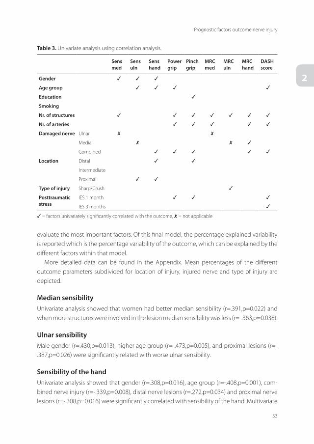
Table 3. Univariate analysis using correlation analysis.
sens med
sens uln
sens hand
Power grip
Pinch grip
mRc med
mRc uln
mRc hand
dAsh score
Gender ✓ ✓ ✓
Age group ✓ ✓ ✓ ✓
education ✓
smoking
Nr. of structures ✓ ✓ ✓ ✓ ✓ ✓ ✓
Nr. of arteries ✓ ✓ ✓ ✓ ✓
damaged nerve Ulnar ✗ ✗
Medial ✗ ✗ ✓
Combined ✓ ✓ ✓ ✓ ✓
Location Distal ✓ ✓
Intermediate
Proximal ✓ ✓
Type of injury Sharp/Crush ✓
Posttraumatic stress
IES 1 month ✓ ✓ ✓
IES 3 months ✓
✓ = factors univariately significantly correlated with the outcome, ✗ = not applicable

Chapter 2
34
regression analysis showed a significant model [F(4,60),16.158 p<0.001], that explained 54% of the variance. Male gender, higher age group, combined nerve injury and proximal nerve lesion were found to be negatively correlated with the sensory recovery of the hand, see Table IV.
Power gripUnivariate correlation analysis showed that age group (r=-.431,p=0.007), number of structures involved (r=-.365,p=0.004), number of arteries involved (r=-.310,p=0.015), combined nerve lesions (r=-.372,p=0.003) and posttraumatic stress at 1 month (IES1) (r=-.394,p=0.002) were significantly correlated with power grip. Lower age group and a lower number of structures and arteries involved were significantly associated with higher power grip. Combined nerve lesions and a higher degree of posttraumatic stress at 1 month were associated with lower power grip. Multivariate regression analysis resulted in a significant model [F(4,53),8.141, p<0.001] that explained 40% of the variance. This model showed that higher power grip was correlated with a lower age group, less arteries involved, a higher level of education and a lower degree of post-traumatic stress at 1 month. Beta coefficients of the multivariate regres-sion analysis are depicted in Table IV.
Table 4. Beta coëfficients related to outcome in multiple regression analysis
senshand
Powergrip
Pinchgrip
mRchand
dAshscore
Gender 20.659
Age group -6.516 -6.280 3.914
education 1.749 3.541
smoking
Nr. of structures -2.241
Nr. of arteries -11.992 -1.934
damagednerve
Ulnar
Medial
Combined -25.798 -2.759
Location
Distal
Intermediate
Proximal -17.505 -16.410
Type of injury Sharp/crush -2.226
Posttraum. stress
IES 1 month -.352 .617
IES 3 months -.368

35
Prognostic factors outcome nerve injury
2
Pinch gripNumber of structures involved (r=-.346,p=0.007), number of arteries involved (r=-.320,p=0.012), combined nerve lesions (r=-.256,p=0.046), distal nerve lesions (r=.372,p=0.003), posttraumatic stress at 1 month (r=-.257,p=0.046) and education (r=.306,p=0.025) were found to significantly correlate with pinch grip. A lower number of structures and arteries injured, combined nerve injuries, proximal nerve lesions, a higher degree of posttraumatic stress at 1 month, and higher educational level significantly correlated with less pinch grip. Multivariate analysis showed a significant model [F(3,51),7.960 p<0.001] that explained 33% of the variance. A proximal nerve lesion, lower educational level and more structures involved were associated with a lower pinch grip, see Table IV.
median motor recoveryFewer structures (r=-.437,p=0.011) and fewer arteries (r=-.344,p=0.046) involved in the lesion were univariate significantly related with a better median motor recovery measured by MRC.
ulnar motor recoveryMore injured structures (r=-.412,p=0.019) and crush nerve injuries (r=-.352,p=0.045) were univariate found to significantly correlated with worse ulnar motor recovery.
motor recovery of the whole handNumber of structures involved (r=-.411,p=0.001), number of arteries involved (r=-.387,p=.002), medial nerve injury (r=.362,p=0.004) and combined nerve injuries (r=-.289,p=0.024) were univariate significantly correlated to better motor recovery. Multivariate regression analysis showed a significant model [F(3,60),5.658 p=0.002] with explained 23% of the variance. The number of arteries involved, combined nerve lesions and crush injuries were related to lower motor recovery of the hand.
dAshAge group (r=.478,p<0.001), number of structures (r=.326,p=0.012) and arteries (r=.262,p=0.040) involved, combined nerve injuries (r=.277,p=0.031) and the degree of posttraumatic stress at 1 (r=.446, p<0.001) and 3 months (r=.423, p=0.001) were significantly correlated to the DASH results analyzed with univariate analysis. Multivariate analysis showed a significant model [F(3,59),7.782 p<0.001] explaining 29% of the variance. Age group and posttraumatic stress at 1 and 3 months were included in this model, see Table IV.
Chapter 2
36
discussiON
Our prospective study revealed that (1) gender, (2) age, (3) education, (4) number of structures involved, (5) number of arteries involved, (6) damaged nerve, (7) location of injury, (8) type of nerve injury and (9) posttraumatic stress at 1 and 3 months after repair of a peripheral nerve injury were all significant predictors for different aspects of functional outcome 12 months after injury. Of all possible prognostic factors only smoking was not found to be significantly correlated to functional outcome in this study.
sensibility of the handFifty-four percent of the variability of the sensibility of the hand can be explained using this prognostic model. Negative prognostic factors are male gender, higher age group, combined nerve injury and proximal nerve lesions. Interpretation of these results show that females have a 20% higher sensibility compared to men, a higher age group lowers the sensibility with 6.5%. As we defined 4 age groups the difference in hand sensibility between the oldest and youngest age group is 19.5%. Combined nerve injuries have a 25.8% lower sensibility compared to the ulnar and medial injuries and a proximal lesion lowers the sensibility with 17.5% compared to the other locations.
Power gripThe difference in return of power grip after peripheral nerve injury in our study population can be explained for 40%. This prognostic model included age group, educational level, number of arteries involved and amount of posttraumatic stress at 1 month. The power grip was 6.3% less in each higher age group. The difference between the oldest and youngest group is 18.85%. Education gives a 1.7% increased power grip per level. When we compare the highest education level with the lowest we found a difference of 10.5% in return of power grip.
Pinch gripThirty-three percent of the differences in the return of pinch grip following peripheral nerve trauma can be explained with our model. Prognostic factors are educational level, number of structures involved and proximal location of the injury. Education level gives an increase in pinch grip of 3.5%. Comparing the highest with the lowest educational level we find a difference of 21.2%. A higher number of structures involved give a 2.3% decrease in pinch grip. Proximal nerve lesions decrease the return of pinch grip with 16.4% when compared with other locations.
motor recovery of the whole handThe difference in motor recovery of the hand can be explained for 23% with this prognostic model. Factors included in this model are number of arteries involved, type of injury, com-

37
Prognostic factors outcome nerve injury
2
bined nerve injuries. On a zero to 10 scale the MRC score will be 1.9 points less for each involved artery. Combined nerve injuries will lower the MRC score with 2.8 points compared to single nerve injuries. Crush injuries decrease the MRC score of the hand with 2.2 points compared to sharp nerve injuries.
dAshThe outcome of the DASH score can be explained for 29% with the age group and amount of posttraumatic stress at 1 and 3 months postoperatively. A higher age group results in a 3.9 point higher DASH score. The older age group -thus- has an 11.7 point higher DASH score. Interestingly, our model showed a discrepancy between the 1 and 3 month posttraumatic stress assessment and the DASH. This is in contrast to our univariate analysis where both time points of posttraumatic stress level show a negative correlation with the DASH score.
Factors which showed to be important in predicting outcome of peripheral nerve injury can be subdivided in general characteristics and injury-specific characteristics. The general characteristics, age, gender, educational level and amount of posttraumatic stress at 1 and 3 months will be discussed first. It is commonly known that age has an important influence throughout all subjects in regenerative medicine[8, 27, 36]. Not unsurprisingly this is also the case in our study.
Gender clearly influences recovery of sensibility after nerve injury. The results of the uni- & multivariate analysis show that gender is only related as a prognostic factor to sensory recov-ery and not motor function recovery. Be that as it may, we can’t find a logic explanation in our data set regarding this prognostic factor. Nevertheless, the results demonstrate women in general will have a better sensory recovery after a peripheral nerve injury.
Educational level proofs to be of strong predictive value for sensibility, power grip and DASH outcome. People in the lower educational level group are more likely to perform (heavy) labor work. And thus, it is likely that their chances to be involved in a more severe hand trauma are higher compared to the white-collar population. Evidently this will influence the functional outcome after recovery. Secondly, it is our experience that educated patients are more willing to train intensively with their hand therapists, which has been reported to affect function, both for sensory and motor recovery[7]. The aid of hand therapy is of paramount importance in the rehabilitation of patients. In this study, all patients were assigned the same postopera-tive rehabilitation program. Amount of visits per participants were not recorded in this study.
Posttraumatic stress level at one and three months postoperatively has significant influ-ence on the recovery of power grip and DASH outcome[37].
Despite the obvious, no relation between smoking and one of the functional outcome factors was found in both the uni- and multivariate analysis. In two retrospective studies healing after nerve transection appeared to be affected adversely by cigarette smoke[12, 38]. However in an experimental study regarding the effects of tobacco smoke on nerve healing after crush injury in a rat model, no significant association between tobacco smoke exposure

Chapter 2
38
and delayed nerve recovery was found[39]. An experimental study in 2011 showed that expo-sure to cigarette smoke was associated with a slower functional recovery following ischemia/reperfusion injury of a peripheral nerve in the rat[40].
The injury-specific characteristics, number of structures and arteries involved, location, type of damaged nerve and the type of nerve trauma are all influencing functional recovery. Obviously, we found that the number of structures and arteries involved were predictors of outcome. Interestingly, studying the multivariate models, the relation was only found for motor recovery (e.g. power grip and MRC), and not for sensory recovery and DASH outcome.
cONcLusiON ANd FuTuRe PeRsPecTiVes
Gender, age, educational level, number of injured structures and arteries, damaged nerve, location of injury, type of nerve injury and posttraumatic stress at 1 and 3 months are strong predictors for functional recovery after peripheral injuries of the median and/or ulnar nerve of the forearm. Sensibility of the hand, powergrip and DASH score have proven to be the best three prognostic factors in our study. Our results may help us to inform patients about their expected recovery during the first year after injury. The information also assists clinicians in un-derstanding the outcome of patients with peripheral nerve injuries of the forearm. However, at this time, posttraumatic stress is probably the only factor which can be influenced in an early phase to optimize functional outcome results. To what extent the latter really improves outcome should be subject of further study.
AcKNOWLedGmeNTsThe authors would like to thank J.B. Jaquet3 MD, PhD, P.H.M. Spauwen4, Professor, MD, PhD, A. Hofman2 MD, PhD, M. Ritt, Professor5 MD, PhD and M. Kon, Professor6 MD, PhD for their contribution to the inclusion of patients and data collection.

39
Prognostic factors outcome nerve injury
2
ReFeReNces
1. Sorock, G.S., et al., Epidemiology of occupational acute traumatic hand injuries: a literature review. Safety Science,
2001. 38(3): p. 241-256.
2. O’Sullivan, M.E. and J. Colville, The economic impact of hand injuries. J Hand Surg Br, 1993. 18(3): p. 395-8.
3. Noble, J., et al., Analysis of upper and lower extremity peripheral nerve injuries in a population of patients with multiple
injuries. J Trauma, 1998. 45(1): p. 116-22.
4. Courtney, T.K. and B.S. Webster, Disabling occupational morbidity in the United States. An alternative way of seeing
the Bureau of Labor Statistics’ data. J Occup Environ Med, 1999. 41(1): p. 60-9.
5. Taylor, C.A., et al., The incidence of peripheral nerve injury in extremity trauma. Am J Phys Med Rehabil, 2008. 87(5):
p. 381-5.
6. Robinson, L.R., Traumatic injury to peripheral nerves. Muscle Nerve, 2000. 23(6): p. 863-73.
7. Bruyns, C.N., et al., Predictors for return to work in patients with median and ulnar nerve injuries. J Hand Surg Am,
2003. 28(1): p. 28-34.
8. Jaquet, J.B., et al., Median, ulnar, and combined median-ulnar nerve injuries: functional outcome and return to
productivity. J Trauma, 2001. 51(4): p. 687-92.
9. Abrams, M. and J. Widenfalk, Emerging strategies to promote improved functional outcome after peripheral nerve
injury. Restor Neurol Neurosci, 2005. 23(5-6): p. 367-82.
10. Desouches, C., et al., [Peripheral nerve repair: 30 centuries of scientific research]. Rev Neurol (Paris), 2005. 161(11): p.
1045-59.
11. Johnson, E.O., A.B. Zoubos, and P.N. Soucacos, Regeneration and repair of peripheral nerves. Injury, 2005. 36 suppl 4: p. S24-9.
12. al-Ghazal, S.K., et al., Results of clinical assessment after primary digital nerve repair. J Hand Surg Br, 1994. 19(2): p.
255-7.
13. Altissimi, M., G.B. Mancini, and A. Azzara, Results of primary repair of digital nerves. J Hand Surg Br, 1991. 16(5): p.
546-7.
14. Baysefer, A., et al., Surgical outcomes of ulnar nerve lesions in children. A retrospective clinical study. Pediatr Neuro-
surg, 2004. 40(3): p. 107-11.
15. Barrios, C. and J. de Pablos, Surgical management of nerve injuries of the upper extremity in children: a 15-year survey.
J Pediatr Orthop, 1991. 11(5): p. 641-5.
16. Makcinnon A, D.L., Surgery of the peripheral nerve. 1988, Thieme: New York. p. 98.
17. Posch, J.L. and F. Dela Cruz-Saddul, Nerve repair in trauma surgery. A ten-year study of 231 peripheral injuries. Ortho-
paedic Review, 1980. 9(3): p. 35-45.
18. Rosen, B., Recovery of sensory and motor function after nerve repair. A rationale for evaluation. J Hand Ther, 1996.
9(4): p. 315-27.
19. Trevett, M.C., et al., The functional results of ulnar nerve repair. Defining the indications for tendon transfer. J Hand
Surg Br, 1995. 20(4): p. 444-6.
20. Vastamaki, M., P.K. Kallio, and K.A. Solonen, The results of secondary microsurgical repair of ulnar nerve injury. J Hand
Surg Br, 1993. 18(3): p. 323-6.
21. Sakellarides, H., A follow-up study of 172 peripheral nerve injuries in the upper extremity in civilians. J Bone Joint Surg
Am, 1962. 44-A: p. 140-8.
22. Birch, R. and A.R. Raji, Repair of median and ulnar nerves. Primary suture is best. J Bone Joint Surg Br, 1991. 73(1): p.
154-7.
23. Kim, D.H., et al., Surgical management and outcomes in patients with median nerve lesions. J Neurosurg, 2001. 95(4):
p. 584-94.

Chapter 2
40
24. Leclercq, D.C., A.J. Carlier, and T. Khuc, Improvement in the results in sixty-four ulnar nerve sections associated with
arterial repair. Journal of Hand Surgery, 1985. 10(6 II): p. 997-999.
25. Kallio, P.K. and M. Vastamaki, An analysis of the results of late reconstruction of 132 median nerves. J Hand Surg Br,
1993. 18(1): p. 97-105.
26. Onne, L., Recovery of sensibility and sudomotor activity in the hand after nerve suture. Acta Chir Scand Suppl, 1962.
suppl 300: p. 1-69.
27. Ruijs, A.C., et al., Median and ulnar nerve injuries: a meta-analysis of predictors of motor and sensory recovery after
modern microsurgical nerve repair. Plast Reconstr Surg, 2005. 116(2): p. 484-94; discussion 495-6.
28. Hudak, P.L., P.C. Amadio, and C. Bombardier, Development of an upper extremity outcome measure: the DASH (dis-
abilities of the arm, shoulder and hand) [corrected]. The Upper Extremity Collaborative Group (UECG). Am J Ind Med,
1996. 29(6): p. 602-8.
29. Horowitz, M., N. Wilner, and W. Alvarez, Impact of Event Scale: a measure of subjective stress. Psychosom Med, 1979.
41(3): p. 209-18.
30. Shalev, A.Y., et al., Predictors of PTSD in injured trauma survivors: a prospective study. Am J Psychiatry, 1996. 153(2):
p. 219-25.
31. Bell-Krotoski, J.A., et al., Threshold detection and Semmes-Weinstein monofilaments. J Hand Ther, 1995. 8(2): p.
155-62.
32. Brandsma, J.W., et al., Manual muscle strength testing: intraobserver and interobserver reliabilities for the intrinsic
muscles of the hand. J Hand Ther, 1995. 8(3): p. 185-90.
33. Petersen, P., et al., Grip strength and hand dominance: challenging the 10% rule. Am J Occup Ther, 1989. 43(7): p.
444-7.
34. Mathiowetz, V., et al., Reliability and validity of grip and pinch strength evaluations. J Hand Surg Am, 1984. 9(2): p.
222-6.
35. Inc., S., SPSS Inc. Statistics for Windows. 2008: Chicago.
36. Guerra, W.K., J. Baldauf, and H.W. Schroeder, Long-term results after microsurgical repair of traumatic nerve lesions of
the upper extremities. Zentralbl Neurochir, 2007. 68(4): p. 195-9.
37. Ultee, J., et al., Early posttraumatic psychological stress following peripheral nerve injury: a prospective study. J Plast
Reconstr Aesthet Surg, 2013. 66(10): p. 1316-21.
38. Pankow, D., et al., Effect of carboxy- or methemoglobinemia on motor conduction velocity. Experientia, 1974. 30(9):
p. 1057-8.
39. Doezie, A.M., et al., Effects of tobacco smoke on recovery after nerve crush injury in rats. Ann Plast Surg, 2002. 49(6):
p. 628-34.
40. Rinker, B., et al., The effect of cigarette smoking on functional recovery following peripheral nerve ischemia/reperfusion
injury. Microsurgery, 2011. 31(1): p. 59-65.

41
Prognostic factors outcome nerve injury
2
APPeNdiXAppendix Table 5. Location of injury in relation to outcome at 12 monthsMean values (M) of the different outcome parameters subdivided for location of injury with standard deviation (SD). Values are mean percentages of recovery in comparison with normalized data.
Sens med
Sens uln
Sens hand
Power grip
Pinch grip
MRCmed
MRC uln
MRC hand
DASH score
M (SD) M (SD) M (SD) M (SD) M (SD) M (SD) M (SD) M (SD) M (SD)
distal48.9% (29.4)
63.5% (28.9)
76.6% (17.5)
67.6% (20.5)
65.3% (23.0)
7.2 (3.4)
4.0 (3.0)
5.9 (3.6)
16.0% (17.2)
intermediate40.0% (56.6)
52.5% (32.0)
72.6% (16.6)
62.8% (14.5)
46.4% (14.7)
4.0 (5.6)
4.6 (2.9)
4.1(3.1)
21.0% (21.1)
Proximate38.6% (35.8)
30.0% (16.2)
58.7% (21.2)
59.9% (29.5)
41.4% (33.0)
4.0(3.7)
4.0 (3.3)
4.0(3.5)
10.9% (10.1)
Appendix Table 6. Injured nerve in relation to outcome at 12 monthsMean values (M) of the different outcome parameters subdivided for injured nerve with standard deviation (SD). Values are mean percentages of recovery in comparison with normalized data.
Sensmed
Sensuln
Sens hand
Power grip
Pinch grip
MRC med
MRC uln
MRC hand
DASH score
M (SD) M (SD) M (SD) M (SD) M (SD) M (SD) M (SD) M (SD) M (SD)
median48.4% (32.0)
X74.1% (16.0)
71.0% (19.5)
63.3% (24.4)
6.6 (3.9)
X X14.9% (18.4)
ulnar X57.3% (30.4)
77.0% (17.9)
65.0% (22.1)
58.6% (24.9)
X4.6
(3.0)X
13.6% (12.4)
combined40.0% (25.7)
43.5% (27.4)
50.0% (21.8)
42.7% (8.9)
36.5% (26.8)
6.0(3.4)
2.3 (1.9)
2.3 (1.9)
30.3% (22.5)
Appendix Table 7. Type of injury in relation to outcome at 12 monthsMean values (M) of the different outcome parameters subdivided for type of injury with standard deviation (SD). Values are mean percentages of recovery in comparison with normalized data.
Sensmed
Sensuln
Sens hand
Power grip
Pinch grip
MRCmed
MRCuln
MRC hand
DASH score
M (SD) M (SD) M (SD) M (SD) M (SD) M (SD) M (SD) M (SD) M (SD)
sharp49.7% (30.5)
55.7% (30.2)
73.8% (19.3)
66.4% (20.0)
60.3% (25.9)
6.9(3.7)
4.4 (2.8)
5.6(3.5)
16.1% (17.5)
crush30.6% (30.4)
40.0% (28.3)
67.1% (14.4)
59.4% (31.0)
45.9% (20.1)
4.0(3.6)
0.0 (0.0)
3.1(3.6)
14.3% (11.4)

Chapter 3early posttraumatic psychological
stress following peripheral nerve injury: a prospective study
Jetske Ultee1 MD, PhD, Caroline A. Hundepool1 MSc, Tim H.J. Nijhuis1 MD, PhD, Anneloes L. van Baar2 PhD and Steven E.R Hovius1 Professor, MD, PhD
1 Dept. of Plastic, Reconstructive and Hand surgery, Erasmus MC, University Medical Center,
Rotterdam, The Netherlands2 Child and Adolescent studies, Utrecht University, Utrecht, The Netherlands
Journal of Plastic Reconstructive & Aesthetic Surgery. 2013 Oct;66(10):1316-21.

Chapter 3
44
ABsTRAcT
Background Psychological symptoms frequently accompany severe injuries of upper extremities and are described to influence functional outcome. As yet little knowledge is available about occurrence of posttraumatic psychological stress and the predictive charac-teristics of peripheral nerve injuries of the upper extremity for such psychological symptoms. In this prospective study the incidence of different aspects of early posttraumatic stress in patients with peripheral nerve injury of the forearm is studied as well as the risk factors for the occurrence of early psychological stress.
methods In a prospective study design patients with a median, ulnar or combined median-ulnar nerve injury were monitored for posttraumatic psychological stress symptoms with the Impact of Event Scale questionnaire (IES) up to three months post-operative.
Results Psychological stress within the first month after surgery occurred in 91.8% of the population (IES mean=22.0, SD=17.3). Three months post-operative, 83.3% (IES mean=13.3, SD=14.1) experienced psychological stress. One month postoperative 24.6 % and three months postoperative 13.3% of the patients had IES scores indicating for the need for psychological treatment. Female gender, adult age and combined nerve injuries were related to occurrence of psychological stress symptoms one month post-operative.
conclusions In the majority of these patients, peripheral nerve injury of the forearm is accompanied by early posttraumatic psychological stress, especially in female adults who suffered from combined nerve injuries.

45
Posttraumatic psychological stress outcome nerve injury
3
iNTROducTiON
Severe injuries of the upper extremity are frequently accompanied by psychological symp-toms like persistent, distressing recollection of trauma as flashbacks and nightmares, disturbed affect and anxiety, conditioned arousal to reminders of the trauma, and avoidance of stimuli reminiscent of the accident [1] [2]. When such symptoms last for more than one month, these are representative of posttraumatic stress disorder (PTSD). Posttraumatic stress disorder is a strongly disabling condition, impairing functionality and physical health of sufferers. PTSD is also associated with a high level of suicidal behavior (19%)[3]. Epidemiological studies indicate that posttraumatic stress disorder is becoming a major health concern; approximately one third of the population will be exposed to severe trauma during their lifetime and in 10-20% of these individuals posttraumatic stress disorder will develop. The incidence of posttraumatic stress disorder in injured adults admitted to a hospital is ranging between 14-46%[4]. It has been estimated that the prevalence of posttraumatic stress disorder in the general population ranges from 3-6%[5-7]. The symptoms of posttraumatic stress disorder reflect a psychological disturbance that often requires medication or psychotherapy. Cognitive-behavioral therapy, relaxation training, imagery techniques and systematic desensitization are all helpful treat-ment strategies to help the patient return to a productive everyday life [3, 5, 8-10]. Although early post-traumatic stress disorder is related to functional outcome after peripheral nerve injury, still little is known about the incidence and predictive factors for posttraumatic stress symptoms [1, 8, 9, 11, 12], how to assess incidence and intensity of post-traumatic psycho-logical stress symptoms and to identify risk factors for early psychological stress. Hence this study was designed to evaluate not only this prevalence after severe nerve injury of the up-per extremity, but also to possibly identify specific risk factors for the development of early psychological stress after a nerve injury.
PATieNTs eN meThOds
study populationBetween 2000 and 2003, patients with a peripheral nerve injury, operated in the University Medical Centre Rotterdam, the University Medical Centre Utrecht (UMCU), the MCRZ Rotter-dam, the Isala Clinics Zwolle, the University Medical Centre Nijmegen “St.Radboud”, the Univer-sity Medical Center Amsterdam “VU” were asked to participate in this study. Exclusion criteria were age under 12 years old, amputations of hand and fingers and insufficient knowledge of the Dutch language. By including amputation victims, not only the diversity of our patient trauma severity would increase but also objectively measuring recovery of the hand function would be a problem. According to these criteria 82 patients were asked to participate in the study. Three patients rejected participation and 16 patients who initially participated were lost

Chapter 3
46
during follow up. Finally, data of 61 patients was used for evaluation. A flowchart of the study population is presented in figure 1. Informed consent was provided by all participants. The psychological evaluation was assessed at one and three months post-operative. The Medical Ethical Committee of the University Medical Center Rotterdam approved this study.
AssessmentsTo measure the current degree of subjective impact of peripheral nerve trauma experienced by a person, the Impact of Event Scale (IES) was used at 1 and 3 months after injury. The IES, designed by Horowitz in 1979, includes 15 items that refer to the “the past seven days,” across the subscales of avoidance and intrusion and taps dimensions that are similar to defining symptoms of PTSD[3, 13]. The patients were invited to rate several questions as: “I have been avoiding things that could remind me of the accident”, “I had trouble falling asleep of stay-ing asleep because of pictures of thoughts of the accident reappeared” and “Each time I was reminded to the accident, I had recurrent feelings about it”. Each item is scored on a 4-point scale. These scores were reduced, weighed and labeled as follows: 0 points = not at all, 1 point = rarely, 3 points = sometimes, 5 points = often. Total IES scores ranges from 0-75 (75= worst score). As a general rule patients displaying scores above 30, have symptoms serious enough to be in need for psychological treatment [14].
Statistical analysis:The relation between type of injury, number of damaged structures, presence of arterial dam-age, age, gender, education level and posttraumatic psychological stress, 1 and 3 months after injury (measured with the IES), was studied with one-way analyses of variance or Pearson product moment correlation analyses. Variables that were found to be associated with early posttraumatic stress were then included in a multivariate regression model to evaluate their independent contribution in the prediction of posttraumatic psychological stress.
Figure 1. Flowchart of study population

47
Posttraumatic psychological stress outcome nerve injury
3
ResuLTs
study populationFor this study 61 patients with median (n=30; 49%), ulnar (n=24; 39%) and combined nerve injuries (n=7; 12%) that were reconstructed in the participating hospitals, were prospectively examined at 1 and 3 months. The study group consisted of 23 children and adolescents (38% ≤ 25 years) and 38 adults (62% ≥ 26 years). Most patients, 84% (n=51), were male and 16% (n=10) were female.
Psychological impactIncidence of psychological posttraumatic stress symptoms:The results from the IES revealed that 91.8% (mean = 22.0, SD = 17.3) of the study population experienced psychological stress within the first month after surgery (Table 1). Three months post operatively psychological stress was experienced by 83.3% (mean = 13.3, SD = 14.1). A score greater than 30, which indicates the need for psychological treatment, was found in 24.6 % of the patients at one month and in 13.3% at three months postoperative. Scores above 40, which is the threshold for severe psychological stress, were found in 14.8% of the patients when measured one month after surgery. At 3 months, 8,3% of the patients still scored above 40. A minor assessment score (<18) was reported by 44.3% (1 month) and 73.3% (3 months). A moderate score was found (18-39) in 36.1% (1 month) and 13.3% (3 months) of the patients. Psychological stress symptoms at one month and three months showed a strong correlation (r=.75, p<0.001).
Table 1. Psychological stress symptoms by gender, age and injury
ies 1 month ies 3 months
Mean SD N Mean SD N
Gender*
males 19,8 14,91 51 12,1 12,98 50
Females 33,3 24,41 10 19,1 18,38 10
Age*a
Adolescents 15,0 13,85 23 7,1 8,88 23
Adults 26,2 18,00 38 17,1 15,43 37
Injury
median 19,5 17,8 28 14,4 16,0 27
ulnar 21,2 15,9 27 11,8 12,4 27
combined*b 37.5 16,2 6 15,5 13,4 6
Total 22,0 17,3 61 13,3 14,1 60
IES, Impact of Event Score;*p<0.05
Chapter 3
48
Predictors for early post-traumatic psychological stress:Significant independent predictors of early post-traumatic psychological stress, one month post operative were gender (F(1,59) 5.45, p=.023) and age (F(1,59) 6.53, p=.013). At three months post operative only age was a predicting factor for psychological stress (F(1,58) 7.68, p=.007). Females had significantly more psychological stress symptoms at one month compared to males. Adults had more symptoms than adolescents, both at one and three months post operative. The one-way analysis of variance for stress symptoms according to type of nerve injury showed a non significant difference (F(1,59) 1.05 p=0.310), but post hoc comparisons indicated that combined nerve injuries differed from median nerve lesions (mean difference=18.0, SE=7.56, p=.021) and that they differed from ulnar nerve lesion (mean difference=16.3, SE=7.58, p=.036). On the other hand, these relationships were not found for the measurements three months postoperative. The mean scores for the psychological stress symptoms in relation to these predictors are presented in Table 1. The predictor education (range 1-7) showed an average of 4.1. The number of damaged structures was averaged with 5.5. 57% of the patients had arterial damage and the location of the injury was in 69% of the patients distally orientated. All of these factors were not associated with the amount of psychological stress symptoms at one and three months post-operatively.
Next, a multivariate backward regression analysis was executed, using psychological stress symptom score at one month postoperative as dependent variable and age, gender and type of nerve injury as predictors. This resulted in a significant model (F(2,58) 5.47 p=.007) that explained 40% of the variance. Both age (β=.28, p=0.028) and gender (β=.25, p=0.048) contributed significantly, whereas type of injury (specifically median nerve injury that was least associated with psychological stress symptoms of the different kinds of injury) did not contribute significantly (β=-.034, 0.780).
discussiON
This study showed that psychological stress symptoms occur in most patients after peripheral nerve injuries of the upper extremities within the first month after surgery. Our prospective study showed that almost all patients (92%) experienced psychological stress symptoms and 83% still experienced psychological stress three months post operatively. Psychological treatment was indicated in 25% one month postoperative and still in 13% at three months postoperative, according to the IES.
In medical practice attention following hand trauma is primarily focused on functional recovery. However, posttraumatic stress disorder has been found to contribute more to patient’s perceived general health than the degree of physical functioning or the severity of injury[15]. Psychological results of hand injuries are immediately apparent; there is fear of potential further injury and the injured hand acts as a constant reminder. An example of a

49
Posttraumatic psychological stress outcome nerve injury
3
hand injury with serious functional consequences and often a disappointing recovery is a peripheral nerve injury of the forearm. Earlier studies demonstrated a mean time of returning to work of 31.9 weeks and 41% of patients did not return to work at all in the first year after peripheral nerve injury[16, 17]. A study that investigated even more severe trauma of the forearm showed that as much as 45,2% of patients did not return to work within a year after injury[18].
In the current prospective study the majority of the patients with peripheral nerve injuries of the upper extremities experienced psychological stress symptoms within the first three months after surgery. One month postoperative a quarter of all patients displayed psycho-logical stress with the severity that was indicative for psychological treatment. Three months postoperative 13% of the patients suffered from severe psychological stress. These findings are in agreement with earlier study results. In a retrospective study of Jaquet et al. 94% of patients with peripheral nerve lesions demonstrated additional posttraumatic psychological stress. In that retrospective study, psychological morbidity, measured with the impact of event scale, was found in 36% of the patients[19].
One should not only be aware of the importance of this high prevalence of posttraumatic psychological stress after peripheral nerve injury. Also, data as part of a bigger prospective study also indicated early psychological stress after peripheral nerve injury as a predictive factor for power grip and daily activities -measured with the daily activity of shoulder and hand questionnaire (DASH)- twelve months after injury[19]. Consequently, a more severe posttraumatic stress one month after injury was associated with greater motor disability.
Early identification of patients with psychological stress symptoms and evidently ap-propriate treatment could improve functional outcome and facilitate earlier return to work in patients with severe hand trauma. Grunert et al. already demonstrated (1991) that early intervention reduced the effects of litigation on stress symptoms[11]. Van der Kolk et al (1994) suggests that patients with more recently detected stress symptoms have better response to treatment[20]. Besides, early detection, support and treatment of people with emotional problems, costs less than the long-term costs associated with failure to provide early and effective treatment for the society[7]. Once actual PTSD has developed, it is often a chronic and recurring condition [3]15. Although effects of therapy in patients with PTSD and peripheral nerve injuries need further study, our results indicate that early support and intervention could optimize surgical treatment results. Earlier research has demonstrated effectiveness of cognitive-behavior therapy, group therapy, and exposure therapy (i.e. the patients repeatedly relive the frightening experience under controlled conditions to help him or her to think through the trauma)[8]. In 80% of people with PTSD, depression, or anxiety disorders, alcohol or other substance abuse occurs[3, 21]. The likelihood of treatment success of peripheral nerve injuries of the upper extremities is therefore increased when these other –psychological- conditions are appropriately diagnosed and treated accordingly. Nevertheless, psychological stress symptoms will not affect all patients suffering from a similar injury. Hence, identification
Chapter 3
50
of risk factors that may lead to psychological stress symptoms is needed for optimal selection and consequently treatment of the specific patient population that suffers from PTSD. Previ-ous exposure to trauma and a personal or family history of psychiatric disorders, particularly depressive disorders were found to be predictors of PTSD.
In our study, significant independent predictors of early post-traumatic psychological stress were female gender, adult age and a combined nerve injury. Education, concomitant arterial bleeding, number of damaged structures and location of injury were not significantly associated with the amount of psychological stress at one and three months postoperatively.
Consequently, female gender was found to be predictive for early posttraumatic stress. Multiple research studies have demonstrated that, in general, women have higher rates of PTSD compared to men[4, 22-27] however the results are mixed regarding the prevalence of PTSD in males or females among individuals who have experienced an injury or disability. Perry et al (1987) [28] found that individuals who had severe burns, the PTSD was more likely to emerge in males. Roca et al (1992) [29] found that gender did not significantly predict posttraumatic stress levels among individuals with burn injuries.
Another factor predictive for early posttraumatic stress in our study was age; Adult age (≥ 26 years) was associated with a higher degree of early post-traumatic psychological stress, one month post-operative. In contrast, several studies on other diseases, the younger age -actually- was indicated as a predictor of PTSD. In a recent study where the role of physi-cal, psychosocial and compensation-related factors in the development of 129 patients of an adult population with PTSD following major trauma were examined, it was found that PTSD was significantly associated with younger age (P < 0.0001)[12]. However, note that in our study children above 12 years and adolescents were included, whereas the other studies concerned adults.
Combined nerve injuries were associated with a higher degree of early psychological stress one month post-operative than median nerve injury. However, the relatively small number of patients that was diagnosed with this type of injury could explain the relatively low correlation. As such, further research on the type of injury in relation to psychological stress symptoms seems indicated. Severity of injury does not appear to be a strong predictor for PTSD. Feinstein in 1991[30] and Michaels in 1999[4] were unable to find a relation between severity of injury and PTSD. The major risk is conveyed by the experience of the fear, horror and helplessness and the ongoing pain and uncertainty caused by the injury[31]. In peripheral nerve injury patients and especially patients with combined nerve injuries helplessness and uncertainty caused by the injury is substantial.
A meta-analysis of 77 studies by Brewin et al., in 2000 showed that both female gender, younger age and severity of injury is associated with a higher risk of developing PTSD[32].
The remaining question is what mechanism is responsible for impaired functional outcome in peripheral nerve patients with psychological stress symptoms. Research has demonstrated that PTSD clearly alters a number of fundamental brain mechanisms[5]. Neurobiological stud-

51
Posttraumatic psychological stress outcome nerve injury
3
ies indicate that three structures in the brain are involved with this disorder: the amygdala, medial prefrontal cortex and hippocampus. Neuroimaging research showed hyperrespon-sivity within the amygdala, reduced neuronal responsivity and cortical volumes in medial prefrontal structures and reduced hippocampal volume[33]. Besides glucocorticoid hyper-sensitivity three neurohormone systems (noradrenegic system, serotonergic system and the hypothalamic-pituitary-adrenal [HPA] axis) are dysregulated in PTSD patients, which represent possible neurochemical substrates for an unbalanced modulation of the limbic circuit[5, 33-35]. Abnormalities or dysfunctional processes in brain chemicals may result in problems with coping, behavior, learning and memory. These skills are particularly important in patients with peripheral nerve injuries, who have to adapt to alterations in recognition of objects and touch perception, due to abnormal innervation and misdirection of nerves.
In a review by Yehuda et al in 2011 little evidence is found for specific genes associated with increased risk of the developing PTSD[36]. Twin studies suggest that PTSD symptoms are moderately heritable[37]. Further research in the field of genetics is recommended to determine the role of genes in psychological stress after trauma.
The accompanying symptoms of PTSD like depression, sleeping disorders and loss of motivation could also be responsible for impaired functional recovery and a late return to work. Furthermore there is evidence that psychological stress adversely affects the immune system and could influence and delay wound healing [5]. When striving for optimal recovery, aside from surgery, psychological stress in particular could be one of the few factors which we can try to influence in order to improve their functional outcome. Hence, early detection of posttraumatic psychological stress symptoms and subsequent support and intervention in this patient group might improve their functional outcome.
cONcLusiONs
In conclusion this study demonstrates a high prevalence of early posttraumatic psychologi-cal stress following peripheral nerve injury of the forearm. In the majority of these patients, peripheral nerve injury of the forearm is accompanied by early posttraumatic psychological stress, especially in female adults who suffered from combined nerve injuries.

Chapter 3
52
ReFeReNces
1. Grunert, B.K., et al., Early psychological aspects of severe hand injury. J Hand Surg Br, 1988. 13(2): p. 177-80.
2. Grob, M., et al., The psychological impact of severe hand injury. J Hand Surg Eur Vol, 2008. 33(3): p. 358-62.
3. Ballenger, J.C., et al., Consensus statement on posttraumatic stress disorder from the International Consensus Group
on Depression and Anxiety. J Clin Psychiatry, 2000. 61 suppl 5: p. 60-6.
4. Michaels, A.J., et al., Posttraumatic stress disorder in injured adults: etiology by path analysis. J Trauma, 1999. 47(5): p.
867-73.
5. Kiecolt-Glaser, J.K., et al., Slowing of wound healing by psychological stress. Lancet, 1995. 346(8984): p. 1194-6.
6. Brunello, N., et al., Posttraumatic stress disorder: diagnosis and epidemiology, comorbidity and social consequences,
biology and treatment. Neuropsychobiology, 2001. 43(3): p. 150-62.
7. Kessler, R.C., Posttraumatic stress disorder: the burden to the individual and to society. J Clin Psychiatry, 2000. 61 suppl 5: p. 4-12; discussion 13-4.
8. Grunert, B.K., et al., Treatment of posttraumatic stress disorder after work-related hand trauma. J Hand Surg Am,
1990. 15(3): p. 511-5.
9. Grunert, B.K., et al., Flashbacks after traumatic hand injuries: prognostic indicators. J Hand Surg Am, 1988. 13(1): p.
125-7.
10. Foa, E.B., Psychosocial treatment of posttraumatic stress disorder. J Clin Psychiatry, 2000. 61 suppl 5: p. 43-8; discus-
sion 49-51.
11. Grunert, B.K., et al., Effects of litigation on maintenance of psychological symptoms after severe hand injury. J Hand
Surg Am, 1991. 16(6): p. 1031-4.
12. Harris, I.A., et al., Predictors of post-traumatic stress disorder following major trauma. ANZ J Surg, 2008. 78(7): p.
583-7.
13. Horowitz, M., N. Wilner, and W. Alvarez, Impact of Event Scale: a measure of subjective stress. Psychosom Med, 1979.
41(3): p. 209-18.
14. Shalev, A.Y., et al., Predictors of PTSD in injured trauma survivors: a prospective study. Am J Psychiatry, 1996. 153(2):
p. 219-25.
15. Michaels, A.J., et al., Posttraumatic stress disorder after injury: impact on general health outcome and early risk assess-
ment. J Trauma, 1999. 47(3): p. 460-6; discussion 466-7.
16. Jaquet, J.B., et al., Median, ulnar, and combined median-ulnar nerve injuries: functional outcome and return to
productivity. J Trauma, 2001. 51(4): p. 687-92.
17. Bruyns, C.N., et al., Predictors for return to work in patients with median and ulnar nerve injuries. J Hand Surg Am,
2003. 28(1): p. 28-34.
18. Jaquet, J.B., et al., Spaghetti wrist trauma: functional recovery, return to work, and psychological effects. Plast Recon-
str Surg, 2005. 115(6): p. 1609-17.
19. Jaquet, J.B., et al., Early psychological stress after forearm nerve injuries: a predictor for long-term functional outcome
and return to productivity. Ann Plast Surg, 2002. 49(1): p. 82-90.
20. van der Kolk, B.A., et al., Fluoxetine in posttraumatic stress disorder. J Clin Psychiatry, 1994. 55(12): p. 517-22.
21. Breslau, N., et al., Trauma and posttraumatic stress disorder in the community: the 1996 Detroit Area Survey of Trauma.
Arch Gen Psychiatry, 1998. 55(7): p. 626-32.
22. Malt, U., The long-term psychiatric consequences of accidental injury. A longitudinal study of 107 adults. Br J Psychia-
try, 1988. 153: p. 810-8.
23. Tsuang, M.T., M. Boor, and J.A. Fleming, Psychiatric aspects of traffic accidents. Am J Psychiatry, 1985. 142(5): p.
538-46.
24. Ehlers, A., R.A. Mayou, and B. Bryant, Psychological predictors of chronic posttraumatic stress disorder after motor
vehicle accidents. J Abnorm Psychol, 1998. 107(3): p. 508-19.

53
Posttraumatic psychological stress outcome nerve injury
3
25. Breslau, N., et al., Psychiatric sequelae of posttraumatic stress disorder in women. Arch Gen Psychiatry, 1997. 54(1):
p. 81-7.
26. Zatzick, D.F., et al., Predicting posttraumatic distress in hospitalized trauma survivors with acute injuries. Am J Psy-
chiatry, 2002. 159(6): p. 941-6.
27. Fullerton, C.S., et al., Gender differences in posttraumatic stress disorder after motor vehicle accidents. Am J Psychia-
try, 2001. 158(9): p. 1486-91.
28. Perry, S.W., et al., Pain perception in burn patients with stress disorders. J Pain Symptom Manage, 1987. 2(1): p. 29-33.
29. Roca, R.P., R.J. Spence, and A.M. Munster, Posttraumatic adaptation and distress among adult burn survivors. Am J
Psychiatry, 1992. 149(9): p. 1234-8.
30. Feinstein, A. and R. Dolan, Predictors of post-traumatic stress disorder following physical trauma: an examination of
the stressor criterion. Psychol Med, 1991. 21(1): p. 85-91.
31. McFarlane, A.C., Predictors of post-traumatic stress disorder after major injury. ANZ J Surg, 2008. 78(7): p. 533-4.
32. Brewin, C.R., B. Andrews, and J.D. Valentine, Meta-analysis of risk factors for posttraumatic stress disorder in trauma-
exposed adults. J Consult Clin Psychol, 2000. 68(5): p. 748-66.
33. Vasterling, J.J. and C.R. Brewin, Neuropsychology of PTSD: Biological, cognitive, and clinical perspectives, in Neuropsy-
chology of PTSD: Biological, cognitive, and clinical perspectives. 2005, Guilford Press; US: New York, NY. p. 27-58.
34. Nutt, D.J., The psychobiology of posttraumatic stress disorder. J Clin Psychiatry, 2000. 61 suppl 5: p. 24-9; discussion
30-2.
35. Yehuda, R., Post-traumatic stress disorder. N Engl J Med, 2002. 346(2): p. 108-14.
36. Yehuda, R., et al., The role of genes in defining a molecular biology of PTSD. Dis Markers, 2011. 30(2-3): p. 67-76.
37. Afifi, T.O., et al., The role of genes and environment on trauma exposure and posttraumatic stress disorder symptoms:
a review of twin studies. Clin Psychol Rev, 2010. 30(1): p. 101-12.


PART IIevaluation


Chapter 4ultrasonographic quantifi cation of intrinsic
hand muscle cross-sectional area; reliability and validity for predicting muscle strength
Behnam Mohseny, MD1, Tim H. Nijhuis, MD, PhD1, Caroline A. Hundepool, MSc1, Wim G., Janssen, MD2 Ruud W. Selles, PhD1,2., Henk Coert, MD, PhD1
1 Departments of Plastic, Reconstructive and Hand Surgery, Erasmus MC, University Medical
Center, Rotterdam, The Netherlands.2 Departments of Rehabilitation Medicine, Erasmus MC, University Medical Center, Rotterdam,
The Netherlands.
Archives of Physical Medicine and Rehabilitation. 2015 May;96(5):845-53.

Chapter 4
58
ABsTRAcT
Background To investigate whether ultrasonographic measurement of the cross-sectional area (CSA) of the intrinsic hand muscles can be used to predict muscle strength in a valid and reliable manner, and to determine if this method can be used for follow-up of pa-tients with peripheral nerve injury between the wrist and elbow. Design: Repeated-measures cross-sectional study.
methods Healthy adults (n=31) and patients with ulnar and median nerve injuries (nZ16) between the wrist and elbow who were visiting the Erasmus Medical Center or Maas-stad Hospital were included in the study (N=47). Correlation between measured muscle CSA and strength and assessment of inter- and intrarater reliability. Ultrasound and strength measurements of the intrinsic hand muscles were conducted bilaterally. To establish validity, the CSA of 4 muscles (abductor digiti minimi, first dorsal interosseus, abductor pollicis brevis, opponens pollicis) was compared with strength measurements of the same muscles con-ducted with the Rotterdam Intrinsic Hand Myometer. Repeated measures were conducted to assess inter- and intrarater reliability.
Results The assessed CSA strongly correlated with strength measurements, with correlations ranging from 0.82 to 0.93 in healthy volunteers and from 0.63 to 0.94 in patients. Test-retest reliability showed excellent intrarater reliability (intraclass correlation coefficient range, 0.99-1.00) in patients and volunteers and good interrater reliability (intraclass correla-tion coefficient range, 0.88-0.95) in healthy volunteers.
conclusions We found that ultrasound is a valid and reliable method to assess the CSA of specific muscles in the hand. Therefore, this technique could be useful to monitor muscle reinnervation in patients suffering from peripheral nerve injury as a valuable addition to strength dynamometers.

59
Ultrasound nerve injury clinical
4
iNTROducTiON
Peripheral nerve injury is disabling and has a prevalence of approximately 3% in trauma pa-tients.1 As a direct result of median or ulnar nerve injury, muscles are denervated and atrophy consequently occurs in the weeks after injury. This can result in severe loss of hand function. The outcome with or without surgery is often unpredictable and disappointing. Most patients suffer considerable posttraumatic psychological stress because of the daily confrontation with their inability to use the injured hand and the uncertainty concerning the functional outcome. This psychological stress is believed to adversely affect functional recovery.2,3 To provide patients and surgeons with more information concerning the outcome of surgery and therapy, muscle strength measurement has become an integral part of the physical examination and longitudinal followup after nerve injury.4-7
The methods most frequently used to evaluate outcome of motor function of the hand are manual muscle testing introduced by the British Medical Research Council (MRC) and grip and pinch strength dynamometers.8,9 In addition, more recently, the Rotterdam Intrinsic Hand Myometer (RIHM) has been developed to measure individual finger or thumb strength, allowing more direct assessment of intrinsic muscle strength.10
However, manual muscle testing and dynamometers have a number of limitations. First, the MRC scale used for manual muscle testing is an ordinal scale with disproportional dis-tances between grades and provides little information on recovery in the 4 to 5 segment.11 Second, strength measurements generally assess several muscles at the same time and do not allow the examiner to measure individual muscles. Because the assessed muscles are rarely innervated by the same nerve, strength measured is not a direct representation of regeneration of one specific nerve. Third, strength measurement is often influenced by other factors than muscle strength alone (eg, injuries of associated soft tissues and tendons, pain inhibition, patient motivation, malingering). Another limitation specific to dynamometers is that they cannot be used by patients who score <3 on the MRC scale. Hence, dynamometers do not allow detection of early muscle recovery.10,12,13 Therefore, new methods that can tackle these limitations are necessary to provide patients and clinicians with more detailed and specific information.
Clinical studies have shown ultrasounds to be highly reliable for measurement of muscle cross-sectional area (CSA) and to have a strong correlation with magnetic resonance imaging findings.14,15 Ultrasonography has also been described as a useful tool in the diagnosis of neuromuscular disorders and for the documentation of the course of muscle atrophy and mesenchymal. abnormalities in neurogenic muscle lesions in a rabbit model.16,17 Addition-ally, a previous study conducted by our research group showed high correlations between the gastrocnemius muscle index and muscle thickness measured with ultrasound in a rat sciatic nerve transection model.18 Muscle atrophy and regeneration, objectified by repeated

Chapter 4
60
measurements of the muscle CSA in time, may therefore serve as a monitoring tool to evalu-ate muscle reinnervation, even in the early stages of recovery.
The purpose of this study was to determine whether ultrasonographic measurement of the CSA of the intrinsic hand muscles can be used as a valid and reliable method to predict muscle strength and whether it can be used to monitor muscle reinnervation in patients suffering from nerve injury.
meThOds
ParticipantsEthical approval for this study was obtained in the Erasmus Medical Center and Maasstad Hos-pital. Subjects visiting either hospital were approached for inclusion. After signing informed consent, 47 subjects participated in this study. A short questionnaire was used to determine hand dominance and the presence of known upper-extremity problems or factors that could influence hand strength. We excluded all subjects with preexisting deformities or myopathies of the hand. Subjects unable to use the RIHMa because of insufficient strength or pain and subjects aged <18 years were also excluded. Subjects were divided into 2 groups: a group of healthy volunteers and a group of patients with nerve injuries of the ulnar or median nerve or both nerves combined (Table 1).
Thirty-one healthy volunteers (15 men, 16 women; mean age, 27.5y; age range, 21-54y) were included for normative reference. Sixteen patients (12 men, 4 women; mean age, 35.9y; age range, 19-61y) had a laceration of the ulnar nerve (n=8), median nerve (n=6), or both nerves (n=2) in the forearm between the elbow and wrist. The patients were operated on by hand surgeons and received hand therapy at the rehabilitation department of one of the participating hospitals. All patients had combined nerve and tendon injuries, except for 1 patient who only had an isolated ulnar nerve injury. The average time period after injury was 15 months (range, 6-39mo). Most injuries were caused by sharp objects causing injury to the forearm between the elbow and wrist.
Table 1. Group characteristics
Characteristic Healthy Volunteers Patients
Sex 15 male 16 female 12 male 4 female
Hand dominance 26 right 5 left 14 right 2 left
Damaged nerve NA 8 median nerve 10 ulnar nerve
Meange age (range) (y) 27.5 (21-54) 35.9 (19-61)
Abbreviation: NA, not applicable
61
Ultrasound nerve injury clinical
4
ultrasound measurementsThe ultrasonographic evaluation was conducted using a SonoSite TITAN ultrasound system,b with a 5- to 10-MHz broadband linear array probe. We used 2 examiners who were blinded to each other’s measurements. The first examiner also performed a second measurement with a pause of at least 15 minutes to minimize the recall bias. The examiner and patient were seated at opposite sides of a table. The patient was asked to rest the arms on the table in a relaxed position, with the dorsal aspect of the hand and forearm in contact with the examination table. The thumb was held at a 45degree angle to the midline of the hand. The array probe was placed directly onto the skin using sufficient transmission gel.c All scans were made from the palmar surface of the hand.
We assessed the CSA of 2 intrinsic hand muscles, abductor digiti minimi (ADM) and first dorsal interosseus (DI), which are innervated by the ulnar nerve, and 2 muscles innervated by the median nerve, abductor pollicis brevis (APB) and opponens pollicis (OP). The positions used to measure the CSA of the muscles were standardized to find the most reproducible measure of the intrinsic hand muscles at, or close to, their thickest point (Fig. 1). The scan planes were determined before the start of the study by pilot ultrasound evaluation of the intrinsic hand muscles in healthy volunteers, and the positions were validated using cadaver models.
All ultrasound measurements were conducted bilaterally followed by strength measure-ments using the RIHM. In the patients, only the muscles innervated by the injured nerve were measured. To assess interrater reliability, a second examiner also conducted ultrasound measurements bilaterally in 10 healthy volunteers and 15 patients. Then all healthy volunteers and 13 patients underwent a second ultrasound measurement by the first examiner to assess interrater reliability. Some patients were unwilling to undergo a second and/or third ultra-sound examination.
Processing and analysis of the ultrasound imagesScans of the CSA of the muscles for each subject were saved as bitmap image files and trans-ferred to a personal computer using SiteLink Image Manager 2.2.1.b The analysis of the images was conducted in Adobe Photoshop CS6 Extended.d The lasso tool was used to precisely select the CSA of the muscle in question. The hyperechoic fasciae surrounding the muscles were excluded from the selection in all measurements. The correct measurement scale was set using the depth scale depicted on the right edge of the ultrasound images and the ruler tool to determine the pixel/ distance ratio. Finally, the CSA was computed using the analysis option. The muscles were then outlined using the stroke option, and the images were saved for future reference (Fig. 2).

Chapter 4
62
strength measurementsStrength measurements were conducted with the RIHM. The intra- and interrater reliability and validity of the RIHM has been proven in, among others, patients with median and ulnar nerve injuries and patients with Charcot-Marie-Tooth disease.10,19 We used the protocol previously described by Schreuders et al.10 In short, the patient was shown how to position the fingers and thumb and was instructed to keep the finger or thumb in that position with maximum strength. Force was applied to the same anatomic reference points as used for
Figure 1. Schematic representation of the 3 different scanning planes indicated with the letters A, B, and C. The letters correlate with the inserts on the right, indicated with the same letters, depicting the probe positions during measurement of the assessed muscles. (A) The thenar muscles were measured at the proximal third of the first metacarpal bone, with the probe positioned on the lateral side of the thenar eminence, moving the muscles to the palmar surface. The flexor pollicis longus tendon was used as an anatomic landmark, placing the probe as perpendicular as possible to the tendon. (B) The first DI muscle was measured at the proximal third of the second metacarpal bone, with the scanning plane as perpendicular as possible to its muscle fibers. (C) The ADM was measured at the proximal third of the fifth metacarpal. The probe was placed on the ulnar side of the hand, moving the hypothenar muscles to the palmar surface of the hand. The scanning plane was as perpendicular as possible to the muscle fibers of the ADM.

63
Ultrasound nerve injury clinical
4
Figure 2. Ultrasound images after analysis; the 4 assessed muscles are outlined.
Chapter 4
64
the manual muscle testing and described by Brandsma et al.4,20 The force applied with the RIHM was slowly increased while the patient was verbally encouraged to hold the finger or thumb in place. Four movements were used to assess the strength of the 4 intrinsic hand muscles also used for the ultrasonography measurements: the muscles innervated by the ulnar nerve were assessed by measuring ulnar abduction of the little finger (mainly the ADM) and by measuring radial abduction of the index finger (mainly the DI). Muscles innervated by the median nerve were assessed by the anteposition of the thumb (mainly the APB) and the opposition of the thumb (mainly the OP). All movements were tested 3 times, and the mean was calculated.
statistical analysisFor all statistical analyses, IBM SPSS Statistics 20e was used. The correlation between the CSAs measured with the ultrasound scans and the strength measurements obtained with the RIHM was determined for each of the assessed muscles separately using the Pearson correlation coefficient. The test-retest reliability of the CSA measurements was determined with the intra-class correlation coefficient (ICC) (2- way mixed model, absolute agreement, single measures), standard error of measurement, smallest detectable difference (SDD), and normalized small-est detectable difference. Bland-Altman plots were used to visualize the reliability.21
The ICC is the ratio of the between-subjects variability over the between-subjects variabil-ity plus the within-subject variability. 22 The SDD is the amount of change needed between repeated tests to detect a real difference. For a 95% confidence level, the SDD is calculated as 1.96 x √2 x standard error of measurement. The standard error of measurement is the square root of the error variance. When 2 repeated measurements differ more than the SDD, the change represents a real change in CSA. The normalized SDD is the SDD expressed as a percentage of the mean CSA, which makes it easier to interpret in clinical use.
In clinical practice, the contralateral side is often used as an internal control, and often the 10% rule is used for side-to-side comparison. This rule indicates that the grip strength is ap-proximately 10% greater in the dominant hand than the nondominant hand.23 A relative low intrasubject side-to-side variability is important when the healthy contralateral side is used as the internal control.24 To see whether the CSAs of the muscles in the uninjured hand can provide a normative reference value for that person, we compared the data collected from the dominant hands with the data collected from the nondominant hands in the healthy volunteers for each muscle separately by using the paired-samples t tests. Also, the differ-ences in muscle CSA between the dominant and nondominant hands were calculated using 1 - (nondominant hand/dominant hand) and were expressed as percentages. For comparison, the same calculations were also conducted for the strength measurements.

65
Ultrasound nerve injury clinical
4
ResuLTs
The assessed muscles could all be identified clearly after a short learning curve. Execution of ultrasound measurements of both hands took an average of 10 minutes. Strength measure-ments took an average of 15 to 20 minutes to complete per subject.
correlation between csA and strength measurementThere was an overall high correlation between the CSA and strength measurements in the healthy volunteers (r=0.82-0.93, P<.01). High correlations were also found between the CSA and strength measurements of the DI and OP muscles in the patients (r=0.94 and r=0.85, P<.01) (table 2). The correlations for both groups are visualized with scatter plots (Fig. 3).
Subanalyses of the dominant (r=0.80-0.93, P<.01) and nondominant (r=0.81-0.93, P<.01) hand showed similar values in the healthy volunteers.
intrarater reliabilityThe reliability between the 2 sessions, expressed as an ICC, was excellent for all 4 assessed muscles. The standard error of measurement was approximately the same for all muscles in the healthy volunteers and ranged between 0.02 and 0.03 in the patients. The normalized SDDs were between 4% and 8% in the healthy volunteers and between 4% and 13% in the patients (table 3).
The Bland-Altman plots clearly show that all data are evenly distributed around the zero line in the healthy volunteers and patients (figs 4 and 5).
Table 2. Correlations between CSAs and strength measurements
Muscle Pearson correlation (r) R2 Significance (2 tailed)
Healthy volunteers
ADM (n=62) 0.87 0.75 <.01
DI (n=62) 0.83 0.70 <.01
OP (n=62) 0.93 0.86 <.01
APB (n=62) 0.82 0.67 <.01
Patients
ADM (n=9) 0.74 0.55 <.05
DI (n=10) 0.94 0.88 <.01
OP (n=8) 0.85 0.71 <.01
APB (n=7) 0.63 0.39 .13

Chapter 4
66
interrater reliabilityThe reliability between the 2 examiners, expressed as an ICC, was good (ICCs range, 0.88-0.95; P<.01) for all 4 assessed muscles in the healthy volunteers. The normalized SDDs were between 15% and 25%.
Among the patients, reliability was acceptable in patients with median nerve injury. The ICC was 0.54 (P=.052) for the ADM, 0.27 (P=.17) for the DI, 0.75 (P<.01) for the OP, and 0.58 (P=.04) for the APB. The normalized SDDs ranged between 51% and 74% (table 4).
Table 3. Intrarater reliability
Muscle Mean CSA ± SD (cm3) ICC SEM (cm3) SDD (cm3) %SDD (cm3)
Healthy volunteers
ADM (n=62) 1.27 ± 0.34 1.00 0.03 0.07 5.5
DI (n=62) 1.85 ± 0.42 1.00 0.03 0.08 4.3
OP (n=62) 1.32 ± 0.34 0.99 0.03 0.09 6.6
APB (n=62) 1.14 ± 0.27 0.99 0.03 0.09 8.3
Patients
ADM (n=9) 0.94 ± 0.05 0.99 0.02 0.07 7.3
DI (n=10) 1.21 ± 0.42 1.00 0.02 0.05 4.4
OP (n=8) 0.81 ± 0.47 1.00 0.02 0.07 8.5
APB (n=7) 0.71 ± 0.35 0.99 0.03 0.09 12.7
Abbreviations: %SDD, normalized SDD; SEM, standard error of measurement.
Figure 3. Scatter plots showing the correlation between the CSA and strength measurement for all muscles in healthy volunteers (left) and patients (right).An overall high correlation between the CSA and strength measurements is seen in the healthy volunteers. High correlations were also found between the CSA and strength measurements of the DI and OP muscles in patients.

67
Ultrasound nerve injury clinical
4
Figure 4. Bland-Altman plots of the mean CSA of the ADM (top left), DI (top right), OP (bottom left), and APB (bottom right) muscles versus the difference between the CSA in test and retest for the patient group. All data are evenly distributed around the zero line.
Figure 5. Bland-Altman plots of the mean CSA of the ADM (top left), DI (top right), OP (bottom left), and APB (bottom right) muscles versus the difference between the CSA in test and retest for the healthy volunteers group. All data are evenly distributed around the zero line.
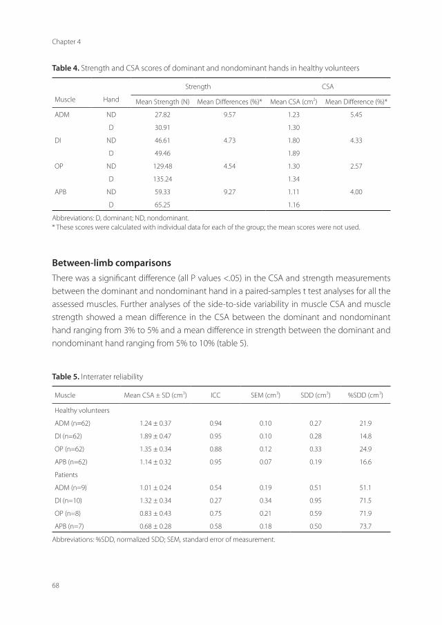
Chapter 4
68
Between-limb comparisonsThere was a significant difference (all P values <.05) in the CSA and strength measurements between the dominant and nondominant hand in a paired-samples t test analyses for all the assessed muscles. Further analyses of the side-to-side variability in muscle CSA and muscle strength showed a mean difference in the CSA between the dominant and nondominant hand ranging from 3% to 5% and a mean difference in strength between the dominant and nondominant hand ranging from 5% to 10% (table 5).
Table 4. Strength and CSA scores of dominant and nondominant hands in healthy volunteers
Muscle Hand
Strength CSA
Mean Strength (N) Mean Differences (%)* Mean CSA (cm2) Mean Difference (%)*
ADM ND 27.82 9.57 1.23 5.45
D 30.91 1.30
DI ND 46.61 4.73 1.80 4.33
D 49.46 1.89
OP ND 129.48 4.54 1.30 2.57
D 135.24 1.34
APB ND 59.33 9.27 1.11 4.00
D 65.25 1.16
Abbreviations: D, dominant; ND, nondominant.* These scores were calculated with individual data for each of the group; the mean scores were not used.
Table 5. Interrater reliability
Muscle Mean CSA ± SD (cm3) ICC SEM (cm3) SDD (cm3) %SDD (cm3)
Healthy volunteers
ADM (n=62) 1.24 ± 0.37 0.94 0.10 0.27 21.9
DI (n=62) 1.89 ± 0.47 0.95 0.10 0.28 14.8
OP (n=62) 1.35 ± 0.34 0.88 0.12 0.33 24.9
APB (n=62) 1.14 ± 0.32 0.95 0.07 0.19 16.6
Patients
ADM (n=9) 1.01 ± 0.24 0.54 0.19 0.51 51.1
DI (n=10) 1.32 ± 0.34 0.27 0.34 0.95 71.5
OP (n=8) 0.83 ± 0.43 0.75 0.21 0.59 71.9
APB (n=7) 0.68 ± 0.28 0.58 0.18 0.50 73.7
Abbreviations: %SDD, normalized SDD; SEM, standard error of measurement.

69
Ultrasound nerve injury clinical
4
discussiON
The aim of this study was to assess the validity and reliability of ultrasound as a noninvasive method to monitor muscle reinnervation in patients suffering from nerve injury by quantifying the CSA of the atrophied intrinsic hand muscles. Values for the CSA of the assessed muscles showed a strong correlation with values found by strength measurements with the RIHM, proving the validity of our ultrasound method for predicting muscle strength. In addition, we found excellent intrarater reliability in both patients and healthy volunteers and a good interrater reliability in the healthy volunteers. The ultrasound even proved to be more reliable than the RIHM. The normalized SDDs ranged between 4% and 8% in the healthy volunteers and between 4% and 13% in patients when measurements were conducted by the same examiner. These values are much lower than the 18% to 27% mentioned for the RIHM in previ-ous studies.25
The measured side-to-side variability in strength between the dominant and nondominant hand using the RIHM is in line with previous studies. However, to our knowledge, no reference values for between-hand comparisons are available for the CSA of the intrinsic hand muscles. Our measured mean difference of 3% to 5% for the CSAs does seem to be in line with the mean difference in strength measured with the RIHM. This study confirmed that ultrasound is an efficient and fast dynamic method to evaluate the CSA of specific muscles in the hand. As such, it could be a useful tool to monitor muscle reinnervation. This method proved to be especially valuable in the evaluation of patients with little strength because of the trouble positioning the fingers for the RIHM measurements.
study limitationsFuture research should include larger patient groups because we believe the small number of patients included in our study was the reason why we did not have significant correlations for all analyses conducted on the patient group.
Some considerations are important when using this method. To perform the ultrasound measurements, knowledge of the anatomic structures of the hand and technical experience with ultrasound is necessary. The use of a standardized protocol with a predetermined set of anatomic landmarks is crucial for the validity and reliability of this method because dif-ferent scan positions will result in different CSA measurements. Furthermore, the ultrasound technique is highly operator dependent, as shown by the analysis for the interrater reliability. This is especially true in the patient group where the muscle outlines are less clear and more subject to interpretation by the examiner. This suggests that follow-up of a patient should be conducted by the same examiner to achieve the most accurate results.
Future research should focus on long-term (ie, longitudinal) follow-up of patients with dif-ferent periods of muscle denervation and different sites of nerve injury to prove the additional benefit of this proposed ultrasound method in a clinical setting. It would be interesting to see

Chapter 4
70
whether the correlations found in this study can be verified in patients with longstanding muscle denervation (eg, patients with Charcot-Marie-Tooth disease) because longstanding muscle denervation causes permanent muscle damage, changing the correlation between the CSA and strength. It could also be an opportunity to increase the number of patients included in the study because we believe that the nonsignificant results for some of the analyses can be explained by the limited patient group size.
cONcLusiONs
We have demonstrated that ultrasound is a valid and reliable method to evaluate the CSA of specific muscles in the hand. As such, ultrasound could allow the observation of muscle rein-nervation in patients suffering from nerve injury noninvasively. The healthy contralateral side can be used as an internal control when consideration is given for between-limb variability. This method is particularly interesting for patients who are incapable of using a dynamom-eter, providing an opportunity to evaluate muscle reinnervation, even in the early stages of recovery and in patients with extended tendon injuries.
suppliersa. The Rotterdam Intrinsic Hand Myometer (RIHM); med.engineers.b. SonoSite Inc.c. Parker Laboratories Inc.d. Adobe Systems Inc.e. IBM Corp.

71
Ultrasound nerve injury clinical
4
ReFeReNces
1. Noble J, Munro CA, Prasad VS, Midha R. Analysis of upper and lower extremity peripheral nerve injuries in a
population of patients with multiple injuries. J Trauma 1998;45:116-22.
2. Jaquet JB, Kalmijn S, Kuypers PD, Hofman A, Passchier J, Hovius SE. Early psychological stress after forearm nerve
injuries: a predictor for long-term functional outcome and return to productivity. Ann Plast Surg 2002;49:82-90.
3. Ultee J, Hundepool CA, Nijhuis TH, van Baar AL, Hovius SE. Early posttraumatic psychological stress following
peripheral nerve injury: a prospective study. J Plast Reconstr Aesthet Surg 2013;66: 1316-21.
4. Brandsma JW, Schreuders TA, Birke JA, Piefer A, Oostendorp R. Manual muscle strength testing: intraobserver
and interobserver reliabilities for the intrinsic muscles of the hand. J Hand Ther 1995;8: 185-90.
5. Florence JM, Pandya S, King WM, et al. Intrarater reliability of manual muscle test (Medical Research Council
scale) grades in Duchenne’s muscular dystrophy. Phys Ther 1992;72:115-22; discussion 122-6.
6. Rosen B, Dahlin LB, Lundborg G. Assessment of functional outcome after nerve repair in a longitudinal cohort.
Scand J Plast Reconstr Surg Hand Surg 2000;34:71-8.
7. Wadsworth CT, Krishnan R, Sear M, Harrold J, Nielsen DH. Intrarater reliability of manual muscle testing and
hand-held dynametric muscle testing. Phys Ther 1987;67:1342-7.
8. Gaul JS Jr. Intrinsic motor recoveryea long-term study of ulnar nerve repair. J Hand Surg 1982;7:502-8.
9. Seddon H. Peripheral nerve injuries. Medical Research Council Special Report Series No. 282. London: Her
Majesty’s Stationary Office; 1954.
10. Schreuders TA, Roebroeck ME, Jaquet JB, Hovius SE, Stam HJ. Measuring the strength of the intrinsic muscles of
the hand in patients with ulnar and median nerve injuries: reliability of the Rotterdam Intrinsic Hand Myometer
(RIHM). J Hand Surg 2004; 29:318-24.
11. Trumble TE, Kahn U, Vanderhooft E, Bach AW. A technique to quantitate motor recovery following nerve grafting.
J Hand Surg 1995; 20:367-72.
12. Geere J, Chester R, Kale S, Jerosch-Herold C. Power grip, pinch grip, manual muscle testing or thenar atrophy
- which should be assessed as a motor outcome after carpal tunnel decompression? A systematic review. BMC
Musculoskelet Disord 2007;8:114.
13. Schreuders TA, Roebroeck ME, Jaquet JB, Hovius SE, Stam HJ. Long-term outcome of muscle strength in ulnar
and median nerve injury: comparing manual muscle strength testing, grip and pinch strength dynamometers
and a new intrinsic muscle strength dynamometer. J Rehabil Med 2004;36:273-8.
14. Morse CI, Thom JM, Reeves ND, Birch KM, Narici MV. In vivo physiological cross-sectional area and specific force
are reduced in the gastrocnemius of elderly men. J Appl Physiol (1985) 2005;99:1050-5.
15. Severinsen K, Obel A, Jakobsen J, Andersen H. Atrophy of foot muscles in diabetic patients can be detected with
ultrasonography. Diabetes Care 2007;30:3053-7.
16. Kullmer K, Sievers KW, Reimers CD, et al. Changes of sonographic, magnetic resonance tomographic, electro-
myographic, and histopathologic findings within a 2-month period of examinations after experimental muscle
denervation. Arch Orthop Trauma Surg 1998;117: 228-34.
17. Pillen S, Arts IM, Zwarts MJ. Muscle ultrasound in neuromuscular disorders. Muscle Nerve 2008;37:679-93.
18. Nijhuis TH, de Boer SA, Wahegaonkar AL, et al. A new approach to assess the gastrocnemius muscle volume in
rodents using ultrasound; comparison with the gastrocnemius muscle index. PLoS One 2013;8: e54041.
19. Selles RW, van Ginneken BT, Schreuders TA, Janssen WG, Stam HJ. Dynamometry of intrinsic hand muscles in
patients with Charcot- Marie-Tooth disease. Neurology 2006;67:2022-7.
20. Brandsma JW, Schreuders TA. Sensible manual muscle strength testing to evaluate and monitor strength of the
intrinsic muscles of the hand: a commentary. J Hand Ther 2001;14:273-8.
21. Bland JM, Altman D. Statistical methods for assessing agreement between two methods of clinical measure-
ment. Lancet 1986;327: 307-10.

Chapter 4
72
22. Weir JP. Quantifying test-retest reliability using the intraclass correlation coefficient and the SEM. J Strength
Cond Res 2005;19: 231-40.
23. Petersen P, Petrick M, Connor H, Conklin D. Grip strength and hand dominance: challenging the 10% rule. Am J
Occup Ther 1989;43:444-7.
24. Tagliafico A, Cadoni A, Fisci E, Bignotti B, Padua L, Martinoli C. Reliability of side-to-side ultrasound cross-sectional
area measurements of lower extremity nerves in healthy subjects. Muscle Nerve 2012;46:717-22.
25. Molenaar HM, Selles RW, Schreuders TA, Hovius SE, Stam HJ. Reliability of hand strength measurements using
the Rotterdam Intrinsic Hand Myometer in children. J Hand Surg 2008;33:1796-801.


Chapter 5motor Nerve Recovery in a Rabbit model:
description and Validation of a Noninvasive ultrasound Technique
Liselotte F. Bulstra1,2 MSc, Caroline A. Hundepool1,2 MSc, Patricia F. Friedrich1 AAS, Tim H.J. Nijhuis2 MD, PhD, Allen T. Bishop1 MD, Alexander Y. Shin1 MD
1. Department of Orthopedic Surgery, Microvascular Research Laboratory, Mayo Clinic,
Rochester, MN, USA2. Departments of Plastic, Reconstructive and Hand Surgery, Erasmus MC, University Medical
Center, Rotterdam, The Netherlands.
Journal of Hand Surgery (American Volume). 2016 Jan;41(1):27-33.
Chapter 5
76
ABsTRAcT
Purpose Currently established outcome measurements to evaluate functional recovery after peripheral nerve reconstruction in animal models are invasive and require ani-mals to be sacrificed. The aim of this study was to develop and validate a novel, non-invasive ultrasound technique for the longitudinal analysis of functional recovery after segmental peroneal nerve reconstruction in a rabbit model.
methods Twelve male New Zealand White rabbits underwent a 1 cm peroneal nerve autograft reconstruction. Ultrasound measurements were performed prior to surgery and at 1, 2, 4, 8, 12 and 16 weeks post-operatively. All rabbits were able to undergo manual restraint for the ultrasound procedure, avoiding the risks of anesthetics. At 12 and 16 weeks, functional recovery was evaluated using Compound Muscle Action Potential (CMAP), iso-metric tetanic force measurements (ITF), wet muscle weight and nerve histomorphometry. Data were compared to the ultrasound measurements by calculating the Pearson correlation coefficient. Additionally, intra- and inter-rater reliability of the ultrasound measurements was determined.
Results Ultrasound demonstrated very good correlations with ITF and wet muscle weight. Good correlations with nerve histomorphometry and a moderate correlation with CMAP were observed. Both intra- and inter-rater reliability of this ultrasound technique were excellent.
conclusions Ultrasound analysis of the tibialis anterior muscle in the rabbit provides a reliable and valid method for analysis of functional recovery in the rabbit peroneal nerve reconstruction model. The non-invasive nature allows for longitudinal follow-up within the same animal and measurement of early recovery without the use of anesthesia.
clinical relevance Application of this non-invasive technique can reduce the variability and sample size necessary in peripheral nerve reconstruction studies and will provide an ideal tool for comparative studies in larger animal models.

77
Ultrasound nerve injury rabbit
5
iNTROducTiON
The treatment of peripheral nerve injury still represents a clinical challenge.[1] To study alternative treatment strategies for peripheral nerve repair, animal models are used.[2] Rats are frequently used and especially useful as a first in vivo step to study short-term nerve regeneration due to their fast neuroregenerative capacity. However, important disadvantages impairing its clinical relevance, include the much faster nerve regeneration compared to humans and limited length of nerve gaps that can be created in rats.[3] Those limitations can be overcome using a larger animal model, the rabbit, where larger nerve gaps up to 8 cm are feasible.[4] Furthermore, the rabbit’s neuroregenerative and immunological properties more closely mimic the human situation, providing an important step prior to research in clinical setting.[5-6]
To evaluate motor nerve recovery after nerve reconstruction in both rat and rabbit, the easily obtainable wet muscle weight is frequently used. Other commonly used and established outcome measurements include isometric tetanic force measurement (ITF), a more accurate measurement of functional recovery, electrophysiological testing and nerve histomorphom-etry.[7-8,10] However, all those techniques require the animal to be euthanized following assessment, restricting longitudinal analysis of outcome over time. A commonly used non-invasive test is the sciatic function index. Unfortunately, this technique correlates poorly with other functional outcome measurement and is only feasible in the rat model.[9]
The use of ultrasound in a rat model has shown to be both reliable and valid.[7, 10] Allow-ing for multiple measurements over time within the same animal, ultrasound can reduce the variability within the studied groups. This would ultimately reduce the required sample size and costs of these studies. We hypothesize that a similar technique could be used in the rabbit model as a non-invasive method for analysis of nerve reconstruction results. Therefore, the aim of this study is to investigate the reproducibility, reliability and validity of a non-invasive ultrasound technique to measure the tibialis anterior muscle cross-sectional area for analysis of the functional recovery in a rabbit peroneal nerve injury model.
mATeRiALs ANd meThOds
After approval by the Institutional Animal Care and Use Committee, 12 male New Zealand White rabbits (Harlan Laboratories Inc., Indianapolis, IN) (weighing 3 to 4 kg) underwent a 1-cm peroneal nerve autograft reconstruction on the left side. Animals were housed individu-ally with a twelve-hour light-dark cycle. Food and water were provided ad libitum.

Chapter 5
78
ultrasound measurementsThe optimal ultrasound protocol was determined in a pilot study. Based on previous studies, different probe positions to obtain the muscle cross-sectional area using ultrasound were compared using cadaveric rabbit legs (data not shown).[7, 10, 11] The protocol that both ob-servers rated as most easily obtainable showed a standard deviation of less than 5% evaluating measurements within and between observers and this protocol was chosen. All rabbits were able to undergo the ultrasound procedure under manual restraint using a “Bunny Snuggle” (Lomir Biomedical Inc., Malone, NY), avoiding the risks of anesthetics. Ultrasound measure-ments were obtained prior to surgery and 1, 2, 4, 8 weeks after surgery and at the sacrifice procedure, 12 and 16 weeks postoperatively. The lower legs were shaved and remaining hair removed using hair removal cream (SURGI-PREP™, Miltex, York, PA). The experimental set-up of the ultrasound measurements of the tibialis anterior muscle in the rabbit is depicted in Figure 1. An instructional video of the ultrasound protocol is available as online supplement on the Journal’s Web site (www.jhandsurg.org). With the ankle joint of the rabbit in a 90° angle, the patella and lateral malleolus were identified. The ultrasound probe was placed perpendicular to the muscle at 1/3 of the distance between those landmarks and cross-sectional images of the muscle obtained using the Philips CX50 ultrasound system with a 12 to 3 MHz linear transducer (Philips Healthcare, Eindhoven, the Netherlands) and ultrasound gel (Aquasonic 100, Parker Laboratories, Fairfield, NJ). Following the ACLAM and IACUC guidelines, animals were monitored for signs of discomfort.[12] At all time points multiple measurements were
Figure 1. experimental set-up. The experimental set-up of the ultrasound recording. The position of the probe and lower leg of the rabbit are depicted.

79
Ultrasound nerve injury rabbit
5
obtained by two similarly trained observers, independent from each other. The cross-sectional area of the tibialis anterior muscle was analyzed in Adobe Photoshop CS5 Extended (Adobe Systems Incorporated, San Jose, CA).
surgical procedureAnesthesia was induced using 35 mg/kg Ketamine (Ketaset®, Fort Dodge Animal Health, Fort Dodge, IA) and 5 mg/kg Xylazine (Vettek, Bluesprings, MO) administered intramuscular (IM). After a subcutaneous (SC) injection of 0.18 mg/kg Buprenorphine (Buprinex®, Reckitt Benckiser Pharmaceuticals Inc., Richmond, VA), animals were intubated and maintained at spontaneous ventilation with the Bain ventilation system (Harvard Apparatus, Holliston, MA) under 1-2% isoflurane. Animals received intravenous (IV) lactated Ringer’s solution and animal temperature was maintained at 37°C. All surgical procedures were performed under standard aseptic conditions. Through a longitudinal incision on the posterolateral aspect of the left thigh, the sciatic nerve and its peroneal branch were exposed by a muscle splitting approach. Under a surgical microscope (Zeiss OpMi 6; Carl Zeiss Surgical, Oberkochen, Germany), a 1 cm segment of the peroneal nerve was dissected at a level 4 cm distal to the border of external obturator muscle. The autograft was reversed and sutured using six 10-0 nylon epineural sutures (Ethicon Inc., Johnson&Johnson, NJ) on each side. All nerves had a similar diameter. Wounds were closed in layers using 4-0 Vicryl rapid bioabsorbable sutures (Ethicon Inc., Johnson&Johnson, NJ). Postoperatively, animals received 5 mg/kg Enrofloxacin (Baytril®, Bayer Animal Health, Shawnee Mission, KS) and 4 mg/kg Carprofen (Rimadyl, Pfizer animal health, NY) SC once daily for 5 days. Elizabethan collars were worn for two weeks after surgery. Rabbits were observed daily until the end of the study.
sacrifice procedureAt 12 and 16 weeks, 6 animals per time point underwent a non-survival procedure. All measure-ments were conducted bilaterally. Similar to the surgical procedure, anesthesia was induced and animals intubated. Anesthesia was maintained using 0.6 mg/kg/min Propofol (Fresenius Kabi, Lake Zurich, IL) IV. Infusion rate was adjusted based on corneal reflex, respiration- and heart-rate. Propofol was used in lieu of isoflurane, which has been shown to interfere with skeletal muscle contraction.[4, 13] Dextrose 5% was administered IV to prevent muscle fatigue.
ElectrophysiologyOn an anesthetized rabbit, a miniature bipolar electrode (Harvard Apparatus, Holliston, MA) was clamped around the exposed sciatic nerve proximal to the autograft and a ground electrode was placed in the surrounding musculature. Two recording electrodes were placed on the tibialis anterior muscle surface, one proximally near the motor endplates and the other distally.[14] Using a VikingQuest Portable EMG (Nicolet Biomedical, Madison, WI) the Compound Muscle Action Potential (CMAP) was measured. Stimulation duration of 0.02 ms
Chapter 5
80
and the minimum intensity to elicit a maximum CMAP signal was used. The responses were analyzed for CMAP amplitude.
Isometric tetanic force measurementIsometric tetanic force (ITF) measurement was performed as previously described by Giusti et al.[4] Briefly, the tibialis anterior tendon was connected to a force transducer (MDB-50, Trans-ducer Techniques, Temecula, CA) using a custom clamp. Using Kirschner wires (DePuy Synthes Vet, West Chester, PA), the knee and ankle joints were secured on a custom made platform. A bipolar stimulator (Grass SD9, Grass Instrument Co., Quincy, MA) was used to stimulate the peroneal nerve distally to the graft and the signal acquired from the force transducer was processed using LabVIEW (National Instruments, Austin, TX). After determining the optimal initial muscle tension, the isometric tetanic muscle force was measured at 100, 125 and 150Hz.
HistomorphometryAnimals were euthanized with an overdose of Fatal-Plus® (Vortech pharmaceuticals, Dearborn, MI) IV and the lower body perfused with 10% formalin through the exposed aorta. A 2.5 mm segment of the peroneal nerve distal from the graft was harvested 2 cm proximal to where the nerve enters the tibial muscle and fixed in Trump’s fixative solution. Samples were cut into 1 μm sections, stained with toluidine blue (Fisher Scientific, Pittsburgh, PA) and analyzed using Image-Pro software (Image-Pro Plus 7.0; Media Cybernetics, Bethesda, MD). The N-ratio was calculated as the total myelinated fiber area divided by the total tissue cable area.[15]
Wet Muscle WeightThe tibialis anterior muscles on both sides were carefully dissected and the tendons were removed. Muscles were directly weighed to determine the wet muscle weight.
statistical analysisThe sample size of this study was based on data from the pilot study showing the highest standard deviation of the ultrasound recordings being 5%. The variability of the other out-come measurements was based on previous studies.[15],[4] Assuming the same variability would occur in this study, the number of animals needed for 80% power to reliably detect a correlation of at least 0.65 was calculated to be 12.
To determine the validity, reliability and reproducibility of the ultrasound technique, the inter- and intra-rater reliability (ICC) (two-way mixed model, absolute agreement, single measure) were calculated. SPSS (SPSS version 17, IBM, Chicago, IL) was used for analysis.[16] Additionally, Pearson correlation coefficients between ultrasound recordings and CMAP, ITF, muscle weight and histomorphometry were calculated. To this end, data were expressed as percentage recovery by dividing the data of the injured side by the data of the healthy side of the same animal. For further confirmation of the correlation between ultrasound and wet

81
Ultrasound nerve injury rabbit
5
muscle weight of the tibial muscle, a Bland Altman plot was created. P-values <0.05 were considered statistically significant.
ResuLTs
All rabbits survived during surgery and follow-up and no major complications occurred. Minor complications that were observed were superficial licking wounds on the dorsum of the foot of the operated leg (n=4) and a superficial skin inflammation on the lower leg (n=1). Topical silver sulfadiazine cream 1% (Thermazene, Ascend Laboratories LLC, Montvale, NJ) was applied until complete wound healing.
The procedure was well tolerated by all animals. Using ultrasound imaging showed the smallest muscle cross-sectional area index at 8 weeks (51% ± 6% (Standard Deviation, SD)) increasing to a mean recovery rate of 60% ± 8% at 12 weeks and 91% ± 10% at 16 weeks. Images of the ultrasound recordings are depicted in Figure 2. To evaluate the reliability and reproducibility of the ultrasound technique, the mean recovery rates as found by both observ-ers at each time point are depicted in Figure 3. Both the intra-rater and inter-rater reliability were found to be excellent as shown in Table 1.
Figure 2. ultrasound recordings of the same animal over time. In the bottom row, the tibialis an-terior muscle is delineated with a white line. The number in each picture indicates the follow-up time point in weeks.
Table 1. Validity and reliability of ultrasound recordings of the tibial muscle.
icc (95% ci) p
intra-rater reliability 0.994 (0.992 – 0.996) < 0.0001*
inter-rater reliability 0.989 (0.984 – 0.992) < 0.0001*
The intra- and inter-rater reliability of the ultrasound technique and the 95% Confidence Intervals. * indicates a statis-tically significant correlation (p<0.05)

Chapter 5
82
Functional outcome measurementsUsing CMAP measurements, a mean recovery rate of 44% ± 8% (SD) at 12 weeks and 67% ± 9% at 16 weeks was found. In three animals (n=1 at 12 weeks and n=2 in the 16 week group) CMAP could not be obtained reliably. In one animal (week 16) the ITF test could not be performed due to mechanical failure of the force transducer. ITF showed 39% ± 24% recovery at 12 weeks recovering up to 104% ± 37% at 16 weeks. The wet muscle weight of the tibial muscle showed a mean recovery rate of 45% ± 8% at 12 weeks, which improved up to 86% ± 13% at 16 weeks. Finally, calculation of the N-ratio revealed a mean recovery at 12 weeks of 61% ± 14% and 83% ± 10% at 16 weeks.
correlations with ultrasound measurementsThe correlations between ultrasound measurements and other outcome measurements at 12 and 16 weeks are shown in Table 2. All correlations were found to be moderate to excellent and are statistically significant (p<0.01). For significance, the Pearson correlation tests the cor-relation that was found against the null hypothesis: ρ = 0 (no correlation). A significant result (p <0.05) means that the correlation is significantly different from 0 (e.g. there is a significant correlation).
A Bland Altman plot of ultrasound measurements and wet muscle weight is shown in Figure 4. The narrow 95% limits of agreement indicate statistical equivalence of the methods.
Figure 4. Bland Altman plot of the ultrasound and muscle Weight measurements. To visualize the difference between the recovery rates calculat-ed using ultrasound (US) measurement and muscle weight (MW), a Bland Altman plot was created. The average of the two measurements is plotted on the x-axis and the difference on the y-axis. The dotted lines indicate the 95% limits of agreement.
Figure 3. muscle cross-sectional area recovery rate over time after nerve reconstruction. The muscle cross-sectional area of the operated leg expressed as percentage of the non-operated side as measured by the two independent observers (Obs 1 and Obs 2) at the different time points.
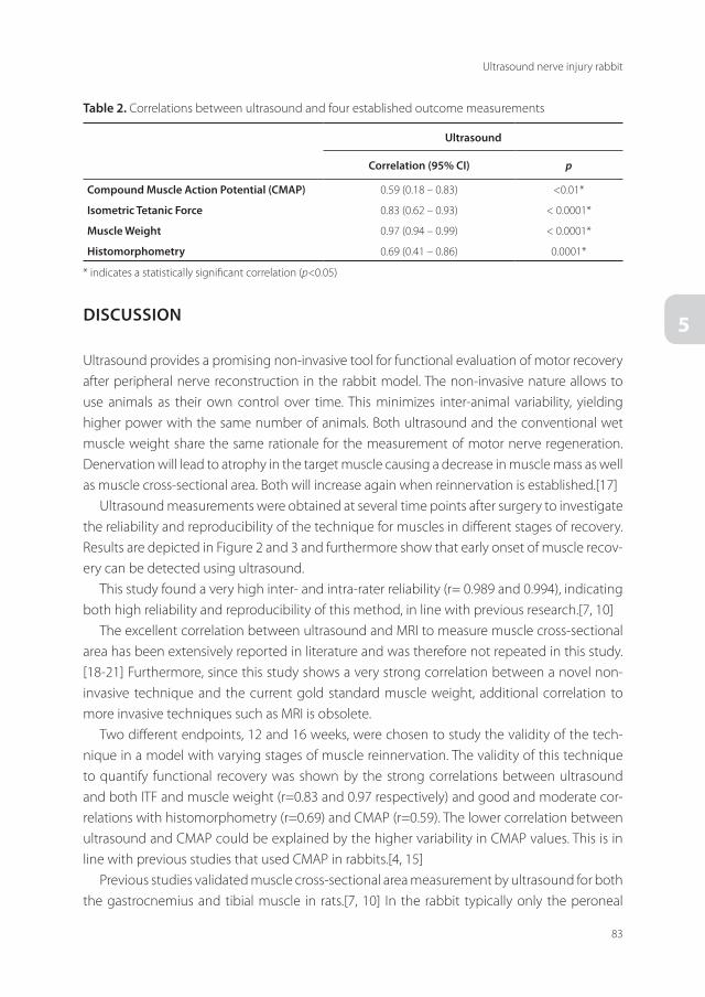
83
Ultrasound nerve injury rabbit
5discussiON
Ultrasound provides a promising non-invasive tool for functional evaluation of motor recovery after peripheral nerve reconstruction in the rabbit model. The non-invasive nature allows to use animals as their own control over time. This minimizes inter-animal variability, yielding higher power with the same number of animals. Both ultrasound and the conventional wet muscle weight share the same rationale for the measurement of motor nerve regeneration. Denervation will lead to atrophy in the target muscle causing a decrease in muscle mass as well as muscle cross-sectional area. Both will increase again when reinnervation is established.[17]
Ultrasound measurements were obtained at several time points after surgery to investigate the reliability and reproducibility of the technique for muscles in different stages of recovery. Results are depicted in Figure 2 and 3 and furthermore show that early onset of muscle recov-ery can be detected using ultrasound.
This study found a very high inter- and intra-rater reliability (r= 0.989 and 0.994), indicating both high reliability and reproducibility of this method, in line with previous research.[7, 10]
The excellent correlation between ultrasound and MRI to measure muscle cross-sectional area has been extensively reported in literature and was therefore not repeated in this study.[18-21] Furthermore, since this study shows a very strong correlation between a novel non-invasive technique and the current gold standard muscle weight, additional correlation to more invasive techniques such as MRI is obsolete.
Two different endpoints, 12 and 16 weeks, were chosen to study the validity of the tech-nique in a model with varying stages of muscle reinnervation. The validity of this technique to quantify functional recovery was shown by the strong correlations between ultrasound and both ITF and muscle weight (r=0.83 and 0.97 respectively) and good and moderate cor-relations with histomorphometry (r=0.69) and CMAP (r=0.59). The lower correlation between ultrasound and CMAP could be explained by the higher variability in CMAP values. This is in line with previous studies that used CMAP in rabbits.[4, 15]
Previous studies validated muscle cross-sectional area measurement by ultrasound for both the gastrocnemius and tibial muscle in rats.[7, 10] In the rabbit typically only the peroneal
Table 2. Correlations between ultrasound and four established outcome measurements
ultrasound
correlation (95% ci) p
compound muscle Action Potential (cmAP) 0.59 (0.18 – 0.83) <0.01*
isometric Tetanic Force 0.83 (0.62 – 0.93) < 0.0001*
muscle Weight 0.97 (0.94 – 0.99) < 0.0001*
histomorphometry 0.69 (0.41 – 0.86) 0.0001*
* indicates a statistically significant correlation (p<0.05)

Chapter 5
84
branch, innervating the tibialis anterior muscle, instead of the entire sciatic nerve is grafted.[15] Therefor measurement of the gastrocnemius muscle like Nijhuis et al.[7] proposed would not be suitable. In both rat ultrasound studies the animals were sacrificed at each interim time point. In this study, ultrasound images were obtained within the same animal at multiple time points, showing that this model is useful to obtain longitudinal data within the same animal. In addition to the previous studies, this study also shows the correlation between ultrasound measurements and CMAP and nerve histomorphometry.
A review of the literature for non-invasive evaluation of functional recovery after nerve reconstruction in the rabbit peroneal nerve model yield few techniques, all with clear limita-tions. The toe spread reflex described by Schmitz et al.[22] indicates onset of motor recovery but does not discriminate in the quality of the muscle function. Meffert et al.[23] described a transcutaneous muscle strength quantification that requires deep sedation. With an over 7% reported risk of death in rabbits due to anesthetics, techniques that can avoid anesthesia are highly favorable.[24] Our ultrasound procedure can be performed without any form of anesthesia and we were able to complete the ultrasound measurements in no longer than 15 minutes per animal, limiting the risks for the animals.
When implementing this technique, a well-defined protocol will allow repeated measure-ments of the same muscle section. In this study, the learning curve of both observers was diminished due to the pilot study that was executed previously. Practice of the technique prior to implementation will yield the best reliability and smallest variability among observers. Although not scientifically proven, after 2 weeks we obtained very good correlations between similarly trained observers, indicating that two weeks provide accurate practice. This is in ac-cordance with previous studies.[7, 10]
The fact that ultrasound measurement of the muscle cross-sectional area is an indirect measure of motor nerve recovery could be considered a limitation of this study. However, this is similar for the muscle weight index that is commonly used and accepted. Furthermore, we found a very good correlation between ultrasound measurements and ITF measurements, a direct measurement for functional motor recovery.[4, 15]
Recovery rates of more than 100% measured using isometric tetanic force are in line with previously reported data and may be explained by a side preference in the rabbit.10,[15],[4, 25]
These limitations notwithstanding, this study demonstrates that this ultrasound technique provides a reliable and valid method for analysis of functional motor nerve recovery in the rabbit peroneal nerve reconstruction model. Naturally, this method can be used for other nerves and innervated muscles as well, such as brachial plexus injury models. The non-invasive nature allows for longitudinal follow-up within the same animal and measurement of early recovery, providing an ideal tool for comparative studies of nerve reconstruction strategies.

85
Ultrasound nerve injury rabbit
5
ReFeReNces
1. Boyd, K.U., A.S. Nimigan, and S.E. Mackinnon, Nerve Reconstruction in the Hand and Upper Extremity. CLIN PLAST
SURG, 2011. 38(4): p. 643-660.
2. Angius, D., et al., A systematic review of animal models used to study nerve regeneration in tissue-engineered scaf-
folds. Biomaterials, 2012.
3. Calvert, G.T., et al., Rat-strain differences in recovery following peripheral-nerve allotransplantation. J Reconstr
Microsurg, 2001. 17(3): p. 185-91.
4. Giusti, G., et al., Description and validation of isometric tetanic muscle force test in rabbits. Microsurgery, 2012. 32(1):
p. 35-42.
5. Amillo, S., R. Yanez, and R.H. Barrios, Nerve regeneration in different types of grafts: experimental study in rabbits.
Microsurgery, 1995. 16(9): p. 621-30.
6. Deumens, R., et al., Repairing injured peripheral nerves: Bridging the gap. Prog Neurobiol, 2010. 92(3): p. 245-76.
7. Nijhuis, T.H., et al., A new approach to assess the gastrocnemius muscle volume in rodents using ultrasound; compari-
son with the gastrocnemius muscle index. PLoS One, 2013. 8(1): p. e54041.
8. Bain, J.R., S.E. Mackinnon, and D.A. Hunter, Functional evaluation of complete sciatic, peroneal, and posterior tibial
nerve lesions in the rat. Plast Reconstr Surg, 1989. 83(1): p. 129-38.
9. Lee, J.Y., et al., Functional evaluation in the rat sciatic nerve defect model: a comparison of the sciatic functional index,
ankle angles, and isometric tetanic force. Plast Reconstr Surg, 2013. 132(5): p. 1173-80.
10. Hundepool, C.A., et al., Non-invasive ultrasound for longitudinal analysis of nerve regeneration. [submitted to
Journal of Neurosurgery], 2014.
11. Mohseny, B., et al., Ultrasonographic quantification of intrinsic hand muscle cross-sectional area; reliability and valid-
ity for predicting muscle strength. Arch Phys Med Rehabil, 2015. 96(5): p. 845-53.
12. Members, A.T.F., et al., Public statement: guidelines for the assessment and management of pain in rodents and rab-
bits. J Am Assoc Lab Anim Sci, 2007. 46(2): p. 97-108.
13. Bouhemad, B., et al., Effects of halothane and isoflurane on the contraction, relaxation and energetics of rat diaphrag-
matic muscle. Br J Anaesth, 2002. 89(3): p. 479-85.
14. Gross, R., ed. The Physiology of Growth. 1978, Academic Pr. 453.
15. Sahakyants, T., et al., Return of motor function after repair of a 3-cm gap in a rabbit peroneal nerve: a comparison
of autograft, collagen conduit, and conduit filled with collagen-GAG matrix. J Bone Joint Surg Am, 2013. 95(21): p.
1952-8.
16. Shrout, P.E. and J.L. Fleiss, Intraclass correlations: uses in assessing rater reliability. Psychol Bull, 1979. 86(2): p. 420-8.
17. Shin, R.H., et al., Isometric tetanic force measurement method of the tibialis anterior in the rat. Microsurgery, 2008.
28(6): p. 452-7.
18. Yi, T.I., et al., Reliability of the supraspinatus muscle thickness measurement by ultrasonography. Ann Rehabil Med,
2012. 36(4): p. 488-95.
19. Takai, Y., et al., Ultrasound method for estimating the cross-sectional area of the psoas major muscle. Med Sci Sports
Exerc, 2011. 43(10): p. 2000-4.
20. Reeves, N.D., C.N. Maganaris, and M.V. Narici, Ultrasonographic assessment of human skeletal muscle size. Eur J Appl
Physiol, 2004. 91(1): p. 116-8.
21. Mendis, M.D., et al., Validity of real-time ultrasound imaging to measure anterior hip muscle size: a comparison with
magnetic resonance imaging. J Orthop Sports Phys Ther, 2010. 40(9): p. 577-81.
22. Schmitz, H.C. and G.M. Beer, The toe-spreading reflex of the rabbit revisited--functional evaluation of complete
peroneal nerve lesions. Lab Anim, 2001. 35(4): p. 340-5.
23. Meffert, R.H., et al., Muscle strength quantification in small animals: a new transcutaneous technique in rabbits. J
Orthop Res, 2008. 26(11): p. 1526-32.

Chapter 5
86
24. Brodbelt, D.C., et al., The risk of death: the confidential enquiry into perioperative small animal fatalities. Vet Anaesth
Analg, 2008. 35(5): p. 365-73.
25. Singh, I., One-sided dominance in the limbs of rabbits and frogs, as evidenced by asymmetry in bone weight. J Anat,
1971. 109(Pt 2): p. 271-5.


Chapter 6Non-invasive ultrasound of the tibial muscle for
longitudinal analysis of nerve regeneration in rats
Caroline A. Hundepool MSc, Tim H. J. Nijhuis MD PhD, Nadia Rbia MD, Liselotte F. Bulstra MSc, Ruud W. Selles PhD, Steven E. R. Hovius MD PhD
Department of Plastic, Reconstructive and Hand Surgery, Erasmus MC, University Medical
Center, Rotterdam, The Netherlands.
Plastic and Reconstructive Surgery 2015 Nov;136(5):633e-9e.

Chapter 6
90
ABsTRAcT
Background Today’s golden standard to measure functional recovery after nerve trauma in experimental studies is the muscle mass ratio and the isometric tetanic force, both of which are invasive and require a sacrificial procedure. We propose ultrasound as a non-invasive method to obtain the amount of muscle atrophy and determine its validity and reliability by comparing it to muscle mass ratio, isometric tetanic force and histology.
methods Fifty rats sustained a 10 mm autograft sciatic nerve reconstruction. With a two-week interval, 5 animals were tested with a total follow-up of 20 weeks. The functional recovery of the hindlimb muscles were measured with ultrasound, muscle mass ratio and isometric tetanic force. Additionally, neuromuscular junctions were histologically analysed. The different evaluation techniques were compared and the reliability of the ultrasound was determined.
Results Four weeks after denervation extensive muscle atrophy resulted in a de-crease of muscle mass up to 30%. Ultrasound showed good correlations with muscle mass ratio for both tibial (r=0.85) and gastrocnemic muscles (r=0.89). The intra- and inter-rater reliability of the ultrasound were high (r=0.97 and 0.88). The correlation with force was lower (0.62), although still statistically significant.
conclusions Ultrasound measurement of muscle atrophy was highly correlated with the golden standard muscle mass ratio and was also significantly correlated with isometric tetanic force. Histology confirmed the regeneration pattern observed with ultrasound. We propose that ultrasound can be used as a valid alternative to muscle mass ratio to study muscle atro-phy after nerve injury in a less-invasive and more animal-friendly manner.

91
Ultrasound nerve injury rat
6
iNTROducTiON
To study peripheral nerve injury, experimental studies are required that are valid and reli-able in measuring successful restoration of nerve function. A wide variety of tests assess the peripheral motor nerve outcome after nerve reconstruction: Electrophysiology, muscle mass ratio, ankle angle, histomorphometry and functional tests as the walking-track analysis and isometric tetanic force[1-4]. Walking track analysis is only used in rats and was previously com-monly used as a measure of motor outcome. It is reported to be fraught with inaccuracies stemming from autotomy of the feet and contractures of the ankle.[5]. The most commonly-used test is presently the post-sacrifice muscle mass ratio. The main reason for the popularity of this test is its simplicity. However, the isometric tetanic force measurement may be the most valid assessment of muscle strength, as it directly evaluates the actual strength capacity and not the indirect muscle mass measure[2]. Several studies already confirmed the lack of association between the different tests available[5-8].
Recently we introduced the use of ultrasound as a valid, non-invasive, and repeatable tool to measure the gastrocnemic muscle thickness and thus quantify muscle atrophy following nerve injury[9]. While we found a high correlation with muscle mass, a number of questions still remained. For example, it is unclear how the ultrasound measurement would compare to a measure of true muscle force, such as the isometric tetanic force measurement. In ad-dition, the method was only validated for a single muscle, the gastrocnemius muscle; the reliability and validity of the visualization and measurements of the tibial muscle has not yet been described. Since many nerve regeneration studies use the tibial muscle, a much smaller muscle, as a reference, an ultrasound protocol to allow assessment of this muscle would be very valuable[2, 10, 11].
The aim of this study is to determine if ultrasound is a valid tool to visualize muscle atrophy in the tibial muscle by comparing the ultrasound assessment to muscle weight and to a more true measure of muscle force, i.e. isometric tetanic force testing. The ultrasound recordings and muscle measurements were also obtained of the bigger, gastrocnemic muscle. In addition, the neuromuscular junction (NMJ) in the muscle was histologically examined by visualizing the denervation and re-innervation of the motor endplates of the tibial and gastrocnemic muscle, and were related to the other outcome assessments. We hypothesize that the early reinnervation, before return of strength can be visualized with this method. The overall goal of this study is thus to create a valid, repeatable, and, most importantly, non-invasive method to analyze functional outcome after nerve injury over time using ultrasound.

Chapter 6
92
meThOds
AnimalsThe experimental protocol was approved by the institutional animal care and use committee. Fifty adult female Lewis rats were used, weighing 175-200 grams (Charles River Laboratories Inc.). Animals were pair-housed in hooded cages on a 12-hour light/dark schedule at room temperature. The rats were given food and water ad libitum. Surgical and sacrificial procedures were performed under general anaesthesia (Isoflurane, 1-2 % in a mixture of O2/N2O).
surgical TechniqueFor this experiment, a reversed nerve autograft reconstruction was made. The surgical pro-cedure was performed by one surgeon and assistant using standard aseptic microsurgical techniques under the operating microscope (Zeiss OP-MI 6-SD; Carl Zeiss, Goettingen, Ger-many) on sciatic nerves of the hind limbs. The operated leg (left or right side) was randomly chosen. The sciatic nerve was exposed by an oblique skin incision. A 10 mm segment of the sciatic nerve was dissected, reversed and sutured back using 10/0 Ethilon sutures (Ethicon, Johnson & Johnson, Amersfoort, the Netherlands). The muscle was approximated using 2 6/0 Vicryl Rapide sutures after which the skin was closed with the same sutures (Ethicon, Johnson & Johnson, Amersfoort, the Netherlands). Buprenorphine (0.5 mL/kg BW) was injected sub-muscular as an analgesic.
evaluation of muscle atrophyEvery other week, functional measurements were performed on both legs on a total of five animals per time period. The total follow-up duration was 20 weeks with a total of 100 legs examined.
ultrasoundAfter initial isoflurane anesthesia, ultrasound images of the muscles were recorded. At first, for better conduction of ultrasound waves, hair of the leg was removed by shaving and applica-tion of depilation cream. For analysis of the tibial muscle, the knee and ankle joint where held in a 90° angle. Transducer gel was applied and the probe was placed in a 45° angle without applying pressure on the muscle. The sagittal plane of the tibial muscle was visualized. The experimental ultrasound set-up is depicted in Figure 1. To perform the ultrasound measure-ments of the gastrocnemic muscle, the rat hind paw was placed in a water basin, as previously described by our group[9]. In this position, images of the sagittal plane of the gastrocnemic muscle were recorded. All measurements were conducted bilaterally. The ultrasound record-ings were performed with the use of the SonoSite Titan Ultrasound (SonoSite Inc., Bothell, USA), with a 5-10 MHz linear array probe. In order to obtain an inter- and intrarater reliability of the ultrasound measurements of the tibial muscle, two independent and blinded inves-

93
Ultrasound nerve injury rat
6
tigators performed each evaluation twice. Images were analyzed to determine the muscle thickness for the gastrocnemic- and the cross-sectional area of the tibial muscle using Adobe Photoshop CS6 Extended (Adobe Systems Incorporated, USA).
isometric tetanic force measurementThe isometric tetanic force measurement was determined for the tibial anterior muscles of both legs as described by Shin et al.[2] The sciatic nerve was exposed by an oblique skin inci-sion. The tendon of the tibial muscle was exposed via a second skin incision and released at its insertion. The leg was fixed to a platform with Kirschner wires, which prevents the leg from moving during muscle stimulation. The Kirschner wires were placed through the knee- and ankle joint. The tendon of the tibial muscle was connected to the force transducer (MDB-50, Transducer Techniques, Temecula, CA) and placed in a horizontal position. A miniature bipolar stimulating electrode (Harvard Apparatus) was placed around the exposed sciatic nerve. A bipolar stimulator (Medtronic, NL) was used to generate the stimulus and the signal acquired from the force transducer was processed on a PC using WinTest (Testometric, Hartech, NL). During testing, body temperature was maintained with a heating pad and the muscles were kept wet and warm with a constant warm saline drip (37˚C). Prior to the maximum muscle force measurements, the optimal values of initial muscle tension (pre load) needed to be determined. This is the muscle tension in which the muscle can generate its maximum muscle force. With this optimal preload, the isometric muscle contraction force was measured with the predetermined frequencies of 100, 120 and 140 Hz. Stimulation was applied for a maximum of 2 seconds or until a force peak was clearly observed.
Figure 1. experimental set-up of the ultrasound recording of the tibial muscle(Published with permission of the Mayo Foundation for Medical Education and Research. All rights reserved, copy-right 2015.)

Chapter 6
94
muscle massAfter the muscle testing, the animal was euthanized using an overdose pentobarbital (1 mL/kg BW) that was injected intracardially. The tibial and gastrocnemic muscles of both the oper-ated and control leg were carefully dissected. The muscle weight was obtained directly after dissection to obtain the wet muscle weight.
histologyFor histological analysis, tibial and gastrocnemic muscles where fast frozen at -40 °C and serial 50-µm frozen sections were cut using a cryostat. A combination stain for the demonstration of motor nerve terminals and cholinesterase at the neuromuscular junctions was used as previ-ously described by Pestronk et al.[12]. This method uses bromoindoxyl acetate staining for cho-linesterase and silver-gold impregnation for nerve terminals. The silver stain reveals individual myelinated axons as black filaments, while the acetylcholinesterase stain labels motor endplates containing the enzyme acetylcholinesterase as well-demarcated transparent blue zones. Unlike acetylcholine receptors, acetylcholinesterase stays permanently after denervation.
statistical AnalysisAll collected data was expressed as a ratio of the operated leg divided by the healthy leg to allow comparison of the percentage of recovery between techniques. Obtained values were compared between the different evaluation techniques. Two comparisons were made: the isometric tetanic force method with the ultrasound and the muscle weight with the ultra-sound. A Pearson correlation coefficient was calculated between the different measurements of the same muscles and the associated confidence intervals were determined. A Bland Alt-man plot was used to visualize the difference between the ultrasound and muscle mass ratio of the tibial muscle. To assess the validity and reliability of these measurements, the inter-rater and intra-rater reliability of this method was calculated using the inter-observer correlation (ICC). A p-value < 0.05 was considered statistically significant.
ResuLTs
After a short learning curve of approximately two weeks, tibial muscle images could easily be obtained using ultrasound by both examiners. The procedure did not take more than 10 minutes per animal. Images of the ultrasound recordings are depicted in Figure 2.
Tibial muscleMuscle atrophy obtained with the golden standard, muscle mass ratio, showed a steep de-crease in the fourth week (32 ± 9% (SD)), followed by an increase of muscle mass with a ratio of 72 ± 6% in the 14th week and 78 ± 5% in the last follow-up week (Fig.3) The recovery of the

95
Ultrasound nerve injury rat
6muscle force, depicted in Figure 4, showed recovery rates of 40 ± 4% in week 10, reaching up to 76 ± 8% in the 20th follow-up week. As the force was not powerful enough to produce mea-sures in the first three follow-up weeks, no recovery rates of these weeks could be obtained. When relating ultrasound to muscle weight and muscle force, we found that ultrasound images from the tibial muscle showed the same pattern of atrophy followed by recovery of the muscle. Similar to muscle mass ratio, with ultrasound most atrophy was observed at week 4 (44% ± 9%), followed by recovery reaching 76 ± 2 percent in the last follow-up week. The correlations between the ultrasound and the muscle mass and muscle force are depicted in Table 1. Ultrasound showed strong significant correlations with muscle weight for the tibial muscle (0.854 ± 0.06). The correlation between ultrasound and muscle force was lower (0.624 ± 0.17) but still highly significant (p<0.001).
Figure 2. ultrasonographic images of the tibial muscle.The dotted line indicates the area of the muscle used to calculate the area of the tibial muscle of the healthy (Above, left) and operated leg (Above, right). The operated legs show a smaller surface area of the muscle than the healthy leg. The tibial muscle was measured 6 weeks postoperatively. At the bottom, the solid line indicates the muscle thickness measured in the gastrocnemic muscle at the healthy (Below, left) and operated side (Below, right).
Figure 3. The recovery of the tibial muscle mea-sured with muscle mass ratio and ultrasound.The recovery of the muscle shows the same trend in both evaluation methods, error bars = SEM.
Figure 4. The recovery of the tibial muscle measured with ultrasound and force.The correlation between ultrasound and force was lower than ultrasound and weight. However, their significant correlation is 0.624, error bars = SEM.

Chapter 6
96
Gastrocnemic muscleThe recovery rates of the gastrocnemic muscle measured with ultrasound and muscle weight are displayed in Figure 5. Ultrasound recordings of the gastrocnemic muscle showed slightly lower values than for muscle weight. The lowest recovery rate was 40 ± 2% in week 4 with the ultrasound and a muscle mass ratio of 28 ±4 percent. The correlation between the ultrasound and muscle weight measurements of the muscle atrophy was high (0.893 ± 0.04), as depicted in Table 1.
Neuromuscular junctionsStaining of the motor endplates and myelinated axons visualized the denervation and re-innervation of the muscles, as shown in Figure 6. Where the ultrasound and muscle mass ratio still shows high values in the second post-operative week, histology shows few axons and free endplates. Also, clubbing was observed, that is axonal sprouting which occurs first as an outgrowth of fine-beaded fibers ending as an axonal dilatation. This is a sign of nerve regrowth. This phenomenon was seen up till week 8. Free endplates were visible up till week 4, in accordance with ultrasound where we observed an increase in the muscle mass starting in week 4. Motor endings formed by several motor arborizations were observed in week 6 and week 8 after nerve injury with reconstruction. Sprouting of the axons and poly-innervated motor endplates could be seen between the 6th and 16th week. Starting from week 14, the innervation of the muscle showed a more normalized pattern. This is also in accordance with our ultrasound finding where we saw a stabilization starting at the 16th week. Up till 20 weeks, poly-innervated motor endplates were observed. The muscle of the control side was also examined end showed no pathological changes in the innervation pattern.
ReliabilityThe reliability of the ultrasound recordings of the tibial muscle are depicted in Table 2. Both the inter-rater and intra-rater reliability were high. The Bland Altman plot (Figure 7) was cre-ated to visualize the comparison between the two methods i.e. the ultrasound and muscle weight. The two techniques generally did not differ more than 10%, which shows a good overall reliability of these measurements.
Table 1. Correlation between Ultrasound, Muscle Weight and Isometric Tetanic Force for both muscles
Tibial muscle
Ultrasound
Correlation (95% CI) p
Muscle weight 0.854 (0.789-0.899) < 0.001*
Isometric Tetanic Force 0.624 (0.428-0.759) < 0.001*
Gastrocnemic muscle
Muscle weight 0.893 (0.844-0.927) < 0.001*
*Statistically significant correlation (p<0.05)

97
Ultrasound nerve injury rat
6
Table 2. Validity and reliability of ultrasound re-cordings of the tibial muscle
ICC (95% CI) p
intrarater reliability 0.974 (0.961-0.983) < 0.001*
interrater reliability 0.882 (0.825-0.921) < 0.001*
*Statistically significant correlation (p<0.05)
Figure 5. The recovery of the gastrocnemic mus-cle measured with ultrasound and weight.The recovery of the muscle shows the same trend in both evaluation methods, error bars = SEM.
Figure 6. Acetylcholinesterase staining to visualize the neuromuscular junctions.(Left) Overview; a wholemount acetylcholinesterase stained rat tibial muscle, showing that this muscle is composed of one belly (original magnification x1.25). Note that the muscle has a single motor endplate band which is located in the middle portion of the muscle. (Center) Clubbing; re-innervated tibial muscle (week 8), showing two endplates on atrophic muscle fibers (original magnification x40) and two growing nerve fibers ending as growth clubs (arrow). (Right) Sprouting; re-innervated tibial muscle (week 14), showing a branching axon forming a double motor arboriza-tion, assumed to supply one muscle fiber (original magnification x40). The thickness of the sections (50μm) causes part of the material to be out of the plane of focus at high magnification.
Figure 7. A Bland Altman plot of the two methods, muscle mass ratio and ultrasound.The average value of the methods is displayed on the x-axis where the difference between the two methods is displayed on the y-axis. The plot shows a good overall reliability of the measurements and reasonably small limits of agreement. SD=standard deviation.
Chapter 6
98
discussiON
New applications for the use of ultrasound are studied and introduced in different research fields for various indications and implications[13, 14] as ultrasound is a non-invasive and an easy to use method. This study investigated the relation between muscle force, muscle weight and muscle diameter obtained with ultrasound to assess the functional motor nerve recovery in a rodent model after nerve injury with reconstruction. Ultrasonographic recordings in this study showed similar recovery of the muscle as shown by the muscle weight; the correlation between both measurements was 0.854 and 0.893, indicating strong relations between the ultrasound and muscle mass. A Bland Altman plot further confirmed that ultrasound and muscle weight have small limits of agreement. The muscle force and ultrasound findings showed lower but still highly-significant correlations (r=0.624). One possible explanation is that force does not relate as directly with ultrasound as muscle weight (i.e. more fibers, more force). This theory is further strengthened by the fact that during the first weeks of recovery the muscle weight gradually increased with delayed response intensity with the isometric te-tanic force evaluation. Staining of the neuromuscular junctions shows the same denervation and re-innervation patterns of the muscles as observed with ultrasound, weight and force. Sprouting of the axons and poly-innervated motor endplates were visible until the sixteenth post-operative week, after which the pattern was normalized.
A systematic review of Wood et al[5] in 2011 explicitly stated the importance of the comparison of different outcome measures of peripheral nerve regeneration. They found an extensive battery of tests but poor correlations between most of them. Today, muscle mass ratio is generally accepted as the golden standard for the evaluation of nerve regeneration in experimental animal studies. Although the assessment is easy, we would like to emphasize that muscle force is closer to the real functional recovery. Shin et al. found that the sciatic foot index (SFI) as a non-invasive assessment did not significantly correlate with the isometric tetanic force or muscle weight[7]. These findings were also confirmed by Urbancheck et al[8]. Several other authors confirmed the lack of specificity of the SFI[4-6]. To our knowledge, no other methods than ultrasound have proven to be non-invasive, making it possible to obtain serial measurements and significantly correlate with other valid outcome measurements.
There are various ways to analyze the functional recovery after nerve trauma in animal experiments, which all have their advantages and disadvantages. This new non-invasive ul-trasound method allows analysing functional motor recovery after nerve trauma in the same animal over time. The assessment is easy and could be performed in less than ten minutes. In future studies, ultrasound assessment can be executed without sacrificing the animal, leading to a reduction in the number of animals required for an experiment. Since the regeneration will be evaluated in the same animal, variation within a group will decrease. This will further reduce the number of animals needed in experimental studies on nerve reconstruction and simultaneously increase the power of the study. A combination can be made with other non-

99
Ultrasound nerve injury rat
6
invasive measurements as the sensory pinprick test, to obtain an overall indication of nerve recovery.
A limitation of this present study is that animals where not measured over time to prove that this can also be reliable. In striving to achieve not only a reduction in the required number of animals needed, but also a reduction in variation, animal tests in nerve regeneration studies should preferably be non-invasive. These limitations notwithstanding, this study is the next step in creating non-invasive tests for the total analysis of the animal after nerve trauma with or without reconstruction.
cONcLusiONs
In conclusion, this study illustrates the purpose of the new, non-invasive method to analyze the functional recovery of the muscle after nerve reconstruction. This method proves to be a valid and reliable method to obtain the muscle atrophy and recovery of both the gastrocne-mic and tibial muscles. Non-invasive evaluation techniques will reduce the required number of animals for experiments and will also reduce the variation within the study groups.
AcKNOWLedGemeNTsThe authors would like to thank dr. A.Y. Shin for generously providing the figure of the experi-mental set-up.
Chapter 6
100
ReFeReNces
1. Bain, J.R., S.E. Mackinnon, and D.A. Hunter, Functional evaluation of complete sciatic, peroneal, and posterior tibial
nerve lesions in the rat. Plast Reconstr Surg, 1989. 83(1): p. 129-38.
2. Shin, R.H., et al., Isometric tetanic force measurement method of the tibialis anterior in the rat. Microsurgery, 2008.
28(6): p. 452-7.
3. Hunter, D.A., et al., Binary imaging analysis for comprehensive quantitative histomorphometry of peripheral nerve. J
Neurosci Methods, 2007. 166(1): p. 116-24.
4. Kanaya, F., J.C. Firrell, and W.C. Breidenbach, Sciatic function index, nerve conduction tests, muscle contraction, and
axon morphometry as indicators of regeneration. Plast Reconstr Surg, 1996. 98(7): p. 1264-71, discussion 1272-4.
5. Wood, M.D., et al., Outcome measures of peripheral nerve regeneration. Ann Anat, 2011. 193(4): p. 321-33.
6. Munro, C.A., et al., Lack of association between outcome measures of nerve regeneration. Muscle Nerve, 1998. 21(8):
p. 1095-7.
7. Lee, J.Y., et al., Functional evaluation in the rat sciatic nerve defect model: a comparison of the sciatic functional index,
ankle angles, and isometric tetanic force. Plast Reconstr Surg, 2013. 132(5): p. 1173-80.
8. Urbanchek, M.S., et al., Rat walking tracks do not reflect maximal muscle force capacity. J Reconstr Microsurg, 1999.
15(2): p. 143-9.
9. Nijhuis, T.H., et al., A new approach to assess the gastrocnemius muscle volume in rodents using ultrasound; compari-
son with the gastrocnemius muscle index. PLoS One, 2013. 8(1): p. e54041.
10. Brenner, M.J., et al., Repair of motor nerve gaps with sensory nerve inhibits regeneration in rats. Laryngoscope, 2006.
116(9): p. 1685-92.
11. Lee, J.Y., et al., The effect of collagen nerve conduits filled with collagen-glycosaminoglycan matrix on peripheral
motor nerve regeneration in a rat model. J Bone Joint Surg Am, 2012. 94(22): p. 2084-91.
12. Drachman, A.P.a.D.B., A new stain for quantitative measurement of sprouting at neuromuscular junctions. Muscle &
Nerve, 1978. 1: p. 70-74.
13. Wittenberg, M., Will ultrasound scanners replace the stethoscope? BMJ, 2014. 348: p. g3463.
14. Solomon, S.D. and F. Saldana, Point-of-care ultrasound in medical education--stop listening and look. N Engl J Med,
2014. 370(12): p. 1083-5.


PART IIIengineering a nerve allograft


Chapter 7Optimizing decellularization techniques to
create a new nerve allograft: an in-vitro study using rodent nerve segments
Caroline A. Hundepool1, 2 MSc, Tim H.J. Nijhuis2 PhD MD, Dimitra Kotsougiani1 MD, Patricia, F. Friedrich1 AAS, Allen T. Bishop1 MD, Alexander Y. Shin1 MD
1. Department of Orthopedic Surgery, Microvascular Research Laboratory, Mayo Clinic,
Rochester, MN, USA2. Department of Plastic, Reconstructive and Hand Surgery, Erasmus Medical Center,
Rotterdam, The Netherlands
Submitted

Chapter 7
106
ABsTRAcT
Purpose Commercially available processed nerve allografts have shown to be inferior to autografts in previous animal studies. We hypothesized that combining different process-ing and storage techniques will result in improved nerve ultrastructure preservation, lower immunogenicity and minimize cellular debris. Different processing protocols were evaluated which included chemical detergents, enzymes and irradiation, with the addition the enzyme Elastase and cold or frozen storage was used. The goal of this study was to create an optimized nerve allograft.
methods Fifty rat nerves were decellularized with modifications of previous protocols and the addition of Elastase. Subsequently, the nerve segments were stored at either 4 or 80°C. Both processed and fresh control nerves were analyzed with confocal microscopy using im-munohistochemical stainings on the basal lamina (laminin γ-1), Schwann cells (S100-protein) and immunogenicity (MHCI). Morphology of the ultrastructure and amount of cellular debris was analyzed on cross sections of the nerves stained using toluidine blue staining, H&E stain-ing and electron microscopy.
Results Nerve ultrastructure was preserved with all decellularization protocols. Storage at -80°C severely altered nerve ultrastructure after any decellularization method. Elastase was found to significantly reduce the immunogenicity and amount of Schwann cells, while maintaining good structural properties.
conclusion Reduced immunogenicity, diminished cellular debris and the elimination of Schwann cells was observed when Elastase was added to the nerve processing while main-taining ultrastructure. Storage at -80°C after the decellularization process heavily damaged the nerve ultrastructure as compared to cold storage. Further in vivo studies need to prove the nerve regenerative capacity of these optimized allografts.
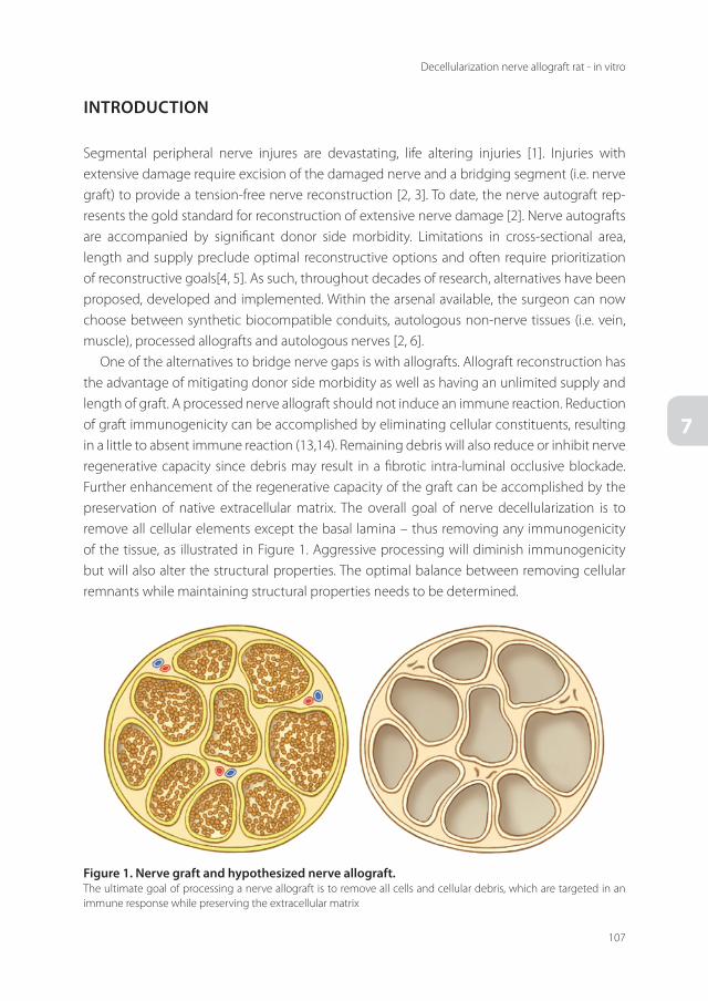
107
Decellularization nerve allograft rat - in vitro
7
iNTROducTiON
Segmental peripheral nerve injures are devastating, life altering injuries [1]. Injuries with extensive damage require excision of the damaged nerve and a bridging segment (i.e. nerve graft) to provide a tension-free nerve reconstruction [2, 3]. To date, the nerve autograft rep-resents the gold standard for reconstruction of extensive nerve damage [2]. Nerve autografts are accompanied by significant donor side morbidity. Limitations in cross-sectional area, length and supply preclude optimal reconstructive options and often require prioritization of reconstructive goals[4, 5]. As such, throughout decades of research, alternatives have been proposed, developed and implemented. Within the arsenal available, the surgeon can now choose between synthetic biocompatible conduits, autologous non-nerve tissues (i.e. vein, muscle), processed allografts and autologous nerves [2, 6].
One of the alternatives to bridge nerve gaps is with allografts. Allograft reconstruction has the advantage of mitigating donor side morbidity as well as having an unlimited supply and length of graft. A processed nerve allograft should not induce an immune reaction. Reduction of graft immunogenicity can be accomplished by eliminating cellular constituents, resulting in a little to absent immune reaction (13,14). Remaining debris will also reduce or inhibit nerve regenerative capacity since debris may result in a fibrotic intra-luminal occlusive blockade. Further enhancement of the regenerative capacity of the graft can be accomplished by the preservation of native extracellular matrix. The overall goal of nerve decellularization is to remove all cellular elements except the basal lamina – thus removing any immunogenicity of the tissue, as illustrated in Figure 1. Aggressive processing will diminish immunogenicity but will also alter the structural properties. The optimal balance between removing cellular remnants while maintaining structural properties needs to be determined.
Figure 1. Nerve graft and hypothesized nerve allograft.The ultimate goal of processing a nerve allograft is to remove all cells and cellular debris, which are targeted in an immune response while preserving the extracellular matrix
Chapter 7
108
The current available processing protocols have been studied extensively and a standard processing protocol is available. Regularly used protocols comprise a complex algorithm of steps including chemical detergents, irradiation and cryopreservation [7]. The basics of this protocol are the essentially the foundation of decellularization of nerves and have only been slightly modified by numerous investigators [8]. Given the fact that current processed allograft studies (both experimental as clinical) demonstrate inferior results to autografts, we sought to determine if the addition of Elastase to the processing would improve decellularization [9]. Elastase is an enzyme, which destructs proteins by deconnecting peptide junctions and breaking down elastin. It has been successfully used in the decellularization of other tissues than nerves; for instance in heart valve and cartilage decellularization [10, 11]. Currently all the clinical available allograft conduits are frozen upon use. In addition to optimizing the decel-lularizing process, the effect of frozen storage versus cold storage on the effect of processed nerve grafts has not been adequately evaluated.
The purpose of this in vitro study was twofold. First, to study the effect of Elastase on the decellularization of rat nerve tissue, and second to evaluate the effect of storage, either frozen or cold. We hypothesized that the addition of Elastase will allow the extracellular matrix to be preserved with a minimal amount of debris. Determination of the optimal storage technique (e.g. cold or frozen) in addition to optimization of the decellularization process would allow for the optimal nerve allograft for the reconstruction of large peripheral nerve defects.
meThOds
After approval by our institutions the Institutional Animal Care and Use Committee (IACUC), twenty-five Sprague-Dawley rats, weighing 250-350 grams (Harlan, Indianapolis, IN), were used. After initial Isoflurane induction, all animals were sacrificed with an overdose of pen-tobarbital. Bilateral, 15 mm nerve segments of the sciatic nerve were aseptically harvested. A total of 50 nerve segments were collected.
experimental designA total of 5 groups were compared in this study. All groups consisted of 10 nerves. The first group was processed following the standard protocol based on previous studies[7-9]. The second and third group underwent the same processing only with the addition of the en-zyme Elastase in two different time periods (i.e. 8 and 16 hours) (group2 & 3). The effect of freeze storage (-80°C) was also studied in group 4. A native unprocessed nerve (group 5) was analyzed as a negative control. An overview of the studied groups is depicted in Table 1.

109
Decellularization nerve allograft rat - in vitro
7
Nerve allograft processingBriefly, nerves segments were immediately after harvest placed in RPMI 1640 solution at 4 °C over night. The next day the nerve tissues were placed in deionized distilled water. After 8 hours, the water was replaced by a solution containing 125 mM sulfobetaine-10 (SB-10), 10 mM phosphate, and 50 mM sodium. The nerves were agitated for 15 h and rinsed for 15 min in a washing solution of 10 mM phosphate and 50 mM sodium. Next the washing solution was replaced by a solution containing 0.14% Triton X-200, 0.6 mM sulfobetaine-16 (SB-16), 10 mM phosphate, and 50 mM sodium and agitated for 24 h. Next the tissues were rinsed with the washing solution 50 mM phosphate and 100 mM sodium. The washing solution was replaced by SB-10 solution and the nerves agitated for 8 h. Next they were washed once using the washing solution and put into a solution of SB-16/Triton X-200. The nerves were agitated for 15 h and then washed in a solution containing 10 mM phosphate and 50 mM sodium. Subsequently, nerves were incubated in a solution containing 2 U/ml Chondroitinase ABC for 16 h at room temperature and then washed in a solution containing 10 mM phosphate and 50 mM sodium. In the Groups 2 – 4 nerves, which underwent Elastase treatment, were incubated in a solution containing 0.05 U/mL Elastase at 37 °C for 8 (group 2) or 16 hours (group 3 and 4). After that, the nerves were sterilized with gamma radiation of 2.5 kGray.
storageAs previously described and successful in other fields, tissue was freeze-stored [9]. Nerve seg-ments were stored in Ringers solution at -80°C for the freeze storage (group 4) for a duration of two weeks before final analysis. The other storage method was cold storage (4°C) where nerves were placed in PBS solution.
Outcome analysisStructureA 5 mm section of each nerve segment was fixed in 2% Trump’s solution (37% formaldehyde and 25% glutaraldehyde). 1 µm thin sections were transversally cut and stained with 1% toluidine blue. Another 5 mm section of each nerve segment was suspended in OCT, fast frozen and 5 µm transverse sections were cut. Nerve sections were stained with hematoxylin
Table 1. Experimental design
Group Treatment storage
I Standard Cold (4 °C)
II Standard + Elastase (short) Cold (4 °C)
III Standard + Elastase (long) Cold (4 °C)
IV Standard + Elastase (long) Freeze (-80 °C)
V Un-processed/native nerve No

Chapter 7
110
and eosin (H&E). Digital images of each sample were taken using a microscope digital camera (Nikon microscopy digital color camera 4.0 mega pixels, Melville, New York). The organization of the basal lamina was visualized with a laminin staining as described below. For electron microscopy, ultra-thin section were cut (500 A), placed on copper grids (200 mesh, EMS, Philadelphia, PA) and stained with uranyl acetate (EMS) and lead citrate (EMS). Sections were examined under a JEOL 1400 transmission electron microscope (JEOL Ltd, Peabody, Mas-sachusetts, USA). All sections were scored for their structural properties on a 1-5 scale with 1 being worst and 5 being optimal. Three independent and blinded investigators performed the analysis. Validity and reliability of the objective analysis was determined with an intra-class correlation of 0.83 (95% CI; 0.71-0.90).
Remnants (Axons and immunogenicity)Intraluminal remnants were examined with immunohistochemical (IHC) stainings on different components of the nerve allograft. Nerve segments were pre-fixed with 4% cold paraformal-dehyde and fast frozen. Transverse frozen sections (5 µm thickness) were cut. To identify the remnant axons left in the graft, a S100 staining was performed. To study the immunogenicity of the graft after processing, MHC-I was stained. Additionally, laminin was stained to identify the basal laminae. The IHC staining procedure was performed using the Leica Bond III Stainer (Leica, Buffalo, IL). The sections were post fixed in 4% paraformaldehyde and retrieved on-line using Epitope Retrieval 1 (Leica, Buffalo, IL) for 5 minutes. The primary antibodies used are as follows: polyclonal S100 anti-rabbit (Dako) was used at 1:5000, polyclonal laminin y1 anti-rabbit (Sigma) was used at 1:200, mouse anti-MHCI (Clone OX18, Novus Biological) was used at 1:100. All antibodies were incubated for 60 minutes. The detection system used was Research detection (Leica DS9455). This system includes the Protein Block (Dako X0909) and secondary antibody AlexFluor488. All sections were nuclear stained with Hoechst33342 (Invitrogen H1399). Once completed, slides were removed from stainer and rinsed for 5 minutes in distilled water. Slides were coverslipped using ProLong Gold antifade media (In-vitrogen). Nerve slides were examined under a fluorescence laser confocal microscope (LSM 780, Zeiss, Germany) and pictures were captured with a camera. The intensity of stainings in the cross section of the nerve was measured with Image J software (NIH, Bethesda, USA) to quantify the different stainings.
statistical AnalysisData were expressed as mean ± SEM. For structural analysis, the results of the three different observers and the three different stainings were averaged to score the structural properties. Statistical analysis of the differences between the groups was performed with one-way analy-sis of variance (ANOVA) followed by Bonferroni post hoc test with GraphPad Prism 5 software (GraphPad Software, CA, USA). P-values < 0.05 were considered to be significant.

111
Decellularization nerve allograft rat - in vitro
7
ResuLTs
An overview of the different stainings is depicted in Figure 2 and a summary of the results in Table 2.
1. enzymatic decellularizationStructure: scoreThe structure of the nerve graft was not significantly influenced by the addition of Elastase to the decellularization protocol. Group I, the standard protocol, showed a score of 3.9 ± 0.2 and group II, with the addition of Elastase, 3.5 ± 0.1. There was no significant difference between the groups. A longer exposure of the enzyme, group III, did not influence the structure of the nerve graft (3.2 ± 0.1). There was no statistical difference between the groups (Fig. 3).
Table 2. Summary of results of the effect of Elastase and freeze storage on different components of the nerve segments
elastase Freeze storage
sTRu
cTu
Re structure = ↓
Laminin = =
Rem
NA
NTs Axons ↓ ↑
immunogenicity ↓ ↑
Figure 2. Overview of different staining for group iii, iV and V.
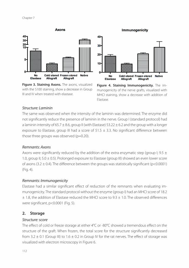
Chapter 7
112
Structure: LamininThe same was observed when the intensity of the laminin was determined. The enzyme did not significantly reduce the presence of laminin in the nerve. Group I (standard protocol) had a laminin intensity of 65.7 ± 8.6, group II (with Elastase) 53.22 ± 6.2 and the group with a longer exposure to Elastase, group III had a score of 51.5 ± 3.3. No significant difference between those three groups was observed (p=0.20).
Remnants: AxonsAxons were significantly reduced by the addition of the extra enzymatic step (group I; 9.5 ± 1.0, group II; 5.0 ± 0.5). Prolonged exposure to Elastase (group III) showed an even lower score of axons (3.2 ± 0.4). The difference between the groups was statistically significant (p<0.0001) (Fig. 4).
Remnants: ImmunogenicityElastase had a similar significant effect of reduction of the remnants when evaluating im-munogenicity. The standard protocol without the enzyme (group I) had an MHCI score of 18.2 ± 1.8, the addition of Elastase reduced the MHCI score to 9.3 ± 1.0. The observed differences were significant, p<0.0001 (Fig. 5).
2. storageStructure: scoreThe effect of cold or freeze storage at either 4°C or -80°C showed a tremendous effect on the structure of the graft. When frozen, the total score for the structure significantly decreased from 3.2 ± 0.1 (Group III) to 1.6 ± 0.2 in Group IV for the rat nerves. The effect of storage was visualized with electron microscopy in Figure 6.
Figure 3. staining Axons. The axons, visualized with the S100 staining, show a decrease in Group III and IV when treated with elastase.
Figure 4. staining immunogenicity. The im-munogenicity of the nerve grafts, visualized with MHCI staining, show a decrease with addition of Elastase.

113
Decellularization nerve allograft rat - in vitro
7
Structure: LamininStorage had no significant effect on the laminin intensity staining. The score of group III (51.49 ± 3.31) was not significantly different compared to the frozen group (IV) 63.67 ± 2.4, p=0.20.
Remnants: AxonsWhen looking at the effect of storage on the intraluminal remnants, it was demonstrated that the cold storage nerves contained a significantly lower amount of axons, stained with S100 compared to the frozen nerves. The effect of storage was statistically significant different. (Group III 3.2 ± 0.3, were the freeze storage (group IV) was higher 5.7 ± 0.3 (Fig. 4).
Remnants: ImmunogenicityThe effect of cold or freeze storage at either 4°C or -80°C showed a severe effect on the struc-ture of the graft. The immunogenicity in the rat nerves of the cold storage group (III), stained with MHCI, were statistically significant different (9.3 ± 1.0) from the frozen storage (group IV 20.8 ± 1.2) as depicted in Figure 5).
Figure 5. staining structure. The structure score of the nerve grafts was not significantly influenced by the enzyme. Frozen storage of the grafts signifi-cantly reduced the score.
Figure 6. scanning electron microscopical images of the nerve graft. Native nerve (left), cold pre-served nerve allograft: group II (middle), frozen nerve allograft: group III (right)
Chapter 7
114
discussiON
This study demonstrated that the enzyme Elastase reduced immunogenicity, diminished cel-lular debris and cleared Schwann cells better while maintaining the ultrastructure compared to standard decellularization protocols. Storage at -80°C after the decellularization process heavily damaged the nerve ultrastructure compared to cold storage. Currently available nerve allograft processing techniques are not sufficient enough to create nerve allografts that can truly challenge the nerve autograft in the clinical setting. Creating a nerve allograft has therefore been the subject of multiple studies[12] [7] [8] [13].
Knowledge of the ultrastructural anatomy of a peripheral nerve is key to the understanding the concept of nerve allograft transplantation. There are two domains in the ultrastructure: an internal basal lamina (BL), and an external fibrillar matrix. The BL faces, and is linked to ,cellular membranes and its components typically include laminin heterotrimers, collagens, nidogen or entactin and poreoglycans. The exact molecular assortment of the BL is cell-specific[14]. Schwann cells, undoubtedly, are essential for myelination and are responsible for the fast conducting signaling. It has been shown that preservation of the basal lamina is necessary to support the host Schwann cells and direct new axon sprouts [15]. Without laminin, Schwann cells cannot differentiate into a myelinating phenotype [16]. Their relation is essential for a structured conducting pattern. Previous studies showed that in mice lacking the laminin γ-1 gene, axon sorting and myelination is derailed, affecting the capacity of axons to regenerate [16]. In addition to its presence, the organization of the basal membrane is of paramount importance in supporting regeneration. Fragmentation of the basal lamina has been demonstrated using electron microscopy after chronic denervation. The progressive fragmentation of the basal membrane is negatively correlated with the re-innervation [17]. Therefore, evaluating the integrity of the basal membrane is fundamental to determine the ideal processing technique to create a nerve allograft.
This study demonstrates the effect of modifications to the standard processing techniques in a rat model. Modifications to the processing protocol should not disrupt the presence and organization of the basal lamina, but should reduce the remnant debris and axons. The stan-dard processing technique was modified by addition of an extra enzymatic step. The enzyme Elastase was hypothesized to remove more of the remnants cells and debris by disrupting the tight junctions[11]. In this study both the organization and the presence of laminin was studied. When evaluating the amount of axons remaining in the nerve graft, the addition of Elastase showed a significant reduction of S100 expression. Longer exposure to the enzyme did not significantly further reduce the cellular remnants in the rat nerves. The immunoge-nicity of the grafts was also reduced by the addition of the enzyme. The elastase processed nerves provided a nerve allograft with reduced cellular remnants resulting and preservation of the ultrastructure.

115
Decellularization nerve allograft rat - in vitro
7
The addition of Elastase reduced the amount of axons in the nerve allograft by 66.3%. The immunogenicity was reduced by 48.9% when the enzyme was added to the decellularization process. By freeze storage of the nerve allograft the structural score was reduced by 50.0% when compared to the cold stored allograft. The effect of both the Elastase and storage on the allograft was significantly different.
Previously produced nerve allografts were preserved at -80°C[10]. In other research fields (bone allograft studies), the allograft structure is less fragile than the nerve tissues and toler-ates freezing preservation better than nerve tissue[10]. The destructive effect of freezing of nerve tissues was found in our study. The freeze stored processed nerve allograft proved to have significant worse structure compared to the cold stored preservation. In Figure 3 the difference between the two studies storage methods is demonstrated. It can be appreciated that the ultrastructure of the nerve, still visible in the cold stored nerve, is severely destructed in the frozen stored nerve. The structure can almost no longer be observed.
We recognize the limitations of this study, in particular that the effect of the processing is only investigated in vitro. The neuroregenerative effect of our proposed processing and stor-age of the allograft still needs to be studied in an in-vivo animal model. Furthermore, it could be argued that more groups need to be investigated. For instance different temperatures could be incorporated in this study. We choose to use two temperatures 4 and -80°C as these are the most commonly used temperatures in clinical setting. To maintain power for this study we only used five different groups.
These limitations notwithstanding, we have clearly demonstrated that the addition of the enzyme Elastase to nerve processing resulted in maintenance of the nerve ultrastructure while significantly reducing immunogenicity, diminished cellular debris and Schwann cells compared to standard decellularization protocols. Additionally, we demonstrated that stor-age at -80°C after the decellularization process heavily damaged the nerve ultrastructure as compared to cold storage at 4°C. We have been able to improve the ultrastructure while re-ducing further the immunogenic capacity of the nerve allografts in this study by the addition of the enzyme Elastase and modification of storage after nerve processing. Further research should focus on the neuroregenerative effect of by this means optimized nerve allografts in an in vivo study.

Chapter 7
116
ReFeReNces
1. Jaquet, J.B., et al., Median, ulnar, and combined median-ulnar nerve injuries: functional outcome and return to
productivity. J Trauma, 2001. 51(4): p. 687-92.
2. Griffin, J.W., et al., Peripheral nerve repair and reconstruction. J Bone Joint Surg Am, 2013. 95(23): p. 2144-51.
3. Sunderland, S., Factors influencing the course of regeneration and the quality of the recovery after nerve suture. Brain,
1952. 75(1): p. 19-54.
4. FF, I.J., J.P. Nicolai, and M.F. Meek, Sural nerve donor-site morbidity: thirty-four years of follow-up. Ann Plast Surg,
2006. 57(4): p. 391-5.
5. Shin, A.Y., et al., Adult traumatic brachial plexus injuries. J Am Acad Orthop Surg, 2005. 13(6): p. 382-96.
6. Houdek, M.T. and A.Y. Shin, Management and complications of traumatic peripheral nerve injuries. Hand Clin, 2015.
31(2): p. 151-63.
7. Hudson, T.W., S.Y. Liu, and C.E. Schmidt, Engineering an improved acellular nerve graft via optimized chemical
processing. Tissue Engineering, 2004. 10(9-10): p. 1346-1358.
8. Neubauer, D., J.B. Graham, and D. Muir, Chondroitinase treatment increases the effective length of acellular nerve
grafts. Exp Neurol, 2007. 207(1): p. 163-70.
9. Giusti, G., et al., Return of motor function after segmental nerve loss in a rat model: comparison of autogenous nerve
graft, collagen conduit, and processed allograft (AxoGen). J Bone Joint Surg Am, 2012. 94(5): p. 410-7.
10. Utomo, L., et al., Preparation and characterization of a decellularized cartilage scaffold for ear cartilage reconstruc-
tion. Biomed Mater, 2015. 10(1): p. 015010.
11. Tedder, M.E., et al., Assembly and testing of stem cell-seeded layered collagen constructs for heart valve tissue engi-
neering. Tissue Eng Part A, 2011. 17(1-2): p. 25-36.
12. Fox, I.K., et al., Prolonged cold-preservation of nerve allografts. MUSCLE NERVE, 2005. 31(1): p. 59-69.
13. Sondell, M., G. Lundborg, and M. Kanje, Regeneration of the rat sciatic nerve into allografts made acellular through
chemical extraction. BRAIN RES, 1998. 795(1-2): p. 44-54.
14. Court, F.A., L. Wrabetz, and M.L. Feltri, Basal lamina: Schwann cells wrap to the rhythm of space-time. Curr Opin
Neurobiol, 2006. 16(5): p. 501-7.
15. Ide, C., T. Osawa, and K. Tohyama, Nerve regeneration through allogeneic nerve grafts, with special reference to the
role of the Schwann cell basal lamina. Prog Neurobiol, 1990. 34(1): p. 1-38.
16. Chen, Z.L. and S. Strickland, Laminin gamma1 is critical for Schwann cell differentiation, axon myelination, and
regeneration in the peripheral nerve. J Cell Biol, 2003. 163(4): p. 889-99.
17. Giannini, C. and P.J. Dyck, The fate of Schwann cell basement membranes in permanently transected nerves. J Neu-
ropathol Exp Neurol, 1990. 49(6): p. 550-63.


Chapter 8Application of elastase in the decellularization
of human sensory and motor nerves
Caroline A. Hundepool1, 2 MSc, Dimitra Kotsougiani1 MD, Patricia F. Friedrich1 AAS, Steven, E.R. Hovius2 PhD MD, Allen T. Bishop1 MD, Alexander Y. Shin1 MD
1. Department of Orthopedic Surgery, Microvascular Research Laboratory, Mayo Clinic,
Rochester, MN, USA2. Department of Plastic, Reconstructive and Hand Surgery, Erasmus Medical Center,
Rotterdam, The Netherlands
Submitted
Chapter 8
120
ABsTRAcT
Background Nerve allograft reconstruction has been extensively studied but clinical results remain inferior to the gold standard of the nerve autograft. Previous studies focused on optimization of nerve allograft processing but were limited to rat nerve tissue, prior to use on human nerve tissue. The purpose of this study was twofold: first, to determine the optimal processing technique using elastase in the both motor and sensory human nerve and second, to determine the effect of storage temperature on the elastase processed human nerves.
methods Seventy human nerve segments were used in this in vitro study. The enzyme elastase was added to the standard decellularization protocol. Additionally, the effect of either cold or frozen storage was tested. All nerve segments were immunohistochemically stained on basal lamina (laminin γ-1), Schwann cells (S100 protein) and immunogenicity (major his-tocompatibility complex class I) and analyzed with confocal microscopy. The preservation of the ultrastructure of the nerve segments was analyzed with toluidine blue, H&E staining and electron microscopy.
Results The addition of the enzyme had a positive effect on the decellularization process of the nerve allograft. The immunogenicity and remnants of the nerve grafts were significantly reduced. Increased concentration of the detergents did not show a beneficial effect on the human nerve allografts. Storage of the nerve segments at minus 80 degrees Celsius resulted in a lower structural score when compared to cold storage. Motor nerve had better structural properties after processing when compared to the sensory nerve allografts.
conclusion This study investigated the effect of elastase on the processing of a human nerve allograft and tested different storage temperatures. The addition of an extra enzymatic step to the processing of the allograft reduced immunogenicity and remnants in the nerve allograft with preservation of the ultrastructure. Frozen storage of the nerve allograft severely altered the structure. These conclusions are in line with the findings of processing in rat tissue. Human motor nerves were better preserved when compared to sensory nerves. We recom-mend a new processing technique, with the addition of elastase, cold storage and use of motor nerves to engineer a human nerve allograft.

121
Decellularization nerve allograft human - in vitro
8
iNTROducTiON
The segmental loss of peripheral nerve may result from open or closed extremity trauma, resection of benign or malignant primary nerve tumors, or limb-sparing tumor surgery. Currently available reconstructive options include vascularized and conventional (non-vascularized) nerve autografts, nerve conduits from a variety of materials, nerve allografts and nerve transfers[1, 2]. Of these, autografting remains the gold standard. However, available autogenous nerves are limited in length, diameter and number limiting their ability to recon-struct segmental loss of major peripheral nerves. In addition, their harvest results in significant donor site morbidity as well[3]. Among the possible options for reconstruction, processed nerve allograft is a promising alternative to nerve autografting[1].
A nerve allograft more closely mimics the structural properties of a nerve without the need to sacrifice healthy nerve tissue with the consequent donor site morbidity. Furthermore, the allograft is theoretically unlimited in both supply and length. During the early introduction of the use of unprocessed nerve allografts the use of immunosuppressive drugs was required[4, 5]. With advances in processing techniques, cellular components have been minimized resulting in substantially decreased immunogenicity of the nerve grafts[6]. This has allowed the use of decellularized allografts without immunosuppression. However, current results of commercially available nerve allografts remain unsatisfactory[1, 7, 8]. The balance between eliminating cellular remnants while maintaining structure is key in the optimization of nerve allograft processing. Our group significantly improved this technique for allograft processing in rodents, stressing this balance. It was found that the addition of an enzyme resulted in a decrease in cellular remnant (i.e. both axons and immunogenicity) while preserving the ultrastructural components of the nerve. The effect of cold storage was compared to freeze storage as commercially available allografts are all frozen upon use. A striking difference with regard to the extracellular matrix was found when freeze stored.
Previous studies evaluating motor nerve reconstruction have demonstrated inferior out-comes with sensory or mixed nerves compared to motor nerves when reconstructing a mo-tor nerve defect[9]. This phenomenon is referred to as modality-specific regeneration[9-11]. Although different researchers addressed this difference, this hasn’t been translated to the production of nerve allografts, and in fact a majority of commercially available nerve allografts are sensory in origin. Additionally, previous studies have focused solely on rodent nerve tis-sues and the processes optimized for the rodent nerves were then applied to human nerves. The purpose of this study was twofold: first, to determine the optimal processing technique using elastase in the human nerve (motor and sensory) and second, to determine the effect of storage temperature on the elastase processed human nerves.

Chapter 8
122
meThOds
The Institutional Review Board of our institution approved this study. For obtaining the hu-man nerve segments, motor and sensory nerves were harvested from recently deceased donors. Exclusion criteria for the donors were diabetes, peripheral neuropathies and previous surgeries or irradiation at the donor site. Also, nerves needed to be harvested within 4 hours post mortem. The sural (sensory), thoracodorsal (motor) and long thoracic (motor) nerves were aseptically harvested and cut into 15 mm segments. A total of 70 human nerve segments were obtained.
experimental designA total of seven groups were compared, each consisting 10 nerves. Within each group 5 nerve segments of a sensory nerve and 5 segments of a motor nerve were included. The first group was processed following the standard protocol based on previous studies[12-15]. Each group has an additive compared to the previous group. Group 2 and 3 underwent the similar pro-cessing with the addition of the enzyme Elastase in two different exposure times (i.e. 8 and 16 hours). The effect of freeze storage (-80°C) was studied in group 4. Group 5 and 6 were treated with a higher concentration of the detergents with either cold (group 5) or freeze (group 6) storage. In group 7 an unprocessed, native nerve was analyzed (negative control). In Table 1 an overview of the experimental groups is depicted.
Nerve allograft processingAfter sterile, aseptic harvest, nerve segments were directly placed in RPMI 1640 solution at 4°C. The following day nerve segments were put in deionized distilled water. After 8 hours, nerves were placed in a solution containing 125 mM sulfobetaine-10 (SB-10), 10 mM phosphate, and 50 mM sodium. Nerves were agitated for 15 hours and rinsed in a washing solution (10 mM
Table 1. experimental design. A total of 7 experimental groups were studied with a different process-ing treatment and cold of frozen storage. Each group contained a total of 10 nerve segments (5 sensory and 5 motor nerve segments).
Group Treatment storage
I Standard Cold (4 °C)
II Standard + Elastase (short) Cold (4 °C)
III Standard + Elastase (long) Cold (4 °C)
IV Standard + Elastase (long) Freeze (-80 °C)
V Standard (high) + Elastase (long) Cold (4 °C)
VI Standard (high) + Elastase (long) Freeze (-80 °C)
VII Un-processed/native nerve No
123
Decellularization nerve allograft human - in vitro
8
phosphate and 50 mM sodium) for 15 min. Next, nerves were placed in a different solution (0.14% Triton X-200, 0.6 mM sulfobetaine-16 (SB-16), 10 mM phosphate, and 50 mM sodium) and agitated for 24 hours. Subsequently tissues were rinsed with the washing solution (50 mM phosphate and 100 mM sodium) after which the nerves were agitated in the SB-10 solution for 8 hours. The nerves were washed with the washing solution again and agitated in the SB-16/Triton X-200 solution for 15 hours and then washed in a solution (10 mM phosphate and 50 mM sodium). For groups 6 and 7 a higher concentration of the detergents was used (250 mM SB-10, 1.2 mM SB-16 and 0.14% Triton X-20). For the first enzymatic step segments were incubated in a solution with 2 U/ml Chondroitinase ABC for 16 hours after which they were washed in a solution (10 mM phosphate and 50 mM sodium). In the groups II – VI, with the enzyme Elastase, nerves were incubated in a solution containing 0.05 U/mL Elastase for 8 (group II and V) or 16 hours (group III and VI) at 37°C. After processing, nerves segments were sterilized with 2.5 kGray gammaradiation.
storageAfter processing nerve segments were either cold (4°C) of freeze (-80°C) stored. For the freeze storage nerve segments were stored in Ringers solution at -80°C (group IV and VI). For the other groups, nerves were placed in PBS solution and cold stored (4°C). Nerves were stored for the duration of two weeks before the analysis.
Outcome analysisStructureNerves were cut in 5 mm segments fixed in 2% Trump’s solution (37% formaldehyde, 25% gluteraldehyde). After embedding, 1 µm thin sections were transversally cut and stained with 1% toluidine blue. Another 5 mm of the nerve segment was suspended in OCT, fast frozen and transverse sections were cut (5 µm). The sections were stained with hematoxylin and eosin (H&E). Digital images of each sample were made using a microscope (Nikon microscopy digital camera, Melville, New York). A laminin staining, described below, was used to visualize the organization of the basal lamina. Electron microscopy was performed on ultra-thin section (500 A) stained with uranyl acetate and lead citrate. Sections were visualized with a JEOL 1400 transmission electron microscope (JEOL Ltd, Peabody, Massachusetts, USA). The structural properties of all sections were scored on a 5-point scale ranging for 1 (worst) to 5 (optimal). Three independent and blinded investigators performed the analysis. Validity and reliability of the objective analysis was determined with an intra-class correlation of 0.74 (95% CI; 0.59-0.83).
Remnants (Axons and immunogenicity)Intraluminal remnants, axons and immunogenicity, were examined with immunohistochemical stainings (IHC) on different elements of the nerve allografts. Pre-fixation with 4% cold parafor-maldehyde was used after which nerve segments were fast frozen and cut in transverse sections

Chapter 8
124
of 5 µm. A S100 staining was performed to identify remnant axons in the nerve segment. A MHC-1 staining was used to analyze the immunogenicity of the nerve after processing. Ad-ditionally, to identify the basal laminae a laminin staining was performed. All IHC stainings were performed with a Leica Bond III Stainer (Leica, Buffalo, IL). Nerve sections were post fixed with 4% paraformaldehyde and retrieved on-line using Epitope Retrieval 1 (Leica, Buffalo, IL) for 5 minutes. The primary antibodies used are: polyclonal S100 anti-rabbit (Dako) was used at 1:5000, polyclonal laminin y1 anti-rabbit (Sigma) at 1:200 and mouse anti-MHCI (Clone EP1395Y, Novus Biological) were used at 1:200. The antibodies were incubated for 1 hour. The Research detection system (Leica DS9455) was used which included the Protein Block (Dako X0909) and second-ary antibody AlexFluor488. A Hoechst33342 nuclear stain was used on all sections (Invitrogen H1399). Slides were coverslipped with ProLong Gold antifade media (Invitrogen). All nerve slides were analyzed with a fluorescence confocal microscope (LSM 780, Zeiss, Germany) and images were captured with a digital camera. The intensity of stainings on the cross section of the nerves were analyzed with Image J software (NIH, Bethesda, USA).
statistical analysisAll data were presented as mean ± SEM. To score the structural properties, the results of the three different observers and the three different stainings were averaged. Statistical analysis of the differences between the groups was performed with one-way analysis of variance (ANOVA) followed by Bonferroni post hoc test. For difference between nerve types additionally a two-way ANOVA analysis was performed. GraphPad Prism 5 software (GraphPad Software, CA, USA) was used. A p-value < 0.05 was considered statistically significant.
ResuLTs
An overview of the results is depicted in Table 2.
1. enzymatic decellularizationThe effect of Elastase on the decellularization process was analyzed by comparing group I (standard protocol) with group II (standard protocol with Elastase). A longer exposure to the enzyme was studied with group III.
Structure: scoreThe structure of the nerve grafts was not significantly reduced by the addition of the extra enzymatic step (p=0.09). In group I, standard protocol, the score was 3.2 ± 0.2 whereas in group II, with Elastase, the score was 3.1 ± 0.3. Longer exposure to the enzyme, group III, did also not significantly influence the structural score (3.2 ± 0.3). The unprocessed nerve segments scored 4.9 ± 0.1 (Fig. 2).

125
Decellularization nerve allograft human - in vitro
8
Structure: LamininLaminin in the processed nerve grafts in the standard protocol group I had a score of 63.0 ± 6.1. Addition of Elastase (group II) had a score of 58.6 ± 4.7. Longer exposure to the enzyme (group III) resulted in a score of 43.17 ± 3.6. These differences were not statistically significant (p=0.02) (Fig. 3).
Remnants: AxonsThe axons in the nerve grafts, identified with the S100 staining had a score of 6.3 ± 0.9 in standard protocol group I (Fig. 4). In group II this was 6.8 ± 1.2 and for group III 5.9 ± 0.7. In comparison, the S100 expression in the native nerve was found to be 44.6 ± 3.0. There were no statistical significant differences between group I-III (p=0.80).
Table 2. effect of processing on different components of the nerve allograft.The effect of Elastase, freeze storage and higher concentration of detergents is visualized for the different outcome measurements structure (i.e. structural score and laminin staining) and remnants (axons stained with S100 and im-munogenicity with MHCI).
elastase Freeze storage high conc. detergents
sTRu
cTu
Re structure= ↓* =
Laminin= = ↓*
Rem
NA
NTs Axons
↓ = =
immunogenicity↓ = =
* for statistical significant differences
Figure 2. structural scoring. The structural proper-ties were scored for the different groups. In group IV and VI a significant lower score was observed.

Chapter 8
126
Remnants: ImmunogenicityThe immunogenicity of the nerve graft processed with the standard protocol (group I) was 6.3 ± 0.5. With addition of the enzyme (group II) this was 6.3 ± 0.7. Longer exposure to the enzyme (group III) resulted in a score of 4.5 ± 0.5. The differences showed a trend with a p value of 0.055 (Fig. 5).
Figure 3. Laminin staining of all nerve segments.The laminin was scored and showed a significantly lower score in the frozen allograft group C/IV (and F/VII).
Figure 4. staining of axons with s100.The amount of axons in the processed allograft were as-sessed with the S100 staining.
Figure 5. staining of immunogenicity on mhci.The immunogenicity of the processed allograft was inves-tigated with the MHCI staining. In the group with a longer exposure to the enzyme Elastase a reduction of the MHCI was observed.
127
Decellularization nerve allograft human - in vitro
8
2. higher concentration detergentsTo evaluate the effect of the higher concentration of detergents group III and V will be com-pared (cold storage with low and high concentration of detergents) and group IV will be compared with group VI (freeze storage with low and high concentration of detergents).
Structure: scoreThe structural score of the processing with a higher concentration of detergents, group V, was lower (2.8 ± 0.2) compared to group III (3.2 ± 0.3). This comparison could also be made in the freeze storage group with high concentration of detergents (group VI) which scored 1.5 ± 0.1 compared to group IV with a score of 1.6 ± 0.2. Both comparisons were not significant different (Fig. 2).
Structure: LamininLaminin was significantly lower in the high concentration detergents group VI (39.1 ± 4.5) compared to group IV (63.7 ± 3.7) (p=0.01). In the two cold storage groups, the high concen-tration group V scored 60.7 ± 5.3 and the lower concentration (group III) scored 43.2 ± 3.6, this comparison was not significant (p=0.17) (Fig. 3).
Remnants: AxonsThe axons in group III scored 5.9 ± 0.7 compared to the higher concentration detergents in group V (4.9 ± 1.1). In the freeze storage group VI the score was 2.8 ± 0.2 compared to Group IV 5.7 ± 1.0. Both comparisons showed no statistical differences between the groups (p=0.9 and 0.13 respectively) (Fig. 4).
Remnants: ImmunogenicityA higher concentration of detergents had no significant influence on the immunogenicity of the nerve grafts (p=0.9). Group III, V, IV and VI scored respectively 4.5 ± 0.5, 4.7 ± 0.8, 4.7 ± 0.5 and 4.5 ± 0.8 (Fig. 5).
3. storageTo evaluate the effect to structure on the nerve grafts group III (cold storage) will be compared with group IV (freeze storage).
Structure: scoreThe effect of cold (4°C) or freeze storage at -80°C showed a tremendous effect on the structure of the graft. The freeze storage (group IV; 1.6 ± 0.2) was lower than group III (3.2 ± 0.3). This difference was statistically significant (p=0.01) (Fig. X). In comparison, an unprocessed, native nerve, had a score of 4.9 ± 0.1 (Fig. 2).

Chapter 8
128
Structure: LamininLaminin scores in the cold storage group III were 43.2 ± 3.6 compared to the freeze group IV with a score of 63.7 ± 3.7. This difference was significant (p<0.01) (Fig. 3).
Remnants: AxonsThe axons in group III were 5.9 ± 0.7 which was not significant different from group IV (5.7 ± 1.0). No significant difference between the groups was observed (p=0.89) (Fig. 4).
Remnants: ImmunogenicityStorage had no significant influence on the immunogenicity, visualized with MHCI (p=0.81). Group III had a score of 4.5 ± 0.5 and group IV showed a score of 4.7 ± 0.5 (Fig. 5).
4. difference motor and sensory nervesEach group consisted 5 sensory and 5 motor nerve segments. The difference between motor and sensory nerves was analyzed with a two-way ANOVA. Regarding the structural proper-ties of the nerve segments a statistically significant difference was found (p=0.03) between the motor and sensory nerve favoring the motor nerve segments. The laminin scores were not statistically different (p=0.71). The axons were also statistically different (p=0.01). The immunogenicity of the nerve segments, stained with MHCI, was also different between the two different nerve types (p<0.001). A greater reduction in immunogenicity was seen in the processed motor nerve segments.
discussiON
This study was conducted to optimize the decellularization protocol to create a human nerve allograft. The effect of the enzyme Elastase and type of storage was investigated. Additionally the differences of processing with motor and sensory nerves were studied. In conclusion, the addition of the enzyme shows a beneficial effect on the decellularization of the human nerve allograft. Longer exposure to the enzyme showed significantly better results. The immuno-genicity of the nerve allograft was significantly reduced as well as the amount of axons, the remnants. The addition of Elastase showed no negative effect of the structure of the allograft.
The storage technique of the allograft was either cold storage, at 4 degrees Celsius or frozen storage, at -80 degrees Celsius. This had no significant influence on either the amount of axons or immunogenicity of the nerve allografts. However, a striking difference was found between the two storage techniques with regard to structure. The cold storage, at 4 degrees Celsius, scored significantly higher whereas the frozen storage, at -80 degrees Celsius, severely altered the ultrastructure of the nerve and is therefore preferable over the cold allograft. After decellularization of a nerve allograft the grafts should be cold stored instead of frozen.

129
Decellularization nerve allograft human - in vitro
8
The addition of Elastase to the decellularization protocol and the investigation of the effect of storage was previously studied in rat nerves. In this study and the in vitro study on rat nerves, the positive effect of the addition of the elastase and the effect of cold storage was found. This shows that the optimization of decellularization of the tissues is similar in both tissues. Previous studies have been focused on rat tissue[7, 14-16]. We sought to test different decellularization protocols on human nerve tissue and determine the optimal protocol.
As rat nerve tissue is different from human nerve tissue (i.e. bigger diameter), different protocols were used. The effect of a higher concentration of the detergents was investigated. It was hypothesized that this would decrease the amount of cellular debris in the grafts after processing. However, the higher concentration of detergents did not significantly reduce the amount of axons stained with S100. The same was observed with regard to immunogenicity. The higher concentration of the detergents did not significantly reduce the MHCI positive cells in the human nerves. No significant effect of the higher concentration was seen on the structure of the grafts.
Next to the addition of an extra enzymatic step to the decellularization process, the ef-fect of storage was investigated as well. Clinically available nerve allografts are stored at -80 degrees Celsius. In clinical practice this makes it difficult as not all operation theaters have a place to store nerve allografts at -80 degrees. More importantly, the effect of storage on nerve allografts has never been studied. Previous allografts were analyzed after the decellulariza-tion process, but not after the storage of the grafts. In this study we found that cold storage showed no negative influence on the axons and immunogenicity. Furthermore, with cold storage the ultrastructure of the allograft was maintained significantly better then compared to frozen storage.
This study strived to elucidate the difference between sensory and motor nerves. Motor nerves were better preserved compared to the sensory nerves. The basal lamina of the motor nerve is thicker and the endoneural tubes are larger in motor nerves as compared to sensory nerves[9]. Also, the specific way of signaling through motor nerves is different from signaling in sensory nerves. Studies have shown that motor nerves have four times higher regenera-tion capacity through motor nerves grafts as compared to sensory nerve grafts[9]. This is an important factor when determining the optimal processing technique for nerve allograft for the reconstruction of motor nerves. Possibly the thicker basal lamina in motor nerves explains why the motor nerves are better preserved when compared to the sensory nerves.
It can be stated that the nerve allograft still does not score better then the gold standard, the autograft. The clinically used decellularized nerve allograft is used for both sensory and motor nerve reconstruction. The results of these nerve allograft on motor nerve regeneration are still not satisfactory[8]. The currently available nerve allograft seems only effective for the reconstruction of short digital, sensory nerve gaps. This study, focusing on optimization of the nerve allograft can improve nerve allograft reconstruction.

Chapter 8
130
One of the limitations of this study is the relatively small sample size and the in vitro nature of this study. Although statistically significant differences between the studied groups were found those could be type II errors due to the relatively low power of this study. Despite this small sample size, this study is the first experimental study to compare different techniques in the optimization of the human nerve allograft. Further studies should be executed to deter-mine the effect of the optimized nerve allograft in an in vivo, animal model.
These limitations notwithstanding, we can conclude that the decellularization of the hu-man nerve allograft can be enhanced by the addition of the enzyme Elastase. Cold storage shows better results with regard to maintaining the ultrastructure of the nerves when com-pared to freeze storage. Using higher concentration of detergents on human nerve tissue did not show a beneficial effect. Motor nerves seem to be better preserved after processing when compared to sensory nerves. The conclusions of this study are in line with the optimization of nerve allograft found in rat tissue. The next step should be to test the optimized nerve allograft in vivo.

131
Decellularization nerve allograft human - in vitro
8
ReFeReNces
1. Cho, M.S., et al., Functional outcome following nerve repair in the upper extremity using processed nerve allograft. J
Hand Surg Am, 2012. 37(11): p. 2340-9.
2. Griffin, J.W., et al., Peripheral nerve repair and reconstruction. J Bone Joint Surg Am, 2013. 95(23): p. 2144-51.
3. FF, I.J., J.P. Nicolai, and M.F. Meek, Sural nerve donor-site morbidity: thirty-four years of follow-up. Ann Plast Surg,
2006. 57(4): p. 391-5.
4. Bain, J.R., et al., The peripheral nerve allograft: an assessment of regeneration across nerve allografts in rats immuno-
suppressed with cyclosporin A. Plast Reconstr Surg, 1988. 82(6): p. 1052-66.
5. Doolabh, V.B. and S.E. Mackinnon, FK506 accelerates functional recovery following nerve grafting in a rat model.
Plastic and reconstructive surgery, 1999. 103(7): p. 1928-36.
6. Evans, P.J., et al., Cold preserved nerve allografts: changes in basement membrane, viability, immunogenicity, and
regeneration. MUSCLE NERVE, 1998. 21(11): p. 1507-22.
7. Szynkaruk, M., et al., Experimental and clinical evidence for use of decellularized nerve allografts in peripheral nerve
gap reconstruction. Tissue Eng Part B Rev, 2013. 19(1): p. 83-96.
8. Brooks, D.N., et al., Processed nerve allografts for peripheral nerve reconstruction: A multicenter study of utilization and
outcomes in sensory, mixed, and motor nerve reconstructions. MICROSURGERY, 2012. 32(1): p. 1-14.
9. Moradzadeh, A., et al., The impact of motor and sensory nerve architecture on nerve regeneration. EXP NEUROL,
2008. 212(2): p. 370-6.
10. Brenner, M.J., et al., Repair of motor nerve gaps with sensory nerve inhibits regeneration in rats. Laryngoscope, 2006.
116(9): p. 1685-92.
11. Hoke, A., et al., Schwann cells express motor and sensory phenotypes that regulate axon regeneration. J Neurosci,
2006. 26(38): p. 9646-55.
12. Neubauer, D., J.B. Graham, and D. Muir, Chondroitinase treatment increases the effective length of acellular nerve
grafts. Exp Neurol, 2007. 207(1): p. 163-70.
13. Hudson, T.W., S.Y. Liu, and C.E. Schmidt, Engineering an improved acellular nerve graft via optimized chemical
processing. Tissue Engineering, 2004. 10(9-10): p. 1346-1358.
14. Hudson, T.W., et al., Optimized acellular nerve graft is immunologically tolerated and supports regeneration. Tissue
Eng, 2004. 10(11-12): p. 1641-1651.
15. Giusti, G., et al., Return of motor function after segmental nerve loss in a rat model: comparison of autogenous nerve
graft, collagen conduit, and processed allograft (AxoGen). J Bone Joint Surg Am, 2012. 94(5): p. 410-7.
16. Moore, A.M., et al., Acellular nerve allografts in peripheral nerve regeneration: A comparative study. MUSCLE NERVE,
2011. 44(2): p. 221-234.


Chapter 9Return of motor function with decellularized nerve allografts using elastase in a rat sciatic
nerve model
Caroline A. Hundepool1,2 MSc, Liselotte F. Bulstra1,2MSc, Patricia F. Friedrich1 AAS, Steven E.R. Hovius2 PhD MD, Allen T. Bishop1 MD, Alexander Y. Shin1 MD
1. Department of Orthopedic Surgery, Microvascular Research Laboratory, Mayo Clinic,
Rochester, MN, USA2. Department of Plastic, Reconstructive and Hand Surgery, Erasmus Medical Center,
Rotterdam, The Netherlands
Submitted

Chapter 9
134
ABsTRAcT
Background For segmental nerve injuries that cannot be directly repaired, a bridging nerve graft is required. Although the nerve autograft remains the gold standard, limited avail-ability and donorsite morbidity remain major disadvantages. Previously, our group evaluated the role of elastase in the decellularization process of nerve allografts and demonstrated improved maintenance of the basement membrane and diminished immunogenicity com-pared to traditional decellularization protocols. The purpose of this study was two fold: 1) to compare the elastase processed nerve allografts and nerve autografts and 2) to compare the type of storage (cold vs frozen) with respect to functional motor outcome in a rat sciatic segmental nerve defect model
methods Sixty-six Lewis rats underwent a 10-mm sciatic nerve reconstruction using either a cold or frozen stored decellularized nerve allograft or a nerve autograft. Sprague-Dawley rats served as full MHC-mismatch donors. The functional motor outcome was evalu-ated after 12 and 16 weeks. Ankle angle, compound muscle action potential (CMAP), isometric tetanic force, wet muscle weight and histomorphometry were tested bilaterally.
Results For CMAP and isometric tetanic force, no statistically significant difference was found between groups at both time points. For ankle angle, histomorphometry and muscle weight, the cold stored allograft was equivalent to the autograft while the autograft showed significantly better results compared to the frozen allograft at both time points. Over-all the frozen stored allograft showed inferior results while the cold stored allograft showed results comparable with the autograft.
conclusions For all outcome measurements, the elastase processed, cold preserved allograft showed no statistically significant differences with the gold standard, the nerve au-tograft, for both early and late regeneration. In three out of the five outcome measurements, cold storage of the processed allograft showed better results than the frozen stored allograft.
clinical Relevance Although further studies in a larger animal are desired, the cold stored nerve allograft decellularized with elastase shows promising results with respect to motor outcome when compared to the gold standard, the nerve autograft.
135
Nerve allograft in vivo
9
iNTROducTiON
Traumatic injuries to the peripheral nerves can cause considerable disability and economic burden[1]. Although highly prevalent in military conflicts, peacetime injuries commonly result from trauma secondary to motor vehicle accidents, penetrating trauma, industrial injuries and falls. 5% of the patients admitted to Level I trauma centers have a peripheral nerve injury[2-4]. The majority of peripheral nerve injuries require surgical reconstruction to restore sensation and function[5]. The gold standard for nerve injuries that cannot be directly repaired is the nerve autograft[6]. Typically, the sural nerve is harvested and sectioned into cables to fit the diameter and length of the defect[7]. However, the autograft is limited by supply, diameter, and length, and is accompanied by donor site morbidity[8]. This has constrained the ability to optimally reconstruct nerves of patients with multiple segmental defects where length of nerve graft needed far exceeds the availability and results in the need to prioritize the nerves to be reconstructed. In the last several decades, different alternatives have been proposed to replace the autograft, each with advantages and disadvantage[9-14]. Currently available reconstruction options comprise of nerve grafts (auto- and allograft), biological conduits (e.g. arterial and venous) and synthetic conduits (collagen, polyglycolic acid and caprolactone)[14].
One of the proposed reconstruction alternatives was the use of a nerve allograft. Theoreti-cally, a nerve allograft will more closely mimic the structure of the autograft than an empty conduit. The first allografts introduced required the use of immunosupressants[15]. Although progress has been made, further development is required prior to using the allograft instead of an autograft, especially in longer nerve gaps. The results of clinically available processed allografts, especially for motor reconstruction have not been optimal or adequately studied. While sensory nerve recovery has shown similar results to autograft, motor recovery remains inferior to the autograft[16, 17]. Previous research by our group has shown that, in a rat model, the allograft nerve processed by AxoGen (AxoGen, Alachua, Florida) showed comparable results to the autograft at 12 weeks. However, at 16 weeks there was a statistically significant difference, where the allograft isometric motor strength degraded while the autograft con-tinued to improve strength[9]. This inferior motor recovery stimulated the search for a better, optimized nerve allograft. The allograft process was improved by the addition of elastase, and demonstrated significant improvements in an in vitro study were different processing techniques and storage protocols were compared[18]. Based on histological outcome, the allograft process was improved and storage (frozen vs cold) was found to have a significant influence on the structure of the grafts.
The purpose of this study was to evaluate the elastase decellularized allograft nerve in a segmental rat sciatic nerve gap model. Specifically, a comparison of the elastase decellular-ized allograft nerve to autograft with respect to motor nerve regeneration was evaluated in addition to the effect of storage of the decellularized allograft.

Chapter 9
136
meThOds
After approval by the Institutional Animal Care and Use Committee, 66 Lewis rats (weighing 250-300 grams) were used and 22 Sprague-Dawley rats served as full major histocompatibility complex mismatch nerve donors. Lewis rats are used as they are known for their reduced tendency for autotomy[19]. Animals were randomly divided in 3 groups, each treated for a 10mm sciatic nerve gap. Group I (n=22) served as a control using a nerve autograft. Group II (n=22) had a unilateral nerve gap reconstructed with a processed nerve allograft that was cold stored (‘Allograft Cold’). Group III (n=22) had a similar procedure except the processed allograft was freeze-stored (‘Allograft Frozen’). The rats were given food and water ad libitum and were individually housed with a 12 hour light-dark cycle.
Nerve processingTwenty-two Sprague-Dawley rats, weighing 300-350 grams, were sacrificed with an intraperi-toneal injection of pentobarbital after which 15 mm nerve segments were harvested asepti-cally. Nerve allografts were prepared as previously described[18, 20]. Briefly, nerve segments were placed in RPMI medium, followed by subsequent steps using different detergents, two enzymatic steps and gamma irradiation[18]. At the end, the processed allografts were stored in phosphate buffered saline at 40C (‘allograft cold’) and the allografts were stored in Ringers Lactate at -800C (‘allograft frozen’) for 14 days.
surgical procedureRats were anesthetized with an intraperitoneal cocktail of ketamine (Ketaset®, 100mg/ml, Fort Dodge Animal Health, Fort Dodge, IA) and xylazine (VettekTM, 100mg/ml, Bluesprings, MO) 10:1 mixture, administering a dose of 1 ml/Kg body weight after initial induction with Isoflurane. Anesthesia was maintained with additional doses of ketamine only. Ringers Lactate was administrated subcutaneously to prevent dehydration, and body temperature was main-tained with a heating pad. The left sciatic nerve was exposed with a mid-gluteal incision. A 10 mm sciatic nerve segment was excised under an operating microscope (Zeiss OpMi6, Carl Zeiss Surgical, Oberkochen, Germany). In group I and II a 10 mm nerve allograft, respectively cold or frozen, was used to bridge the 10 mm nerve gap using 10-0 nylon epineural sutures. In the control group, group III, the nerve segment was reversed and put back. The muscle was approximated and the skin was closed with 5-0 Vicryl rapid sutures. Postoperatively trim-ethoprim/sulfadiazine 30 mg/Kg (Tribrissen, Five Star Compounding Pharmacy, Clive, IA) was administered to prevent infection and buprenorphine (Buprinex®, 0.1 ml/kg, Reckitt Benckiser Pharmaceuticals Inc., Richmond, VA) served as an analgesic.
137
Nerve allograft in vivo
9
evaluation of motor Functional OutcomeFor the sacrificial procedure, at 12 and 16 weeks, the animals were anesthetized as previous described. The passive ankle angle, compound muscle action potential (CMAP), isometric tetanic force and wet muscle weight were tested bilaterally. Distal nerve segments were ana-lyzed for histomorphometry. A detailed description of the sacrificial procedure is presented in the Appendix.
Ankle AngleThe maximum passive plantar flexion angle of the ankle was measured bilaterally in all animals to determine the ankle contracture angle as previously described[21].
ElectrophysiologyThe sciatic nerve was exposed as previously described. A miniature bipolar electrode (Harvard Apparatus, Holliston, MA) was attached proximal to the nerve graft. Recording electrodes were placed subcutaneous to the tibialis anterior muscle and a ground electrode was placed in the surrounding tissue. CMAP was measured using an EMG (VikingQuest, Nicolet Biomedi-cal, Madison, WI). The maximal amplitude was recorded. In a similar fashion the contralateral side was measured.
Maximum Isometric Tetanic ForceFor obtaining the maximum isometric tetanic force, the peroneal nerve, distal of the nerve graft was exposed. The force measurements were executed as previously described[22]. Briefly, the distal tendon of the tibial muscle was attached to a force transducer. The peroneal nerve was stimulated and the stimulus intensity was increased until maximum tetanic muscle force was reached.
Wet Muscle MassAfter bilateral force testing animals were sacrificed with an overdose of pentobarbital in-traperitoneally. The tibial muscles of both hindlimbs were carefully dissected and weighed immediately to obtain muscle mass ratio.
HistomorphometryNerve segments of the peroneal nerve were excised and stored in Trumps solution (37% form-aldehyde and 25% glutaraldehyde) and subsequently embedded in spur resin. 1µm sections were cut and stained with 1% Toluidine Blue. Images were acquired with a digital camera and analysis was performed using Image ProPlus Software (Media Cybernetics Inc, Bethesda, MD) where nerve area, total myelin area, number of axons and total axon area were obtained in semi-automatic fashion.

Chapter 9
138
statistical methodsThe groups were compared with respect to ankle contracture, electrophysiology, maximum isometric tetanic force, muscle weight and histomorphometry. Data were expressed as a percentage of the contralateral (healthy) side to diminish intra-animal differences. One-way analysis of variance (ANOVA) followed by a Bonferroni correction for multiple testing was used for statistical analysis. All results are presented as mean +- standard deviation (SD). A p-value of 0.05 was considered significant.
ResuLTs
All animals survived the surgical procedure and no complications were observed. A summary of all results is presented in the Appendix Table 1.
Ankle AngleThe percentage of recovery of the ankle angle contracture of the experimental side compared to the contralateral side was 80.2 ± 3.1% in group I, 73.7 ± 3.9% in group II and 73.1 ± 4.2% in group III at 12 weeks. At 16 weeks the recovery was 88.0 ± 3.1% in group I, 77.4 ± 3.6% in group II and 74.1 ± 3.1% in group III. Significant difference was observed between group I and III (p<0.001) at both 12 and 16 weeks postoperatively (Fig. 1).
ElectrophysiologyRecovery of the compound muscle action potentials (CMAP) at 12 weeks was 41.9 ± 14.4% in group I and 44.5 ± 15.1% and 40.8 ± 5.3% in group II and III respectively. At 16 weeks the recovery increased to 44.0 ± 21.9% in group I and 56.2 ± 14.0% and 53.5 ± 12.7% in group II
Figure 1. Group comparison for ankle angle contracture and CMAP (compound muscle action poten-tial). Results are expressed as a percentage of the contralateral, normal side and are presented as mean +- standard deviation. *Indicates significance (p<0.05).

139
Nerve allograft in vivo
9
and III respectively. Group comparison showed no statistically significant difference between all groups at both time points (Fig. 1).
Isometric Tetanic ForceIn group I, the percentage of muscle force recovery was found to be 42.3 ± 5.8%. Group II showed the highest recovery with 48.7 ± 7.6% and group III was 43.2 ± 10.1%. In the late follow-up time, at 16 weeks, the muscle force was recovered to 53.9 ± 12.0% in group I and 55.4 ± 12.7% and 50.0 ± 11.4% in group II and III respectively. No statistical significant differ-ence was found when the groups were compared at the early and late follow-up times (Fig. 2).
Muscle WeightThe muscle mass ratio of the tibial muscle at the 12th follow-up week was 63.7 ± 4.9%, 60.2 ± 4.7%, 58.3 ± 4.1% in group I, II and III respectively. At 16 weeks, the muscle weight showed a recovery up to 71.1 ± 4.8% in the autograft group, 67.0 ± 6.6% in the cold allograft group and 16 64.7 ± 3.7% in the frozen allograft group. At both time points, a statistically significant differ-ence was observed between groups I and III (p<0.05 at both weeks). The autograft performed better than the frozen allograft. No difference was found when comparing the autograft to the cold allograft (Fig. 2).
HistomorphometryFigure 3 shows the normalized results of the histomorphometry of the different groups 16 weeks postoperatively. Total nerve area, myelin area and axon area were statistically lower in the frozen allograft group when compared to the autograft (p<0.05). The cold stored allograft did not significantly differ from the nerve autograft (Figs. 3 and 4).
Figure 2. Group comparison for isometric tetanic force and muscle weight. Results are expressed as a percentage of the contralateral, normal side and are presented as mean +- standard deviation. *Indi-cates significance (p<0.05).

Chapter 9
140
Figure 3. Group comparison of histomorphometry of nerve area, myelin area, number of axons and total axon area at 16 weeks postoperative. Results are expressed as a percentage of the contralateral, normal side and are presented as mean +- standard deviation. *Indicates significance (p<0.05).
Figure 4. Histological transverse sections of the peroneal nerve at 20 X magnification. Fig 3-A: Autograft group at 12 weeks Fig 3-B: Cold-stored allograft at twelve weeks Fig 3-c: Freeze-stored allograft at 12 weeks Fig 3-d: Autograft group at 16 weeks Fig 3-e: Cold-stored allograft at 16 weeks Fig 3-F: Freeze-stored allograft at 16 weeks. Nerve size was similar between the three groups at both time point.

141
Nerve allograft in vivo
9
discussiON
The goal of this study was to evaluate the functional outcome of the elastase processed al-lograft in an animal segmental motor nerve defect[18]. Autograft nerve has remained the gold standard for segmental nerve reconstruction despite the well-known donor site morbidity and limited available length and diameter[8]. Therefore the surgeon is often forced to priori-tize in the case of extensive peripheral nerve reconstruction[23]. Developing a nerve allograft that is unlimited in supply without the harmful donor side morbidity and with equivalent outcomes as the autograft would be a tremendous advance in peripheral nerve surgery. It is essential in the optimization of longitudinal nerve regeneration that this alternative has a three dimensional structure[24]. Additionally, this scaffold should cause no immunological reaction, thus leaving the necessity for administering immunosuppressive agents.
Previous studies were executed to evaluate the allograft’s performance, both in vitro and in vivo. Hudson described one of the first processing protocols for the nerve allograft[25, 26] which has been modified by several others. Moore et al. modified and tested the protocol but unfortunately, investigated only 4 animals per group and lacked power to draw any conclusions[27]. Whitlock et al. studied the effect of the first commercially processed allograft (AxoGen, Alachua, Florida) in 14- and 28 mm gaps in rats and evaluated walking track analysis, muscle mass and histomorphometry. They found comparable results between the allograft and a collagen conduit, but the allograft was not able to outperform the autograft 12 weeks post-operatively[28]. Interestingly the study of Whitlock was the basis for the clinical study with the nerve allograft (i.e. the Ranger study) without the confirmation of comparable results at late recovery. Giusti et al. evaluated early and late recovery at 12 and 16 weeks, using Axo-Gen processed allograft rat nerves and compared these to autograft nerves. While both the allograft and the autograft had comparable results at the early recovery time of 12 weeks, 16 weeks post-surgery the autograft outperformed the AxoGen processed rat allograft nerve[9]. This could be explained by the fact that the allografts still showed intraluminal remaining debris. This could reduce or inhibit the nerve regenerative capacity since the debris results in a fibrotic intra-luminal occlusive blockade. This degradation of motor function could be secondary to the inflammatory reaction as a consequence of the immunogenicity. As such, the ultimate goal is to process a nerve allograft by removing all cells and cellular debris, which are targeted in an immune response, while preserving the extracellular matrix. In addition to finding the novel processing technique, the optimal storage technique was studied. Therefore we challenged different processing and storage protocols in vitro and described our elastase processed nerve allograft.
In this in vivo study the regenerative capacity of the elastase processed nerve allograft was evaluated. Motor function analysis is the most reliable and important way to evaluate the neuroregenerative capacity of different nerve grafts[22]. Motor nerve regeneration was evaluated using ankle contracture, electrophysiology (CMAP), isometric tetanic force, wet
Chapter 9
142
muscle weight and histomorphometry of the distal nerve segment at 12 and 16 weeks to study both early and late regeneration. An interesting observation is the superior significant result (i.e. 3 out of 5 assessments) of the cold stored graft compared to the frozen graft. This was observed in both early and late nerve regeneration. The cold preserved allograft showed no statistically significant difference -and thus comparable results- with the nerve autograft. The difference between the two storage techniques was in line with the previous in vitro study[18]. First, the results of this study confirm the superior effect of the cold preservation as found in our in-vitro study. Secondly, when comparing the results of this study to the in vivo study with the AxoGen processed allograft (Guisti et al), as described above, improved regeneration was observed at 16 weeks, favoring the elastase processed allograft, compared to the degradation in motor function which was observed at the 16 week time point in the previous in vivo study[9].
Ankle contracture, although minimal, was observed in all animals at both time points. Ankle contracture has shown to be correlated with muscle force and muscle weight[21],[29]. In our study a difference was found in ankle contracture between the autograft group and the frozen allograft group at both time points. No difference was observed when comparing the cold allograft group to the autograft.
Isometric tetanic muscle force testing is a reliable method to test motor nerve recovery[22]. In this study the differences between the groups were, with regard to muscle force, not sta-tistically different. This suggests that the processed allograft can produce results similar to the autograft. The results of the electrophysiological testing were similar to the isometric tetanic force testing. Wet muscle mass showed, independent of recovery time, comparable results between the nerve autograft and the cold preserved allograft. On the other hand, significantly lower scores were found for the frozen allograft when compared to the autograft, suggest-ing an inferior regeneration potential. Histomorphometrical analysis of the peroneal nerve, distal to the nerve graft, showed statistically comparable results between the cold preserved allograft and the autograft for all parameters investigated. The frozen allograft showed inferior values to the autograft, which is in line with the functional testing.
Clinically, the use of commercially available decellularized human nerves (AxoGen, Alachua, Florida) for motor nerve reconstruction have been reported in three case reports and recently in a larger multicenter study17,32,33. The case reports show results that are not compared to any other treatment and therefore lack the ability to show superiority of the allograft to the autograft. The results of the multicenter study did not provide satisfying results despite the positive conclusions made[16, 17]. Similar to the case reports, no control patients were included, and thus lack the possibility to put the results in perspective. Another study with this allograft was used for the evaluation of reconstruction of sensory nerve defects but also did not compare the nerve allograft with the nerve autograft[10]. As such, we believe that the introduction of a new allograft needs careful evaluation in both in vitro and in vivo studies to

143
Nerve allograft in vivo
9
thoroughly investigate neuroregenerative capacity. Not before non-inferiority or superiority has been proven, clinical testing is justified.
We recognize the limitations of this study in particular the use of the rat model. The rat has a rapid neuroregenerative capacity, a limited gap size and its size prohibits serial immunological testing. Although the regeneration is argued to be too fast, significant differences between groups have been demonstrated in previous studies as well as with this study. The limitation of the nerve gap length and the immunological testing could be overcome by using a larger animal.
These limitations notwithstanding, we can conclude that there is a superior regeneration potential of the cold stored elastase processed nerve allograft compared to the frozen stored allograft with respect to motor functional outcome. The elastase processed cold preserved allograft showed statistically similar results to the nerve autograft. Further testing in a larger animal model is required prior to application in the clinical setting.
Chapter 9
144
ReFeReNces
1. Jaquet, J.B., et al., Median, ulnar, and combined median-ulnar nerve injuries: functional outcome and return to
productivity. J Trauma, 2001. 51(4): p. 687-92.
2. Noble, J., et al., Analysis of upper and lower extremity peripheral nerve injuries in a population of patients with multiple
injuries. J Trauma, 1998. 45(1): p. 116-22.
3. Taylor, C.A., et al., The incidence of peripheral nerve injury in extremity trauma. Am J Phys Med Rehabil, 2008. 87(5):
p. 381-5.
4. Campbell, B.T., et al., Epidemiology of trauma at a level 1 trauma center. Conn Med, 2009. 73(7): p. 389-94.
5. Kovachevich, R., et al., Complications of intercostal nerve transfer for brachial plexus reconstruction. J Hand Surg Am,
2010. 35(12): p. 1995-2000.
6. Millesi, H., Nerve grafting. CLIN PLAST SURG, 1984. 11(1): p. 105-13.
7. Berger, A. and H. Millesi, Nerve grafting. Clin Orthop Relat Res, 1978(133): p. 49-55.
8. FF, I.J., J.P. Nicolai, and M.F. Meek, Sural nerve donor-site morbidity: thirty-four years of follow-up. Ann Plast Surg,
2006. 57(4): p. 391-5.
9. Giusti, G., et al., Return of motor function after segmental nerve loss in a rat model: comparison of autogenous nerve
graft, collagen conduit, and processed allograft (AxoGen). J Bone Joint Surg Am, 2012. 94(5): p. 410-7.
10. Karabekmez, F.E., A. Duymaz, and S.L. Moran, Early clinical outcomes with the use of decellularized nerve allograft for
repair of sensory defects within the hand. Hand (N Y), 2009. 4(3): p. 245-9.
11. Li, S.T., et al., Peripheral nerve repair with collagen conduits. Clin Mater, 1992. 9(3-4): p. 195-200.
12. Whitlock, E.L., et al., Processed allografts and type I collagen conduits for repair of peripheral nerve gaps. MUSCLE
NERVE, 2009. 39(6): p. 787-99.
13. Shin, R.H., et al., Treatment of a segmental nerve defect in the rat with use of bioabsorbable synthetic nerve conduits:
a comparison of commercially available conduits. The Journal of bone and joint surgery. American volume, 2009.
91(9): p. 2194-204.
14. Griffin, J.W., et al., Peripheral nerve repair and reconstruction. J Bone Joint Surg Am, 2013. 95(23): p. 2144-51.
15. Mackinnon, S.E., et al., Clinical outcome following nerve allograft transplantation. Plast Reconstr Surg, 2001. 107(6):
p. 1419-29.
16. Cho, M.S., et al., Functional outcome following nerve repair in the upper extremity using processed nerve allograft. J
Hand Surg Am, 2012. 37(11): p. 2340-9.
17. Brooks, D.N., et al., Processed nerve allografts for peripheral nerve reconstruction: a multicenter study of utilization and
outcomes in sensory, mixed, and motor nerve reconstructions. Microsurgery, 2012. 32(1): p. 1-14.
18. C.A. Hundepool, T.H.J.N., D. Kotsougiani, P.F. Friedrich, A.T. Bishop, A.Y. Shin, Optimizing decellularization techniques
to create a novel nerve allograft: Part 1: an in-vitro study evaluating the use of elastase in decellularization of rodent
nerve segments. Submitted.
19. Carr, M.M., et al., Strain differences in autotomy in rats undergoing sciatic nerve transection or repair. Ann Plast Surg,
1992. 28(6): p. 538-44.
20. Giusti, G., et al., The influence of vascularization of transplanted processed allograft nerve on return of motor function
in rats. Microsurgery, 2014.
21. Lee, J.Y., et al., Functional evaluation in the rat sciatic nerve defect model: a comparison of the sciatic functional index,
ankle angles, and isometric tetanic force. Plast Reconstr Surg, 2013. 132(5): p. 1173-80.
22. Shin, R.H., et al., Isometric tetanic force measurement method of the tibialis anterior in the rat. Microsurgery, 2008.
28(6): p. 452-7.
23. Shin, A.Y., et al., Adult traumatic brachial plexus injuries. J Am Acad Orthop Surg, 2005. 13(6): p. 382-96.

145
Nerve allograft in vivo
9
24. Lee, J.Y., et al., The effect of collagen nerve conduits filled with collagen-glycosaminoglycan matrix on peripheral
motor nerve regeneration in a rat model. The Journal of bone and joint surgery. American volume, 2012. 94(22): p.
2084-91.
25. Hudson, T.W., et al., Optimized acellular nerve graft is immunologically tolerated and supports regeneration. Tissue
Eng, 2004. 10(11-12): p. 1641-1651.
26. Hudson, T.W., S.Y. Liu, and C.E. Schmidt, Engineering an improved acellular nerve graft via optimized chemical
processing. Tissue Engineering, 2004. 10(9-10): p. 1346-1358.
27. Moore, A.M., et al., Acellular nerve allografts in peripheral nerve regeneration: A comparative study. MUSCLE NERVE,
2011. 44(2): p. 221-234.
28. Whitlock, E.L., et al., Processed allografts and type I collagen conduits for repair of peripheral nerve gaps. MUSCLE
NERVE, 2009. 39(6): p. 787-799.
29. Lin, F.M., et al., Ankle stance angle: a functional index for the evaluation of sciatic nerve recovery after complete
transection. J Reconstr Microsurg, 1996. 12(3): p. 173-7.

Chapter 9
146
APPeNdiX
evaluation of motor Functional Outcome
Maximum Isometric Tetanic ForceFor obtaining the maximum isometric tetanic force, the peroneal nerve, distal of the nerve graft was exposed. The force measurements were executed as previously described22. The tibial muscle was carefully freed from its insertion while preserving the neurovascular pedicle. The hindlimb was stabilized with 2 Kirschner wires (DePuy Orthopedics) in the distal femur and ankle joint. The distal tendon of the tibial muscle was attached to a force transducer (MDB-0.5, Transducer Techniques, Temecula, CA) using a custom clamp with the tendon aligned in the anatomical position. The force transducer signal was processed and analyzed with a computer using LabView software (National Instruments). A miniature bipolar electrode (Harvard Appa-ratus) was attached to the peroneal nerve. The nerve was stimulated with a stimulator (Grass SD9, Grass Instrument Co., Quincy, MA). After establishing the optimal preload or muscle resting length, the stimulus intensity was increased until maximum tetanic muscle force was reached.
statistical methods
Power analysisThe sample size of the groups was based on the results of muscle force test obtained from previous studies showing the highest standard deviation being approximately 10%. Assum-ing that same variability will occur (two tailed distribution, α=0.05), the number of animals to provide an 80% power to detect 10% difference between the groups was estimated to be 19. To guard against potential attrition and to overpower the study, the sample size was increased to 22 per group.

147
Nerve allograft in vivo
9
Appendix Table 1. Summary of results of test for all groups
surgical intervention
Group i Group ii Group iii
Autograft Cold stored Allograft Frozen stored Allograft
No. of animals tested 11 11 11 11 11 11
sacrifice time (wk) 12 16 12 16 12 16
maximum passive plantar flexion ankle angle (%)
80.2 ± 1.0 88.0 ± 0.9 73.7 ± 1.2 77.4 ± 1.1 73.1 ± 1.3 74.1 ± 0.1
cmAP (%) 41.9 ± 4.6 44.0 ± 6.9 44.6 ± 4.8 56.2 ± 4.4 40.8 ± 1.7 53.5 ± 4.0
maximum isometric tetanic tension (%)
42.3 ± 1.8 53.9 ± 3.8 48.7 ± 2.4 55.4 ± 4.0 43.2 ± 3.2 50.0 ± 3.6
Tibialis anterior wet muscle weight (%)
63.7 ± 1.5 71.1 ± 1.5 60.2 ± 1.5 67.0 ± 2.1 58.3 ± 1.3 64.7 ± 1.2
his
tom
orph
omet
ryof
per
onea
l ner
ve
(%)
Nerve area 74.6 ± 4.9 92.2 ± 4.4 86.9 ± 5.9 78.7 ± 5.6 76.7 ± 3.6 71.4 ± 2.7
Myelin area 55.5 ± 4.1 72.8 ± 3.3 75.7 ± 6.9 66.1 ± 4.9 57.3 ± 3.2 53.4 ± 2.3
No. of axons 120.1 ± 11.5 130.0 ± 5.4 138.4 ± 9.5 130.3 ± 10.4 101.8 ± 4.7 101.0 ± 6.6
Axon area 29.8 ± 3.7 35.0 ± 4.0 30.7 ± 3.0 35.0 ± 3.1 18.9 ± 11.3 23.6 ± 1.5


PART IVimproving the nerve allograft


Chapter 10The eff ect of stem cells in bridging peripheral
nerve defects: a meta-analysis.
Caroline A. Hundepool MSc1, Tim H.J. Nijhuis MD, PhD1, Behnam Mohseny MD1, Ruud W. Selles PhD1,2, Steven E.R. Hovius MD, PhD1
1. Dept. of Plastic, Reconstructive and Hand surgery, Erasmus MC, University Medical Center,
Rotterdam, The Netherlands2. Department of Rehabilitation Medicine and Physical Therapy, Erasmus MC, University
Medical Center, Rotterdam, The Netherlands
Journal of Neurosurgery. 2014 Jul;121(1):195-209.

Chapter 10
152
ABsTRAcT
Background For decades the gold standard for reconstructing a large peripheral nerve defect has been and still is the nerve autograft. Alternatives to the nerve autograft include biological conduits and vessels. Adding stem cells in the lumen of a nerve conduit has been the subject of multiple studies. The purpose of this meta-analysis is to summarize animal experimental studies on the effect of stem cells as a luminal additive when reconstructing a peripheral nerve defect with a nerve graft.
methods A literature search in the Medline and Embase databases was performed from inception to April 2012, searching for animal experiments on peripheral nerve recon-struction models using a nerve conduit with and without the support of three different types of stem cells. Stem cells were analyzed according to their origin: bone marrow, adipose and other origins. Included studies had consistent outcome measurements: walking track analysis, muscle mass ratio and electrophysiology.
Results Forty-four studies were included in the final analysis. Forest plots of the three outcome measurements (walking track analysis, muscle mass ratio and electrophysiol-ogy) showed positive effects of stem cells on the regeneration of peripheral nerves at differ-ent time points. Almost all comparisons showed significant difference for all three stem cells groups with a control group without stem cells.
conclusions This study systematically analyzed the different studies using stem cells as a luminal additive when bridging a large peripheral nerve defect. All three different stem cell groups showed a beneficial effect compared to reconstruction without stem cells.

153
Meta-analysis stem cells
10
iNTROducTiON
Peripheral nerve trauma is a disabling injury with an incidence of 2,8% in trauma patients.[1] Reconstructing the lacerated or injured nerve stumps using a direct end-to-end technique without any tension results in the best recovery. If a gap between both nerve ends cannot be bridged without tension, a graft or guide should be used. For decades the autologous nerve graft has been the gold standard[2] and while this still is the case, using a donor nerve as a nerve graft has several limitations. The most outstanding limitation is the need for a secondary surgical intervention with corresponding donor site morbidity and consequently symptoms such as loss of sensibility (hypaesthesia), scar formation, and possible neuroma formation can arise. The prolonged duration of the surgery and the limited donor nerve length are also limitations which should not be underestimated when using a donor nerve.[3]
Due to the above-mentioned limitations of nerve grafts, experimental research has focussed on the use of alternative conduits. Materials used are biological- (muscle, artery, vein and tendon), nondegradable- (silicone tube) and biodegradable synthetic materials (collagen, polyglycolic acid, polyesters, copolyesters and chitosan).[4, 5] In recent years, within this field the introduction of supporting growth factors and other cells received increased interest, aiming to supply substitutes which promote nerve regeneration. A few examples of these additives uses are Nerve Growth factor (NGF), Schwann cells and several types of stem cells.[5-10] A number of different types of stem cells have been used in animal experiments focussing on nerve regeneration in a nerve reconstruction model. Bone marrow stromal cells (BMSCs), adipose-derived stromal cells (ADSCs), hair follicle stem cells, skin-derived mesen-chymal stem cells, and amniotic fluid derived mesenchymal stem cells can all be applied.[7, 9, 11-13] The different stem cells have different advantages and disadvantages. Stem cells, such as BMSCs from mesenchymal origin, can differentiate successfully into neural cells.[14] The BMSCs in particular can be easily harvested from bones and with their apparent plasticity they can transdifferentiate into a Schwann cell-like phenotype.[15] Another theory, however, for the beneficial effect of BMSCs is that they function as growth promoting factors.[16] ADSCs are less invasive to harvest as they can be derived from belly fat or other adipocyte-containing tissue. ADSCs have a comparable phenotypic profile as the BMSCs and some studies even describe that ADSCs have a more profound tendency to transdifferentiate towards a Schwann cell-like phenotype.[7] More recently applied stem cells, such as hair follicle pluripotent stem cells and skin derived stem cells, are also easily accessible and can transdifferentiate into Schwann Cell-like cells as well.[12]
Although most studies investigating the use of stem cell to enhance nerve regeneration are enthusiastic and promote further research,[11, 17] researchers have not compared their data to other studies nor did anyone propose an evaluation protocol which allows a correct comparison of the results. Therefore the purpose of this meta-analysis is to summarize the

Chapter 10
154
effect of different types of stem cells in animal experimental studies to enhance regeneration after reconstructing a peripheral nerve injury.
meThOds
Following PRISMA guidelines this meta-analysis was conducted by three reviewers who as-sessed each reference and determined study eligibility. The ARRIVE guideline was not used.
Criteria for included studiesThe criteria for inclusion and exclusion of studies are depicted in Table 1.
Search method for identification of studiesA literature search in Medline and Embase (from inception to April 2012) was performed with assistance of a professional librarian. The following terms were applied: Nerve reconstruc-tion, repair, injury, transection, regeneration and stem cells (different kind of stem cells) in animal experiments (the full search-term is presented in Figure 1). Three reviewers (C.H., T.N., B.M.) independently examined all articles by reading the title and, when the title seemed relevant, the abstract was examined. Disagreement about the inclusion or exclusion of articles was solved by consensus. When multiple journals published the same study, or parts of the same study, only one study was included and multiple reports of the same study were merged together.
Data collection and extractionAfter examination of the included studies, data was extracted. Collected characteristics were: study design, sample size, animal- weight/gender/age/species and -strain, stem cell type, scaffold type, peripheral nerve, gap length, assessment modality, and follow-up time. The col-lected outcome data was: sciatic functional index, muscle mass ratio and electrophysiology (nerve conduction velocity, amplitude and latency). If data was presented in graphs or figures
Table 1. Inclusion and exclusion criteria.
inclusion criteria exclusion criteria
1. Peripheral nerve reconstruction2. Transection injury, full nerve defect with gap
created3. Animal and in vivo experiments4. Experiments comparing a nerve conduit with and
without stem cells as luminal additive5. Functional tests and electrophysiology evaluation
as outcome
1. Central nerve reconstruction2. End to end-coaptation, crush or traction-injury
model, denervation or own epineurium used as scaffold
3. Clinical studies and reviews

155
Meta-analysis stem cells
10
only, values were estimated using the Adobe® Photoshop CS5 measure tool. Data was entered in ReviewManager version 5.1[18] for further examination and analysis. Characteristics of included studies are shown in Table 2, data of included studies are projected in the Table 3.
Data and statistical analysisData analysis was performed using ReviewManager. Since the primary objective was to anal-yse the effect of stem cells on nerve regeneration, two groups needed to be compared. In both groups the nerve defect should have been bridged with a conduit but only one group should have received the cellular therapy. Because of discrepancy and inconsistencies, not all outcome measurements could be used for analysis. Based on the frequency of the outcome measurement, three different outcome measurements were chosen for analysis, included were the Sciatic Functional Index (SFI), the muscle mass ratio (MM) and electrophysiology. These three outcome measurements were all functional whereas, for instance, histology differed too much to summarize the effect of different studies. The extreme heterogeneity makes it impossible to summarize all these varying results for a meta-analysis. From the included outcome measures forest plots were created (see Figures 3-5). The SFI is a measure-ment which can be derived from the walking track analysis and is displayed in values ranging from 0 (good) to -100 (no function).[19] The muscle mass ratio or gastrocnemius muscle index is calculated by comparing the muscle mass of the operated leg with the muscle mass in the non-operated leg. The electrophysiology is obtained by applying electrical stimulation at the nerve trunk proximal of the nerve graft and at the distal part. Consequently nerve conduction velocity (NCV), amplitude and latency’s can be recorded.
Figure 1. Search terms in Med-line and Embase databases (with number of articles identified)

Chapter 10
156 157
Meta-analysis stem cells
10
Tabl
e 2.
Cha
ract
erist
ics o
f inc
lude
d st
udie
s
stud
yst
em c
ell t
ype
sam
ple
size
(n)
Ani
mal
sp
ecie
sA
ge, W
eigh
t, G
ende
rm
ater
ial
Peri
pher
al
nerv
eG
ap
leng
thA
sses
smen
t m
odal
ity
Follo
w-
up ti
me
SFI
MM
Elec
tro
1ch
en, X
; et a
l. 20
06[2
3]BM
SCs
30SD
rats
Adul
t, 25
0-30
0 g
Silic
one
Scia
tic
nerv
e5
mm
✓5
wee
ks
2ch
en, c
J; e
t al.
2007
[28]
BMSC
s40
SD ra
tsAd
ult,
200-
250
g, ♂
Silic
one
Scia
tic
nerv
e15
mm
✓✓
✓10
wee
ks
3d
ing,
F; e
t al.
2010
[29]
BMSC
s12
Beag
le d
ogs
8-10
kg,
adu
lt,
♂PL
GA
Scia
tic
nerv
e50
mm
✓✓
6 m
onth
s
4h
ou, s
y; e
t al.
2006
[30]
BMSC
s14
F344
rats
160-
200
g, ♀
PLG
ASc
iatic
ne
rve
10 m
m✓
✓3
mon
ths
5h
u, J;
et a
l. 20
07[3
1]BM
SCs
12Rh
esus
m
onke
ys3-
4 ye
ars o
ld, 2
,7-
3,9
kg, 8
♂ 5
♀Ac
ellu
lar g
raft
Uln
ar n
erve
40 m
m✓
6 m
onth
s
6Ji
a, h
; et a
l. 20
12[3
2]BM
SCs
40W
istar
rats
200-
250
g, ♀
& ♂
ANA,
AN
XSc
iatic
ne
rve
10 m
m✓
✓2
mon
ths
7Ke
ilhoff
, G; e
t al.
2006
[33]
BMSC
s21
Wist
ar ra
ts♀
Mus
cle
Scia
tic
nerv
e20
mm
✓6
wee
ks
8Li
ao, c
; et a
l. 20
12[3
4]BM
SCs
36SD
rats
Adul
t, 20
0 gr
+
- 20
Chito
san
Scia
tic
nerv
e4
mm
✓2
mon
ths
9m
imur
a, T
; et a
l. 20
04[3
5]BM
SC-D
SC(=
BMSC
-de
rived
Sch
wan
n ce
lls13
Wist
ar ra
tsAd
ult,
250-
300
g, ♂
Dia
fro h
ollo
w
fiber
sSc
iatic
ne
rve
12 m
m✓
✓6
mon
ths
10m
oham
mad
i, R;
et a
l. 20
11[3
6]BM
SCs
36W
istar
rats
270
g, ♂
Vein
Scia
tic
nerv
e10
mm
✓✓
3 m
onth
s
11m
oham
mad
i, R;
et a
l. 20
11[3
7]BM
SCs
36W
hite
alb
ino
rats
270
g, ♂
Vein
gra
ft in
side
out
Scia
tic
nerv
e10
mm
✓✓
3 m
onth
s
12m
oham
mad
i, R;
et a
l. 20
12b[
11]
BMSC
s36
Wist
ar ra
ts27
0 g,
♂Ve
in g
raft
insid
e ou
tSc
iatic
ne
rve
10 m
m✓
3 m
onth
s
13N
ijhui
s, T
h; e
t al.
2010
[5]
BMSC
s32
Lew
is ra
ts24
0-28
0 g
Vein
Scia
tic
nerv
e20
mm
✓3
mon
ths
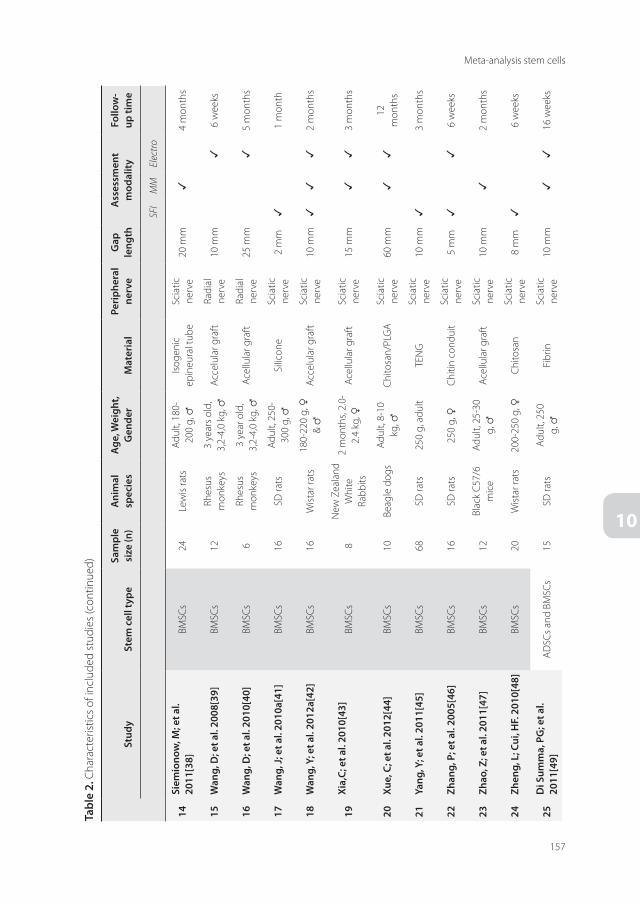
Chapter 10
156 157
Meta-analysis stem cells
10
Tabl
e 2.
Cha
ract
erist
ics o
f inc
lude
d st
udie
s (co
ntin
ued)
stud
yst
em c
ell t
ype
sam
ple
size
(n)
Ani
mal
sp
ecie
sA
ge, W
eigh
t, G
ende
rm
ater
ial
Peri
pher
al
nerv
eG
ap
leng
thA
sses
smen
t m
odal
ity
Follo
w-
up ti
me
SFI
MM
Elec
tro
14si
emio
now
, m; e
t al.
2011
[38]
BMSC
s24
Lew
is ra
tsAd
ult,
180-
200
g, ♂
Isoge
nic
epin
eura
l tub
eSc
iatic
ne
rve
20 m
m✓
4 m
onth
s
15W
ang,
d; e
t al.
2008
[39]
BMSC
s12
Rhes
us
mon
keys
3 ye
ars o
ld,
3,2-
4,0
kg, ♂
Acce
lula
r gra
ftRa
dial
ne
rve
10 m
m✓
6 w
eeks
16W
ang,
d; e
t al.
2010
[40]
BMSC
s6
Rhes
us
mon
keys
3 ye
ar o
ld,
3,2-
4,0
kg, ♂
Acel
lula
r gra
ftRa
dial
ne
rve
25 m
m✓
5 m
onth
s
17W
ang,
J; e
t al.
2010
a[41
]BM
SCs
16SD
rats
Adul
t, 25
0-30
0 g,
♂Si
licon
eSc
iatic
ne
rve
2 m
m✓
1 m
onth
18W
ang,
y; e
t al.
2012
a[42
]BM
SCs
16W
istar
rats
180-
220
g, ♀
& ♂
Acce
lula
r gra
ftSc
iatic
ne
rve
10 m
m✓
✓✓
2 m
onth
s
19Xi
a,c;
et a
l. 20
10[4
3]BM
SCs
8N
ew Z
eala
nd
Whi
te
Rabb
its
2 m
onth
s, 2.
0-2.
4 kg
, ♀Ac
ellu
lar g
raft
Scia
tic
nerv
e15
mm
✓✓
3 m
onth
s
20Xu
e, c
; et a
l. 20
12[4
4]BM
SCs
10Be
agle
dog
sAd
ult,
8-10
kg
, ♂Ch
itosa
n/PL
GA
Scia
tic
nerv
e60
mm
✓✓
12
mon
ths
21ya
ng, y
; et a
l. 20
11[4
5]BM
SCs
68SD
rats
250
g, a
dult
TEN
GSc
iatic
ne
rve
10 m
m✓
3 m
onth
s
22Zh
ang,
P; e
t al.
2005
[46]
BMSC
s16
SD ra
ts25
0 g,
♀Ch
itin
cond
uit
Scia
tic
nerv
e5
mm
✓✓
6 w
eeks
23Zh
ao, Z
; et a
l. 20
11[4
7]BM
SCs
12Bl
ack
C57/
6 m
ice
Adul
t, 25
-30
g, ♂
Acel
lula
r gra
ftSc
iatic
ne
rve
10 m
m✓
2 m
onth
s
24Zh
eng,
L; c
ui, h
F. 2
010[
48]
BMSC
s20
Wist
ar ra
ts20
0-25
0 g,
♀Ch
itosa
nSc
iatic
ne
rve
8 m
m✓
6 w
eeks
25d
i sum
ma,
PG
; et a
l. 20
11[4
9]AD
SCs a
nd B
MSC
s15
SD ra
tsAd
ult,
250
g, ♂
Fibr
inSc
iatic
ne
rve
10 m
m✓
✓16
wee
ks

Chapter 10
158
Tabl
e 2.
Cha
ract
erist
ics o
f inc
lude
d st
udie
s (co
ntin
ued)
stud
yst
em c
ell t
ype
sam
ple
size
(n)
Ani
mal
sp
ecie
sA
ge, W
eigh
t, G
ende
rm
ater
ial
Peri
pher
al
nerv
eG
ap
leng
thA
sses
smen
t m
odal
ity
Follo
w-
up ti
me
SFI
MM
Elec
tro
26La
dak,
A; e
t al.
2011
[50]
uBM
SCs a
nd d
BMSC
s16
SD ra
ts25
0-27
5 g
Colla
gen
Scia
tic
nerv
e12
mm
✓3
mon
ths
27W
ang,
y; e
t al.
2012
[51]
BMSC
-SCs
and
AD
SC-
SCs
39SD
rats
Adul
t, 20
0-25
0 g,
♂Ac
ellu
lar g
raft
Scia
tic
nerv
e15
mm
✓3
mon
ths
28Li
u,G
; et a
l. 20
11a[
52]
ADSC
s32
Wist
ar ra
ts3-
4 w
eeks
old
, 20
0-25
0 g,
ANA
Scia
tic
nerv
e10
mm
✓✓
✓3
mon
ths
29Li
u, G
B; e
t al.
2011
[17]
ADSC
s20
Wist
ar ra
tsAd
ult,
200-
250
g, ♀
Acce
lula
r gra
ftSc
iatic
ne
rve
15 m
m✓
✓✓
3 m
onth
s
30O
rbay
, h; e
t al.
2011
[7]
(d-)A
DSC
S12
Fish
er ra
ts25
0-30
0 g,
♂Si
licon
eSc
iatic
ne
rve
10 m
m✓
✓6
mon
ths
31sa
ntia
go, L
y; e
t al.
2009
[21]
ADSC
s15
Athy
mic
rats
8 w
eeks
old
, 22
3.5±
49,2
g ♂
PCL
Scia
tic
nerv
e6
mm
✓✓
3 m
onth
s
41sh
en, c
c; e
t al.
2012
[53]
undi
ffere
ntia
ted
ADSC
s12
SD ra
tsAd
ult,
250-
300
gG
GT
Scia
tic
nerv
e10
mm
✓2
mon
ths
32A
o, Q
; et a
l. 20
11[5
4]O
ther
: SCs
18SD
rats
Adul
t, 19
0-21
0 g,
♂Ch
itosa
nSc
iatic
ne
rve
12 m
m✓
3 m
onth
s
33Fu
, Ky;
et a
l. 20
11[5
5]O
ther
: NSC
s12
SD ra
tsAd
ult,
250-
300
g, ♂
PLA
Scia
tic
nerv
e15
mm
✓2
mon
ths
34h
su, s
h; e
t al.
2009
[56]
Oth
er: N
CSCs
8SD
rats
250-
300
g, ♂
PLA
Scia
tic
nerv
e10
mm
✓6
wee
ks
35Li
n, h
; et a
l. 20
09[1
2]O
ther
: Hai
r fol
licle
ne
ural
cre
st st
em c
ell
50SD
rats
Adul
t, 18
0-20
0 g,
♂AN
XSc
iatic
ne
rve
40 m
m✓
12
mon
ths
36m
arch
esi,
c; e
t al.
2007
[57]
Oth
er: S
DSC
s10
0W
istar
rats
2 m
onth
s, 15
0-20
0 g,
♂PL
A an
d Co
llage
nSc
iatic
ne
rve
16 m
m✓
3 m
onth
s
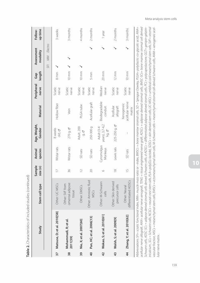
159
Meta-analysis stem cells
10
Tabl
e 2.
Cha
ract
erist
ics o
f inc
lude
d st
udie
s (co
ntin
ued)
stud
yst
em c
ell t
ype
sam
ple
size
(n)
Ani
mal
sp
ecie
sA
ge, W
eigh
t, G
ende
rm
ater
ial
Peri
pher
al
nerv
eG
ap
leng
thA
sses
smen
t m
odal
ity
Follo
w-
up ti
me
SFI
MM
Elec
tro
37m
atsu
se, d
; et a
l. 20
10[5
8]O
ther
: UC-
MSC
s17
Wist
ar ra
ts8
wee
ks
old,
♂H
ollo
w fi
ber
Scia
tic
nerv
e8
mm
✓3
wee
ks
38m
oham
mad
i, R;
et a
l. 20
11[5
9]
Oth
er: S
VF fr
om
omen
tal a
dipo
se
tissu
e36
Wist
ar ra
ts27
0 g,
♂Ve
inSc
iatic
ne
rve
10 m
m✓
✓3
mon
ths
39N
ie, X
; et a
l. 20
07[6
0]O
ther
: EM
SCs
12SD
rats
Adul
t, 20
0 g,
♂PL
GA
tube
Scia
tic
nerv
e10
mm
✓4
mon
ths
40Pa
n, h
c; e
t al.
2006
[13]
Oth
er: A
mni
otic
flui
d M
SCs
20SD
rats
250-
300
gAc
ellu
lar g
raft
Scia
tic
nerv
e5
mm
✓2
mon
ths
42W
akao
, s; e
t al.
2010
[61]
Oth
er: M
-Sch
wan
n ce
lls6
Cyno
mol
gus
Mon
keys
Adul
t (3-
4 ye
ars)
, 3,7
-4,2
kg
, ♂
Biod
egra
dabl
e co
ndui
tM
edia
n ne
rve
20 m
m✓
1 ye
ar
43W
alsh
, s; e
t al.
2009
[9]
Oth
er: S
kin
deriv
ed
prec
urso
r cel
ls18
Lew
is ra
ts22
5-25
0 g,
♂Ac
ellu
lar
allo
graf
tSc
iatic
ne
rve
12 m
m✓
2 m
onth
s
44Zh
ang,
y; e
t al.
2010
[62]
Oth
er: N
eura
l di
ffere
ntia
ted
ADSC
s15
SD ra
ts--
Xeno
gene
ic
acel
lula
r ner
ve
mat
rix
Scia
tic
nerv
e10
mm
✓3
mon
ths
Abbr
evia
tions
SFI
= sc
iatic
func
tiona
l ind
ex, M
M=
mus
cle
mas
s-ra
tio o
r –in
dex,
BMSC
s = b
one
mar
row
stro
mal
cel
ls, S
D =
Spr
ague
Daw
ley,
PLG
A= p
oly(
lact
ic-c
o-gl
ycol
ic a
cid)
, AN
A=
acel
lula
r ner
ve a
llogr
aft,
ANX=
ace
llula
r ner
ve x
enog
raft,
TEN
G =
tiss
ue e
ngin
eere
d ne
rve
graf
t, AD
SCs
= a
dipo
se d
eriv
ed s
trom
al c
ells,
BM
SC-S
Cs=
bon
e m
arro
w s
trom
al c
ell d
eriv
ed
Schw
ann
cells
, AD
SC-S
Cs =
adi
pose
der
ived
stro
mal
cel
ls de
rived
Sch
wan
n ce
lls, d
-AD
SCs =
diff
eren
tiatie
d ad
ipos
e de
rived
stro
mal
cel
ls, P
CL=
pol
ycap
rola
cton
e, G
GT=
gen
ipin
-gel
atin
-tri
calc
ium
, SCs
= S
chw
ann
cells
, NCS
Cs =
neu
ral c
rest
stem
cells
, PLA
= p
oly(
D,L-
lact
ide)
, SD
SCs =
skin
der
ived
stem
cells
, UC-
MSC
s = u
mbi
lical
cord
mes
ench
ymal
stem
cells
, SVF
= st
rom
al
vasc
ular
frac
tion,
MSC
s = m
esen
chym
al st
em ce
lls, E
MSC
s = e
ctom
esen
chym
al st
em ce
lls, M
-Sch
wan
n ce
lls =
mes
ench
ymal
stro
mal
cell-
deriv
ed S
chw
ann
cells
, AN
M =
xeno
gene
ic a
cel-
lula
r ner
ve m
atrix
.

Chapter 10
160 161
Meta-analysis stem cells
10
Tabl
e 3.
Dat
a of
incl
uded
stud
ies.
Mea
n di
ffere
nces
bet
wee
n ex
perim
enta
l and
con
trol
gro
up a
s ent
ered
in fo
rest
plo
ts
Scia
tic F
unct
iona
l Ind
ex
Bmsc
sA
dsc
sO
ther
expe
rim
enta
lco
ntro
lex
peri
men
tal
cont
rol
expe
rim
enta
lco
ntro
l
Auth
orM
ean
SDM
ean
SDAu
thor
Mea
nSD
Mea
nSD
Auth
orM
ean
SDM
ean
SD
Shor
t fol
low
-up
time
< 1
mon
th
Wan
g 20
10a[
41]
-93.
24
-98.
110
Shen
201
2[53
]-6
2.57
5.53
-69.
452.
83M
atsu
se 2
010[
58]
-82
3.2
-96
2.9
Liao
201
2[34
]-4
74
-68
5Li
u 20
11a[
52]
-75.
28.
0-9
2.8
6.9
Moh
amm
adi
2012
a[59
]-7
3.3
2.05
-92.
81.
24
Chen
200
6[23
]-6
7.9
10.5
-83.
910
.6Li
u 20
11[1
7]-9
6.18
0.57
-97.
50.
12Ao
201
1[54
]-7
9.2
2.21
-84.
172.
75
Zhen
g 20
10[4
8]-3
92
-52.
41
Mar
ches
i 200
7(1)
[57]
-73.
663.
13-7
73.
2
Hou
200
6[30
]-8
6.12
12.5
8-8
9.47
11.5
6M
arch
esi 2
007(
2)[5
7]-7
3.5
2.4
-77.
70.
5
Moh
amm
adi
2012
[36]
-74.
41.
28-9
2.8
1.24
Wan
g 20
12a[
4]-7
8.4
4-8
8.4
1.6
Yang
201
1[45
]-8
38.
7464
-86
11.6
619
Mim
ura
2004
[35]
-92.
53.
65-9
3.3
8.33
Chen
200
7[60
]-7
3.3
30.8
577
-89.
724
.149
5
Mid
dle
follo
w-u
p tim
e 1-
3 m
onth
s
Liao
201
2[34
]-1
04
-19
4Sh
en 2
012[
53]
-56.
814.
12-6
4.96
1.9
Hsu
200
9[56
]-4
710
-69
10
Chen
200
6[23
]-6
7.1
7.5
-82.
88.
5Li
u 20
11a[
52]
-58.
15.
3-8
4.8
5.0
Moh
amm
adi
2012
a[59
]-5
8.6
1.23
-74.
61.
44
Zhen
g 20
10[4
8]-1
9.4
1-3
4.7
2Li
u 20
11[1
7]-8
2.2
0.49
-90.
70.
09Ao
201
1[54
]-7
5.02
2.34
-83.
792.
71

Chapter 10
160 161
Meta-analysis stem cells
10
Hou
200
6[30
]-7
1.21
7.67
-78.
6810
.95
Mar
ches
i 200
7(1)
[57]
-75
0.5
-74.
30.
9
Moh
amm
adi
2012
[36]
-59.
81.
14-7
4.6
1.44
Mar
ches
i 200
7(2)
[57]
-71
0.01
-72.
90.
01
Wan
g 20
12a49
-73
3.6
-82.
22.
4
Yang
201
1[45
]-8
07.
5802
-82
12.2
45
Chen
200
7[60
]-4
6.3
26.8
328
-74.
331
.752
2
Long
follo
w-u
p tim
e >
3 m
onth
s
Hou
200
6[30
]-5
8.73
9.44
-72.
137.
45O
rbay
201
2 (3
)[7]
-57
50.8
-86.
820
.8M
oham
mad
i 20
12a[
59]
-44.
81.
83-6
4.1
2.1
Moh
amm
adi
2012
[36]
-41.
91.
12-6
4.1
2.1
Orb
ay 2
012
(4)[7
]-5
9.2
7.06
-86.
820
.8N
ie 2
007[
60]
-66.
23.
05-8
4.3
4
Yang
201
1[45
]-6
88.
1633
-78
18.6
59Li
u 20
11a[
52]
-45.
37.
1-7
9.1
7.5
Ao 2
011[
54]
-73.
572.
09-7
6.96
3.32
Mim
ura
2004
[35]
-76.
56.
31-8
1.9
6.65
Liu
2011
[17]
-80.
510.
09-8
6.79
0.4
Mar
ches
i 200
7(1)
[57]
-69.
40.
21-7
5.1
2.6
Mar
ches
i 200
7(2)
[57]
-74.
91.
1-8
2.9
1.1
(1) M
arch
esi 2
007
= C
olla
gen
tube
, (2)
Mar
ches
i 200
7 =
PLA
tube
, (3)
Orb
ay 2
012
= d
iffer
entia
ted
ASC,
(4) O
rbay
201
2 =
ASC
Mus
cle
Mas
s Rat
io
Bmsc
sA
dsc
sO
ther
expe
rim
enta
lco
ntro
lex
peri
men
tal
cont
rol
expe
rim
enta
lco
ntro
l
Auth
orM
ean
SDM
ean
SDAu
thor
Mea
nSD
Mea
nSD
Auth
orM
ean
SDM
ean
SD
Follo
w-u
p tim
e <
3 m
onth

Chapter 10
162 163
Meta-analysis stem cells
10
Jia 2
012(
1)[3
2]43
.29
3.71
29.4
455
6
Jia 2
012(
2)[3
2]40
.57
4.82
27.9
22.
11
Wan
g 20
12a[
4]43
.29
3.71
29.4
35.
55
Zhao
201
1[47
]91
.91
5.38
43.3
5.11
Chen
200
7[60
]40
.75.
3224
.73.
86
Xia
2010
[43]
43.7
60.
9142
.71
1.05
Keilh
off 2
006(
3)[3
3]25
624
7
Keilh
off 2
006(
4)[3
3]30
424
7
Nijh
uis 2
010[
5]33
.05
5.96
29.7
4.18
Siem
iono
w 2
011[
38]
279
377
Follo
w-u
p tim
e >
3 m
onth
s
Di S
umm
a 20
11[4
9]43
.22.
9069
343.
1305
Sant
iago
200
9[21
]52
.210
.848
.715
.1M
oham
mad
i 20
12a[
59]
68.4
27.7
27.8
23.5
Moh
amm
adi
2012
[36]
77.1
414
.42
27.3
4.04
Di S
umm
a 20
11[4
9]43
.76.
7082
343.
1305
Moh
amm
adi
2012
b[11
]68
.44.
927
.78
7.97
Liu
2011
a[52
]48
.82.
937
.64.
1
Lada
k 20
11(5
)[50]
25.9
2.82
8429
.74.
5255
Wan
g 20
12[5
1]38
.314
.934
.54.
3
Lada
k 20
11(6
)[50]
28.4
4.24
2629
.74.
5255
Liu
2011
[17]
44.5
4.63
38.1
33.
76
Wan
g 20
12[5
1]37
.79.
934
.54.
3
Xia
2010
[43]
65.0
11.
0966
.36
1.28
Nijh
uis 2
010[
5]21
.68
1.89
17.2
22.
32
Siem
iono
w 2
011[
38]
3710
3933
Din
g 20
10[2
9]76
960
15
Xue
2012
(7)[4
4]46
2032
11
Xue
2012
(8)[4
4]72
2270
7

Chapter 10
162 163
Meta-analysis stem cells
10
(1) J
ia 2
012
= A
NA,
(2) J
ia 2
012
= A
NX,
(3) K
eilh
off 2
006
= n
on-tr
ansd
iffer
entia
ted
MSC
, (4)
Kei
lhoff
200
6 =
tran
sdiff
eran
tiate
d M
SC, (
5) La
dak 2
011
= u
n-di
ffere
ntia
ted
MSC
, (6)
Lada
k 20
11 =
diff
eren
tiate
d M
SC, (
7) X
ue 2
012
= Ti
bial
is an
terio
r, (8
) Xue
201
2 =
Gas
trocn
emiu
s
Elec
trop
hysio
logy
– L
aten
cy
Bmsc
sA
dsc
sO
ther
expe
rim
enta
lco
ntro
lex
peri
men
tal
cont
rol
expe
rim
enta
lco
ntro
l
Auth
orM
ean
SDM
ean
SDAu
thor
Mea
nSD
Mea
nSD
Auth
orM
ean
SDM
ean
SD
Mid
dle
follo
w-u
p tim
e 1-
3 m
onth
s
Jia 2
012(
1)[3
2]1.
560.
121.
680.
1Pa
n 20
06[1
3]1.
70.
472.
50.
45
Jia 2
012
(2)[3
2]1.
440.
271.
720.
13
Wan
g 20
08[3
9]3.
820.
755.
030.
66
Wan
g 20
12a[
4]1.
330.
091.
870.
13
Chen
200
7[60
]2.
250.
8497
2.87
1.11
8
Long
follo
w-u
p tim
e >
3 m
onth
s
Wan
g 20
10[4
0]2.
380.
523.
370.
56Li
u 20
11a[
52]
1.38
0.14
2.85
0.21
Hu
2007
[31]
7.82
1.05
39.
971.
244
Liu
2011
[17]
1.39
0.07
1.85
0.05
Elec
trop
hysio
logy
– A
mpl
itude
Bmsc
sA
dsc
sO
ther
expe
rim
enta
lco
ntro
lex
peri
men
tal
cont
rol
expe
rim
enta
lco
ntro
l
Auth
orM
ean
SDM
ean
SDAu
thor
Mea
nSD
Mea
nSD
Auth
orM
ean
SDM
ean
SD
Mid
dle
follo
w-u
p tim
e 1-
3 m
onth
s
Jia 2
012(
1)[3
2]6.
031.
463.
750.
65Fu
201
1[55
]0.
460.
020.
40.
02
Jia 2
012(
2)[3
2]5.
870.
324.
961.
12W
alsh
200
9[9]
0.39
0.24
0.19
0.09
Wan
g 20
08[3
9]2.
950.
752
1.97
0.74
Wan
g 20
12a[
4]5.
120.
443.
010.
28
Chen
200
7[60
]26
.118
.783
13.4
18.3
358
Long
follo
w-u
p tim
e >
3 m
onth
s
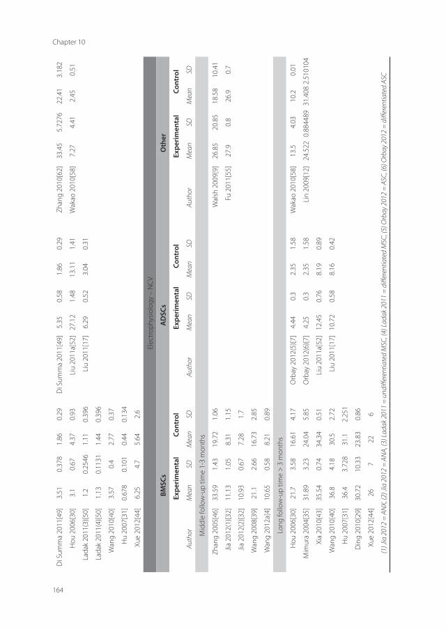
Chapter 10
164
Di S
umm
a 20
11[4
9]3.
510.
378
1.86
0.29
Di S
umm
a 20
11[4
9]5.
350.
581.
860.
29Zh
ang
2010
[62]
33.4
55.
7276
22.4
13.
182
Hou
200
6[30
]3.
10.
674.
370.
93Li
u 20
11a[
52]
27.1
21.
4813
.11
1.41
Wak
ao 2
010[
58]
7.27
4.41
2.45
0.51
Lada
k 20
11(3
)[50]
1.2
0.25
461.
110.
396
Liu
2011
[17]
6.29
0.52
3.04
0.31
Lada
k 20
11(4
)[50]
1.13
0.11
311.
440.
396
Wan
g 20
10[4
0]3.
570.
42.
770.
37
Hu
2007
[31]
0.67
80.
101
0.44
0.13
4
Xue
2012
[44]
6.25
4.7
5.64
2.6
Elec
trop
hysio
logy
– N
CV
Bmsc
sA
dsc
sO
ther
expe
rim
enta
lco
ntro
lex
peri
men
tal
cont
rol
expe
rim
enta
lco
ntro
l
Auth
orM
ean
SDM
ean
SDAu
thor
Mea
nSD
Mea
nSD
Auth
orM
ean
SDM
ean
SD
Mid
dle
follo
w-u
p tim
e 1-
3 m
onth
s
Zhan
g 20
05[4
6]33
.59
1.43
19.7
21.
06W
alsh
200
9[9]
26.8
520
.85
18.5
810
.41
Jia 2
012(
1)[3
2]11
.13
1.05
8.31
1.15
Fu 2
011[
55]
27.9
0.8
26.9
0.7
Jia 2
012(
2)[3
2]10
.93
0.67
7.28
1.7
Wan
g 20
08[3
9]21
.12.
6616
.73
2.85
Wan
g 20
12a[
4]10
.65
0.58
8.21
0.89
Long
follo
w-u
p tim
e >
3 m
onth
s
Hou
200
6[30
]21
.73.
5816
.61
4.17
Orb
ay 2
012(
5)[7
]4.
440.
32.
351.
58W
akao
201
0[58
]13
.54.
0310
.20.
01
Mim
ura
2004
[35]
31.8
93.
2324
.04
5.85
Orb
ay 2
012(
6)[7
]4.
250.
32.
351.
58Li
n 20
09[1
2]24
.522
0.88
4489
31.4
082.
5101
04
Xia
2010
[43]
35.5
40.
7434
.34
0.51
Liu
2011
a[52
]12
.45
0.76
8.19
0.89
Wan
g 20
10[4
0]36
.84.
1830
.52.
72Li
u 20
11[1
7]10
.72
0.58
8.16
0.42
Hu
2007
[31]
36.4
3.72
831
.12.
251
Din
g 20
10[2
9]30
.72
10.3
323
.83
0.86
Xue
2012
[44]
267
226
(1) J
ia 2
012
= A
NX,
(2) J
ia 2
012
= A
NA,
(3) L
adak
201
1 =
und
iffer
entia
ted
MSC
, (4)
Lada
k 201
1 =
diff
eren
tiate
d M
SC, (
5) O
rbay
201
2 =
ASC
, (6)
Orb
ay 2
012
= d
iffer
entia
ted
ASC
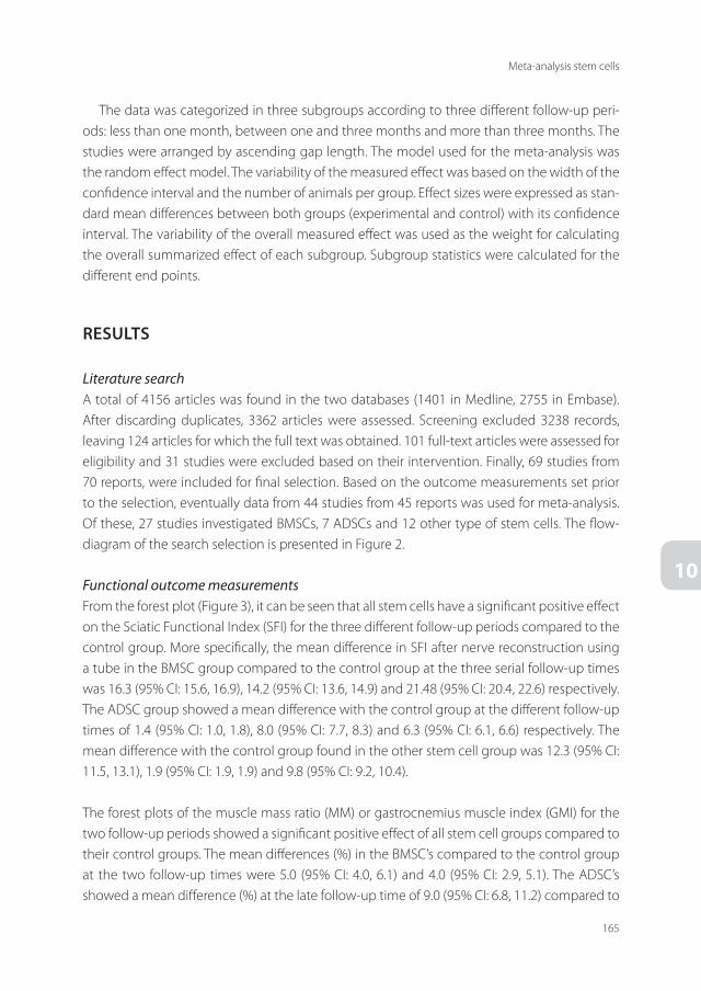
165
Meta-analysis stem cells
10
The data was categorized in three subgroups according to three different follow-up peri-ods: less than one month, between one and three months and more than three months. The studies were arranged by ascending gap length. The model used for the meta-analysis was the random effect model. The variability of the measured effect was based on the width of the confidence interval and the number of animals per group. Effect sizes were expressed as stan-dard mean differences between both groups (experimental and control) with its confidence interval. The variability of the overall measured effect was used as the weight for calculating the overall summarized effect of each subgroup. Subgroup statistics were calculated for the different end points.
ResuLTs
Literature searchA total of 4156 articles was found in the two databases (1401 in Medline, 2755 in Embase). After discarding duplicates, 3362 articles were assessed. Screening excluded 3238 records, leaving 124 articles for which the full text was obtained. 101 full-text articles were assessed for eligibility and 31 studies were excluded based on their intervention. Finally, 69 studies from 70 reports, were included for final selection. Based on the outcome measurements set prior to the selection, eventually data from 44 studies from 45 reports was used for meta-analysis. Of these, 27 studies investigated BMSCs, 7 ADSCs and 12 other type of stem cells. The flow-diagram of the search selection is presented in Figure 2.
Functional outcome measurementsFrom the forest plot (Figure 3), it can be seen that all stem cells have a significant positive effect on the Sciatic Functional Index (SFI) for the three different follow-up periods compared to the control group. More specifically, the mean difference in SFI after nerve reconstruction using a tube in the BMSC group compared to the control group at the three serial follow-up times was 16.3 (95% CI: 15.6, 16.9), 14.2 (95% CI: 13.6, 14.9) and 21.48 (95% CI: 20.4, 22.6) respectively. The ADSC group showed a mean difference with the control group at the different follow-up times of 1.4 (95% CI: 1.0, 1.8), 8.0 (95% CI: 7.7, 8.3) and 6.3 (95% CI: 6.1, 6.6) respectively. The mean difference with the control group found in the other stem cell group was 12.3 (95% CI: 11.5, 13.1), 1.9 (95% CI: 1.9, 1.9) and 9.8 (95% CI: 9.2, 10.4).
The forest plots of the muscle mass ratio (MM) or gastrocnemius muscle index (GMI) for the two follow-up periods showed a significant positive effect of all stem cell groups compared to their control groups. The mean differences (%) in the BMSC’s compared to the control group at the two follow-up times were 5.0 (95% CI: 4.0, 6.1) and 4.0 (95% CI: 2.9, 5.1). The ADSC’s showed a mean difference (%) at the late follow-up time of 9.0 (95% CI: 6.8, 11.2) compared to

Chapter 10
166
the control group. The effect of the other stem cells (SVF) was based on only one study and showed a mean difference (%) of 40.6 (95% CI: 23.9, 57.4) at the long follow-up time.
Regarding the electrophysiology, all stem cell groups showed significant positive effects compared to the control groups for the two follow-up periods except for the other stem cells at the long follow-up period in nerve conduction velocity. The forest plots are depicted in Figure 3. The mean difference in BMSC’s showed a significant positive effect on the NCV (mm/ms) at the two follow up times of 4.7 (95% CI: 4.2, 5.3) and 1.9 (1.1, 2.7). The mean difference in amplitude (mV) was 1.9 (95% CI: 1.6, 2.2) and 0.3 (95% CI: 0.2, 0.4) in the two follow up times. The latency (ms) showed a mean difference of -0.4 (95% CI: -0.4, -0.3) and -1.3 (95% CI: -2.1, -0.6), compared to the control group.
The mean difference in the ADSC group was only measured at the long follow up time and showed a significant positive difference of 2.9 (95% CI: 2.6, 3.3) in NCV (mm/ms). The mean difference at the long follow up time in amplitude (mV) was 3.9 (95% CI: 3.6, 4.2). The latency (ms) showed a mean difference of -0.6 (95% CI: -0.6, -0.5) compared to the control group at the long follow up time.
The mean difference with the control group in the other stem cell group (amniotic fluid derived stem cells and skin-derived stem cells) showed a positive effect on the NCV (mm/ms) at the two follow up times of 1.0 (95% CI: 0.2, 1.9) and -6.2 (95% CI: -7.2, -5.2). The mean
Figure 2. Flowchart of study selection.

167
Meta-analysis stem cells
10
Figu
re 3
. Sci
atic
Fun
ctio
nal I
ndex
. Abb
revi
atio
ns: S
FI =
scia
tic fu
nctio
nal i
ndex
, BM
SCs =
bon
e m
arro
w st
rom
al c
ells,
AD
SCs =
adi
pose
der
ived
stro
mal
cel
ls
Figu
re 4
. Mus
cle
Mas
s Rat
io. A
bbre
viat
ions
: mm
=m
uscl
e m
ass,
BMSC
= b
one
mar
row
stro
mal
cel
ls, A
DSC
s = a
dipo
se d
eriv
ed st
rom
al c
ells

Chapter 10
168
Figu
re 5
. Ele
ctro
phys
iolo
gy: l
aten
cy (m
s), a
mpl
itude
(mV)
and
ner
ve c
ondu
ctio
n ve
loci
ty (N
CV) (
mm
/ms)
. Abb
revi
atio
ns: B
MSC
s =
bon
e m
arro
w s
trom
al
cells
, AD
SCs =
adi
pose
der
ived
stro
mal
cel
ls.
169
Meta-analysis stem cells
10
difference in amplitude (mV) was 0.1 (95% CI: 0.0, 0.1) at the middle follow up time and 6.0 (95% CI: 2.1, 9.9) at the long follow up time. The effect of the other stem cells on the latency was based on only one stud and showed a mean difference (ms) of -0.8 (95% CI: -1.2, -0.4) at the middle follow-up time.
discussiON
This meta-analysis aimed to summarize the beneficial regenerative effect of the different types of stem cells. Comparing animal studies investigating nerve reconstruction using a nerve guide with additional support of stem cells to a control group without the support of stem cells. Analyzing functional outcome, the contribution of stem cells is apparent and thus suffices an even better neuronal regeneration. The regeneration effect is summarized for three different functional outcome measurements.
The first outcome measurement evaluated was the SFI. The SFI showed significantly better results in all stem cell groups for the different follow up periods compared to a control group. Since the SFI is detracted from the walking track analysis, which is a valid tool for evaluating the regeneration of the nerve, the positive effect found in all studies using this technique is strong evidence for the supporting role of stem cells.[19]
The second functional outcome, the muscle mass ratio of gastrocnemius muscle index, also showed positive results for both the adipose and bone marrow derived stem cells. For the ADSCs (n=7) and BMSCs (n=27), multiple studies were used in this analysis. Like the SFI, the MM or GMI are also valid, well-accepted, tools for evaluation nerve regeneration. The standardized protocol for assessing the muscle mass allows an easy comparison between the different studies.
The third functional outcome measurement is electrophysiology. Using electrophysiology, nerve conduction latency, amplitude and velocity can be recorded. The mean differences showed the beneficial effect of the BMSCs and the ADSCs for all three parameters at the three different follow up periods. The other stem cells (amniotic fluid derived stem cells and the skin derived stem cells) showed a positive effect for the three different measurements for both the latency and amplitude at the three different follow up periods. Only with evaluation of nerve conduction velocity in the long follow up period in the other stem cell group, no significant beneficial effect of the intra luminal injection of those stem cells was found.
Interpretation of resultsRegarding survival some studies investigated the survival of stem cells in the graft.[20] The main conclusion was that the cells survived for follow up periods up to three months[15, 21]. The proportion of cells that survived was reported to be up to 38%.[22] Furthermore the transition from stem cell to Schwann cell has been encountered previously and thus indicates

Chapter 10
170
that this could be a vital step in explaining the beneficial effect.[23-26] Schwann cells however not only myelinate, Hoke et al showed that the Schwann cell phenotype can be modulated, for example into motor or sensory Schwann cells[27]. Also the stem cells extent a more sup-porting and productive role, producing neural growth stimulating factors.[24, 25]
LimitationsThis study has a number of limitations. The first limitation is that the duration of investigation for the different types of stem cells varies significantly. BMSCs are subject to investigation for a longer period of time compared to, for example, the use of hair follicle-derived stem cells. As can be expected, the number of articles used for analysis for the different types of cells -thus- varies and possibly warrant the differences in regenerative effect.
Regarding the evaluation tools used, it should be noted that some studies evaluated nerve regeneration at multiple time points using the walking track analysis to determine the SFI. To allow a comparison between all included studies, we decided to set a maximum of three end points for this evaluation. For the other two outcome measurements, muscle mass and electrophysiology, only one end point was used. When a study compared two experimental groups with one control group, the data of this control group was used twice in the analysis. This made the actual sample size of the control group smaller than the factual one.
ImplicationsThe studies investigated in this review are all animal related studies and do not warrant suc-cess in clinical setting. Although multiple studies have been conducted and stimulate the use of stem cells for nerve reconstruction, the bridge towards clinical implementation has to be made yet. At this point undoubtedly strong evidence is presented for the beneficial effect of the stem cells. Therefore this study can help progress the translational approach for implementing stem cells in further clinical research. The focus of the present study was on the beneficial effect of stem cells on nerve regeneration. A next step could be to compare the overall effect of the stem cells to the regeneration after reconstructing a peripheral nerve defect with a nerve autograft.
cONcLusiONs
This study is the first meta-analysis executed in the field of experimental nerve reconstruction studies to illustrate a beneficial effect of the stem cells used as a luminal additive. Taking our findings into consideration, we may conclude that reconstructing a peripheral nerve gap using a guide with a luminal additive (i.e. cellular therapy) results in better regeneration as compared to using a conduit alone.

171
Meta-analysis stem cells
10
AcKNOWLedGmeNTs
The authors would like to thank W.M. Bramer for his help with the study search.

Chapter 10
172
ReFeReNces
1. Noble, J., et al., Analysis of upper and lower extremity peripheral nerve injuries in a population of patients with multiple
injuries. J Trauma, 1998. 45(1): p. 116-22.
2. Dellon, A.L. and S.E. Mackinnon, An alternative to the classical nerve graft for the management of the short nerve
gap. Plast Reconstr Surg, 1988. 82(5): p. 849-56.
3. Pettersson, J., et al., Muscle recovery after repair of short and long peripheral nerve gaps using fibrin conduits. Neuro-
sci Lett, 2011. 500(1): p. 41-6.
4. Angius, D., et al., A systematic review of animal models used to study nerve regeneration in tissue-engineered scaf-
folds. Biomaterials, 2012.
5. Nijhuis, T.H., et al., Isogenic venous graft supported with bone marrow stromal cells as a natural conduit for bridging
a 20 mm nerve gap. Microsurgery, 2010. 30(8): p. 639-645.
6. Boyd, J.G. and T. Gordon, Neurotrophic factors and their receptors in axonal regeneration and functional recovery
after peripheral nerve injury. Mol Neurobiol, 2003. 27(3): p. 277-324.
7. Orbay, H., et al., Differentiated and undifferentiated adipose-derived stem cells improve function in rats with peripheral
nerve gaps. J Plast Reconstr Aesthet Surg, 2011.
8. Yan, Q., Y. Yin, and B. Li, Use new PLGL-RGD-NGF nerve conduits for promoting peripheral nerve regeneration. Biomed
Eng Online, 2012. 11(1): p. 36.
9. Walsh, S., et al., Supplementation of acellular nerve grafts with skin derived precursor cells promotes peripheral nerve
regeneration. Neuroscience, 2009. 164(3): p. 1097-1107.
10. Hood, B., H.B. Levene, and A.D. Levi, Transplantation of autologous Schwann cells for the repair of segmental periph-
eral nerve defects. Neurosurg Focus, 2009. 26(2): p. E4.
11. Mohammadi, R., et al., Transplantation of undifferentiated bone marrow stromal cells improves sciatic nerve regen-
eration and functional recovery through inside-out vein graft in rats. Turk J Med Sci, 2012. 42(1): p. 127-136.
12. Lin, H., et al., Pluripotent hair follicle neural crest stem-cell-derived neurons and schwann cells functionally repair
sciatic nerves in rats. Mol Neurobiol, 2009. 40(3): p. 216-223.
13. Pan, H.C., et al., Enhanced regeneration in injured sciatic nerve by human amniotic mesenchymal stem cell. J Clin
Neurosci, 2006. 13(5): p. 570-575.
14. Hermann, A., et al., Efficient generation of neural stem cell-like cells from adult human bone marrow stromal cells. J
Cell Sci, 2004. 117(Pt 19): p. 4411-22.
15. Walsh, S. and R. Midha, Use of stem cells to augment nerve injury repair. Neurosurgery, 2009. 65(4 Suppl): p. A80-6.
16. Nijhuis, T.H., Bridging the gap in relation to nerve injury, in Plastic, reconstructive and handsurgery. 2011, Erasmus
University Rotterdam: Rotterdam.
17. Liu, G.B., et al., Adipose-derived stem cells promote peripheral nerve repair. Arch Med Sci, 2011. 7(4): p. 592-596.
18. Collaboration, T.C., Review Manager. 2011, The Nordic Cochrane Centre: Copenhagen.
19. Bain, J.R., S.E. Mackinnon, and D.A. Hunter, Functional evaluation of complete sciatic, peroneal, and posterior tibial
nerve lesions in the rat. Plast Reconstr Surg, 1989. 83(1): p. 129-38.
20. Gartner, A., et al., Use of poly(DL-lactide-epsilon-caprolactone) membranes and mesenchymal stem cells from the
Wharton’s jelly of the umbilical cord for promoting nerve regeneration in axonotmesis: in vitro and in vivo analysis.
Differentiation, 2012. 84(5): p. 355-65.
21. Santiago, L.Y., et al., Delivery of adipose-derived precursor cells for peripheral nerve repair. Cell Transplant, 2009. 18(2):
p. 145-158.
22. Shen, J., et al., In vivo MR imaging tracking of transplanted mesenchymal stem cells in a rabbit model of acute periph-
eral nerve traction injury. J Magn Reson Imaging, 2010. 32(5): p. 1076-85.
23. Chen, X., et al., Study of in vivo differentiation of rat bone marrow stromal cells into schwann cell-like cells. Microsur-
gery, 2006. 26(2): p. 111-115.
173
Meta-analysis stem cells
10
24. Cuevas, P., et al., Peripheral nerve regeneration by bone marrow stromal cells. Neurol Res, 2002. 24(7): p. 634-8.
25. Sanchez-Ramos, J., et al., Adult bone marrow stromal cells differentiate into neural cells in vitro. Exp Neurol, 2000.
164(2): p. 247-56.
26. Tomita, K., et al., Glial differentiation of human adipose-derived stem cells: implications for cell-based transplantation
therapy. Neuroscience, 2013. 236: p. 55-65.
27. Hoke, A., et al., Schwann cells express motor and sensory phenotypes that regulate axon regeneration. J Neurosci,
2006. 26(38): p. 9646-55.
28. Chen, C.J., et al., Transplantation of bone marrow stromal cells for peripheral nerve repair. Exp Neurol, 2007. 204(1):
p. 443-453.
29. Ding, F., et al., Use of tissue-engineered nerve grafts consisting of a chitosan/poly(lactic-co-glycolic acid)-based scaf-
fold included with bone marrow mesenchymal cells for bridging 50-mm dog sciatic nerve gaps. Tissue Eng Part A,
2010. 16(12): p. 3779-3790.
30. Hou, S.Y., et al., Tissue-engineered peripheral nerve grafting by differentiated bone marrow stromal cells. Neurosci-
ence, 2006. 140(1): p. 101-110.
31. Hu, J., et al., Repair of extended peripheral nerve lesions in rhesus monkeys using acellular allogenic nerve grafts
implanted with autologous mesenchymal stem cells. Exp Neurol, 2007. 204(2): p. 658-666.
32. Jia, H., et al., Sciatic nerve repair by acellular nerve xenografts implanted with BMSCs in rats xenograft combined with
BMSCs. Synapse, 2012. 66(3): p. 256-269.
33. Keilhoff, G., et al., Peripheral nerve tissue engineering: autologous Schwann cells vs. transdifferentiated mesenchymal
stem cells. Tissue Eng, 2006. 12(6): p. 1451-1465.
34. Liao, C.D., et al., Peripheral nerve repair: monitoring by using gadofluorine M-enhanced MR imaging with chitosan
nerve conduits with cultured mesenchymal stem cells in rat model of neurotmesis. Radiology, 2012. 262(1): p. 161-
171.
35. Mimura, T., et al., Peripheral nerve regeneration by transplantation of bone marrow stromal cell-derived Schwann cells
in adult rats. J Neurosurg, 2004. 101(5): p. 806-812.
36. Mohammadi, R., et al., The use of undifferentiated bone marrow stromal cells for sciatic nerve regeneration in rats. Int
J Oral Maxillofac Surg, 2011.
37. Mohammadi, R., et al., Comparison of beneficial effects of undifferentiated cultured bone marrow stromal cells and
omental adipose-derived nucleated cell fractions on sciatic nerve regeneration. Muscle Nerve, 2011. 43(2): p. 157-
163.
38. Siemionow, M., et al., Peripheral nerve defect repair with epineural tubes supported with bone marrow stromal cells: a
preliminary report. Ann Plast Surg, 2011. 67(1): p. 73-84.
39. Wang, D., et al., Bridging small-gap peripheral nerve defects using acellular nerve allograft implanted with autologous
bone marrow stromal cells in primates. Brain Res, 2008. 1188: p. 44-53.
40. Wang, D., et al., Repairing large radial nerve defects by acellular nerve allografts seeded with autologous bone marrow
stromal cells in a monkey model. J Neurotrauma, 2010. 27(10): p. 1935-1943.
41. Wang, J., et al., No synergistic effect of mesenchymal stem cells and exercise on functional recovery following sciatic
nerve transection. Funct Neurol, 2010. 25(1): p. 33-43.
42. Wang, Y., et al., Synergistic Effects of Bone Mesenchymal Stem Cells and Chondroitinase ABC on Nerve Regeneration
After Acellular Nerve Allograft in Rats. Cell Mol Neurobiol, 2012. 32(3): p. 361-371.
43. Xia, C., et al., Platelet-rich plasma-induced bone marrow mesenchymal stem cells versus autologous nerve grafting for
sciatic nerve repair. Neural Regen Res, 2010. 5(17): p. 1291-1295.
44. Xue, C., et al., Joint use of a chitosan/PLGA scaffold and MSCs to bridge an extra large gap in dog sciatic nerve. Neuro-
rehabil Neural Repair, 2012. 26(1): p. 96-106.
45. Yang, Y., et al., Repair of rat sciatic nerve gap by a silk fibroin-based scaffold added with bone marrow mesenchymal
stem cells. Tissue Eng Part A, 2011. 17(17-18): p. 2231-2244.
Chapter 10
174
46. Zhang, P., et al., Bridging small-gap peripheral nerve defects using biodegradable chitin conduits with cultured
schwann and bone marrow stromal cells in rats. J Reconstr Microsurg, 2005. 21(8): p. 565-571.
47. Zhao, Z., et al., Repair of nerve defect with acellular nerve graft supplemented by bone marrow stromal cells in mice.
Microsurgery, 2011. 31(5): p. 388-394.
48. Zheng, L. and H.F. Cui, Use of chitosan conduit combined with bone marrow mesenchymal stem cells for promoting
peripheral nerve regeneration. J Mater Sci Mater Med, 2010. 21(5): p. 1713-1720.
49. di Summa, P.G., et al., Long-term in vivo regeneration of peripheral nerves through bioengineered nerve grafts. Neuro-
science, 2011. 181: p. 278-291.
50. Ladak, A., et al., Differentiation of mesenchymal stem cells to support peripheral nerve regeneration in a rat model. Exp
Neurol, 2011. 228(2): p. 242-252.
51. Wang, Y., et al., Recellularized nerve allografts with differentiated mesenchymal stem cells promote peripheral nerve
regeneration. Neurosci Lett, 2012. 514(1): p. 96-101.
52. Liu, G., et al., Transplantation of adipose-derived stem cells for peripheral nerve repair. Int J Mol Med, 2011. 28(4): p.
565-572.
53. Shen, C.C., Y.C. Yang, and B.S. Liu, Peripheral nerve repair of transplanted undifferentiated adipose tissue-derived stem
cells in a biodegradable reinforced nerve conduit. J Biomed Mater Res A, 2012. 100(1): p. 48-63.
54. Ao, Q., et al., The regeneration of transected sciatic nerves of adult rats using chitosan nerve conduits seeded with bone
marrow stromal cell-derived Schwann cells. Biomaterials, 2011. 32(3): p. 787-796.
55. Fu, K.Y., et al., Sciatic nerve regeneration by microporous nerve conduits seeded with glial cell line-derived neurotrophic
factor or brain-derived neurotrophic factor gene transfected neural stem cells. Artif Organs, 2011. 35(4): p. 363-372.
56. Hsu, S.H., C.H. Su, and I.M. Chiu, A novel approach to align adult neural stem cells on micropatterned conduits for
peripheral nerve regeneration: a feasibility study. Artif Organs, 2009. 33(1): p. 26-35.
57. Marchesi, C., et al., Skin-derived stem cells transplanted into resorbable guides provide functional nerve regeneration
after sciatic nerve resection. Glia, 2007. 55(4): p. 425-438.
58. Matsuse, D., et al., Human umbilical cord-derived mesenchymal stromal cells differentiate into functional Schwann
cells that sustain peripheral nerve regeneration. J Neuropathol Exp Neurol, 2010. 69(9): p. 973-985.
59. Mohammadi, R., et al., Transplantation of uncultured omental adipose-derived stromal vascular fraction improves
sciatic nerve regeneration and functional recovery through inside-out vein graft in rats. J Trauma Acute Care Surg,
2012. 72(2): p. 390-396.
60. Nie, X., et al., Improvement of peripheral nerve regeneration by a tissue-engineered nerve filled with ectomesenchymal
stem cells. Int J Oral Maxillofac Surg, 2007. 36(1): p. 32-38.
61. Wakao, S., et al., Long-term observation of auto-cell transplantation in non-human primate reveals safety and effi-
ciency of bone marrow stromal cell-derived Schwann cells in peripheral nerve regeneration. Exp Neurol, 2010. 223(2):
p. 537-547.
62. Zhang, Y., et al., A nerve graft constructed with xenogeneic acellular nerve matrix and autologous adipose-derived
mesenchymal stem cells. Biomaterials, 2010. 31(20): p. 5312-5324.


Chapter 11General discussion


179
General Discussion
11
1. PART i – investigating the clinical problemNerve injury may cause severe, life-altering disabilities including the loss of motor function, sensory loss, hyperesthesia and cold intolerance. These disabilities will have a great impact on a patient’s quality of life as they diminish an individual’s capacity to work and exercise hobbies. The outcome after surgery of peripheral nerve injuries of the upper extremities is often unpredictable. In part I of this thesis the clinical outcome after surgical reconstruction of upper extremity nerve injury was investigated. In particular we focused on prognostic factors for a better outcome after reconstructive surgery and the psychological stress involved with nerve trauma.
Several factors were shown to predict functional outcome 1 year after injury (chapter 1 and 2). These included gender, age, education, number of structures and arteries involved, (type of ) damaged nerve, location of injury, type of nerve injury and posttraumatic stress at 1 and 3 months post trauma. The factors sensibility of the hand, power grip and DASH score are the strongest predictors for the outcome after nerve reconstruction. Interestingly and against our hypothesis smoking had no significant correlation with functional outcome. We expected that smoking would diminish the recovery since microcirculation is significantly reduced in the smoking population. The low number of smoking patients in our study group could bias this finding.
Posttraumatic stress at 1 and 3 months are factors that can actually be influenced. Psy-chological symptoms frequently accompany severe injuries of the upper extremities. . Post-traumatic psychological stress was assessed with the Impact of Event Scale (IES) questionnaire, at month 1 and 3 following nerve repair. This questionnaire included 15 items that refer to ‘the past seven days’, across the subscales of avoidance and intrusion and taps dimensions that are similar to defining symptoms of posttraumatic stress disorder In our study, 92% of patients experienced psychological stress 3 months post-operatively Based on IES scores, psychological treatment was indicated in 25% and 13% of patients, 1 and 3 months post-operatively, respectively. Higher IES scores (i.e. more psychological stress) were negatively cor-related with functional outcome. Early recognition of psychological stress could potentially improve outcome. To what extent this really will improve the outcome should be subject of further study. The relation between posttraumatic stress and outcome can be influenced by many factors. Possibly the amount of stress will decrease the person’s ability and enthusiasm to participate in rehabilitation programs. Fear or anxiety can also have a negative effect on the engagement in rehabilitation.
Although the study design was a prospective multicenter study including 7 large hospitals in the Netherlands, it still represents a relatively small sample size. It is known that it is difficult to study nerve injuries. They often have a higher percentage of loss to follow-up. A long-term prospective study should be executed with an automated electronical inclusion of all

Chapter 11
180
patients visiting the hospitals/national registry to include more patients. That would increase the power of these studies allowing for more factors to be studied.
The findings of our study were in line with the findings of other research groups[1, 2]. Bailey et al. highlighted the relationship between nerve injury of the upper extremity and activity participation,pain, depression, and perceived quality of life[2]. They reported a percentage of 39 on the depression scale indicating probable clinical depression. They suggest that patients should be screened before and after surgery. The identification of patients with higher scores on the depression scale could be identified as this may improve the compliance of patients with rehabilitation programs and improve long-term outcome[2].
The results of this study may help us to inform patients about their expected recovery the first year after injury. The information can also assist clinicians in understanding the outcome of patients with peripheral nerve injuries of the forearm.
2. PART ii – evaluationAfter nerve injury with subsequent reconstruction, the process of nerve regeneration begins. The outcome of peripheral nerve injury can be measured in various ways[3]. Validated out-come measurements are important to aid clinical diagnosis, to assess and compare surgical techniques, to follow rehabilitation and to inform patient, surgeon and therapist about the outcome of treatment. In the case of peripheral nerve injury both sensory and motor function should be evaluated.
Clinical use of ultrasound;
In chapter 4, a new, non-invasive ultrasound method to longitudinally assess both early and late nerve injury recovery was investigated. Ultrasound is non-invasive, quick, low in cost and not painful for patients recovering from nerve injuries. The possibility to evaluate motor func-tion recovery using ultrasound was first described by our group in 2013[4]. Confident with the good results a clinical study was established to assess motor function recovery in patients.
In the clinical setting, the method most frequently used to evaluate outcome of motor function of the hand is manual muscle testing (MRC), powergrip, pinchgrip and individual finger strength (by use of the intrinsic Rotterdam myometer)[5, 6]. However, these methods have some limitations. For instance, they can not be used with patients with a MRC score lower than 3 nor can they detect early muscle recovery. Therefore, there is a need for a diag-nostic tool that can provide both patient and clinician with information regarding the early recovery phase. Non-invasive measurements to assess muscle reinnervation after nerve injury are needed for the longitudinal follow up of muscle reinnervation. Ultrasound may serve such a purpose. Other evaluation methods to visualize the muscle, as CT-scan or a MRI scan, are more expensive and time-consuming. The ultrasound measurements can be made in the outpatient clinic in less than 10 minutes and are inexpensive.
181
General Discussion
11
In both healthy adults and patients with ulnar and median nerve injuries a strong correlation was found between the muscle cross-section area, as analyzed with ultrasound, and muscle force. This showed the usefulness of this technique to monitor muscle reinnervation in pa-tients suffering from peripheral nerve injury.
Experimental use of ultrasound;
After establishing this method in the clinical setting we wanted to use this method in the experimental setting as well. For animal experiments, non-invasive methods to assess mo-tor function are highly favorable. They allow for multiple measurements and longitudinal follow-up within the same animal without the need to sacrifice the animal. This minimizes inter-animal variability, yielding higher power with the same number of animals per studied group. This will ultimately reduce the required sample size and costs of those studies and improve animal welfare. The new technique is also easy and could be performed in less than 10 minutes. This makes the new technique an easy applicable method.
To study functional motor recovery after nerve reconstruction in animal models the easily obtainable wet muscle weight is frequently used. Other commonly used established outcome measurements include electrophysiological testing, isometric tetanic force measurement and nerve histomorphometry [7-10]. However, all of those techniques require the animal to be killed for assessment, making a longitudinal analysis of the recovery impossible.
In chapters 5 and 6, the potential of ultrasound was investigated in the two most commonly used animal models in nerve injury research: the rat and the rabbit[11, 12]. Their fast neuro-regenerative capacity makes the rat an efficient model to study nerve regeneration. However, the nerve gap length in rats is limited. The larger rabbit model overcomes many of these limitations, as nerve gaps up to 8 cm are feasible [7, 13]. Also, the rabbit’s neuro-regenerative properties more closely mimic the human setting [14]. Therefore, the rabbit is an important model to study new nerve reconstruction techniques before the step towards the clinical setting can be made.
To establish ultrasound as a new and valid tool to analyze functional recovery after nerve trauma, this technique was compared to the commonly used outcome measurements muscle weight and isometric tetanic force in a rat model. In addition, the neuromuscular junction in the muscle was examined histologically, to visualize denervation and reinnervation of the motor endplates. The intra- and interrater reliability of ultrasound was found to be strong (r=0.97 and r=0.88). The correlation between ultrasonographic recordings and the muscle weight were 0.85 and 0.89, again indicating a strong relation between ultrasound and muscle mass. The correlation between muscle strength, analyzed with isometric tetanic force mea-surements, and ultrasound was lower, but still significant with 0.62. The explanation for the
Chapter 11
182
better higher correlation between ultrasound and muscle weight compared with ultrasound and muscle strength may be that force does not directly relate to weight (i.e. more fibers does not mean more force). This hypothesis was further strengthened by the observation that during the first weeks of recovery, muscle weight gradually increased with delayed response intensity with the isometric tetanic force evaluation.
The ultrasound method was further validated in the rabbit model where a bigger nerve gap of 3 cm could be created (chapter 5). The inter- and intra-rater reliability of the ultrasound recordings in the rabbit were, like in the rat model, high (0.989 and 0.99 respectively). Impor-tantly, the ultrasound measurements could be performed on the rabbits without the need for anesthesia by manual restrain with a harness. This may reduce the risk of anesthesia-related death of the rabbits, which is as high as 7%[15].
The importance of valid outcome measurements in experimental studies investigating peripheral nerve injuries was already stated by others. Several reviews showed the wide variety of different outcome measurements used and the lack of correlation between those measurements[9, 10, 16, 17]. Also, none of these outcome measurements could be used for longitudinal follow-up as all outcome measurements require the animal to be euthanized[3, 7-10].
In summary, these studies demonstrate the value of ultrasound as a diagnostic tool. Ultrasound has a better discriminative power and lower variability, allows for repeated measurements in animal studies and reduces animal suffering. Therefore, we believe that ultrasound should be included in the standard diagnostic armamentarium of both experimental and human studies on nerve regeneration.
3. PART iii - engineering a nerve allograftIn Part III of this thesis, nerve reconstruction using a newly-engineered nerve allograft was studied both in vitro and in vivo. Within the wide variety of available surgical techniques to reconstruct a peripheral nerve gap, the nerve allograft has shown the greatest potential and most closely mimics the gold standard, the autograft. Nerve allografts have the advantage of mitigating donor side morbidity and potentially have an unlimited supply and length of the grafts.
Nerve allografts were first used in the clinical setting when immunosuppressive drugs were not available. Over the years, processing protocols for nerve allografts were improved. Hudson et al. described one of the first processing protocols for the nerve allograft[18, 19] in 2004, which has later been modified by several others[18-22]. After several improvements by Neu-bauer et al. a nerve allograft was introduced in the clinic [20, 22, 23]. However, the currently available nerve allograft shows results that are not satisfactory, especially when focusing on motor nerve reconstruction[24]. Up to this day, there have been no clinical studies or high-
183
General Discussion
11
level evidence (e.g. level I or II) studies published that challenged the allograft to the autograft or to conduits for the reconstruction of large peripheral (motor) nerve defects. The conclu-sions are based on experimental animal studies and level III clinical studies[25]. In processing nerve allografts it is difficult to find the balance between remaining the structure of a graft while removing all cellular debris, which can cause an immune reaction after transplantation.
In chapter 7, the optimization of a novel nerve allograft was studied in vitro in rat sciatic nerve segments. The aim of this experiment was to decellularize the nerves more aggressively by re-moving more cellular debris. Improvement on currently used decellularization protocols were tested by adding an extra enzymatic step, and by studying different storage temperatures. The enzyme Elastase was added to the decellularization process and was hypothesized to better decellularize the nerves by removing more cellular debris. In addition to the rat nerves, the nerve allograft process was also tested in human motor and sensory nerves (chapter 8).
Reduced immunogenicity by diminished cellular debris and the elimination of Schwann cells was observed when Elastase was added to the nerve processing while maintaining ultrastructure. Storage at -80°C after the decellularization process heavily damaged the nerve ultrastructure as compared to cold storage.
The same effects were observed when different processing techniques were tested on hu-man nerves. Human motor nerves were better preserved when compared to sensory nerves. Especially the structure was better preserved in motor nerves. This could be explained by the fact that the basal lamina of the motor nerve is thicker and the endoneural tubes are bigger in motor nerves as compared to sensory nerves[26]. That could make the motor nerve more robust to the processing techniques.
After establishing the optimal processing and storage techniques for the nerve allograft, the next step was to implement the allograft in a rat model (chapter 9). The purpose of this study was to evaluate the elastase-decellularized allograft nerve in a 1-cm segmental rat sciatic nerve gap model. In these experiments the outcome was motor functional outcome at 12 and 16 weeks post-surgery using different storage conditions of the decellularized allograft. When comparing our optimized nerve allografts we found that for all outcome measure-ments investigated, the elastase processed cold preserved allograft showed no statistically significant differences with the gold standard, the nerve autograft, for both early and late regeneration at 12 and 16 weeks. In three out of five outcome measurements, cold storage of the processed allograft showed better results than the frozen stored allograft. We found an improvement at the 16-week time point compared to the 12 week follow-up. This may seem like a logical outcome, however in previous studies with nerve allografts this was not always observed. Giusti et al. evaluated early and late recovery at 12 and 16 weeks, using AxoGen processed allograft rat nerves and compared these to autograft nerves[27]. While
Chapter 11
184
both the allograft and the autograft had comparable results at the early recovery time of 12 weeks, 16 weeks post-surgery the autograft outperformed the AxoGen processed rat allograft nerve[27]. The hypothesis is that this could have been caused by fibrosis. If a nerve allograft is not entirely clear of cellular debris this can cause an reaction after implementation. Possibly fibrosis caused the decrease in muscle force at the late time point of 16 weeks.
In summary, the novel, elastase-processed cold stored nerve allograft has statistically similar functional outcomes compared with the gold standard, the nerve autograft, at both 12 and 16 weeks follow-up. The regeneration potential of the cold stored elastase processed nerve allograft is superior to the frozen stored allograft with respect to motor functional outcome.
4. PART iV – improving the nerve allograftIn the quest to further improve nerve reconstruction techniques investigating nerve allograft transplantation, the next step was to investigate the effect of different cellular additives (chapter 10). We conducted a systematic review with meta-analysis to study the beneficial effect of stem cells in nerve regeneration with a nerve graft by looking at any nerve conduit with and without the addition of stem cells to solemnly evaluate the effect of the stem cells.
A total of 44 studies were investigated in this analysis. We found that the contribution of stem cells had a positive effect on the functional outcome compared to regeneration us-ing a conduit alone. This effect was positive for bone marrow derived stem cells, adipose derived stem cells and other stem cells such as skin-derived stem cells. The mechanism be-hind this positive effect is debatable. The transition from stem cell to Schwann cell has been encountered previously and thus indicates that this could be a vital factor in explaining the beneficial effect[28-31]. Schwann cells however, not only myelinate; Höke et al showed that the Schwann cell phenotype can be modulated, for example, into motor or sensory Schwann cells[32]. Others believe that the stem cells offer a more supporting and productive role by producing neural growth stimulating factors[29]. Also, mesenchymal stem cells are found to be immunosuppressive and could therefore have a positive effect on nerve regeneration us-ing allografts[33]. The mechanism of the effect of stem cells should be investigated in further studies. These questions need to be answered before the step toward the clinical setting can be made.
5. Future perspectivesAs demonstrated in this thesis the nerve allograft can be optimized by better decellularization and storage techniques. Further steps need to be taken to develop and further improve a new nerve allograft. After testing the nerve allograft in a rat model, the step towards a bigger ani-mal is required when striving for clinical implementations. The rabbit model could be an ideal model for further testing. The rabbit model allows for a larger nerve gap to be reconstructed, i.e. nerve gaps up to 8 cm. Also, the rabbit is said to have a neuro-regenerative pace that more closely mimics the human nerve regeneration. The rabbit model furthermore allows for

185
General Discussion
11
immunological testing, which is in particular important for nerve allograft reconstruction. By investigating the immunological effect of implementation of a nerve allograft in the model, the hypothesis that the new allograft is immunological inert could be tested. This would be an important step in the development of a new nerve allograft. In this future study the use of non-invasive ultrasound in the longitudinal follow-up of motor nerve recovery should be used.
After testing in bigger animal models the next step in the development of the nerve al-lograft would be to test the graft in clinical cases to examine functional recovery.
Future research should also be focused on other ways to create a nerve conduit that mimics the structure of a human nerve, as is the case in nerve allograft reconstruction. The introduc-tion of new, innovative techniques can help in this development. With the aid of a 3D printer these nerve grafts can be created on a very small scale. A 3D-printed nerve conduit that resembles the structure of the inert nerve would potentially provide the ideal structure for a nerve graft. An artificial conduit with the same structural properties as the nerve autograft should be challenged by the nerve allograft. The addition of supportive cells like stem cells to the artificial graft and to the Elastase processed allograft should be executed to determine the beneficial effect. Interaction between the allograft and an artificial nerve conduit should give us more insight in the interaction between stem cells and nerve grafts.
Finally those two concepts should be combined: a structural conduit with the same struc-tural properties as the inert nerve with the addition of stem cells as supportive cells or growth factors. The combination of these two factors should be the focus of future studies and could potentially revolutionize peripheral nerve reconstruction.

Chapter 11
186
mAiN cONcLusiONs OF This Thesis
- Sensibility of the hand, power grip, and DASH score (DASH, Disabilities of Arm, Shoulder and Hand) are the three best prognostic factors for outcome after peripheral nerve injury
- Peripheral nerve injury of the forearm is often accompanied by early posttraumatic psy-chological stress, especially in female adults who suffered from combined nerve injuries
- Ultrasound is a valid and reliable method to assess the cross-sectional area of muscle in the hand to monitor muscle reinnervation in patients suffering from peripheral nerve injury.
- Ultrasound is a valid, non-invasive method to study motor nerve reconstruction on both the rat and rabbit model
- Ultrasound allows for a longitudinal follow-up within the same animal
- Nerve allograft processing in rat nerve can be optimized by the addition of the enzyme Elastase and by the use of cold storage
- Motor nerves are preferred over sensory nerves when optimizing nerve decellularization techniques
- he elastase processed, cold preserved, allograft showed similar outcome to the gold standard, the nerve autograft, with regard to motor function outcome in a rat model
- The use of stem cells as a luminal additive has a beneficial effect on peripheral nerve reconstruction
187
General Discussion
11
ReFeReNces
1. Novak, C.B., et al., Biomedical and psychosocial factors associated with disability after peripheral nerve injury. J Bone
Joint Surg Am, 2011. 93(10): p. 929-36.
2. Bailey, R., et al., Effect of upper extremity nerve damage on activity participation, pain, depression, and quality of life. J
Hand Surg Am, 2009. 34(9): p. 1682-8.
3. Wang, Y., M. Sunitha, and K.C. Chung, How to measure outcomes of peripheral nerve surgery. Hand Clin, 2013. 29(3):
p. 349-61.
4. Nijhuis, T.H., et al., A new approach to assess the gastrocnemius muscle volume in rodents using ultrasound; compari-
son with the gastrocnemius muscle index. PLoS One, 2013. 8(1): p. e54041.
5. Schreuders, T.A., et al., Strength measurements of the intrinsic hand muscles: a review of the development and evalu-
ation of the Rotterdam intrinsic hand myometer. J Hand Ther, 2006. 19(4): p. 393-401; quiz 402.
6. Schreuders, T.A., et al., Measuring the strength of the intrinsic muscles of the hand in patients with ulnar and median
nerve injuries: reliability of the Rotterdam Intrinsic Hand Myometer (RIHM). J Hand Surg Am, 2004. 29(2): p. 318-24.
7. Giusti, G., et al., Description and validation of isometric tetanic muscle force test in rabbits. Microsurgery, 2012. 32(1):
p. 35-42.
8. Shin, R.H., et al., Isometric tetanic force measurement method of the tibialis anterior in the rat. Microsurgery, 2008.
28(6): p. 452-7.
9. Wood, M.D., et al., Outcome measures of peripheral nerve regeneration. Ann Anat, 2011. 193(4): p. 321-33.
10. Vleggeert-Lankamp, C.L., The role of evaluation methods in the assessment of peripheral nerve regeneration through
synthetic conduits: a systematic review. Laboratory investigation. J Neurosurg, 2007. 107(6): p. 1168-89.
11. Bulstra, L.F., et al., Motor Nerve Recovery in a Rabbit Model: Description and Validation of a Noninvasive Ultrasound
Technique. J Hand Surg Am, 2016. 41(1): p. 27-33.
12. Hundepool, C.A., et al., Noninvasive Ultrasound of the Tibial Muscle for Longitudinal Analysis of Nerve Regeneration
in Rats. Plast Reconstr Surg, 2015. 136(5): p. 633e-9e.
13. Sahakyants, T., et al., Return of motor function after repair of a 3-cm gap in a rabbit peroneal nerve: a comparison
of autograft, collagen conduit, and conduit filled with collagen-GAG matrix. J Bone Joint Surg Am, 2013. 95(21): p.
1952-8.
14. Amillo, S., R. Yanez, and R.H. Barrios, Nerve regeneration in different types of grafts: experimental study in rabbits.
Microsurgery, 1995. 16(9): p. 621-30.
15. Brodbelt, D.C., et al., The risk of death: the confidential enquiry into perioperative small animal fatalities. Vet Anaesth
Analg, 2008. 35(5): p. 365-73.
16. Dijkstra, J.R., et al., Methods to evaluate functional nerve recovery in adult rats: walking track analysis, video analysis
and the withdrawal reflex. J Neurosci Methods, 2000. 96(2): p. 89-96.
17. Lee, J.Y., et al., Functional evaluation in the rat sciatic nerve defect model: a comparison of the sciatic functional index,
ankle angles, and isometric tetanic force. Plast Reconstr Surg, 2013. 132(5): p. 1173-80.
18. Hudson, T.W., et al., Optimized acellular nerve graft is immunologically tolerated and supports regeneration. Tissue
Eng, 2004. 10(11-12): p. 1641-1651.
19. Hudson, T.W., S.Y. Liu, and C.E. Schmidt, Engineering an improved acellular nerve graft via optimized chemical
processing. Tissue Engineering, 2004. 10(9-10): p. 1346-1358.
20. Neubauer, D., J.B. Graham, and D. Muir, Chondroitinase treatment increases the effective length of acellular nerve
grafts. Exp Neurol, 2007. 207(1): p. 163-70.
21. Moore, A.M., et al., Acellular nerve allografts in peripheral nerve regeneration: A comparative study. MUSCLE NERVE,
2011. 44(2): p. 221-234.
22. Szynkaruk, M., et al., Experimental and clinical evidence for use of decellularized nerve allografts in peripheral nerve
gap reconstruction. Tissue Eng Part B Rev, 2013. 19(1): p. 83-96.

Chapter 11
188
23. Brooks, D.N., et al., Processed nerve allografts for peripheral nerve reconstruction: A multicenter study of utilization and
outcomes in sensory, mixed, and motor nerve reconstructions. MICROSURGERY, 2012. 32(1): p. 1-14.
24. Cho, M.S., et al., Functional outcome following nerve repair in the upper extremity using processed nerve allograft. J
Hand Surg Am, 2012. 37(11): p. 2340-9.
25. Isaacs, J., Major peripheral nerve injuries. Hand Clin, 2013. 29(3): p. 371-82.
26. Moradzadeh, A., et al., The impact of motor and sensory nerve architecture on nerve regeneration. EXP NEUROL,
2008. 212(2): p. 370-6.
27. Giusti, G., et al., Return of motor function after segmental nerve loss in a rat model: comparison of autogenous nerve
graft, collagen conduit, and processed allograft (AxoGen). J Bone Joint Surg Am, 2012. 94(5): p. 410-7.
28. Chen, X., et al., Study of in vivo differentiation of rat bone marrow stromal cells into schwann cell-like cells. Microsur-
gery, 2006. 26(2): p. 111-5.
29. Cuevas, P., et al., Peripheral nerve regeneration by bone marrow stromal cells. Neurol Res, 2002. 24(7): p. 634-8.
30. Sanchez-Ramos, J., et al., Adult bone marrow stromal cells differentiate into neural cells in vitro. Exp Neurol, 2000.
164(2): p. 247-56.
31. Tomita, K., et al., Glial differentiation of human adipose-derived stem cells: implications for cell-based transplantation
therapy. Neuroscience, 2013. 236: p. 55-65.
32. Hoke, A., et al., Schwann cells express motor and sensory phenotypes that regulate axon regeneration. J Neurosci,
2006. 26(38): p. 9646-55.
33. Hoogduijn, M.J., et al., Advancement of mesenchymal stem cell therapy in solid organ transplantation (MISOT).
Transplantation, 2010. 90(2): p. 124-6.


Chapter 12summary
Nederlandse samenvatting


193
Summary
12
In chapter 1 a general introduction on peripheral nerve injury and an overview of the aims of this thesis called ‘the road to optimized nerve reconstruction’ is provided.
In the first part of this thesis the clinical problem of peripheral nerve injury of the median and ulnar nerves was investigated.
In chapter 2 prognostic factors predicting functional outcome after median, ulnar and com-bined nerve injuries were analyzed. This study was based on a prospective, multi-center study that was conducted in the Netherlands and included a total of 61 patients. Gender, age, level of education, number of injured arteries and structures, the specific nerve damaged, the loca-tion and type of nerve injury and the presence of post-traumatic stress at 1 and 3 months after repair of the peripheral nerve injury, were all found to be predictors of functional recovery. Of these factors, the sensibility of the hand, power grip and DASH (Disabilities of Arm, Shoulder and Hand) score were identified as the strongest predictors of the outcome 12 months after the peripheral nerve injury.
The objective of chapter 3 was to investigate early post-traumatic psychological stress after peripheral nerve injury. The same study population as in chapter 2 was investigated. Post-traumatic psychological stress was assessed with the Impact of Event Scale (IES) ques-tionnaire, at month 1 and 3 following nerve repair. This questionnaire included 15 items that refer to ‘the past seven days’, across the subscales of avoidance and intrusion and taps dimensions that are similar to defining symptoms of posttraumatic stress disorder. The main finding was that as much as 92% of the study population experienced psychological stress in the first post-operative month. Three months after surgery this percentage decreased to 83%. However, not all patients who experienced stress had IES scores indicating the need for psychological treatment. Psychological treatment is advised in 25% and 13% of the patients 1 and 3 months post-operative, respectively. In addition, we found that other factors that are associated with the functional outcome of nerve injury including female gender, adult age and combined nerve injury were all associated with higher IES scores. Taken together, this study demonstrates that post-traumatic psychological stress frequently complicates periph-eral nerve injury and suggests that it negatively influences the rehabilitation of these patients. Of all identified prognostic factors for peripheral nerve injury outcome, this is the only one that can potentially be influenced. Therefore we argue that early discovery of post-traumatic stress and timely psychological intervention has the potential to improve the outcome for these patients.
In the second part of this thesis the use of ultrasound as a non-invasive measurement method /tool of muscle mass and therefore nerve reinnervation, was studied. This technique was validated in both the clinical setting as well as in the experimental setting (rat and rabbit model).

Chapter 12
194
In chapter 4 ultrasound was used to assess the cross-sectional area of the intrinsic muscles of the hand, which was then correlated with muscle strength. A major limitation of the currently used muscle strength measurements is that patients need to have a MRC (Medical Research Council) score of 3 or higher to be able to perform the test. This makes this test inadequate to observe early nerve regeneration. Ultrasound has the advantage of being a non-invasive method and can be used directly after surgery. In the study described in chapter 4, healthy adults and patients suffering from ulnar and median nerve injuries were analyzed. In all par-ticipants, the cross-sectional area of 4 muscles (the abductor digiti minimi, first dorsal interos-seus, abductor pollicis brevis and oppones pollicis) was assessed using ultrasound. Strength was measured by use of a myometer. The correlation between the muscle strength and the ultrasound measurements was found to be strong in the healthy adults (r=0.82-0.93) and in patients (0.63-0.94). The intra-rater reliability was high (0.99-1.00) as well as the inter-rater reliability (0.88-0.95). This study shows that ultrasound is a valid and reproducible method to assess the strength of the muscles in the hand. We feel that ultrasound should therefore be included in the clinical care of patients suffering from peripheral nerve injuries of the upper extremity to assess early muscle reinnervation.
After establishing this method in patients, the efficacy of ultrasound to analyze functional recovery after nerve reconstruction in the experimental setting was investigated. In chapter 5 the ultrasound technique was used for analysis of functional recovery after segmental nerve reconstruction of the peroneal nerve in a rabbit model. At several time points up to 16 weeks post-operatively, ultrasound measurements of the tibial muscle of the rabbit were obtained. At 12 and 16 weeks, the functional recovery of the tibial muscle was also evaluated with elec-trophysiology, isometric tetanic strength, wet muscle weight and nerve histomorphometry. All these different outcome assessments of functional recovery after nerve reconstruction were compared to the newly-developed ultrasound technique. Good correlations were found between ultrasound and the force measurements, muscle weight and nerve histomorphom-etry. In line with the findings described in Chapter 4, excellent intra-rater and inter-rater reli-ability of the ultrasound technique was found (intra-class correlation 0.99). Another important finding of this study was that the ultrasound measurements of the rabbit tibial muscle could be obtained without the need for anesthesia. Especially in rabbit studies, with an anesthesia-related risk of death of 7% this is a valuable finding as it may improve experimental animal welfare.
In addition to establishing the ultrasound method in rabbits, this method was validated in a rat model. In the study described in chapter 6, rats underwent an autograft sciatic nerve reconstruction. Every two weeks, up to a total follow-up period of 20 weeks, the animals un-derwent ultrasound recordings of the tibial muscle. Functional recovery was tested by means
195
Summary
12
of wet muscle weight and isometric tetanic force. Furthermore, the neuromuscular junction in the tibial muscle was examined histologically, to visualize denervation and reinnervation of the motor endplates. Good correlations were observed between ultrasound and wet muscle weight (r=0.85). The correlation with force was lower but still statistically significant (r=0.62). In line with what we observed in our human and rabbit studies, also in the rat model, a high intra- (0.97) and inter-rater reliability (r=0.88) was found.
Taken together, the work described in Chapters 5 and 6 demonstrates that an ultrasound measurement of the tibial muscles is a valid and animal-friendly method to examine muscle atrophy in both rabbits and rats. An additional advantage of ultrasound over conventional techniques is the high reproducibility. This suggests that fewer animals are required per experiment without compromising the statistical power of such studies.
The goal of part three of this thesis was to investigate a newly-engineered nerve allograft. Ideally, this allograft could be implemented in clinical care. Nerve reconstruction using an engineered allograft has the advantage of a theoretically unlimited supply in terms of both length and diameter of the grafts without the donor site morbidity, which may occur when using nerve autografts. The novel nerve allograft was investigated first by studying its de-cellularization in vitro (both rat and human nerves) and second by studying the functional outcome after grafting in a rat model.
In chapter 7, rat sciatic nerve segments were decellularized using different protocols and dif-ferent storage temperatures. The main difference between these protocols was the addition of the enzyme elastase. By adding elastase as an extra enzymatic step of the decellularization protocol, we aimed to remove more cellular debris from the allografts. We postulated that this would lower the immunogenicity of the nerve grafts and that this would result in better results when implanted. Indeed, we found that the addition of elastase resulted in reduced im-munogenicity, diminished cellular debris and elimination of Schwann cells while maintaining the ultrastructural properties of a nerve. Storage at -80 °C after the decellularization process, heavily damaged the nerve ultrastructure as compared to cold storage at -4 °C.
Next, the decellularization protocols were optimized for human nerve segments (chapter 8). Again, the addition of elastase improved the decellularization process and cold storage (-4 °C) was more beneficial compared with storage at -80°C. In this experiment, both human sensory and motor nerve were investigated. The latter were found to be better preserved under dif-ferent cold storage conditions as compared to sensory nerves, especially in terms of structure.
After finding the best method to decellularize and store the nerve allograft (in vitro) the graft was tested in a rat model to study functional outcome following repair of a segmental nerve

Chapter 12
196
defect (chapter 9). At 12 and 16 weeks post-surgery, the allograft was challenged to the gold standard, the nerve autograft. The newly-engineered elastase-processed, cold-preserved allograft produced results for all outcome measures that were comparable with the gold standard, the nerve autograft, for both early and late regeneration at 12 and 16 weeks. These results suggest that the allograft may result in comparable outcomes without the compli-cations associated with the use of nerve autografts. Nonetheless, despite these promising results, further testing in a larger animal model is required prior to application in the clinical setting.
The fourth part of this thesis focused on the proposed beneficial effect of using stem cells for peripheral nerve reconstruction.
The possible beneficial effects of the addition of stem cells as a cellular adjunct to nerve grafts for the reconstruction of peripheral nerve defects was investigated in chapter 10 by means of a systematic review with meta-analysis. The systematic review included 44 studies, which used different types of stem cells, which were analyzed according to their origin (being either bone marrow, adipose tissue or from another source like skin, amniotic fluid or hair follicle). The functional outcomes of reconstruction using a nerve graft with stem cells was compared to a nerve graft alone. The outcome measurements studied were the sciatic functional index, muscle mass ratio and electrophysiology. The conclusion of this meta-analysis was that the addition of stem cells (independent to their source) in a nerve graft improved functional outcomes as compared with a “standard” nerve graft. Future research should focus on the use of stem cells to further improve the nerve allograft we developed and which are described in part III of this thesis.
In chapter 11 the results of our studies described in this thesis are discussed, conclusions are drawn and suggestions for future research are made. The road to optimized nerve reconstruc-tion is far from reaching a dead end but is wide open.

197
Nederlandse Samenvatting
12
NedeRLANdse sAmeNVATTiNG
hoofdstuk 1 vormt een algemene introductie en beschrijft het klinisch probleem van peri-feer zenuwletsel. De doelen van het promotieonderzoek zoals beschreven in dit proefschrift getiteld “De weg naar optimale zenuw reconstructie” worden uiteengezet.
In het eerste gedeelte van dit proefschrift word het klinisch probleem ontstaan door perifeer letsel van de nervus medianus en nervus ulnaris onderzocht.
Het onderzoek zoals beschreven in hoofdstuk 2 had tot doel om factoren te identificeren die het functionele herstel voorspellen van letsel van de nervus medianus, nervus ulnaris of een combinatie van deze twee. Dit onderzoek vormde een onderdeel van een Nederlandse, prospectieve, multicenter studie waaraan in totaal 61 patiënten deelnamen. Er werden di-verse prognostische factoren geïdentificeerd, te weten: leeftijd, geslacht, opleidingsniveau, het aantal beschadigde arteriën en weefselstructuren, de specifieke (beschadigde) zenuw, de locatie en het type zenuwletsel en de aanwezigheid van post-traumatische stress 1 en 3 maanden na chirurgisch herstel van het zenuwletsel. Van al deze factoren waren de sensibili-teit van de hand, de knijpkracht van de hand en de zogenaamde DASH score (hetgeen staat voor “Disabilities of Arm, Shoulder and Hand”) de sterkste voorspellers voor het functionele herstel 12 maanden na het perifeer zenuwletsel.
Het doel van het onderzoek zoals beschreven in hoofdstuk 3 was het inventariseren van vroege post-traumatische stress na perifeer zenuwletsel. Dit werd onderzocht in dezelfde onderzoekspopulatie als in hoofdstuk 2. De aanwezigheid van post-traumatische stress 1 en 3 maanden na chirurgisch herstel van het zenuwletsel werd onderzocht met behulp van een vragenlijst, de zogenaamde Impact of Event Scale (IES). Deze vragenlijst bestaat uit 15 vragen over ‘de afgelopen 7 dagen’ waarop gescoord word op de subschaal van vermijding en op-dringen andere dimensies die gelijk zijn aan de symptomen van een post-traumatische stress stoornis. De belangrijkste bevinding van dit onderzoek was dat 92% van de onderzoekspo-pulatie last had van psychologische stress in de eerste maand na de operatie. Drie maanden na de ingreep was dit percentage gedaald naar 83%. Echter, niet alle patiënten hadden een dusdanig hoge score op de IES schaal dat psychotherapie nodig was. Een psychologische behandeling was wel geïndiceerd in 25% en 13% van de patiënten na respectievelijk 1 en 3 maanden na de reconstructie. Samenvattend toont dit onderzoek aan dat perifeer zenuwlet-sel zeer frequent wordt gecompliceerd door post-traumatische psychologische stress. Deze stress lijkt het functioneel herstel na perifeer zenuwletsel negatief te beïnvloeden. Van alle factoren die het functioneel herstel na een dergelijk letsel voorspellen, is psychologische stress de enige die mogelijk kan worden beïnvloed. Wij denken daarom dat het nuttig is om

Chapter 12
198
routinematig te onderzoeken of er sprake is van psychologische stress bij patiënten met een perifeer zenuwletsel. Indien dit het geval is, kan psychologische begeleiding worden aange-boden. Hiermee kan de prognose van deze patiënten mogelijk verbeterd worden.
In het tweede gedeelte van het proefschrift werd de waarde onderzocht van echografie als een niet-invasief diagnosticum voor het meten van de spiermassa en daarmee zenuw reïnnervatie. Deze echografische techniek werd zowel in de kliniek als dierexperimenteel (in een konijn en rat proefdiermodel) onderzocht.
In het onderzoek zoals beschreven in hoofdstuk 4, werd de oppervlakte van de intrinsieke handspieren gemeten (in dwarsdoorsnede) en werd deze vervolgens gecorreleerd aan spier-kracht. De meetmethoden die heden ten dage in de kliniek worden gebruikt om spierkracht te meten hebben als belangrijk nadeel dat de patiënten een zekere kracht moeten hebben om tot een betrouwbaar test resultaat te komen. Een score van 3 of hoger volgens de zoge-naamde MRC (Medical Research Council) is daarom een vereiste. De huidige testmethoden zijn derhalve niet geschikt om vroege zenuw regeneratie te meten. Het voordeel van echo-grafie is dat dit een niet-invasieve techniek is en dat deze direct na een zenuw herstel operatie kan worden gebruikt. In dit onderzoek (hoofdstuk 4) werden patiënten met een letsel van de nervus ulnaris en/of nervus medianus onderzocht. De oppervlakte van 4 intrinsieke hand-spieren (de musculus abductor digiti minimi, interosseus dorsalis I, abductor pollicis brevis en opponens pollicis) werd bij alle deelnemers aan het onderzoek gemeten (in dwarsdoor-snede). Vervolgens werd de spierkracht gemeten met behulp van een myometer. Wij vonden dat de correlatie tussen de echografische metingen en de spierkracht sterk was, zowel bij gezonde volwassenen (met een correlatie coëfficiënt, r van 0.82 – 0.93) als bij patiënten (r = 0.63 – 0.94). De meting was zeer reproduceerbaar, zowel binnen één observator als tus-sen verschillende observatoren. De zogenaamde intra-rater en inter-rater betrouwbaarheid was hoog met waarden van respectievelijk 0.99 – 1.00 en 0.88 – 0.95. Samenvattend toont dit onderzoek aan dat echografie een betrouwbare en reproduceerbare methode is om de kracht van de intrinsieke handspieren te meten. Wij zijn van mening dat echografie daarom moeten worden toegevoegd aan het klinisch arsenaal om vroege reïnnervatie van de spieren na perifeer zenuwletsel van de bovenste extremiteit vast te kunnen stellen.
Nadat de meetmethode was gevalideerd in patiënten met perifeer zenuwletsel, werd de echografische techniek getest in een tweetal proefdiermodellen. In hoofdstuk 5 werd het functioneel herstel na een operatieve segmentele zenuwreconstructie van de nervus pero-neus onderzocht in het konijn. Op gezette tijden na de operatie (tot en met week 16 posto-peratief ) werd het oppervlak van de musculus tibialis van deze konijnen gemeten met behulp van echografie. Het functionele herstel van deze spier werd na 12 en 16 weken gemeten met elektrofysiologie, isometrische tetanische kracht, spiergewicht en zenuw histomorfometrie. Al
199
Nederlandse Samenvatting
12
deze maten van functioneel herstel werden gecorreleerd aan de echografische meetresulta-ten. De correlaties tussen de echo resultaten enerzijds en de krachtmeting, het spiergewicht en de zenuw histomorfometrie anderzijds waren sterk. Net als bij patiënten (zie Hoofdstuk 4) bleken de intra– en inter-observer reproduceerbaarheid van de echografische techniek hoog (een intra-class correlatie van 0.99). Een andere belangrijke bevinding van dit onderzoek was dat de echografische metingen konden worden verricht zonder dat de konijnen onder anes-thesie hoefden te worden gebracht. Dit is namelijk een risicovolle procedure en ongeveer 7% van de konijnen overlijdt ten gevolge van de anesthesie. Door het gebruik van echografie kan daarom het welzijn van proefdieren worden verbeterd.
De echografische techniek werd ook onderzocht in een rattenmodel (hoofdstuk 6). In dit onderzoek werd een reconstructie uitgevoerd van de nervus ischiadicus met behulp van een zenuw autograft. Gedurende een periode van 20 weken werd elke 2 weken de m. tibialis gemeten met echografie. Het functionele herstel na de operatie werd gemeten door het nat spiergewicht en de isometrische tetanische kracht te bepalen. Daarnaast vond er histo-logisch onderzoek plaats van de neuromusculaire verbindingen in de m. tibialis om de de –en reïnnervatie van de motorische eindplaten te visualiseren. Er werd een goede correlatie gevonden tussen de echografische meting en het nat spiergewicht (r = 0.85) en een minder sterke correlatie met spierkracht (r = 0.62). Net als bij de humane en konijnen studies waren de intra– en inter-observer betrouwbaarheid hoog (respectievelijk 0.97 en 0.88).
Samengevat toont het onderzoek zoals beschreven in Hoofdstukken 5 en 6 aan dat echogra-fie van de m. tibialis een betrouwbare en diervriendelijke methode is om spieratrofie in zowel konijnen als ratten te meten. Een additioneel voordeel van echografie ten opzichte van con-ventionele meetmethoden is de hoge reproduceerbaarheid. Hierdoor zijn er relatief minder proefdieren nodig voor dit soort experimenten zonder dat er aan statistisch onderscheidend vermogen wordt ingeboet.
In het derde deel van het proefschrift onderzoeken we een nieuw-ontwikkelde zenuw al-lograft. Het uiteindelijke doel van dit onderzoek is de klinische introductie van deze zenuw allograft. Het grote voordeel van zenuw reconstructie met een dergelijke allograft is dat er theoretisch gezien een onuitputtelijke voorraad is van zenuwweefsel (zowel voor wat betreft de lengte als de diameter). Daarnaast resulteert het gebruik van een allograft niet in morbidi-teit ter plaatse van het donorgebied, zoals deze kan optreden wanneer er een autograft wordt gebruikt. De zenuw allograft werd op twee manieren onderzocht: als eerste werd de decel-lularisatie van zowel ratten als humane allografts in vitro onderzocht. Ten tweede werd het functionele resultaat na zenuwreconstructie met behulp van de allograft in een rattenmodel bestudeerd.

Chapter 12
200
hoofdstuk 7 beschrijft het onderzoek naar verschillende decellularisatie en bewaartech-nieken voor het optimaliseren van de zenuw allograft. Verschillende protocollen werden vergeleken met als belangrijkste verschil de toevoeging van het enzym elastase. Het doel van het toevoegen van elastase was om meer cellulaire débris uit de allograft te verwijderen. De hypothese was dat hiermee de immunogeniciteit van de allograft zou worden verlaagd en dat dit zou leiden tot een beter functioneel resultaat. Deze hypothese bleek correct: de toevoeging van elastase verminderde de hoeveelheid cellulaire débris en het aantal Schwann cellen, verlaagde de immunogeniciteit van de allograft terwijl de kenmerkende ultrastructuur van een zenuw behouden bleef. Verder bleek een bewaartemperatuur van -4 °C beter te zijn dan een temperatuur van -80 °C. Opslag van de allograft bij laatstgenoemde temperatuur leidde tot een ernstige beschadiging van de ultrastructuur van het zenuwweefsel.
Vervolgens werd het decellularisatie protocol voor humane zenuwen geoptimaliseerd (hoofdstuk 8). Andermaal bleek de toevoeging van elastase een gunstig effect te hebben en bleek opslag bij -4 °C beter dan opslag bij -80 °C. In deze experimenten werden zowel sensibele als motorische (humane) zenuwen onderzocht. Motorische zenuwen bleken beter bestand tegen verschillende bewaartemperaturen dan sensorische zenuwen, met name voor wat betreft zenuwstructuur.
Nadat de optimale methode voor het decellulariseren en opslaan van de zenuwallograft in vitro was vastgesteld, werd deze functioneel getest in een proefdier model (hoofdstuk 9). In dit experiment werd een perifeer zenuw defect in de rat gerepareerd met ófwel de nieuw-ontwikkelde allograft ófwel met de gouden standaard, te weten een autograft. De nieuw-ontwikkelde, met elastase gedecellulariseerde en bij -4 °C bewaarde allograft bleek een vergelijkbaar goede zenuw regeneratie te geven 12 en 16 weken na de hersteloperatie. Dit suggereert dat zenuwreconstructie met behulp van deze allograft in een vergelijkbaar functioneel herstel resulteert maar dan zonder de complicaties die worden gezien bij het gebruik van een autograft. Desalniettemin dienen deze bevindingen te worden bevestigd in een groter proefdiermodel alvorens zij in de kliniek kunnen worden getest.
Het vierde deel van dit proefschrift richt zich op de potentieel gunstige effecten van stamcel-len op perifere zenuw reconstructie.
De toevoeging van stamcellen aan zenuwgrafts ten behoeve van de reconstructie van peri-feer zenuwletsel heeft mogelijk een gunstig effect. hoofdstuk 10 beschrijft een systematisch review met een meta-analyse naar de effecten van stamcellen. Er werden in totaal 44 onder-zoeken in het systematisch review geïncludeerd. Deze studies werden onderverdeeld naar het type stamcel dat werd onderzocht (stamcellen afkomstig uit het beenmerg, vetweefsel, de huid, amnion vloeistof of een haarfollikel). Het functionele herstel na zenuwreconstructie

201
Nederlandse Samenvatting
12
werd vergeleken tussen een “gewone” zenuwgraft en een zenuwgraft met toegevoegde stamcellen. De bestudeerde uitkomstmaten waren de zogenaamde sciatic functional index, de spiermassa ratio en elektrofysiologische metingen. De conclusie van deze meta-analyse is dat de toevoeging van stamcellen aan een zenuwgraft, ongeacht hun origine, leidt tot een beter functioneel herstel in vergelijking met een zenuwgraft zonder toegevoegde stamcellen. Toekomstig onderzoek zal moeten uitwijzen of de toevoeging van stamcellen aan de nieuw-ontwikkelde zenuw allograft (zoals beschreven in deel III van dit proefschrift) de gunstige resultaten behaald met deze graft nog verder zal kunnen verbeteren.
In hoofdstuk 11 worden de bevindingen van het onderzoek zoals beschreven in dit proef-schrift bediscussieerd, worden conclusies getrokken en suggesties gedaan voor toekomstig onderzoek. De weg naar een optimale zenuwreconstructie is allesbehalve doodlopend maar strekt zich wijd voor ons uit.


APPeNdices
List of publications
Phd Portfolio
curriculum Vitae
dankwoord

205
List of publications
A
LisT OF PuLicATiONs
1. Prognostic factors following peripheral nerve injury: a prospective study.
J. Ultee, C.A. Hundepool, A.L. van Baar, T.H.J. Nijhuis, S.E.R. Hovius. Journal of Plastic Reconstructive & Aesthetic
Surgery. 2015 Jan;68(1):1-8.
2. Early posttraumatic psychological stress following peripheral nerve injury: a prospective study.
J. Ultee, C.A. Hundepool, A.L. van Baar, T.H.J. Nijhuis, S.E.R. Hovius. Journal of Plastic Reconstructive & Aesthetic
Surgery. 2013 Oct;66(10):1316-21.
3. Ultrasonographic quantification of intrinsic hand muscle cross-sectional area; reliability and validity for predict-
ing muscle strength.
B. Mohseny, T.H.J. Nijhuis, C.A. Hundepool, W.G.M. Janssen, R.W. Selles, J.H. Coert. Arch Phys Med Rehabil. 2015
May;96(5):845-53.
4. Motor Nerve Recovery in a Rabbit Model: Description and Validation of a Noninvasive Ultrasound Technique.
L.F. Bulstra, C.A. Hundepool, P.F. Friedrich, T.H.J. Nijhuis, A.T. Bishop, A.Y. Shin. J Hand Surg Am. 2016 Jan;41(1):27-33.
5. Noninvasive Ultrasound of the Tibial Muscle for Longitudinal Analysis of Nerve Regeneration in Rats.
C.A. Hundepool, T.H.J. Nijhuis, N. Rbia, L.F. Bulstra, R.W. Selles, S.E.R. Hovius. Plast Reconstr Surg 2015
Nov;136(5):633e-9e.
6. The effect of stem cells for bridging peripheral nerve defects: a meta-analysis.
C.A. Hundepool, T.H.J. Nijhuis, B. Mohseny, R.W. Selles, S.E.R. Hovius. Journal of Neurosurgery. 2014 Jul;121(1):195-
209
7. Alcoholgebruik onder geneeskunde- en niet-geneeskunde studenten.
C. Hundepool, E. van Leest, E. Poels, D. Verboom, J. Visser en A. Koornstra. Tijdschrift voor gezondheidswetenschap-
pen 2012 Jan Volume 90, Number 1, 18-20.
8. Recipient-derived angiogenesis with short term immunosuppression increases bone remodeling in bone VCA:
A pilot study in a swine tibial defect model.
D. Kotsougiani, C.A. Hundepool, L.F. Bulstra, P.F. Friedrich, A.Y. Shin, A.T. Bishop. Journal of Orthopaedic Research
2016 Jul 29.
9. Influence of postoperative vasoactive agent administration on free-flap outcomes.
D. Kotsougiani, C. Banz, C.A. Hundepool, T. Kremer, U. Kneser, M. Lehnhardt, L. Harhaus. European Journal of Plastic
Surgery 2016 August
10. The learning rate in three dimensional high definition video assisted microvascular anastomosis in a rat model.
D. Kotsougiani, C.A. Hundepool, L.F. Bulstra, D.M. Shin, A.Y. Shin, A.T. Bishop. Journal of Plastic, Reconstructive &
Aesthetic Surgery 2016 September
11. Safety and suitability of finger replantations in residency training: A retrospective cohort study with analysis of
the initial postoperative outcomes.
D. Kotsougiani, F. Ringwald, C.A. Hundepool, F. Neubrech, T. Kremer, B. Bickert, U. Kneser, C. Hirsche. Annals of
Plastic Surgery 2016 October


207
PhD Portfolio
A
Phd PORTFOLiO
Name PhD student: Caroline Anna HundepoolErasmus MC department: Plastic and Reconstructive Surgery and Hand SurgeryPhD period: 2012 – 2016Promotor: Prof. dr. S.E.R. HoviusCo-promotor: Dr. T.H.J. Nijhuis
1. Phd training year Workload
General academic skills
NIHES Master of Clinical Epidemiology 2012-2013 70 ECTS
in-depth courses
Microsurgery; Mevr. JM Hekking, Skillslab- Plastic and reconstructive surgery 2012-2015 164 hours
Microsurgery; Mayo Microvascular Surgery Training Center, Mayo Clinic College of Medicine, Rochester, Minnesota, US10/28/2013-11/01/2013
2013-2014 60 hours
EndNote, Pubmed and other databases, Medical Library, Erasmus MC Rotterdam 2012 30 hours
Course Animal Experimentation Article 9 Wod 2013 3 ECTS
Animal course Mayo Clinic, human specimen course Institutional Review Board (IRB), Mayo Clinic College of Medicine, Rochester, MN, USA
2013 24 hours
Presentations
Maastricht Medical Students Research Conference “MSRC” –Poster with oral presentation
2011 20 hours
XVIIth International Handsurgery congress “FESSH” Antwerp, Belgium – Oral presentation
2012 20 hours
XVIIIth International Handsurgery congress “FESSH” Antalya, Turkey – Oral presentation 2013 20 hours
Symposium for Experimental Research for all Surgical Specialisms “SEOHS” Groningen, The Netherlands, – Poster presentation (best poster session)
2014 20 hours
Symposium for Experimental Research for all Surgical Specialisms “SEOHS” Groningen, The Netherlands, - Oral presentation
2014 20 hours
American Society of Peripheral Nerve ‘ASPN’, Bahama’s, USA– Poster presentation 2015 20 hours
American Society of Peripheral Nerve ‘ASPN’, Bahama’s, USA –Oral presentation 2015 20 hours
XXth International Handsurgery congress “FESSH” Milaan, Italië – Oral presentation 2015 20 hours
Annual meeting: American Association for Surgery of the Hand, Seattle – Oral presentation
2015 20 hours
Wetenschappelijke vergadering NVPC, Eindhoven, The Netherlands– Oral presentation 2016 20 hours
(inter)national conferences and seminars
23th Symposium for Experimental Research for all Surgical Specialisms “SEOHS” Rotterdam
2010 6 hours
Regenerative medicine: Today and Tomorrow, Center for regenerative medicine, Mayo Clinic, Rochester, MN, USA
2013 6 hours
ASSH: American Association for Surgery of the Hand, Boston, USA 2014 30 hours
Esser Nose, Rotterdam, The Netherlands 2014 6 hours
Esser Nerve Rotterdam, The Netherlands 2014 6 hours

APPENDICES
208
NVSCA najaarsvergadering, Rotterdam, The Netherlands 2014 6 hours
WCH World Congenital Hand Conference 2015 6 hours
NVPC 2013-2015 24 hours
Kortjakje 2012-2015 12 hours
Hesperis Course, European Society for Organ Transplantation, Rome, Italy 2016 36 hours
Other
Organizing the 19th Esser Course: CMC1 osteoarthritis 2013 50 hours
Organizing the 20th Esser Course: Masterclass neuropathic pain 2013 50 hours
2. Teaching activities
Lecturing
Course “Chronic diseases of the hand” (2th year students) 2013 12 hours
Anatomy of the hand and arm (3th year students) 2013 12 hours
Course “Chronic diseases of the hand” (2th year students) 2015 12 hours
supervising practicals and excursions
Minor studenten review schrijven: zenuw transfers 2015 3 days
Supervision Microsurgery course, Skillslab, Erasmus MC Rotterdam 2012-2014 5 days
Course “Basics in suture techniques” (2th year students) 2013 12 hours
Course “Suture techniques” (3th year students) 2013 24 hours
Minor studenten review schrijven: zenuwreconstructies 2012 3 days
supervising master thesis
Master thesis students: J. Hagemans, N. Rbia, L. Bulstra, P. Edomskis 2013-2015 120 hours
Grants
Trustfonds € 3000
Michaël Van Vloten € 12.000
Esser foundation n.a.
Mayo Clinic; Regenerative Medicine $ 200.000
Mayo Clinic; Bacterin $ 200.000
ASSH grant $ 20.000
Grants received from Gerrit Jan Mulder Stichting, Erasmus Trustfonds, ASRM Scholarship and van Walree to visit conferences

209
Curriculum Vitae
A
cuRRicuLum ViTAe
Caroline Anna Hundepool was born on April 13th, 1989 in Leidschen-dam, the Netherlands. After graduating from the Gymnasium Haganum in Den Haag in 2007, she started her medicine study at the Erasmus University in Rotterdam in 2008. In 2012 she started the PhD project resulting in this thesis at the Department of Plastic and Reconstructive Surgery and Hand Surgery at the Erasmus MC University Medical Center in Rotterdam (Prof. S.E.R. Hovius). In 2013, she attained a Master’s degree in clinical epidemiology at the Netherlands Institute for Health Sciences (NIHES). In 2013 she did a research fellowship at the Microvascular research laboratory at the department of Orthopedic Surgery, division of Hand Surgery at the Mayo Clinic, Rochester, Minnesota, USA under the supervision of dr. A.Y. Shin and dr. A.T. Bishop. She expects to finish her MD training in the beginning of 2017. It is her dream to pursue a carrier in the field of Plastic and Reconstructive Surgery and Hand Surgery.

211
Dankwoord
A
dANKWOORd
Geachte professor hovius, best prof, dank voor uw fantastische begeleiding van dit proef-schrift en alle projecten daaromheen. U weet altijd de essentie uit het onderzoek te halen en trekt hiermee het onderzoek naar een hoger niveau. Uw uitspraak: “ik hoor je” terwijl u ‘de advocaat van de duivel’ was waren bekende geluiden tijdens onze onderzoeksbesprekingen. Dank voor alles wat ik van u heb mogen leren.
Dear dr. shin, dear Alex, thank you for giving me the opportunity to work in your lab at the Mayo Clinic. I’m truly blessed to have met such an inspiring great mentor. You gave me all the freedom to perform the research and gave me so many opportunities. And, next to being a great surgeon I know that you can fix cars to, thank you! I will never forget that year and I’m grateful that we keep working together. As you said when I left Mayo, never say goodbye, our lives will mingle!
Dear dr. Bishop, thank you for allowing me to work in the best lab of the USA. You and dr. Shin are such a great team and I feel lucky to have been able to learn for you. Thank you for all the lovely diners at your house with Gail and I really enjoyed the concerts that we went to. When the World Doctors Orchestra will play in the Netherlands in 2018 I’ll be in the front row! Thank you for everything.
Beste Tim Nijhuis, toen ik jou ontmoette als tweedejaars student gaf je mij direct het ver-trouwen en de mogelijkheden binnen ons onderzoek waarvoor ik je enorm dankbaar ben. Je enthousiasme en motivatie voor het onderzoek werkt aanstekelijk. Jij gaat altijd voor perfectie en weet een manuscript telkens nog te verbeten. Dankjewel dat je me voorstelde aan dr. Shin, wat een waanzinnige tijd heb je me daarmee gegeven. Ik kijk uit naar onze volgende projecten!
Graag wil ik professor malessy, professor sillevis smitt, professor Ritt, professor Kleinren-sink en dr. Ruigrok danken voor hun bereidheid zitting te nemen in de grote commissie, ik kijk ernaar uit om met u van gedachten te wisselen.
Dear Patricia, dear Pat, thank you for making me feel home at Mayo the minute I walked into the lab. I loved doing research and thank you so much for all your help. I’ll never forget the camp fire, the four-wheel and the State Fair. And thank you for coming all the way to Rotterdam to be at my thesis defense!
Beste mevrouw Vermeij. Wie had vroeger, toen u in het LUMC samen met mijn groot-vader bij prof. Landsmeer werkte, ooit gedacht dat wij elkaar laten zouden treffen. Ik wil u hartelijk danken voor uw zoektocht naar een tekening van mijn grootvader, ik denk dat hij dat fantastisch had gevonden dat zijn tekening op de cover staat.
Beste carin, lieve C., formulier 1, 2, 3 of 4?? Zonder jou was dit boekje nooit ingeleverd!Lieve ineke, de micro-ochtenden op woensdag waren altijd het hoogtepunt van de
onderzoeks-week! Dank voor de gezellige en leerzame sessies.

APPENDICES
212
staf, assistenten en onderzoekers van de afdeling plastische chirurgie, in het bijzonder michelle en eveline, dank voor jullie interesse in mijn onderzoek, het was ontzettend leuk om als collega’s samen onderzoek te doen op de 15de maar vooral om daarna een biertje te drinken in de Ari, op de piste of op congres.
Dear friends at Mayo, especially Juliana, dimitra, catalina, Nicholas, charlotte, maurits and chris, thanks for making our year at Mayo absolutely unforgettable. Our paths will cross again!
Lieve Robert, wat hebben we gelachen in dat kleine lelijke kamertje in de toren. Onze variabele muzieksmaak werd niet door iedereen gewaardeerd maar trok ons door menig dood punt in het onderzoek. Dank dat je vandaag mijn paranimf bent. Doen we snel weer een drankje?
Lieve Liselotte, wie had er vandaag beter naast mij kunnen staan dan jij? Samen hebben we veel onderzoek gedaan. Ik waardeer de precisie waarmee jij altijd werkt, maar ik waardeer je goede vriendschap nog veel meer. We gaan nog vele mooie avonturen beleven samen!
Lieve (schoon-)familie, vrienden, club, BVe en huis. Dank voor jullie in de interesse in mijn onderzoek de afgelopen jaren.
Lieve papa en mama, het klinkt vast cliché maar dank voor jullie onvoorwaardelijke steun, zonder jullie was dit nooit gelukt. Pa, elke statistische analyse in dit boekje heb ik denk ik met jou besproken, dank voor alles wat je altijd voor ons doet. Lieve mama, jij staat altijd voor ons klaar, dank je wel. Arnaud en maarten, jullie zien mij nog steeds als het kleine zusje en ik zal altijd tegen jullie blijven opkijken. esther, als iemand mijn onderzoek op de voet heeft gevolgd ben jij het. Er zijn weinig mensen met wie je harder kunt lachen. Ik ben super trots op jou!
Lieve Thomas, met jou is echt alles leuker. Dankjewel!


