112
Case Presentation & Discussion on Abdominal Vascular Emergency Oliver S. Leyson, MD Surgery Resident Department of Surgery Ospital Ng Maynila Medical Center
Case Presentation& Discussion on
Abdominal Vascular EmergencyOliver S. Leyson, MD
Surgery ResidentDepartment of Surgery
Ospital Ng Maynila Medical Center

General Data:
O.L, 34 y/o, MTondo, Manila
Chief Complaint:
Abdominal pain
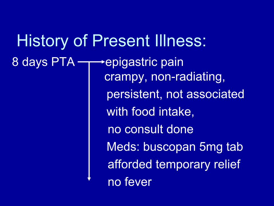
History of Present Illness:8 days PTA epigastric pain
crampy, non-radiating, persistent, not associated with food intake,no consult doneMeds: buscopan 5mg tabafforded temporary reliefno fever

7 day PTA Persistence of painprompted consult OMMCDx: Non-ulcerative dyspepsiawith no alarm signsMeds: AlMgOH syrupafforded some reliefHAMA
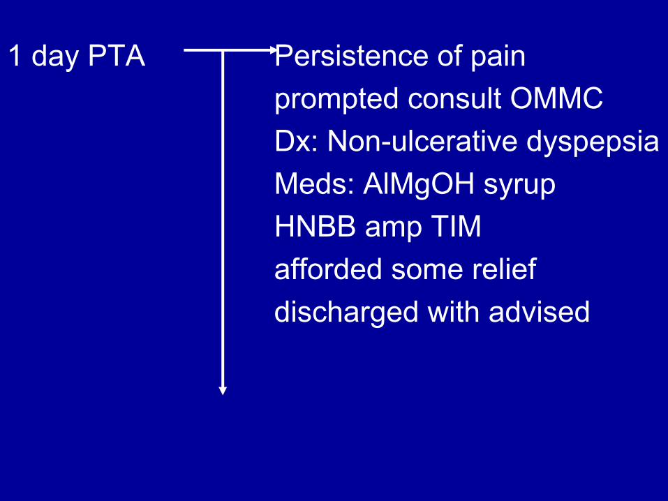
1 day PTA Persistence of painprompted consult OMMCDx: Non-ulcerative dyspepsiaMeds: AlMgOH syrupHNBB amp TIMafforded some reliefdischarged with advised
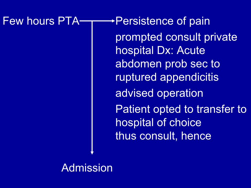
Few hours PTA Persistence of painprompted consult private hospital Dx: Acute abdomen prob sec to ruptured appendicitisadvised operationPatient opted to transfer to hospital of choicethus consult, hence
Admission
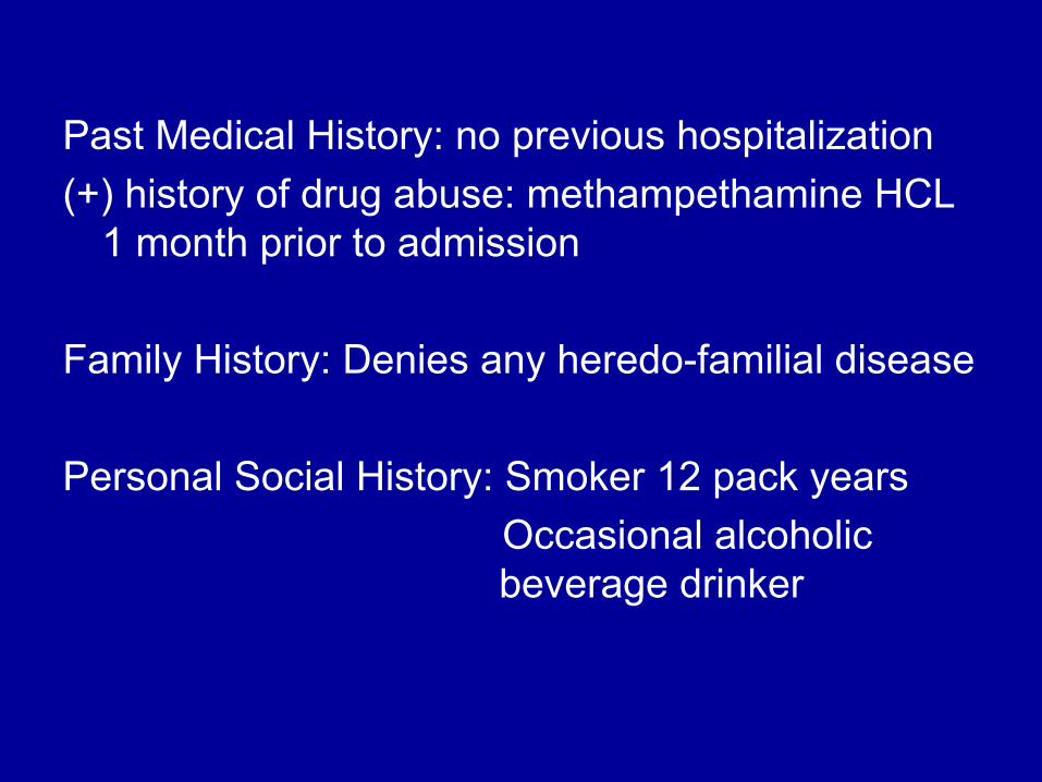
Past Medical History: no previous hospitalization(+) history of drug abuse: methampethamine HCL
1 month prior to admission
Family History: Denies any heredo-familial disease
Personal Social History: Smoker 12 pack yearsOccasional alcoholic beverage drinker
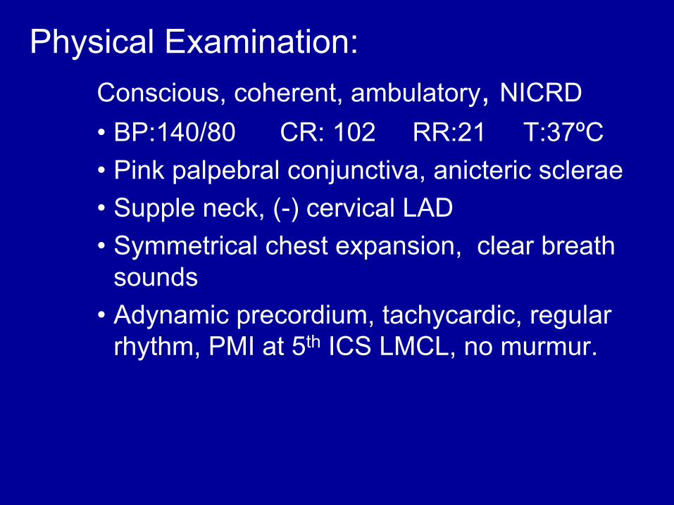
Physical Examination:Conscious, coherent, ambulatory, NICRD• BP:140/80 CR: 102 RR:21 T:37ºC• Pink palpebral conjunctiva, anicteric sclerae• Supple neck, (-) cervical LAD• Symmetrical chest expansion, clear breath
sounds• Adynamic precordium, tachycardic, regular
rhythm, PMI at 5th ICS LMCL, no murmur.

Abdomen:
Flat, hypoactive bowel sounds, soft, with severe direct and rebound tenderness on all quadrants RLQ > LLQNo guarding

Physical Examination• Rectal exam: no skin tags, no fissure good
sphincteric tone, with brown stool on examining finger, with parectal tenderness
• Grossly normal extremities, no cyanosis, no pallor
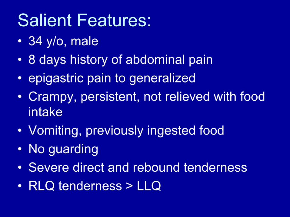
Salient Features:• 34 y/o, male• 8 days history of abdominal pain• epigastric pain to generalized • Crampy, persistent, not relieved with food
intake• Vomiting, previously ingested food• No guarding• Severe direct and rebound tenderness• RLQ tenderness > LLQ
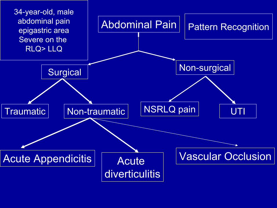
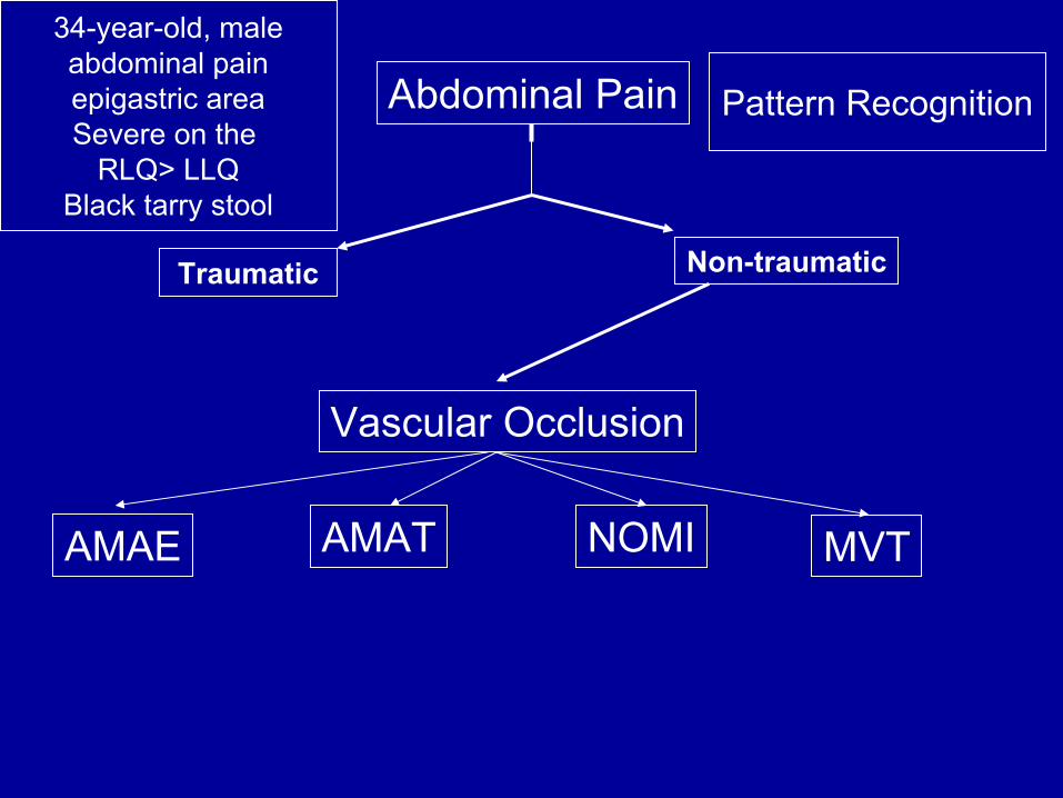
Abdominal Pain
Surgical Non-surgical
Traumatic NSRLQ pain UTI
Pattern Recognition
Non-traumatic
Acute Appendicitis Acute diverticulitis
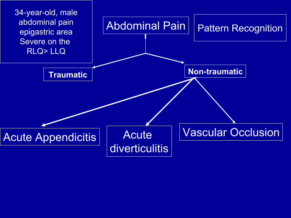
34-year-old, maleabdominal painepigastric areaSevere on the
RLQ> LLQ
Vascular Occlusion
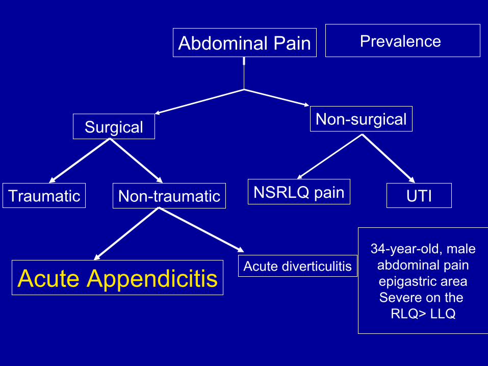
Abdominal Pain
Surgical Non-surgical
Traumatic NSRLQ pain UTI
Prevalence
Non-traumatic
Acute Appendicitis Acute diverticulitis34-year-old, maleabdominal painepigastric areaSevere on the
RLQ> LLQ
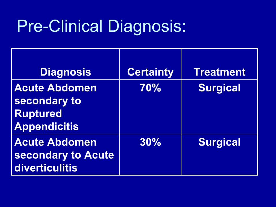
Pre-Clinical Diagnosis:
Surgical30%Acute Abdomen secondary to Acute diverticulitis
Surgical70%Acute Abdomen secondary to Ruptured Appendicitis
TreatmentCertaintyDiagnosis

Do I need a para-clinical diagnostic procedure?
No, I do not need any para-clinical diagnostic procedure
Reasons:1) My treatment option for my primary and
secondary diagnosis are the same.2) Presence of persistent abdominal pain

Goal of treatment
• Resolved the peritonitis• Remove the source of infection
(appendix)• Treat the infection

Pre-op preparation:
• Informed consent secured• Psychosocial support provided• Optimized patient’s physical health• Patient screened for any health
condition• Operative materials secured

Pre-op preparation:
• Large bore gauge 18 hypodermic needle inserted for venoclysis
• Foley catheter and naso-gastric tube inserted.
• Antibiotics were started– Metronidazole 500mg TIV every 8 hrs– Choramphenicol 500mg TIV q 8 hrs

Pre-op preparation:
• Scheduled for exploratory laparotomy• Adequate hydration
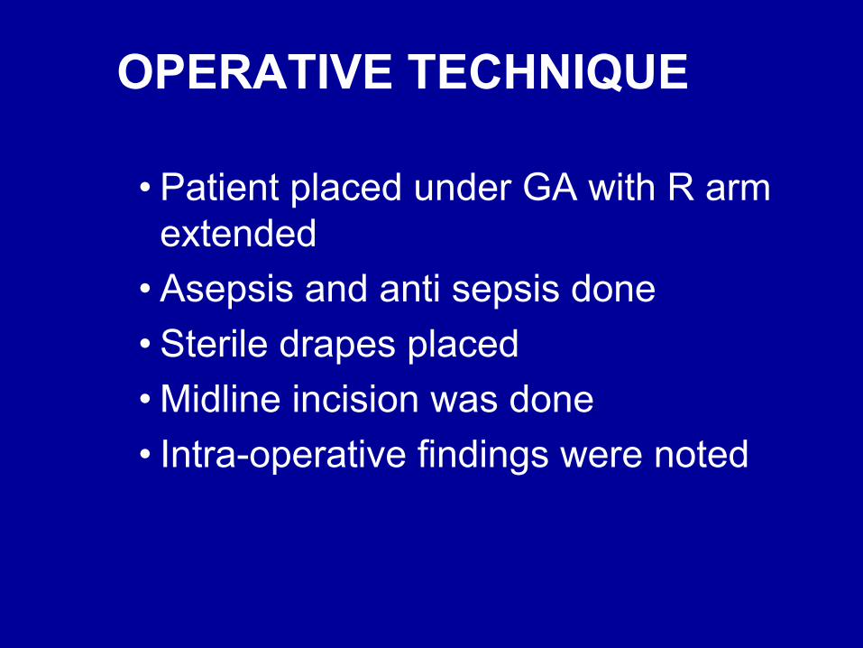
• Patient placed under GA with R arm extended
• Asepsis and anti sepsis done• Sterile drapes placed• Midline incision was done• Intra-operative findings were noted
OPERATIVE TECHNIQUE
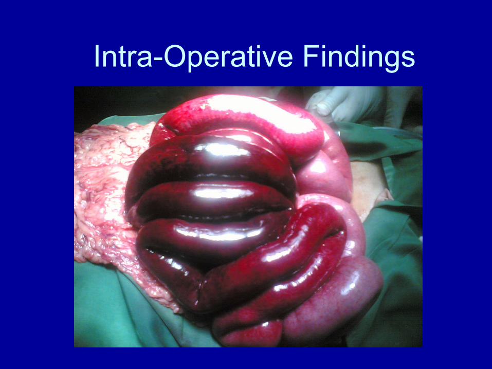
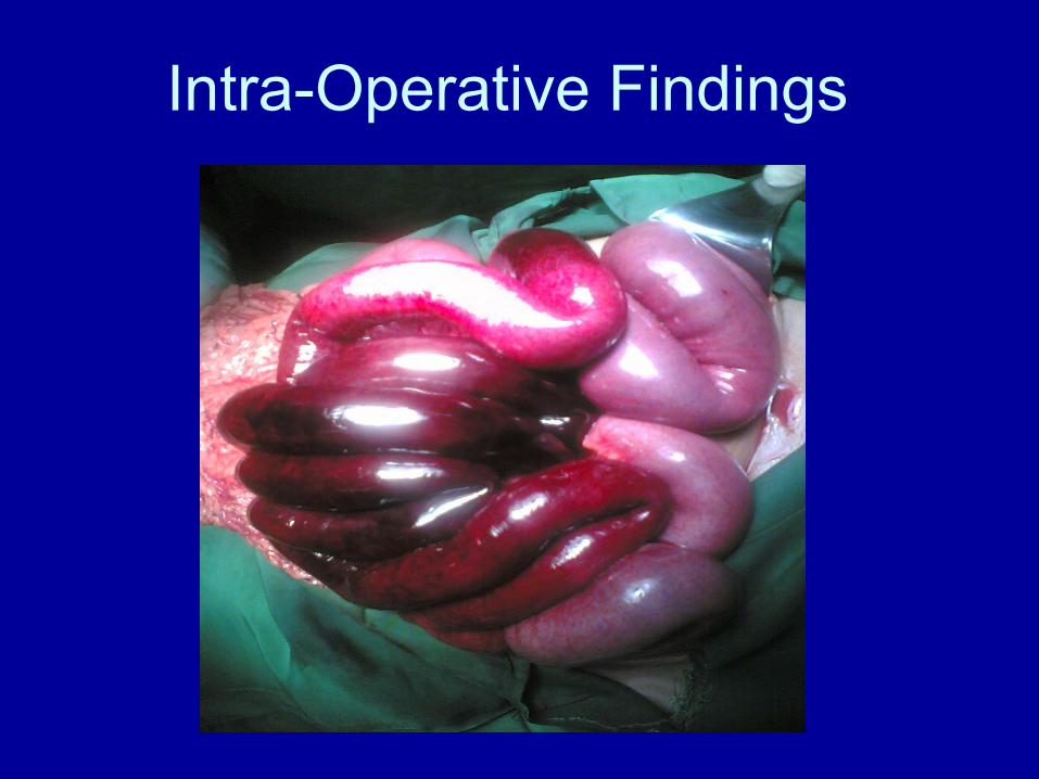
Intra-Operative Findings
Intra-Operative Findings
Intra-operative findings:Upon doing a midline incision, we noted
a sero-purulent peritoneal fluid, there was a gangrenous segment of the small intestine about 90 cm from the ligament of treitz, about 110 cm from the ileo-cecal valve.

Intra-operative findings:With no pulse on the gangrenous
segment while with poor pulse on the adjacent segment.
Small bowel exhibited good capillary refill was noted on the unaffected segment.
Liver, stomach, colon were grossly normal
Appendix was normal.

Abdominal Pain
Surgical Non-surgical
Traumatic NSRLQ pain UTI
Pattern Recognition
Non-traumatic
Acute Appendicitis Acute diverticulitis
34-year-old, maleabdominal painepigastric areaSevere on the
RLQ> LLQ
Vascular Occlusion
Abdominal Pain
Traumatic Non-traumatic
Pattern Recognition
Acute Appendicitis Acute diverticulitis
34-year-old, maleabdominal painepigastric areaSevere on the
RLQ> LLQ
Vascular Occlusion
Abdominal Pain
Traumatic Non-traumatic
Pattern Recognition
34-year-old, maleabdominal painepigastric areaSevere on the
RLQ> LLQBlack tarry stool
Vascular Occlusion
AMAE AMAT NOMI MVT

Intra-operative Diagnosis:
Surgical10%Acute Abdomen secondary to Gangrenous jejuno-ilealsegment prob 20 Acute mesenteric arterial embolus
Surgical90%Acute Abdomen secondary to Gangrenous jejuno-ilealsegment prob 20
Superior Mesenteric vein thrombosis
TreatmentCertaintyDiagnosis

Goal of treatment
1. Maintain bowel continuity2. Resolution of bowel gangrene with
adequate and viable tissue margin3. No complication

Intra-operative Management
• Plan: resection-anastomosis of affected bowel

• Intra-operative assesment was done• Referred to service consultant• Bowels were observed for pulsation
from the roots of the mesentery, the mesenteric proper and bowel segment.
OPERATIVE TECHNIQUE

Open Method
Clamps are applied in such aWay that the tissue removedPlaced crushing clamps and
The bowels which remains forthe anastomosis was held by
Non-traumatic clamps
Intestinal clamps( Non-traumatic)
Oschners clamp(crushing clamps)
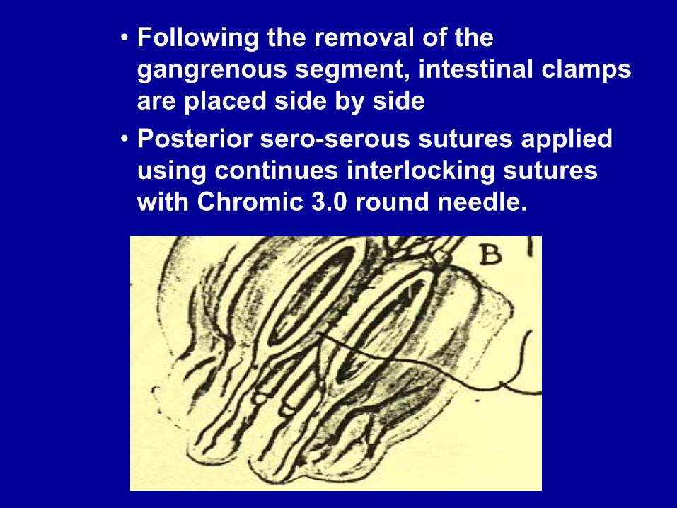
• Following the removal of the gangrenous segment, intestinal clamps are placed side by side
• Posterior sero-serous sutures applied using continues interlocking sutures with Chromic 3.0 round needle.

• Angle sutures applied using silk 2.0 (Maunsel mesenteric stich) and traction stich applied
• Posterior layer of through-and-through sutures using chromic 3.0 applied

• Anterior layer through-and-through with continous interlocking sutures using Chromic 3.0 round needle.
• Anterior serous sutures simple interrupted using silk 3.0
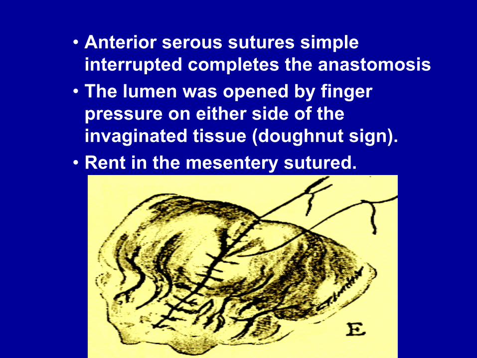
• Anterior serous sutures simple interrupted completes the anastomosis
• The lumen was opened by finger pressure on either side of the invaginated tissue (doughnut sign).
• Rent in the mesentery sutured.

• Formal exploration done• Omentum placed over the remaining
segment• Correct instrument, needle and sponge
count• Layer by layer sutures applied on the
– Fascia closed using Vicryl 0 round needle continous suturing.
– Reinforced External bolster applied pre-peritoneally using double Silk 0 strands on a carabao needle.
• Povidone-iodine paint• DSD
Post-operative Management
• Maintained on NPO.• NGT and foley catheter monitored hourly.• Vital signs every hour.• Adequate analgesia given.• Venoclysis and IV antibiotics were
continued.• Input and output monitoring were recorded

Post-operative Diagnosis:
Surgical10%Acute Abdomen secondary to Gangrenous jejuno-ilealsegment prob 20 Acute mesenteric arterial throbosis
Surgical90%Acute Abdomen secondary to Gangrenous jejuno-ilealsegment prob 20
Superior Mesenteric arterial embolus
TreatmentCertaintyDiagnosis
Post-operative problems encountered
1. Fever 2. Dehydration 3. LBM4. Phlebitis
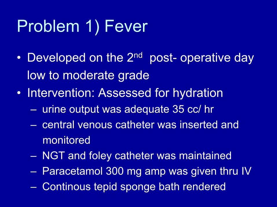
Problem 1) Fever
• Developed on the 2nd post- operative daylow to moderate grade
• Intervention: Assessed for hydration – urine output was adequate 35 cc/ hr– central venous catheter was inserted and
monitored – NGT and foley catheter was maintained– Paracetamol 300 mg amp was given thru IV– Continous tepid sponge bath rendered

Problem 2) Dehydration
• Developed on the 2nd post- operative day• Intervention: Assessed for hydration
– CVP line was inserted and monitored– Urine output was monitored– Fast drip of 200 cc of PLRS
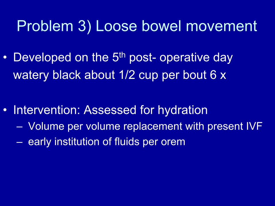
Problem 3) Loose bowel movement
• Developed on the 5th post- operative daywatery black about 1/2 cup per bout 6 x
• Intervention: Assessed for hydration – Volume per volume replacement with present IVF– early institution of fluids per orem

Problem 4) Phlebitis
• Developed on the 5th post- operative daytenderness on the IVF site
• Intervention: – re-insertion of IV fluid– warm compressed on affected site
Expected complications
1. Short bowel syndrome:
Short Bowel Syndrome
• average length adult human small intestine 600 cm (260-800 cm).
• Any disease, traumatic injury, vascular accident, or other pathology that leaves less than 200 cm of viable small bowel places the patient at risk for developing short-bowel syndrome
Lennard-Jones JE: Review article: practical management of the short bowel. Aliment Pharmacol Ther 1994 Dec; 8(6): 563-77[Medline]

Short Bowel Syndrome• clinically defined by malabsorption, diarrhea,
fluid and electrolyte disturbances, and malnutrition.
• The final common etiologic factor in all causes of short-bowel syndrome is the functional or anatomic loss of extensive segments of small intestine so that absorptive capacity is severely compromised.
Lennard-Jones JE: Review article: practical management of the short bowel. Aliment Pharmacol Ther 1994 Dec; 8(6): 563-77[Medline]

Short Bowel Syndrome
• Massive small intestinal resection compromises digestive and absorptive processes.
• Adequate digestion and absorption cannot take place, and proper nutritional status cannot be maintained without supportive care.
Lennard-Jones JE: Review article: practical management of the short bowel. Aliment Pharmacol Ther 1994 Dec; 8(6): 563-77[Medline]
Short Bowel Syndrome• Examples
– mesenteric ischemia– Trauma– inflammatory bowel disease– Cancer– radiation enteritis– congenital short small bowel– midgut volvulus– multiple small bowel atresias– Gastroschisis– meconium peritonitis– necrotizing enterocolitis.

Short Bowel Syndrome
• Not all patients with loss of significant amounts of small intestine develop the short-bowel syndrome.
Lennard-Jones JE: Review article: practical management of the short bowel. Aliment Pharmacol Ther 1994 Dec; 8(6): 563-77[Medline]
Short Bowel Syndrome• Important cofactors that help to
determine whether the syndrome will develop or not include– premorbid length of small bowel– segment of intestine – age– remaining length of small bowel and colon– Presence or absence of the ileocecal
valve.
Lennard-Jones JE: Review article: practical management of the short bowel. Aliment Pharmacol Ther 1994 Dec; 8(6): 563-77[Medline]

Short Bowel Syndrome• Significant weight loss• Fatigue, malaise, and lethargy. • Dehydration• Electrolyte imbalance• Protein-calorie malnutrition• Loss of critical vitamins and minerals.
Prevention of Small Bowel Syndrome
• Early institution of Total Parenteral Nutrition

Total Parenteral Nutrition
• an important therapy in the care of the patient with short-bowel syndrome.
• Provides adequate protein, calories, other macronutrients, and micronutrients until the bowel has had time to adapt
• Bowel compensation occurs after 3 months.
Carbonnel F, Cosnes J, Chevret S, et al: The role of anatomic factors in nutritional autonomy after extensive small bowel resection. JPEN J Parenter Enteral Nutr.20(4): 275-80;1996 [Medline].
Total Parenteral Nutrition
• Wilmore and colleagues (1971) suggest that supplementing enteral intake with parenteral nutrition early in the postoperative course results in better overall bowel adaptation.
• This is most likely because it facilitates provision of adequate calorie and nitrogen sources.
Wilmore DW, Dudrick SJ, Daly JM, Vars HM: The role of nutrition in the adaptation of the small intestine after massive resection. Surg Gynecol Obstet.132(4): 673-80;1971[Medline].
Total Parenteral Nutrition
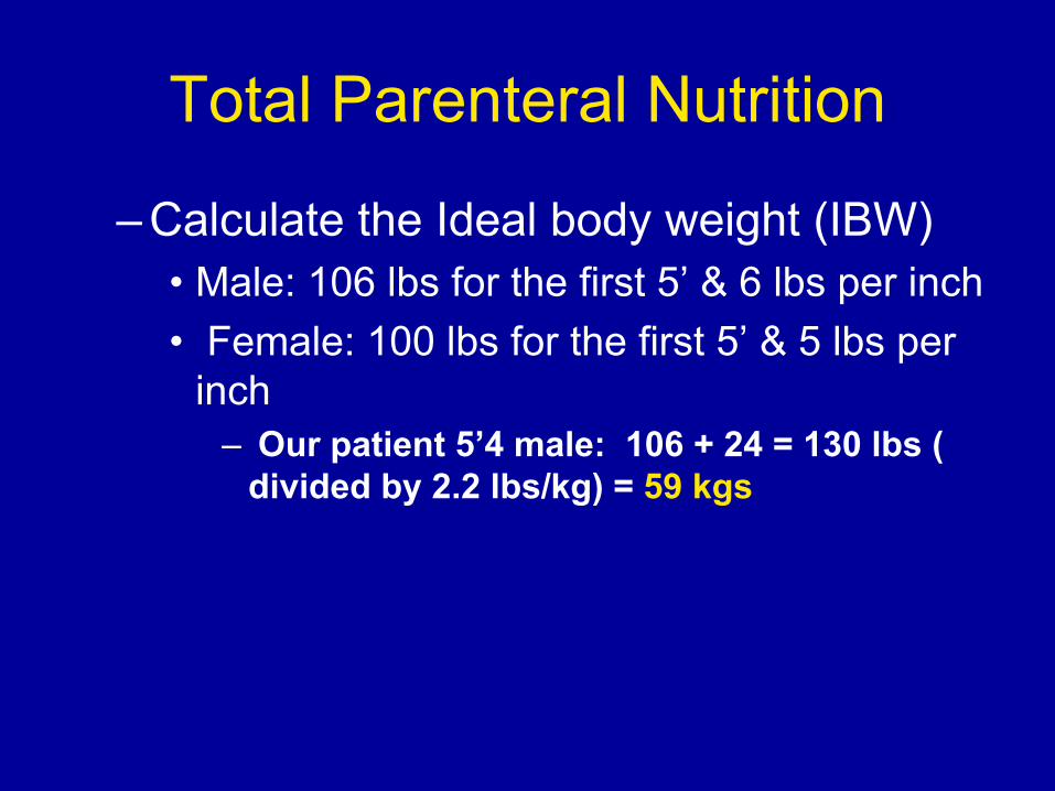
– Calculate the Ideal body weight (IBW)• Male: 106 lbs for the first 5’ & 6 lbs per inch • Female: 100 lbs for the first 5’ & 5 lbs per
inch– Our patient 5’4 male: 106 + 24 = 130 lbs (
divided by 2.2 lbs/kg) = 59 kgs
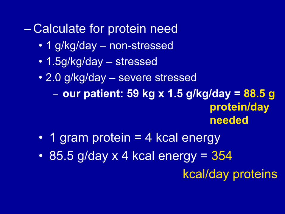
– Calculate for protein need• 1 g/kg/day – non-stressed• 1.5g/kg/day – stressed• 2.0 g/kg/day – severe stressed
– our patient: 59 kg x 1.5 g/kg/day = 88.5 g protein/day needed
• 1 gram protein = 4 kcal energy• 85.5 g/day x 4 kcal energy = 354
kcal/day proteins
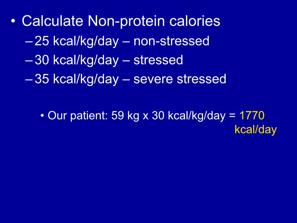
• Calculate Non-protein calories– 25 kcal/kg/day – non-stressed– 30 kcal/kg/day – stressed– 35 kcal/kg/day – severe stressed
• Our patient: 59 kg x 30 kcal/kg/day = 1770 kcal/day
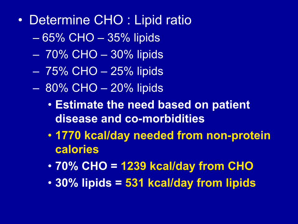
• Determine CHO : Lipid ratio– 65% CHO – 35% lipids– 70% CHO – 30% lipids– 75% CHO – 25% lipids– 80% CHO – 20% lipids
• Estimate the need based on patient disease and co-morbidities
• 1770 kcal/day needed from non-protein calories
• 70% CHO = 1239 kcal/day from CHO• 30% lipids = 531 kcal/day from lipids
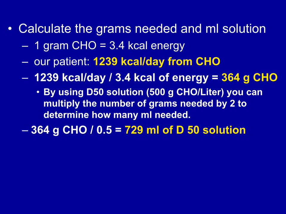
• Calculate the grams needed and ml solution– 1 gram CHO = 3.4 kcal energy– our patient: 1239 kcal/day from CHO– 1239 kcal/day / 3.4 kcal of energy = 364 g CHO
• By using D50 solution (500 g CHO/Liter) you can multiply the number of grams needed by 2 to determine how many ml needed.
– 364 g CHO / 0.5 = 729 ml of D 50 solution

• 1 gram lipids = 9 kcal energy/– 531 kcal lipids needed/day = 59 g Lipids/day
• If using a 20% solution, 1 cc = 2 kcal energy• If using a 10% solution, 1 cc = 1 kcal energy
– take the number of kcal needed and divide by 2 to determine the number of ml of a 20% lipid solution
• 531 kcal/day needed = 531 / 2 = 266 cc of a 20% lipid sol
(11 cc/hr x 24 hrs)
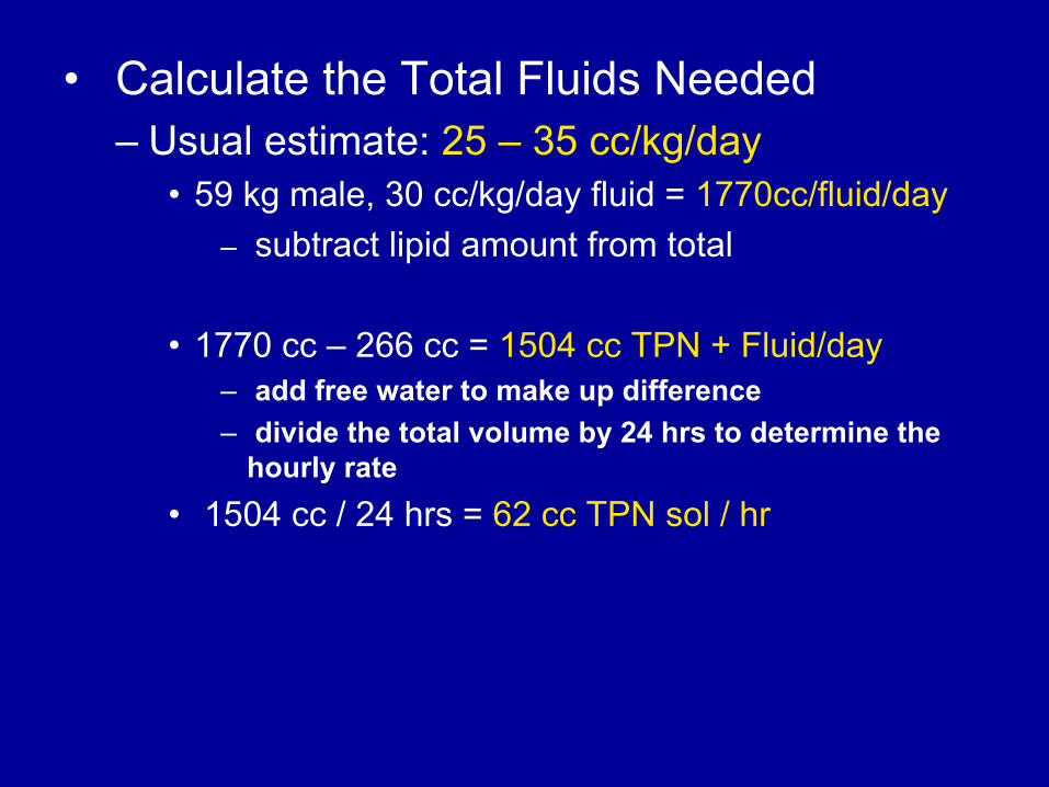
• Calculate the Total Fluids Needed– Usual estimate: 25 – 35 cc/kg/day
• 59 kg male, 30 cc/kg/day fluid = 1770cc/fluid/day– subtract lipid amount from total
• 1770 cc – 266 cc = 1504 cc TPN + Fluid/day– add free water to make up difference– divide the total volume by 24 hrs to determine the
hourly rate• 1504 cc / 24 hrs = 62 cc TPN sol / hr

Course in the wards
• TPN was started on the 8th HD with Nutripack 1900 kcal at 62 cc TPN/hr
• IVF and IV meds were continued• Early enteral feeding were started and
tolerated• Rest of his stay was unremarkable• Patient was discharged on the 21st HD

Follow-Up Care
• Advised to come back at Out Patient Department 1 week after discharged
• Daily bathing including the wound• Daily wound care• Small stiches were removed after 1 week• Bolster sutures will to be removed after 1
month
Follow-Up Care
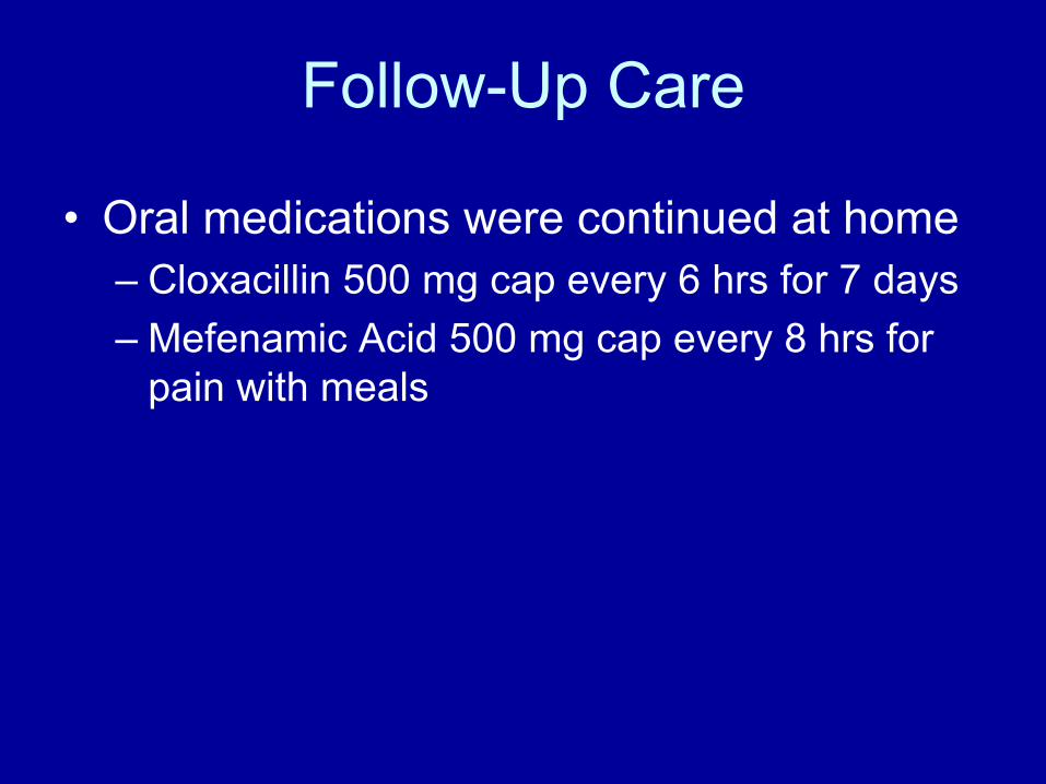
• Oral medications were continued at home– Cloxacillin 500 mg cap every 6 hrs for 7 days– Mefenamic Acid 500 mg cap every 8 hrs for
pain with meals
Follow-up plan:
• Patients require lifetime follow-up for subsequent complications
• Patients should be weighed regularly to assure that they are not losing weight on the nutritional regimen they are receiving.
• Several smaller feedings per day are usually advisable
Prevention and Health Promotion
• Changes diet • More physically active • Lose weight • Taking medications • Quit smoking • Stop using drugs • Educate patients who survive to discharge
about short-bowel syndrome

Outcome:• Resolution of the bowel gangrene• Live patient• Discharged • Happy and contented with the outcome• No complications• Satisfied patient• No medico-legal suit

Final Histopathologic Diagnosis
• to be discussed by Dr Jane Nicoza

Sharing of Information:
Acute Mesenteric Ischemia
• is a syndrome in which inadequate blood flow through the mesenteric circulation causes ischemia and eventual gangrene of the bowel wall.
• The syndrome can be classified generally as arterial or venous disease.
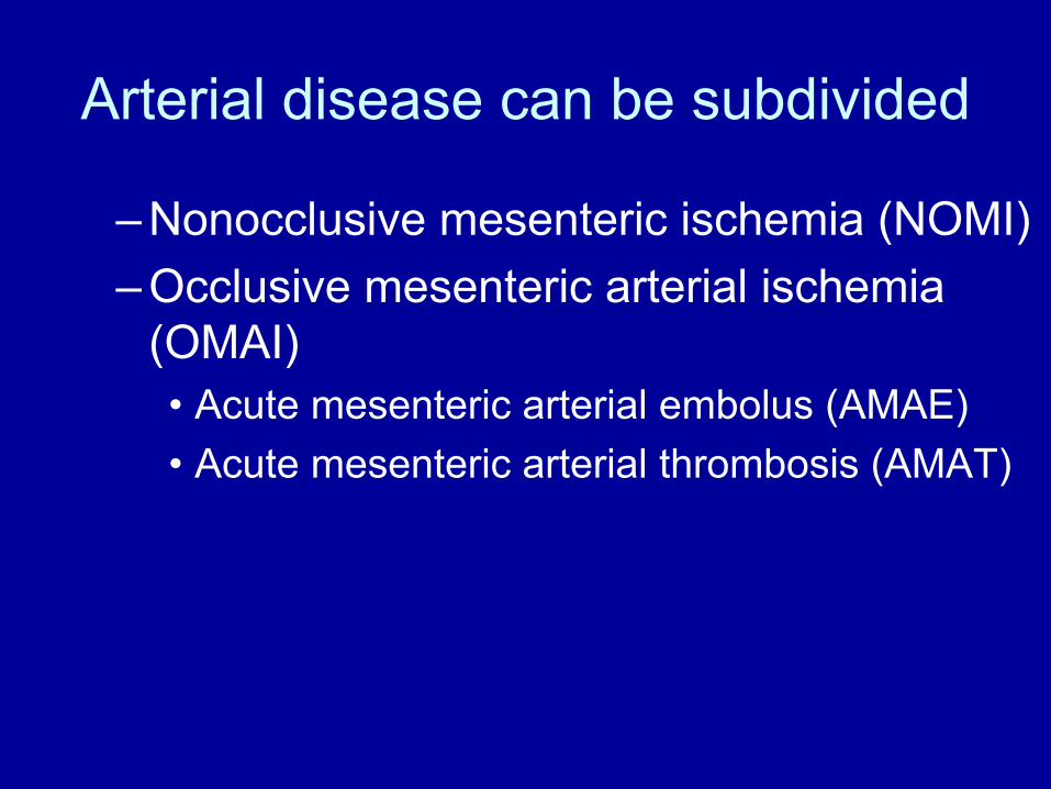
Arterial disease can be subdivided
– Nonocclusive mesenteric ischemia (NOMI)– Occlusive mesenteric arterial ischemia
(OMAI)• Acute mesenteric arterial embolus (AMAE)• Acute mesenteric arterial thrombosis (AMAT)
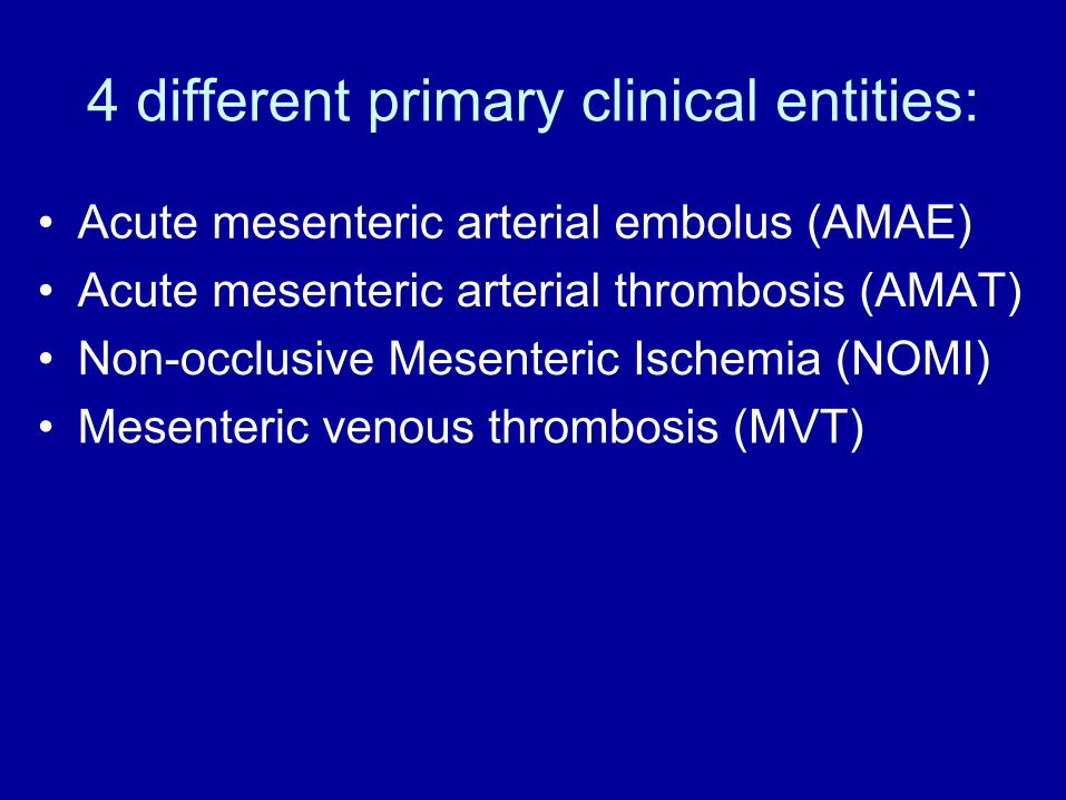
4 different primary clinical entities:
• Acute mesenteric arterial embolus (AMAE)• Acute mesenteric arterial thrombosis (AMAT)• Non-occlusive Mesenteric Ischemia (NOMI)• Mesenteric venous thrombosis (MVT)

Anatomy• Celiac artery (CA) supplies the foregut,
hepatobiliary system, and spleen• Superior mesenteric artery (SMA) supplies
the midgut (ie, small intestine and proximal mid colon)
• Inferior mesenteric artery (IMA) supplies the hindgut (ie, distal colon and rectum), but multiple anatomic variants are observed.
• Venous drainage is through the superior mesenteric vein (SMV), which joins the plenicvein to become the portal vein.
Acute Mesenteric Ischemia
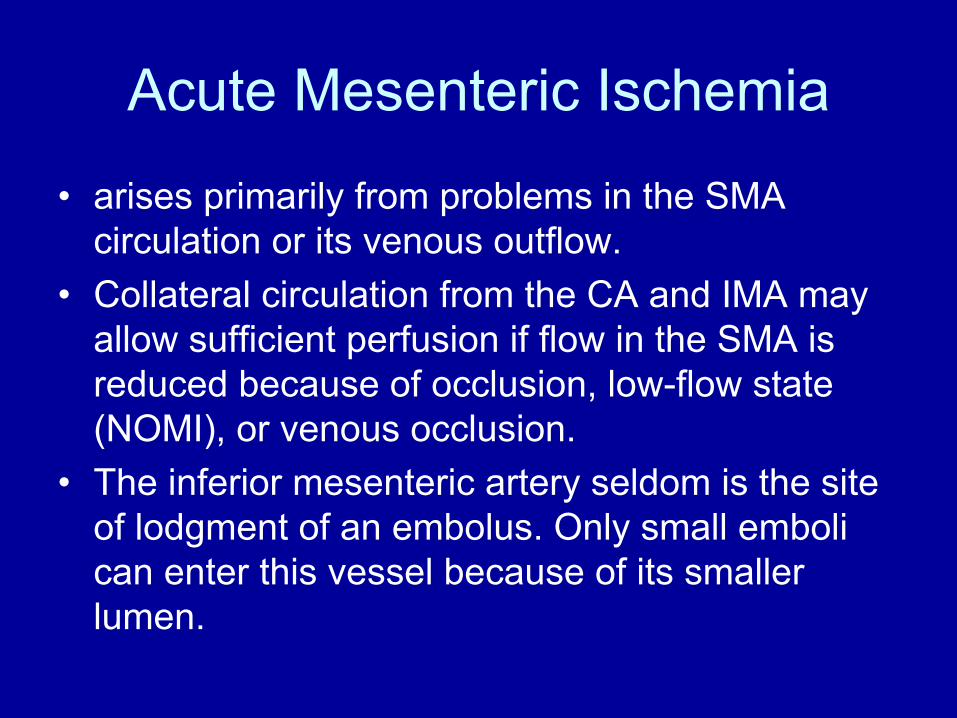
• arises primarily from problems in the SMA circulation or its venous outflow.
• Collateral circulation from the CA and IMA may allow sufficient perfusion if flow in the SMA is reduced because of occlusion, low-flow state (NOMI), or venous occlusion.
• The inferior mesenteric artery seldom is the site of lodgment of an embolus. Only small emboli can enter this vessel because of its smaller lumen.

Pathophysiology:
• Insufficient blood perfusion to the small bowel and colon may result from arterial occlusion by embolus or thrombosis (AMAE or AMAT), thrombosis of the venous system (MVT), or nonocclusiveprocesses such as vasospasm or low cardiac output (NOMI).
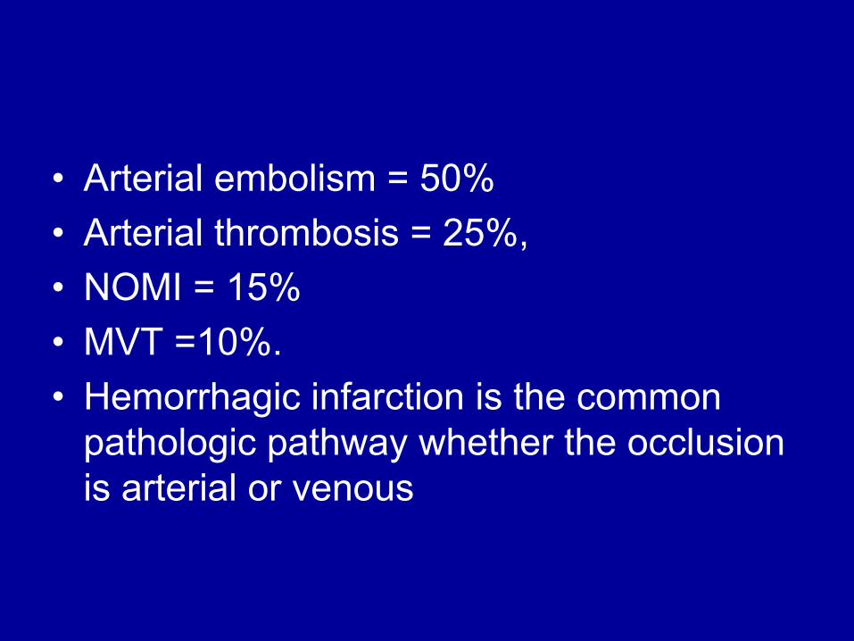
• Arterial embolism = 50% • Arterial thrombosis = 25%, • NOMI = 15%• MVT =10%. • Hemorrhagic infarction is the common
pathologic pathway whether the occlusion is arterial or venous

• Damage to the affected bowel portion may range from reversible ischemia to transmural infarction with necrosis and perforation.
• The injury is complicated by reactive vasospasm in the SMA region after the initial occlusion.
• Arterial insufficiency causes tissue hypoxia, leading to initial bowel wall spasm.
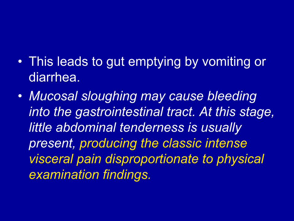
• This leads to gut emptying by vomiting or diarrhea.
• Mucosal sloughing may cause bleeding into the gastrointestinal tract. At this stage, little abdominal tenderness is usually present, producing the classic intense visceral pain disproportionate to physical examination findings.
• The mucosal barrier becomes disrupted as the ischemia persists, and bacteria, toxins, and vasoactive substances are released into the systemic circulation.
• This can cause death from septic shock, cardiac failure, or multisystem organ failure before bowel necrosis actually occurs.
• As hypoxic damage worsens, the bowel wall becomes edematous and cyanotic.
• Fluid is released into the peritoneal cavity, explaining the sero-sanguinous fluid
• Bowel necrosis can occur in 8-12 hours from the onset of symptoms.
• Transmural necrosis leads to peritoneal signs and heralds a much worse prognosis.
Superior Mesenteric Vein Thrombosis
• 2 types– Primary – no predisposing factor– Secondary - 80% result of predisposing factor

Predisposing factor
• Intra-abdominal infection• phlebitis or pylephlebitis (portal pyemia)• diverticulitis, • Appendicitis• infected carcinoma of the bowel• hypercoagulable states (polycythemia vera)
• oral contraceptives• protein C or S deficiency• mesenteric venous stasis from portal
hypertension or mass effect of abdominal tumors
• direct trauma to the mesenteric veins from a surgical procedure.
Predisposing factor
Predisposing factor
• after ligation of the portal vein or the superior mesenteric vein as part of "damage-control surgery" for severe penetrating abdominal injuries.
• Other associated causes include pancreatitis, sickle cell disease, and hypercoagulability caused by malignancy.
• 30-40-years-old• Symptoms may be present longer than in
the typical cases of AMI, (>30 days)• Infarction from MVT is rarely observed
with isolated SMV thrombosis, unless collateral flow in the peripheral arcades or vasa recta is compromised as well.
• Fluid sequestration and bowel wall edema are more pronounced than in arterial occlusion.
Frequency:
• 0.1% of all hospital admissions

Mortality/Morbidity
• MVT has a 30-day mortality rate of 13-15%.

Race:
• No racial predilections are known for AMI• However, people of races with a higher
rate of conditions leading to atherosclerosis, such as black people, might be at higher risk.
Sex
• No overall sex preference exists for AMI.• Men might be at higher risk for occlusive
arterial disease because they have a higher incidence of atherosclerosis.
• Conversely, women who are on oral contraceptives or are pregnant are at higher risk of MVT.
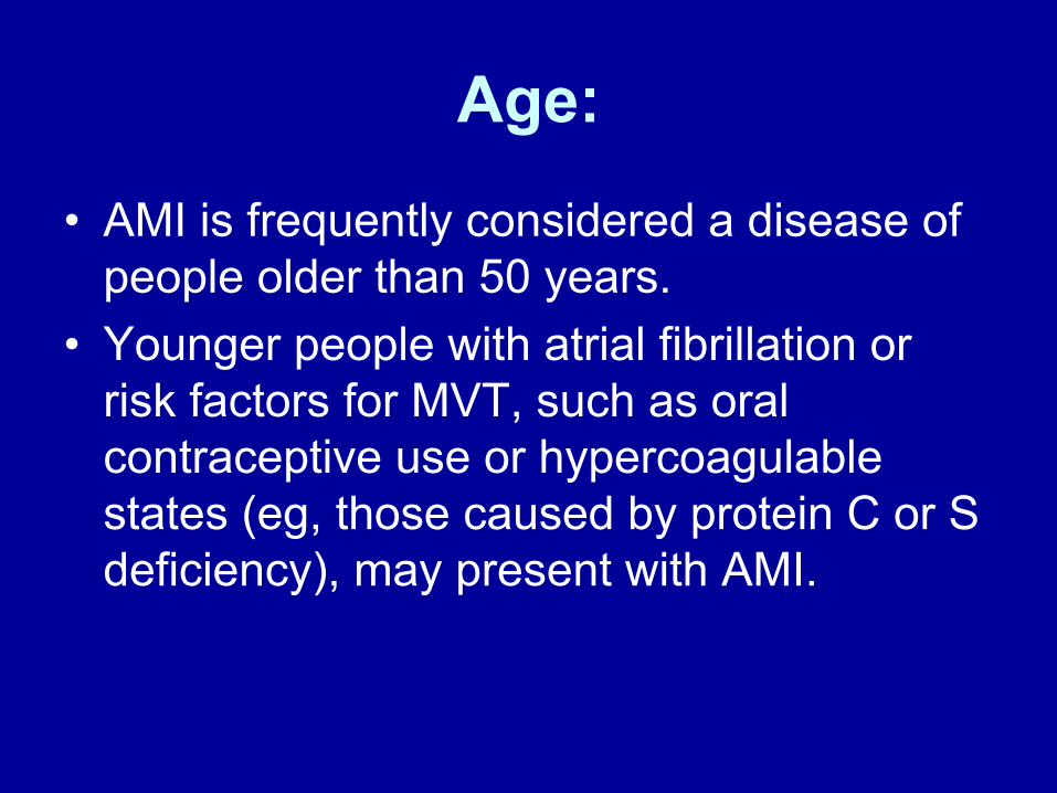
Age:
• AMI is frequently considered a disease of people older than 50 years.
• Younger people with atrial fibrillation or risk factors for MVT, such as oral contraceptive use or hypercoagulablestates (eg, those caused by protein C or S deficiency), may present with AMI.

History:
• The most important finding is pain disproportionate to physical examination findings.
• Typically, pain is moderate to severe, diffuse, nonlocalized, constant, and sometimes colicky.
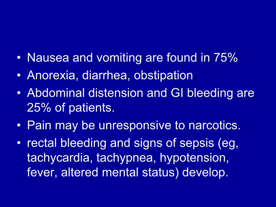
• Nausea and vomiting are found in 75% • Anorexia, diarrhea, obstipation• Abdominal distension and GI bleeding are
25% of patients. • Pain may be unresponsive to narcotics. • rectal bleeding and signs of sepsis (eg,
tachycardia, tachypnea, hypotension, fever, altered mental status) develop.

Lab Studies: • CBC count: Leukocytosis and/or leftward
shift are (50%)• Plain abdominal films: ileus, small bowel
obstruction, edematous/thickened bowel walls, and paucity of gas in the intestines. More specific signs, such as pneumatosisintestinalis, ie, submucosal gas thumb printing of bowel wall; and portal vein gas, are late findings.
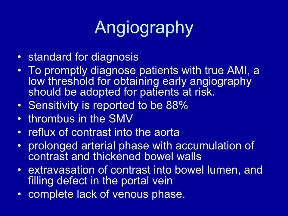
Angiography • standard for diagnosis • To promptly diagnose patients with true AMI, a
low threshold for obtaining early angiography should be adopted for patients at risk.
• Sensitivity is reported to be 88% • thrombus in the SMV• reflux of contrast into the aorta• prolonged arterial phase with accumulation of
contrast and thickened bowel walls• extravasation of contrast into bowel lumen, and
filling defect in the portal vein• complete lack of venous phase.

Surgical Care:
• exploratory laparotomy and resection of infarcted bowel is indicated.
Further Outpatient Care:
• Heparin - For patients with MVT or after revascularization

Salamat Po……..

References:1. Lennard-Jones JE: Review article: practical management of the
short bowel. Aliment Pharmacol Ther; 8(6): 563-77,1994.
2. Wilmore DW, Dudrick SJ, Daly JM, Vars HM: The role of nutrition in the adaptation of the small intestine after massive resection. Surg Gynecol Obstet.132(4): 673-80;1971.
3. Carbonnel F, Cosnes J, Chevret S, et al: The role of anatomic factors in nutritional autonomy after extensive small bowel resection. JPEN J Parenter Enteral Nutr.20(4): 275-80;1996.
4. Howard L, Heaphey L, Fleming CR, et al: Four years of North American registry home parenteral nutrition outcome data and their implications for patient management. JPEN J Parenter Enteral.15(4): 384-93;1991.

References:1. Boley SJ, Brandt LJ, Sammartano RJ: History of mesenteric
ischemia. The evolution of a diagnosis and management. SurgClin North Am.77(2): 275-88;1997.
2. Klempnauer J, Grothues F, Bektas H: Long-term results after surgery for acute mesenteric ischemia. Surgery. 121(3): 239-43 1997.
3. Hassan HA, Raufman JP: Mesenteric venous thrombosis. South Med J. 92(6): 558-62;1999.
4. McKinsey JF, Gewertz BL: Acute mesenteric ischemia. Surg Clin North Am. 77(2): 307-18;1997.

Questions
1. Among the four distinct pathophysiologicmechanism of Acute Mesenteric ischemia, the most common caused is?
a. Arterial thrombib. Arterial embolic. Venous thrombosisd. NOMI
Questions
1. Among the four distinct pathophysiologicmechanism of Acute Mesenteric ischemia, the most common caused is?
a. Arterial thrombib. Arterial embolic. Venous thrombosisd. NOMI
2. Mesenteric Venous Thrombosis accounts for only 5-10% of acute mesenteric ischemia and in 95%of cases this vessel is involved?
a. Splenic veinb. Portal veinc. Superior mesenteric veind. Inferior mesenteric vein

2. Mesenteric Venous Thrombosis accounts for only 5-15% of acute mesenteric ischemia and in 95%of cases this vessel is involved?
a. Splenic veinb. Portal veinc. Superior mesenteric veind. Inferior mesenteric vein
3. Regardless on the pathophysiologic mechanism of acute mesenteric ischemia. What is the hallmark findings on physical examination of the abdomen?
a. Abdominal distentionb. Abdominal pain out of proportion to the degree of
tendernessc. Muscle guardingd. Rebound tenderness
3. Regardless on the pathophysiologic mechanism of acute mesenteric ischemia. What is the hallmark findings on physical examination of the abdomen?
a. Abdominal distentionb. Abdominal pain out of proportion to the degree of
tendernessc. Muscle guardingd. Rebound tenderness

4. The two distinct clinical syndromes of acute mesenteric ischemia are ?
a. Acute mesenteric ischemia b. Mesenteric Venous thrombosisc. Chronic mesenteric ischemiad. Non-occlusive Mesenteric ischemia

4. The two distinct clinical syndromes of acute mesenteric ischemia are ?
a. Acute mesenteric ischemiab. Mesenteric Venous thrombosisc. Chronic mesenteric ischemiad. Non-occlusive Mesenteric ischemia
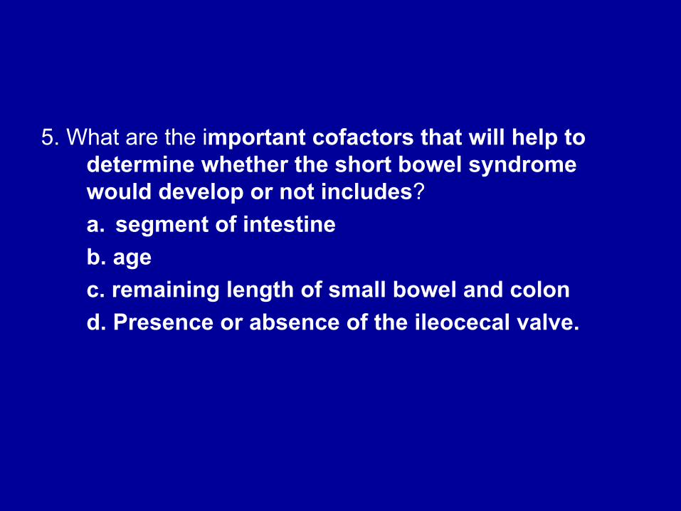
5. What are the important cofactors that will help to determine whether the short bowel syndrome would develop or not includes?a. segment of intestine b. agec. remaining length of small bowel and colond. Presence or absence of the ileocecal valve.
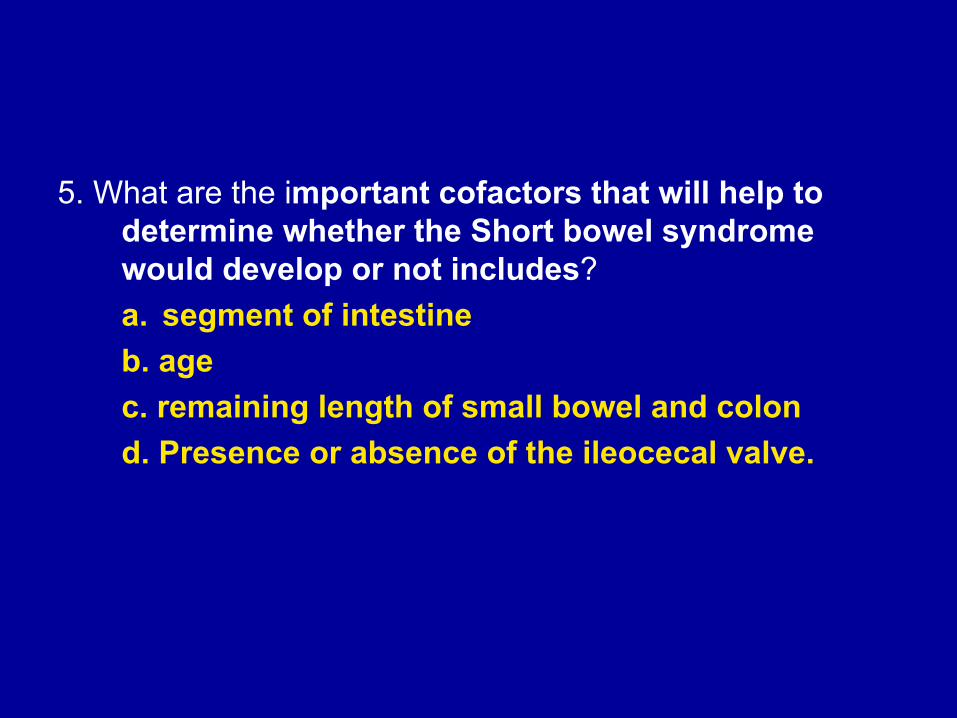
5. What are the important cofactors that will help to determine whether the Short bowel syndrome would develop or not includes?a. segment of intestine b. agec. remaining length of small bowel and colond. Presence or absence of the ileocecal valve.
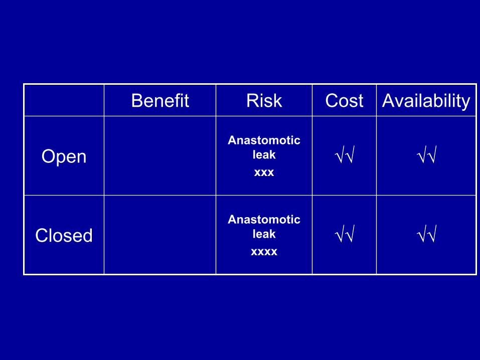
√√√√Anastomotic
leakxxxx
Closed
√√√√Anastomotic
leakxxx
Open
AvailabilityCostRiskBenefit


