736 Srp Arh Celok Lek. 2014 Nov-Dec;142(11-12):736-739 DOI: 10.2298/SARH1412736R ПРИКАЗ БОЛЕСНИКА / CASE REPORT UDC: 615.356.099-053.31 Correspondence to: Nedeljko RADLOVIĆ University Children’s Hospital Tiršova 10, 11000 Belgrade Serbia [email protected]SUMMARY Introduction Vitamin D intoxication represents a rare and potentially serious pathological condition caused by the excess of calcium and phosphorus. We are presenting an infant with vitamin D intoxication due to excessive daily administration, as well as therapeutic procedures that prevented its adverse effects. Case Outline A 1.5-month-old female infant, born at term, exclusively breastfed and without any com- plaints and abnormalities of physical findings, was observed due to the data that during the preceding month, by her mother’s mistake, she had received about 200,000 IU of vitamin D3. Laboratory analyses showed a high serum level of 25(OH)D (>400 nmol/L) and calcium (2.72 mmol/L), lowered PTH (6.6 pg/ml) and high urinary calcium/creatinine ratio (1.6), while other findings, including urotract ultrasonography image, were within normal limits. Treatment based on the discontinuation of vitamin D administration, infant’s forced water intake, as well as the application of 2-month prednisolone and 4-month pheno- barbitone and furosemide, resulted in complete normalization of the laboratory indicators of vitamin D overdose, as well as the prevention of its adverse effects. Conclusion By timely recognition and adequate treatment, including triple therapy with prednisolone, phenobarbitone and furosemide, adverse effects of acute vitamin D intoxication can be prevented. Keywords: vitamin D intoxication; infant; therapy Case Report of Acute Vitamin D Intoxication in an Infant Nedeljko Radlović 1,2 , Zoran Leković 2 , Dragana Ristić 2 , Vladimir Radlović 2 , Goran Djuričić 2 , Aleksandar Dimitrijević 2 , Biljana Vuletić 3 1 School of Medicine, University of Belgrade, Belgrade, Serbia; 2 University Children’s Hospital, Belgrade, Serbia; 3 Faculty of Medical Sciences, University of Kragujevac, Kragujevac, Serbia INTRODUCTION Vitamin D (calciferol) is the precursor of 1,25(OH) 2 D (calcitriol), a liposolubile hor- mone of essential importance for the home- ostasis of calcium and phosphorus, bone and tooth mineralization, as well as regulation of cell proliferation, differentiation and apopto- sis, immunoregulation, hormonogenesis and other physiological processes in organism [1- 4]. Therefore, its lack, beside negative effects caused by calcium and phosphorus deficiency, has essential participation in the pathogenesis of various malignant, autoimmune, allergic and other diseases [1-6]. Viewed from the biologi- cal aspect, physiological needs in vitamin D in humans are primarily accomplished by cutane- ous synthesis, i.e. photolysis of 7-dehydrocho- lesterol into cholecalciferol (D3), while, gen- erally viewed, food is its very scarce resource [4, 5]. However, the modern life-style followed by insufficient exposure to sun, as well as the fear of skin malignancies, above all melanoma, has mostly deprived the man of the natural and basic source of vitamin D [5, 7]. Accordingly, as well as based on numerous epidemiological studies indicating an increase in diseases with underlying essential participation of vitamin D deficiency, has enforced the necessity for its additional intake [5, 8, 9]. The recommended daily vitamin D intake, either in the form of supplements and/or as addition to food, which can satisfy optimal body needs is 400 IU for age 0-18 years, 600 IU for 19-70 years and 800 IU for age over 70 years [4, 8, 9]. Since the physi- ological effect of vitamin D as well as of other bioactive substances is accomplished within strictly defined limits, it is clear that its excess can also seriously endanger health [4, 9]. Al- though the negative balance of vitamin D is very much present worldwide, both in children and adults, in rare cases the opposite condi- tion can be also seen [4, 5, 9, 10-14]. With this aim, we are presenting an infant with vitamin D intoxication due to excessive daily adminis- tration, as well as therapeutic procedures that have prevented its adverse effect. CASE REPORT A female infant aged 1.5 months hospitalized on November 2012 due to excessive admin- istration of vitamin D 3 (Vigantol oil Merck KGaA, 10 drops daily, total about 200,000 IU per month). She was born after an uneventful term pregnancy, with body weight (BW) 3600 g and body length (BL) 54 cm. Since birth the infant has been exclusively breastfed. The aforesaid dose of vitamin D3, which was the consequence of misunderstanding between the mother and the pediatrician, the infant had re- ceived between the mid-first and mid-second month after birth. The problems which would have pointed at the excess of vitamin D had not been registered. On admission: a normally
Transcript
736
Srp Arh Celok Lek. 2014 Nov-Dec;142(11-12):736-739 DOI: 10.2298/SARH1412736R
ПРИКАЗ БОЛЕСНИКА / CASE REPORT UDC: 615.356.099-053.31
SUMMARYIntroduction Vitamin D intoxication represents a rare and potentially serious pathological condition caused by the excess of calcium and phosphorus. We are presenting an infant with vitamin D intoxication due to excessive daily administration, as well as therapeutic procedures that prevented its adverse effects.Case Outline A 1.5-month-old female infant, born at term, exclusively breastfed and without any com-plaints and abnormalities of physical findings, was observed due to the data that during the preceding month, by her mother’s mistake, she had received about 200,000 IU of vitamin D3. Laboratory analyses showed a high serum level of 25(OH)D (>400 nmol/L) and calcium (2.72 mmol/L), lowered PTH (6.6 pg/ml) and high urinary calcium/creatinine ratio (1.6), while other findings, including urotract ultrasonography image, were within normal limits. Treatment based on the discontinuation of vitamin D administration, infant’s forced water intake, as well as the application of 2-month prednisolone and 4-month pheno-barbitone and furosemide, resulted in complete normalization of the laboratory indicators of vitamin D overdose, as well as the prevention of its adverse effects.Conclusion By timely recognition and adequate treatment, including triple therapy with prednisolone, phenobarbitone and furosemide, adverse effects of acute vitamin D intoxication can be prevented.Keywords: vitamin D intoxication; infant; therapy
Case Report of Acute Vitamin D Intoxication in an InfantNedeljko Radlović1,2, Zoran Leković2, Dragana Ristić2, Vladimir Radlović2, Goran Djuričić2, Aleksandar Dimitrijević2, Biljana Vuletić3
1School of Medicine, University of Belgrade, Belgrade, Serbia;2University Children’s Hospital, Belgrade, Serbia;3Faculty of Medical Sciences, University of Kragujevac, Kragujevac, Serbia
INTRODUCTION
Vitamin D (calciferol) is the precursor of 1,25(OH)2D (calcitriol), a liposolubile hor-mone of essential importance for the home-ostasis of calcium and phosphorus, bone and tooth mineralization, as well as regulation of cell proliferation, differentiation and apopto-sis, immunoregulation, hormonogenesis and other physiological processes in organism [1-4]. Therefore, its lack, beside negative effects caused by calcium and phosphorus deficiency, has essential participation in the pathogenesis of various malignant, autoimmune, allergic and other diseases [1-6]. Viewed from the biologi-cal aspect, physiological needs in vitamin D in humans are primarily accomplished by cutane-ous synthesis, i.e. photolysis of 7-dehydrocho-lesterol into cholecalciferol (D3), while, gen-erally viewed, food is its very scarce resource [4, 5]. However, the modern life-style followed by insufficient exposure to sun, as well as the fear of skin malignancies, above all melanoma, has mostly deprived the man of the natural and basic source of vitamin D [5, 7]. Accordingly, as well as based on numerous epidemiological studies indicating an increase in diseases with underlying essential participation of vitamin D deficiency, has enforced the necessity for its additional intake [5, 8, 9]. The recommended daily vitamin D intake, either in the form of supplements and/or as addition to food, which can satisfy optimal body needs is 400 IU for age
0-18 years, 600 IU for 19-70 years and 800 IU for age over 70 years [4, 8, 9]. Since the physi-ological effect of vitamin D as well as of other bioactive substances is accomplished within strictly defined limits, it is clear that its excess can also seriously endanger health [4, 9]. Al-though the negative balance of vitamin D is very much present worldwide, both in children and adults, in rare cases the opposite condi-tion can be also seen [4, 5, 9, 10-14]. With this aim, we are presenting an infant with vitamin D intoxication due to excessive daily adminis-tration, as well as therapeutic procedures that have prevented its adverse effect.
CASE REPORT
A female infant aged 1.5 months hospitalized on November 2012 due to excessive admin-istration of vitamin D3 (Vigantol oil Merck KGaA, 10 drops daily, total about 200,000 IU per month). She was born after an uneventful term pregnancy, with body weight (BW) 3600 g and body length (BL) 54 cm. Since birth the infant has been exclusively breastfed. The aforesaid dose of vitamin D3, which was the consequence of misunderstanding between the mother and the pediatrician, the infant had re-ceived between the mid-first and mid-second month after birth. The problems which would have pointed at the excess of vitamin D had not been registered. On admission: a normally
737Srp Arh Celok Lek. 2014 Nov-Dec;142(11-12):736-739
www.srp-arh.rs
developed and well nourished infant (BL 56 cm, P50; BW 4600 g, P50), with normal physical findings. Laboratory analyses showed a high serum level of 25(OH)D, milder hypercalcemia, decreased serum PTH level and significant hypercalciuria (Table 1). ECG, urotract ultrasonography image, serum level of inorganic phosphorus, creatinine, acid-base status of blood, liver and urine tests, and other standard analyses were normal. As D hypervitaminosis was confirmed, beside withdrawal of its administration and forced water intake, the infant was initiated on therapy with prednisolone (2 mg/kg per day), phenobarbitone (3 mg/kg per day) and furosemide (2 mg/kg per day). In or-der to compensate renal potassium loss caused by furo-semide and prednisolone, KCl (2 mmol/kg oral daily) was also administered. In addition, ranitidine was adminis-tered as well, not only to prevent steroid ulcer but also for its negative effect on intestinal absorption of calcium. Having in mind the significance of mother’s milk at this age, as well as a low concentration of vitamin D and a rela-tively low content of calcium and phosphorus, exclusive breastfeeding of the infant was further continued (Table 1).
After 17 days of treatment, the level of 25(OH)D in serum was still >400 nmol/L, while other laboratory indi-cators of vitamin D overdose normalized, except high cal-cium to creatinine ratio (mg/mg) in 24-hour urine (UCa/UCr) (1.8). On the next checkup that followed after 1.5 months of unchanged treatment, serum levels of 25(OH)D an PTH were considerably improved, hypercalciuria re-mained high, while other relevant laboratory findings were within normal limits (Table 1). Control urotract ultra-sonography images were also normal so that prednisolone was slowly tapered and discontinued, while other thera-peutic measures were continued. As control findings after the next months were within the referent values [4, 15, 16], except for slightly higher values of UCa/UCr (0.8), the treatment of the infant was continued with lower dosages of phenobarbitone and furosemide; 1.5 mg/kg daily for 30 days and then 1 mg/kg daily over the next 30 days. After 4 months of treatment all relevant laboratory findings were normal (Table 1), as well as urotract unltrasonography im-age, so that complete therapy was interrupted. During the whole period of observation the child’s development was optimal and without clinical indicators either of vitamin D intoxication or adverse effects of the treatment. Serum level of 25(OH)D, calcium, phosphorus and PTH, UCa/UCr, urotract ultrasonography image and X-ray findings of the wrist 2 months later were within normal limits. Be-side breastfeeding and complementary food introduced at 5 months, vitamin D3 (400 IU daily) was also included
at age 7.5 months. On the last checkup at age 10 months, a complete infant’s clinical status, serum levels of calcium and phosphorus, UCa/UCr and urotract ultrasonography image were also normal.
DISCUSSION
Due to the presence of melanin, a limited transport capac-ity of transcalciferin and photoisomerisation of cholecal-ciferol into inactive metabolites (lumisterol, tachysterol, suprasterol I and II and 5.6-trans-cholecalciferol), and permanent skin desquamation, vitamin D intoxication by exposure to sun is not possible [4, 9, 17, 18]. Contrarily, excessively high oral intake of vitamin D is followed nei-ther by limited intestinal absorption and transport nor, in the victim’s condition of excess by its over-high activation [4, 17]. These facts, as well as a highly deficient capacity of 25(OH)D and 1.25(OH)2D inactivation and elimination, are the basis of the pathogenesis of vitamin D intoxica-tion in its excessive oral and/or parenteral application [4, 17, 19]. The upper tolerance level of vitamin D daily oral intake is 1000 IU for age 6 months, 1500 IU from 6-12 months, 2500 IU from 1-3 years, 3000 IU from 4-8 years and 4000 IU for ages over 9 years [4, 9]. However, in some pathological conditions these values can be much lower or higher. Thus, for example, in chronic granulomatous and some malignant diseases, because of uncontrolled lo-cal hyperproduction of 1.25(OH)D, they are considerably lower, i.e. higher as is the case of vitamin D-dependent and resistant forms of rickets [4, 9, 18, 20]. Increased sensitivity to vitamin D is also present in the conditions of massive osteolysis, primary hyperparathyroidism and some famil-ial forms of hypercalcemia and hypercalciuria [4, 9, 21]. Also, some medications modulate the biological effect of vitamin D. In this sense, negative influence have glucocor-ticoids and phenobarbitone that induce 24-hydroxylase, and thus inactivation and elimination of 25(OH)D and 1.25(OH)2D [4, 18, 22].
Vitamin D intoxication occurs due to renal hyper-production and excessively high calcitropic effect of 1.25 (OH)2D [4]. This metabolite of vitamin D, a derivate of 25(OH)2D and by all characteristics a liposoluble hor-mone, stimulates in enterocytes and tubulocytes the syn-thesis of calcium channels, calbindin, Ca2+ATP-ase, 3Na+/Ca2+ ion-exchanger and 2Na+/HPO4
2 cotransporter, thus enabling intestinal absorption and renal reabsorption of these ions as well as their transfer into circulation [1-5]. The optimal level of calcium and phosphorus in body
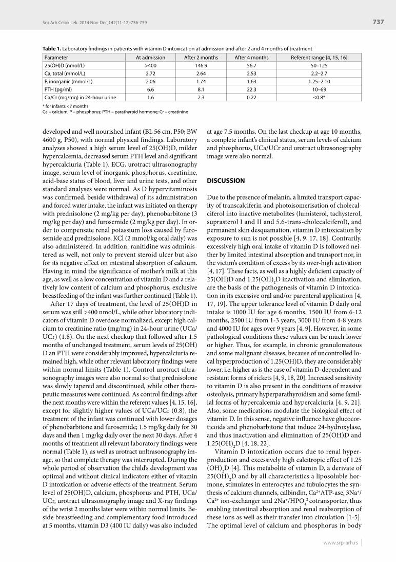
Table 1. Laboratory findings in patients with vitamin D intoxication at admission and after 2 and 4 months of treatment
Parameter At admission After 2 months After 4 months Referent range [4, 15, 16]
25(OH)D (nmol/L) >400 146.9 56.7 50–125
Ca, total (mmol/L) 2.72 2.64 2.53 2.2–2.7
P, inorganic (mmol/L) 2.06 1.74 1.63 1.25–2.10
PTH (pg/ml) 6.6 8.1 22.3 10–69
Ca/Cr (mg/mg) in 24-hour urine 1.6 2.3 0.22 ≤0.8*
* for infants <7 monthsCa – calcium; P – phosphorus; PTH – parathyroid hormone; Cr – creatinine
738
doi: 10.2298/SARH1412736R
Radlović N. et al. Case Report of Acute Vitamin D Intoxication in an Infant
fluids is of essential significance for numerous metabolic processes, neuromuscular bioelectric transmission and mineralization of skeleton and teeth [1, 4]. However, in vitamin D intoxication exactly on this characteristic of 1.25 (OH)2D followed by increased balance and elevated levels of serum calcium and phosphorus its toxic effects are based [4, 23]. Due to hypercalcemia, the initial phase of intoxication is dominated by the signs of neuromuscular dysfunction (hypotonia, constipation, cardiovascular dis-turbances), while its prolonged combination with hyper-phosphatemia is complicated with nephrocalcinosis and/or urinary lithiasis, as well as calcifications in blood ves-sels, myocardium and other soft tissues [4, 9, 13, 24]. Hav-ing in mind a wide differential diagnosis of hypercalcemia, hyperphosphatemia and hypercalciuria, a reliable confir-mation of vitamin D overdose or intoxication requires the verification of 25(OH)D serum levels above 125 nmol/L [4]. This parameter has a key diagnostic significance as 25(OH)D, with half-life in circulation of about 15 days, and it is the best indicator of balance and effect of vitamin D in organism [4, 5, 19]. With the aim to prevent immedi-ate and postponed complications of vitamin D intoxica-tion, the basis of treatment involves the interruption of its intake, maximal restriction of calcium and phosphorus in food, forced diuresis and administration of glucocorti-coids [4, 13, 23, 24]. In more severe intoxication, calcitonin and bisphosphonates are administered, and in the cases of
life-threatening medically unmanageable hypercalcemia hemodialysis or exchange transfusions [13, 23].
Vitamin D intoxication of the presented infant was due to its excessive daily administration. Although the total dose of vitamin D taken during one month was about 200,000 IU, i.e. serum level of 24(OH)D >400 nmol/L, symptoms and clinical signs of intoxication were not reg-istered. The explanation lies in the fact that the signs of vitamin D excess usually manifest just after 1-3 months, which is also supported by the value of calcemia in our patient which never exceeded 3 mmol/L [4, 18]. Beside standard measures intended for the treatment of this con-dition, with the goal of additional inactivation and better elimination of vitamin D active metabolites in our patient, we also administered phenobarbitone [4, 22]. In addition, we also introduced ranitidine which, by its suppressive ef-fect on gastric secretion, besides preventing peptic ulcer within a longer-lasting glucocorticoid therapy, contributes to a lower intestinal absorption of calcium as well [25]. The administered therapy passed without adverse effects and with full effect.
In conclusion, vitamin D intoxication represents a po-tentially most serious pathologic condition followed by numerous immediate and later complications. If timely registered and adequately treated, including triple therapy with prednisolone, phenobarbitone and furosemide, pos-sible complications can be prevented.
1. Norman AW. From vitamin D to hormone D: fundamentals of the vitamin D endocrine system essential for good health. Am J Clin Nutr. 2008; 88(2):491S-499S.
2. Zhang R, Naughton DP. Vitamin D in health and disease: Current perspectives. Nutr J. 2010; 9:65-77.
3. Radlović N, Mladenović M, Simić D, Radlović P. Vitamin D in the light of current knowledge. Srp Arh Celok Lek. 2012; 140(1-2):110-4.
4. Institute of Medicine, Food and Nutrition Board. Dietary Reference Intakes for Calcium and Vitamin D. Washington, DC: National Academy Press; 2011.
5. Wacker M, Holick MF. Vitamin D – effects on skeletal and extraskeletal health and the need for supplementation. Nutrients. 2013; 5(1):111-48.
6. Di Rosa M, Malaguarnera M, Nicoletti F, Malaguarnera L. Vitamin D3: a helpful immuno-modulator. Immunology. 2011; 134(2):123-39.
7. Prentice A. Vitamin D deficiency: a global perspective. Nutr Rev. 2008; 66(2):S153-S64.
8. Wagner CL, Greer FR, American Academy of Pediatrics Section on Breastfeeding, American Academy of Pediatrics Committee on Nutrition. Prevention of rickets and vitamin D deficiency in infants, children, and adolescents. Pediatrics. 2008; 122(5):1142-52.
9. U.S. Department of Agriculture, Agricultural Research Service. 2011. USDA National Nutrient Database for Standard Reference, Release 24. Nutrient Data Laboratory Home Page. Available from: http://www.ars.usda.gov/ba/bhnrc/ndl.
10. Pramyothin P, Holick MF. Vitamin D supplementation: guidelines and evidence for subclinical deficiency. Curr Opin Gastroenterol. 2012; 28(2):139-50.
11. Chambellan-Tison C, Horen B, Plat-Wilson G, Moulin P, Claudet I. Severe hypercalcemia due to vitamin D intoxication. Arch Pediatr. 2007; 14(11):1328-32.
12. Joshi R. Hypercalcemia due to hypervitaminosis D: report of seven patients. J Trop Pediatr. 2009; 55(6):396-8.
13. Ozkan B, Hatun S, Bereket A. Vitamin D intoxication. Turk J Pediatr. 2012; 54(2):93-8.
14. Vanstone MB, Oberfield SE, Shader L, Ardeshirpour L, Carpenter TO. Hypercalcemia in children receiving pharmacologic doses of vitamin D. Pediatrics. 2012; 129(4):e1060-3.
15. Nicolson JF, Pesce MA. Reference ranges for laboratory tests and procedures. In: Kliegman RM, Stanton BF, Schol NF, St Geme III JW, Behrman RE, editors. Nelson Textbook of Pediatrics. 19th ed. Philadelphia: Elsevier; 2011. p.2396-427.
16. So NP, Osorio AV, Simon SD, Alon US. Normal urinary calcium/creatinine ratios in African-American and Caucasian children. Pediatr Nephrol. 2001; 16(2):133-9.
17. de Paula FJ, Rosen CJ. Vitamin D safety and requirements. Arch Biochem Biophys. 2012; 523(1):64-72.
18. Greenbaum LA. Rickets and hypervitaminosis D. In: Kliegman RM, Stanton BF, Schol NF, St Geme III JW, Behrman RE, editors. Nelson Textbook of Pediatrics. 19th ed. Philadelphia: Elsevier; 2011. p.200-9.
19. Jones G. Pharmacokinetics of vitamin D toxicity. Am J Clin Nutr. 2008; 88(2):582S-6S.
22. Kurahashi I, Matsunuma A, Kawane T, Abe M, Horiuchi N. Dexamethasone enhances vitamin D-24-hydroxylase expression in osteoblastic (UMR-106) and renal (LLC-PK1) cells treated with 1alpha, 25-dihydroxyvitamin D3. Endocrine. 2002; 17(2):109-18.
23. Barrueto F Jr, Wang-Flores HH, Howland MA, Hoffman RS, Nelson LS. Acute vitamin D intoxication in a child. Pediatrics. 2005; 116(3):e453-6.
24. Bothra M, Jain V. Vitamin D intoxication: too much of a good thing! Indian Pediatr. 2013; 50(4):429-30.
25. Kopic S, Geibel JP. Gastric acid, calcium absorption, and their impact on bone health. Physiol Rev. 2013; 93(1):189-268.
REFERENCES
739Srp Arh Celok Lek. 2014 Nov-Dec;142(11-12):736-739
www.srp-arh.rs
КРАТАК САДРЖАЈУвод Тро ва ње ви та ми ном Д је рет ко и по тен ци јал но те шко па то ло шко ста ње узро ко ва но пре ви со ким би лан сом кал ци-ју ма и фос фо ра. При ка зу је мо одој че ко је је до жи ве ло акут но тро ва ње ви та ми ном Д, као и те ра пиј ске по ступ ке ко ји ма су пред у пре ђе не не же ље не по сле ди це.При каз бо ле сни ка Де вој чи ца уз ра ста од ме сец и по да на, ро ђе на у тер ми ну, хра ње на са мо мај чи ним мле ком и без ика квих те го ба и по ре ме ћа ја у фи зи кал ном на ла зу, при мље-на је на пре глед због по да тка да је то ком прет ход ног ме се ца гре шком мај ке уне ла око 200000 ИЈ ви та ми на Д
3. Ла бо ра то-
риј ске ана ли зе су по ка за ле ви сок се рум ски ни во 25(OH)D (>400 nmol/l) и кал ци ју ма (2,72 mmol/l), сни же не вред но сти PTH (6,6 pg/ml) и ви сок од нос кал ци ју ма и кре а ти ни на у мо-
кра ћи (1,6), док су оста ли на ла зи, укљу чу ју ћи и ул тра звуч ни из глед мо краћ ног трак та, би ли нор мал ни. Ле че ње, ко је се за сни ва ло на об у ста ви да љег уно са ви та ми на Д, по ја ча ном по је њу де те та во дом, као и дво ме сеч ној при ме ни пред ни зо-ло на и че тво ро ме сеч ној при ме ни фе но бар би то на и фу ро се-ми да, до ве ло је до пот пу не нор ма ли за ци је ла бо ра то риј ских по ка за те ља тро ва ња ви та ми ном Д, те спре чи ло не же ље не ефек те ин ток си ка ци је.За кљу чак Уз бла го вре ме но пре по зна ва ње и од го ва ра ју ће ле че ње, укљу чу ју ћи при ме ну пред ни зо ло на, фе но бар би-то на и фу ро се ми да, не же ље не по сле ди це акут ног тро ва ња ви та ми ном Д мо гу се пред у пре ди ти.
Кључ не ре чи: ин ток си ка ци ја ви та ми ном Д; одој че; ле че ње
Приказ одојчета с акутним тровањем витамином ДНедељко Радловић1,2, Зоран Лековић2, Драгана Ристић2, Владимир Радловић2, Горан Ђуричић2, Александар Димитријевић2, Биљана Вулетић3
1Медицински факултет, Универзитет у Београду, Београд, Србија;2Универзитетска дечја клиника, Београд, Србија;3Факултет медицинских наука, Универзитет у Крагујевцу, Крагујевац, Србија