11
In the mammalian heart, Ca¥ influx through voltage-dependent Ca¥ channels in the sarcolemma triggers Ca¥-induced Ca¥ release from the sarcoplasmic reticulum (SR;Bers, 1991; Stern & Lakatta, 1992; Eisner et al. 1998).Under various conditions that result in increased cellularCa¥ content Ca¥ release can occur spontaneously in theform of regenerative Ca¥ waves (Kort et al. 1985; Wier etal. 1987; Takamatsu & Wier, 1990; Lipp & Niggli, 1994;Trafford et al. 1995; Engel et al. 1995; Wussling & Salz,1996; Cheng et al. 1996; Lukyanenko et al. 1996, 1999).Spontaneous Ca¥ waves have been implicated in certaincardiac dysfunctions such as triggered arrhythmias (Lakatta,1992; Ishide, 1996). Nevertheless, the mechanisms ofgeneration of Ca¥ waves and their relationship to the Ca¥release process during normal excitation—contraction (E—C)coupling are not precisely understood. Recent studies usingconfocal Ca¥ imaging revealed that Ca¥ release during bothnormal E—C coupling and Ca¥ waves is a result of thesummation of elementary release events, Ca¥ sparks(Cannell et al. 1994; Lopez-Lopez et al. 1995; Cheng et al.1996). Ca¥ sparks can arise spontaneously and in responseto electrical stimulation of the cell. Under normal cellularCa¥ loading conditions sparks remain localized. When thecellular Ca¥ content is elevated they give rise topropagating Ca¥ waves (Cheng et al. 1996; Lukyanenko etal. 1996, 1999). The factors which could potentiallyinfluence the activity of individual Ca¥ release sites as wellas the interaction between adjacent sites include Ca¥ levelsin both cytosolic and SR luminal compartments, intra-cellular Ca¥ buffering, Ca¥ activation and inactivation
properties of the release channels, and the presence of intra-cellular modulatory agents.
Permeabilization that allows rapid equilibration of varioussubstances between extracellular fluid and cytosol has beena valuable tool for studying E—C coupling in both skeletal andcardiac muscle cells. In particular, Fabiato, by measuringforce and aequorin light signals in permeabilized cardiacmyocytes, has defined the Ca¥ dependence of SR Ca¥release activation and inactivation (Fabiato, 1985), layingthe foundation of the theory of Ca¥-induced Ca¥ release.In the present study we used confocal Ca¥ imaging toexplore local Ca¥ signalling in permeabilized cardiac cells.We investigated the role of such factors as Ca¥ buffering,[Ca¥]é as well as calmodulin and cADPR in the modulationof local release events (Ca¥ sparks) and propagating Ca¥waves.
METHODS
Cell isolation, permeabilization and experimental solutions
Adult Sprague—Dawley rats (200—300 g) were killed by lethalinjection of Nembutal (Abbott Laboratories, 100 mg kg¢, i.p.), andsingle ventricular myocytes were obtained by enzymaticdissociation as described previously (Gy�orke et al. 1997). The cellswere loaded with fluo-3 by a 20 min incubation with 5 ìÒ fluo-3 AM (acetoxymethyl ester form, Molecular Probes) at roomtemperature. The Tyrode solution contained (mÒ): 140 NaCl, 5·4KCl, 0·5 MgClµ, 1—5 CaClµ, 10 Hepes, 0·25 NaHµPOÚ, 5·6 glucose;pH 7·3. The cardiac myocytes were permeabilized with saponin(0·01% for 45—60 s) in an ‘internal’ solution containing (mÒ): 120potassium aspartate, 3 MgATP (free [Mg¥] •1 mÒ), 0·1 EGTA,
Journal of Physiology (1999), 521.3, pp.575—585 575
Ca¥ sparks and Ca¥ waves in saponin-permeabilized rat
ventricular myocytes
Valeriy Lukyanenko and Sandor Gy�orke
Department of Physiology, Texas Tech University Health Sciences Center, Lubbock,TX 79430, USA
(Received 30 April 1999; accepted after revision 28 September 1999)
1. We carried out confocal Ca¥ imaging in myocytes permeabilized with saponin in ‘internal’solutions containing: MgATP, EGTA and fluo-3 potassium salt.
2. Permeabilized myocytes exhibited spontaneous Ca¥ sparks and waves similar to thoseobserved in intact myocytes loaded with fluo-3 AM.
3. In the presence of ‘low’ [EGTA] (0·05 mÒ), Ca¥ waves arose regularly, even at relatively low[Ca¥] (50—100 nÒ, free). Increasing [EGTA] resulted in decreased frequency andpropagation velocity of Ca¥ waves. Propagating waves were completely abolished at[EGTA] > 0·3 mÒ.
4. The frequency of sparks increased as a function of [Ca¥] (50—400 nÒ range) with no sign ofa high affinity Ca¥-dependent inactivation process.
5. The rate of occurrence of Ca¥ sparks was increased by calmodulin and cyclic adenosinediphosphate-ribose (cADPR).
9575

10 phosphocreatine, 5 U ml¢ creatine phosphokinase, and 8%dextran (40000, to prevent osmotic swelling of the cells); pH 7·2.The control experimental solution contained (mÒ): 120 potassiumaspartate, 3 MgATP, 0·5 EGTA, 0·114 CaClµ (free [Ca¥] •100 nÒ),10 phosphocreatine, 0·03 fluo-3 potassium salt (TefLabs, Austin,TX, USA) and 5 U ml¢ creatine phosphokinase; pH 7·2. Solutionswith different buffering strengths and free [Ca¥] were prepared byadding appropriate amounts of KµEGTA and CaCalµ. The free[Ca¥] at given total Ca¥, Mg¥, ATP and EGTA concentrations wascalculated using a computer program (WinMAXC 1.80, StanfordUniversity, CA, USA) and verified by measurements with aspectrofluorometer D_Scan (PTI, Monmouth Junction, NJ, USA)and the Ca¥ indicator fura_2 (TefLabs, Austin, TX, USA). Thedrugs were applied through a gravity-driven perfusion system. Allexperiments were performed at room temperature (21—23°C). Allchemicals except fluo-3 and fura_2 were from Sigma.
Confocal microscope
Experiments were performed as described previously (Gy�orke et al.1997), using an Olympus laser scanning confocal microscope(LSM_GB200) equipped with an Olympus ²60, 1·4 NA objective.Fluo-3 was excited by light at 488 nm using a 25 mW argon laserwith intensity attenuated to 1—3%. Fluo-3 fluorescence wasmeasured at wavelengths of >515 nm. Images were acquired in theline-scan mode of the microscope at a rate of 2·1 or 8·3 ms per scan,with the scan line oriented along the longitudinal axis of the cell.An analog recording of fluorescence intensity was digitized into640 pixels, giving a nominal pixel dimension of 0·41 ìm. To reducecell damage by the laser illumination, the position of the line scanwas changed after acquiring three to six images from eachparticular location. Thus, in a typical cell the measurements couldbe performed for 10—15 min without significant alterations in Ca¥spark properties.
V. Lukyanenko and S. Gy�orke J. Physiol. 521.3576
Figure 1. The effects of permeabilization on Ca¥ sparks and Ca¥ waves in ventricular myocytes
A, images of a cardiac myocyte obtained in transmitted light before and after permeabilization withsaponin. B, line-scan images of fluorescence in a portion of the same cell pre-loaded with fluo-3 AMmeasured before permeabilization (a, [Ca¥]ï = 5 mÒ), after permeabilization in an internal solution withno dye (b) and after addition to the internal solution 30 ìÒ fluo-3 potassium salt in the presence of 0·1 (c) or0·5 mÒ EGTA (d) (pCa 7). Calibration bars: horizontal 10 ìm, vertical 0·4 s, the colour bar representschanges in units of absolute fluorescence. C, surface plots of Ca¥ sparks measured before permeabilization(a) and after permeabilization in the presence of 0·1 or 0·5 mÒ EGTA (b and c, respectively). Each plot wasobtained by averaging 10 individual events.

Ca¥ sparks were detected and measured using a computeralgorithm similar to that described previously (Song et al. 1997;Cheng et al. 1999). The program defines Ca¥ sparks as regions ofelevated fluorescence relative to the standard deviation (s.d.) ofbackground noise of the fluorescence image. The performance of theprogram at different event detection threshold settings was testedby using standard Ca¥ sparks of various intensities contaminatedwith appropriate amounts of random noise (Song et al. 1997). Withthe detection threshold set at a level of 2·6 ² s.d., the amplitude ofevents detected with 50% efficiency was about 1·3FÏFï (where F isthe recorded fluorescence intensity and Fï is backgroundfluorescence) while the probability of false events was 1—2%. Theaverage propagation velocity of Ca¥ waves was determined byfitting a linear function to the position of the wave (defined at half-maximal amplitude) in the x—t plane (Lukyanenko et al. 1999).Image processing and analysis were performed using NIH Image(NIH, Bethesda, MD, USA) and IDL software (Research SystemsInc., Boulder, CO, USA).
RESULTS
Effects of permeabilization
Figure 1 illustrates the main steps of a permeabilizationexperiment in a single isolated rat ventricular myocyte pre-loaded with fluo-3 AM. The series of line-scan images in Bwere acquired before permeabilization (a), afterpermeabilization with 0·01% saponin for 1 min in aninternal solution containing no dye (b) and after addition tothe internal solution of 30 ìÒ fluo-3 potassium salt in thepresence of 0·1 mÒ (c) or 0·5 mÒ EGTA (d). Photographicwide field images of the myocyte before and after
permeabilization are illustrated at the top (A). As describedpreviously (Cheng et al. 1993, 1996; Lukyanenko et al.1996), the intact cell spontaneously exhibited sporadic Ca¥sparks and occasional Ca¥ waves. Permeabilization wasconfirmed by the disappearance of all the fluorescencesignals in the bathing solution containing no dye and re-emergence of the signals after introduction of the free acidform of the dye into the bathing solution. Note that theoverall appearance of the cell and the Ca¥ signals before andafter permeabilization are very similar. Figure 1C showssurface plots of Ca¥ sparks in intact and permeabilized cellsin the presence of 0·1 or 0·5 mÒ EGTA. Table 1 summarizesspark statistics for the same conditions. As can be seen,permeabilization had no significant impact on the frequency,amplitude, width or length of the events (for all groups,P > 0·05). The similarities in the spatio-temporal propertiesof Ca¥ sparks at 0·1 and 0·5 mÒ EGTA are likely to be dueto the slow rate of Ca¥ binding by EGTA. The overallsimilarities between Ca¥ sparks and waves in intact andpermeabilized cells suggest that our permeabilizationprocedure does not significantly alter the Ca¥ signallingmechanisms in the cell.
Effects of EGTA
Theoretical studies predict that soluble intracellular Ca¥buffers must have a strong effect on interaction betweenrelease sites by lowering the rate of effective diffusion ofCa¥ (Keizer et al. 1998). We experimentally tested theeffects of intracellular Ca¥ buffering on Ca¥ waves in
Ca¥ sparks and waves in permeabilized cardiomyocytesJ. Physiol. 521.3 577
Figure 2. Effects of calcium buffering on Ca¥ waves in saponin-permeabilized myocytes
A, B, C and D, representative line-scan images of fluorescence recorded in a permeabilized myocyte in thepresence of 0·05 mÒ (A), 0·1 mÒ (B), 0·2 mÒ (C) and 0·3 mÒ EGTA (D). Free [Ca¥] in all cases wasadjusted to 100 nÒ. Calibration bars: horizontal 15 ìm, vertical 0·35 s.

V. Lukyanenko and S. Gy�orke J. Physiol. 521.3578
Figure 3. For legend see facing page.
permeabilized cardiac myocytes. In these experiments Ca¥buffering strength was varied by addition of differentconcentrations of EGTA to the bathing solution, whileadjusting basal Ca¥ to a constant value of 100 nÒ. Ininternal solutions with low Ca¥ buffering strength (0·05 mÒEGTA), Ca¥ waves arose relatively frequently (0·06 ±0·01 s¢, n = 21) and propagated typically through theentire cell with a velocity of about 70 ìm s¢ (66 ± 6 ìm s¢,n = 21; Fig. 2A). Elevation of EGTA concentration to0·1 mÒ resulted in decreased frequency and propagationvelocity of waves (0·03 ± 0·01 s¢ and 53 ± 1 ìm s¢,respectively, n = 12; Fig. 2B). Further increase in [EGTA]resulted in fragmentation of Ca¥ waves into abortiveresponses (Fig. 2C and D). Images obtained at high [EGTA]also clearly show that Ca¥ waves arise from sequentialactivation of discrete release events, revealing the saltatorynature of wave propagation. Propagating Ca¥ waves werecompletely abolished at [EGTA] > 0·3 mÒ. The fast Ca¥buffer BAPTA prevented Ca¥ waves even at lowerconcentrations (•0·1 mÒ, not shown). These results indicatethat Ca¥ buffering has a profound influence on Ca¥ waves.
Effects of [Ca¥]
Ca¥ is the principal regulator of the activity of ryanodinereceptors (RyRs) in the heart. Although the Ca¥dependence of RyR activation and inactivation has been
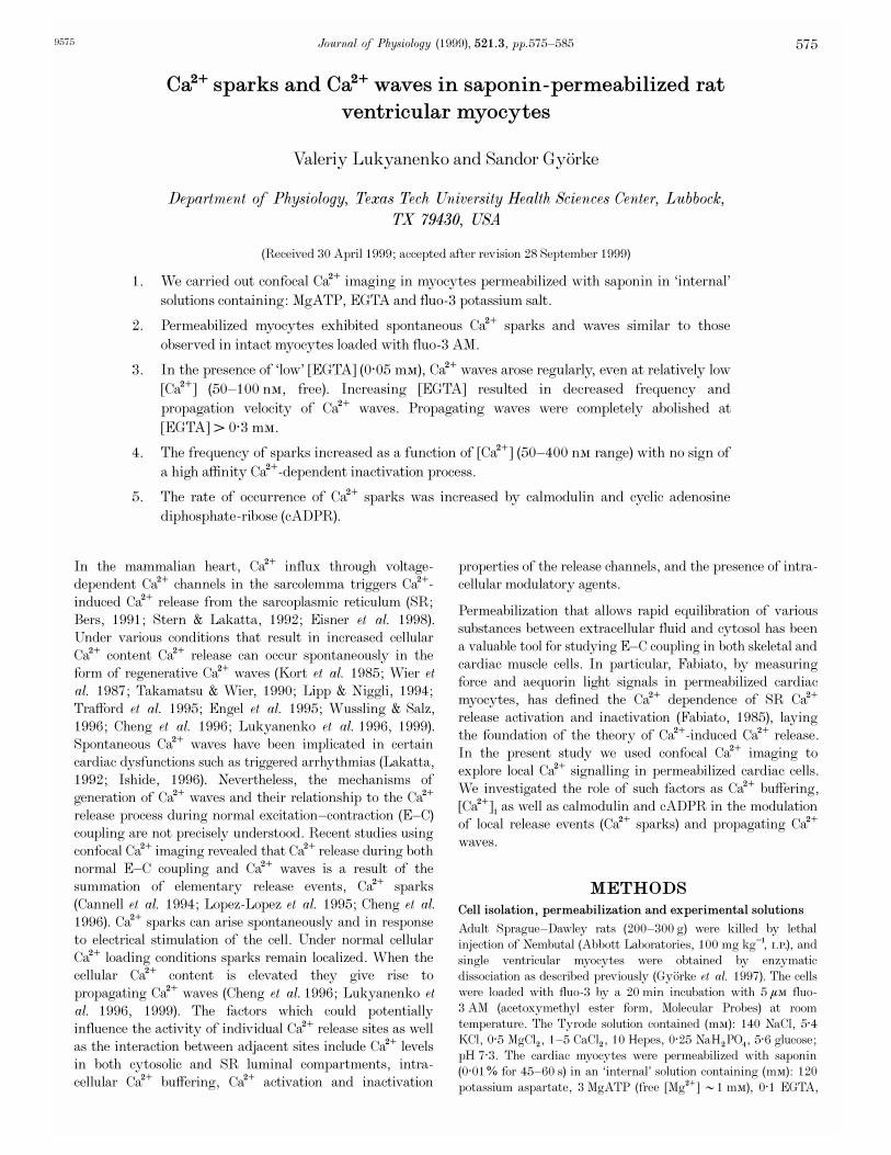
described in in vitro experiments (Coronado et al. 1994), theCa¥ dependency of RyRs in situ remains uncertain becauseof the difficulties in measuring and controlling [Ca¥] in thediadic cleft during E—C coupling. We took advantage of ourpermeabilized myocyte preparation to investigate therelationship between Ca¥ and the activity of Ca¥ releasesites. Figure 3A illustrates the effects of increasing [Ca¥]from 50 nÒ to 100, 150 or 200 nÒ in the presence of 0·1 mÒEGTA. Elevating [Ca¥] resulted in increased frequency ofCa¥ waves. In addition, at elevated [Ca¥] multi-focal Ca¥waves arising simultaneously from several independent sitesbecame evident. Quantitative assessments of spark propertiesat high [Ca¥] were impaired by the presence of Ca¥ wavesand elevated background fluorescence. Increases inbackground fluorescence also limited the Ca¥ concentrationsin the experimental solution to a rather limited rangebecause of saturation of the photomultiplier at high [Ca¥]é.Therefore, we employed bathing solutions containing high[EGTA] to abolish spontaneous Ca¥ waves. We also correctedthe images for changes in background fluorescence atdifferent [Ca¥]é by adjusting the fluorescence of the bathingsolution outside the permeabilized cells to the same level.Figure 3B shows representative images from such anexperiment recorded during successive increases of [Ca¥]from 100 to 150, 250 and 400 nÒ and a subsequent return
Ca¥ sparks and waves in permeabilized cardiomyocytesJ. Physiol. 521.3 579
Figure 3. Effects of [Ca¥]é on Ca¥ sparks and Ca¥ waves in saponin-permeabilized myocytes
A, representative line-scan images of fluorescence recorded in a permeabilized myocyte at various [Ca¥]élevels (indicated at the top of the respective images) in the presence of 0·1 mÒ EGTA. B, line-scan imagesof Ca¥ sparks corrected for increases of background fluorescence at various [Ca¥]é levels (indicated at thetop of the respective images) in the presence of 0·5 mÒ EGTA. Calibration bars: horizontal 15 ìm (A) and20 ìm (B), vertical 0·5 s (A) and 0·1 s (B). C, Ca¥ spark frequency (blue) and amplitude (light grey) as afunction of time before and after elevating [Ca¥]é to indicated levels for the experiment shown in B.D, Ca¥ spark frequency as a function of [Ca¥]é. The values are represented as means ± s.e.m. obtained in5 experiments. The lines were obtained by fitting the data according to the equationf = fmax{[Ca¥]
n
Ï([Ca¥]n
+ KD
n
)}, where fmax = 10000 events s¢ (100 ìm)¢, KD = 9·9 ìÒ and n = 1·6(blue line); fmax = 20000 events s¢ (100 ìm)¢; KD = 15 ìÒ and n = 1·6 (red line); andfmax = 30000 events s¢ (100 ìm)¢; KD = 20 ìÒ and n = 1·6 (green line).
––––––––––––––––––––––––––––––––––––––––––––––––––––––––––––––––––––––––––––––––––––––––––––Table 1. Spatio-temporal properties of Ca¥ sparks in intact and permeabilized ventricular cells
––––––––––––––––––––––––––––––––––––––––––––––Frequency
a
Amplitude Duration Width(FÏFï) (ms)
b
(ìm)c
––––––––––––––––––––––––––––––––––––––––––––––Intact cells [Ca¥]ï = 1 mÒ 4·60 ± 0·48 1·69 ± 0·02 27·7 ± 1·1 1·75 ± 0·07
Skinned cells [EGTA] = 0·1 mÒ 4·25 ± 0·88 1·72 ± 0·02 26·1 ± 0·7 1·85 ± 0·05[EGTA] = 0·5 mÒ 6·36 ± 0·65 1·71 ± 0·03 25·3 ± 0·6 1·96 ± 0·06
––––––––––––––––––––––––––––––––––––––––––––––Data represented as means ± s.e.m. of measurements, n = 30—235.
a
Spark frequency was defined as thenumber of events per second per 100 ìm line scanned.
b
Duration andc
width of spark were measured athalf-maximal amplitude.––––––––––––––––––––––––––––––––––––––––––––––––––––––––––––––––––––––––––––––––––––––––––––

V. Lukyanenko and S. Gy�orke J. Physiol. 521.3580
Figure 4. The effects of elevating [Ca¥]é on sparking activity in the presence of thapsigargin
A, representative line-scan images of fluorescence acquired before (a and b) and at different times (2 and3 min) after increasing [Ca¥]é from 80 nÒ to 250 nÒ in the presence of thapsigargin (c and d, respectively).Thapsigargin (10 ìÒ) was introduced into the bathing solution 1 min before elevating [Ca¥]é. Calibrationbars: horizontal 20 ìm, vertical 0·15 s. B, Ca¥ spark frequency as a function of time before and after theaddition of thapsigargin into the bathing solution in the same experiment. The experimental protocol ispresented schematically at the top.
––––––––––––––––––––––––––––––––––––––––––––––––––––––––––––––––––––––––––––––––––––––––––––Table 2. Spatio-temporal properties of Ca¥ sparks in permeabilized ventricular cells before and
after addition of calmodulin or cADPR
––––––––––––––––––––––––––––––––––––––––––––––Frequency
a
Amplitude Duration Width(FÏFï) (ms)
b
(ìm)c
––––––––––––––––––––––––––––––––––––––––––––––[Calmodulin] 0 7·7 ± 1·2 1·8 ± 0·02 26 ± 0·9 2·02 ± 0·06
5 ìÒ 15·5 ± 2·2 * 1·9 ± 0·03 27 ± 0·6 1·93 ± 0·04
[cADPR] 0 5·1 ± 1·1 1·5 ± 0·02 29 ± 1·4 1·92 ± 0·115 ìÒ 8·9 ± 1·8 * 1·5 ± 0·01 29 ± 1·3 1·94 ± 0·09
––––––––––––––––––––––––––––––––––––––––––––––Data represented as means ± s.e.m. of measurements, n = 24—329.
a
Spark frequency was defined as thenumber of events per second per 100 ìm line scanned.
b
Duration andc
width of spark were measured athalf-maximal amplitude. *Significantly different from control at P < 0·05.––––––––––––––––––––––––––––––––––––––––––––––––––––––––––––––––––––––––––––––––––––––––––––

to 100 nÒ. The effects of [Ca¥] on the frequency andamplitude of Ca¥ sparks from the same experiment aredocumented in Fig. 3C. Increasing [Ca¥] in the range100—400 nÒ resulted in a gradual increase in frequency ofsparks. Changing back to the original solution with 100 nÒCa¥ resulted in restoration of spark frequency to thecontrol level, thus indicating no significant signs of a run-down in sparking activity. The changes in the frequency ofsparks were accompanied only by insignificant alterations inthe amplitude of sparks (10% at 150 nÒ [Ca¥], grey bars).The results of five experiments are summarized in Fig. 3D,which plots the frequency of Ca¥ sparks as a function of[Ca¥]. It is unlikely that the amplitude of Ca¥ sparks wassaturated at elevated [Ca¥]. The KD of fluo-3 in themyoplasm has been estimated to be near 1 ìÒ (Harkins etal. 1993). Ca¥ sparks with an amplitude of 200—300 nÒwould rise above a background [Ca¥] of 400 nÒ only to alevel of 600—700 nÒ, which still would be in the linearrange of the indicator.
The observed potentiation of Ca¥ sparks could be attributedalso to a possible increase in the SR Ca¥ content (Fabiato,1992; Orchard et al. 1998) affecting the activity of the Ca¥release channels at luminal sites (Gy�orke & Gy�orke, 1998).We assessed the potential role of this mechanism, by usingthapsigargin to prevent accumulation of extra Ca¥ into theSR upon elevation of cytosolic Ca¥. Representative imagesfrom such an experiment are shown in Fig. 4A. The effectsof [Ca¥] on spark frequency in the same experiment arequantified in Fig. 4B. We applied 10 ìÒ thapsigargin for1 min before elevating [Ca¥] from 100 nÒ to 250 nÒ. Atthis concentration and exposure time thapsigargin inhibitsSR Ca¥ uptake without causing a significant loss in the SRCa¥ content (Bassani et al. 1993; Lukyanenko et al. 1999).Under these experimental conditions increasing [Ca¥]éresulted in an about 6-fold (from 7·1 ± 0·9 to 39·8 ±1·9 event s¢ (100 ìm)¢, n = 5) increase in sparkingfrequency, which is similar to that observed in experimentswithout thapsigargin. Following more than 2—3 min ofcontinuous exposure to thapsigargin, the frequency of sparksgradually decreased, apparently due to a loss of Ca¥ fromthe SR (Bassani et al. 1993; Lukyanenko et al. 1999). Takentogether, these results suggest that under our experimentalconditions, the increase in sparking activity cannot beattributed to increased SR Ca¥ load.
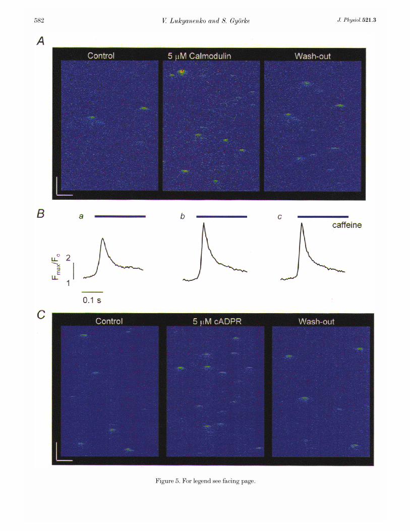
Effects of calmodulin and cADPR
We tested the effects on release site activity of certainmembrane-impermeable putative regulators of the SR Ca¥release such as cyclic adenosine diphosphate-ribose (cADPR,MW 541·3) and calmodulin (MW 16680), which would bedifficult to study in cells with an intact sarcolemma. Figure5 illustrates representative images of Ca¥ sparks before andafter exposure of two permeabilized cells to calmodulin(A, 5 ìÒ) and cADPR (C, 5 ìÒ), while Table 2 summarizesthe effects of these drugs on Ca¥ spark properties. Bothdrugs caused dramatic increases in the frequency of Ca¥
sparks with no significant changes in the amplitude andspatio-temporal properties of the events. The effects ofcADPR developed over a period of 2—6 min, and they werereadily reversible. The onset of potentiation of Ca¥ sparksby calmodulin was considerably slower, 10—15 min. Inaddition, the cells showed no significant recovery uponreverting to the control solution during a period of 5—10 min.The slow onset and lack of reversibility of calmodulin effectscould be attributed to the slow diffusion rate of this highmolecular weight agent as well as to the fact that the effectsare likely to involve long-lasting biochemical changes(phosphorylation of SR proteins). Incubation of the cellswith calmodulin also resulted in a significant increase in theamplitude of caffeine-induced Ca¥ transients (31·2 ± 4·4%,n = 5; Fig. 5B), suggesting that the effects of this agent onCa¥ sparks might be mediated by changes in the SR Ca¥load. These results further illustrate our ability tomanipulate the environment of the release sites in ways notpossible in intact cells.
DISCUSSION
In the present study we investigated the Ca¥-releasingactivity of the SR in saponin-permeabilized cardiacmyocytes using confocal Ca¥ imaging. In essential internalsolutions designed to maintain the basic functional integrityof Ca¥ stores, permeabilized cardiac myocytes exhibitedCa¥ sparks and Ca¥ waves similar to those observed inintact cells. During permeabilization, the content of thecells is diluted in the internal bathing solution (•100000-fold dilution). Thus, it appears that no endogenous factorssuch as cytosolic kinases and phosphatases that could be lostthrough equilibration with the bathing solution are essentialto maintaining basic activation and inactivation propertiesof the release sites.
We described explicitly the dependency of spark frequencyupon [Ca¥] in the range 50—400 nÒ. It is interesting tospeculate how these results might relate to the overall Ca¥dependency of Ca¥ spark activation in cardiomyocytes. Ifwe assume that all of the the approximately 200 releaseunits (Sommer, 1995; Franzini-Armstrong & Protasi, 1997)contained in the volume of a rectangular block ofapproximately 1 ìm width (diameter of the laser beam spot),1 ìm height (depth of field of the microscope) and 100 ìmlength scanned along a myocyte can be activated within10 ms (Cannell et al. 1994), the maximal frequency of sparkswould be 20 000 events s¢ (100 ìm)¢. This maximalsparking rate would be consistent with the estimatedfrequency of sparks during action potential stimulation,when presumably most of the release units becomeactivated (•15000 events s¢ (100 ìm)¢, Cannel et al. 1994).We fitted our data presented in Fig. 3D to Hill functionswith three different maximal sparking rates of 10000,20000 and 30000 events s¢ (100 ìm)¢ (Fig. 3D). The bestfits to the data for the three specified maximal sparkingrates yielded Ca¥ dissociation constants of 9·9, 15·2 and
Ca¥ sparks and waves in permeabilized cardiomyocytesJ. Physiol. 521.3 581
V. Lukyanenko and S. Gy�orke J. Physiol. 521.3582
Figure 5. For legend see facing page.

19·6 ìÒ and Hill coefficients equal to 1·6 in all cases. Theestimated values of KD of Ca¥ sensitivity of release sitesare in the range of Ca¥ dependency of RyR openprobability measured in lipid bilayer experiments in thepresence of physiological concentrations of Mg¥ and ATP(KD � 30 ìÒ; Gy�orke & Gy�orke, 1998). The relatively lowKD and supra-linear dependency of release site activation on[Ca¥] could explain why most release events normallyremain localized and do not initiate calcium-induced calciumrelease (CICR) in the neighbouring release sites (Stern et al.1999). We should point out, however, that because of thelimited range of [Ca¥] over which the sparks could bemeasured the obtained characteristics of Ca¥ dependency ofrelease sites represent only approximate estimates.
In mechanically skinned Purkinje cells, Fabiato showedthat sub-micromolar basal Ca¥ resulted in a substantialdecrease in the macroscopic CICR, suggesting that Ca¥release is inhibited at a high affinity inactivation site (Fabiato,1985). Under our experimental conditions, sparking activitydoes not appear to be influenced by such a high Ca¥ affinityinactivation process because spark frequency only increasedsteadily upon elevations of [Ca¥]. Our results, however, donot rule out the possibility that local release events arecontrolled by high local Ca¥ through a low-affinityinactivation process. Inhibition of RyRs by Ca¥ in a rangeof 50 ìÒ to 10 mÒ has been reported in both lipid bilayer(Laver et al. 1995; Copello et al. 1997; Gy�orke & Gy�orke,1988; Marengo et al. 1998) and vesicle flux experiments(Chamberlain et al. 1984; Zimanyi & Pessah, 1991; Chu etal. 1993). Such a low affinity inactivation, as well as apossible use-dependent inactivation induced by prioractivation of the channel (Sham et al. 1998; Zahradnikovaet al. 1999), could contribute to local release terminationwithout influencing the stationary frequency of sparks.Indeed, at the sub-micromolar [Ca¥] employed in ourexperiments, the probability of reactivation of individualrelease units must be quite low (P = 0·09 assuming 200independent release units sparking at a rate of60 events s¢ (100 ìm)¢) for time periods compatible withthe time of recovery from such inactivated conditions (•1 s).
The propagating Ca¥ waves were profoundly influenced byCa¥ buffering. At low Ca¥ buffering strength (EGTA
< 100 ìÒ), waves arose regularly even at low [Ca¥].Increasing EGTA resulted in decreased wave generation.Propagating Ca¥ waves were completely abolished atEGTA > 0·3 mÒ. These results reveal the importance ofintracellular Ca¥ buffering in the confinement of CICR incardiac cells. The high sensitivity of Ca¥ wave propagationto Ca¥ buffering is consistent with a saltatory mechanismof Ca¥ wave propagation (Keizer et al. 1998; Lukyanenko etal. 1999). In contrast to a continuous model of Ca¥ waves ina saltatory model, the release sites are spatially separatedand thus are sensitive to interventions impairing the freediffusion of Ca¥. In fact, site-to-site propagation bysequential activation of Ca¥ sparks was evident in thepresence of intermediate EGTA concentrations (Fig. 2C andD).
The activity of release sites was affected by the endogenoussignalling molecules calmodulin and cADPR. Interestingly,calmodulin, which has been shown to inhibit RyRs (bydirect interaction or through the activation ofCa¥—calmodulin-dependent protein kinase) in most RyRreconstitution studies (Meissner & Henderson, 1987;Takasago et al. 1991; Lokuta et al. 1995; Hain et al. 1995),enhanced sparking activity in cardiac cells. We attributethis potentiation to increased accumulation of Ca¥ withinthe SR due to stimulation of the SR Ca¥-ATPase(Narayanan & Xu, 1997) and subsequent activation of RyRsby Ca¥ at luminal sites (Gy�orke & Gy�orke, 1988). Indeed,exposure of the cells to calmodulin resulted in a significantincrease in the SR Ca¥ content (Fig. 5B). These results arein line with the previous studies suggesting that luminalCa¥ is a critical determinant of the functional state of Ca¥release in cardiac muscle (Bassani et al. 1995; Gy�orke et al.1997; Eisner et al. 1998). The demonstrated potentiation ofCa¥ sparks by cADPR supports the previous studiessuggesting that cADPR can enhance Ca¥ releasepresumably through potentiation of RyRs (Meszaros et al.1993; Iino et al. 1997; Galione et al. 1998). Taken together,our results show that saponin-permeabilized cells can be auseful model system for studying both spatial and temporalaspects of Ca¥ signalling in the heart under conditions inwhich the environment surrounding the Ca¥ releasechannels can be controlled precisely.
Ca¥ sparks and waves in permeabilized cardiomyocytesJ. Physiol. 521.3 583
Figure 5. Effects of calmodulin and cADPR on Ca¥ sparks
A, representative line-scan images of fluorescence changes acquired under control conditions (left-handpanel), 15 min after exposure of the cells to 5 ìÒ calmodulin (middle panel), and 10 min after changingback to the control solution (right-hand panel). Calibration bars: horizontal 10 ìm, vertical 0·2 s.B, caffeine-induced Ca¥ transients measured in the same cell at the same stages of the experiment as in A.Caffeine (20 mÒ) was applied for 2 s. C, representative line-scan images of fluorescence changes measuredunder control conditions (left-hand panel), 2 min after exposure of the cells to 5 ìÒ cADPR (middle panel),and 5 min after reverting back to the control solution (right-hand panel). Calibration bars: horizontal10 ìm, vertical 0·2 s.

Bassani, J. W., Bassani, R. A. & Bers, D. M. (1993). Twitch-dependent SR Ca accumulation and release in rabbit ventricularmyocytes. American Journal of Physiology 265, C533—540.
Bassani, J. W., Yuan, W. & Bers, D. M. (1995). Fractional SR Ca¥release is regulated by trigger Ca¥ and SR Ca¥ content in cardiacmyocytes. American Journal of Physiology 268, C1313—1329.
Bers, D. M. (1991). Excitation—Contraction Coupling and CardiacContractile Force. Kluwer Academic Publishers, Dordrecht, TheNetherlands.
Cannell, M. B., Cheng, H. & Lederer, W. J. (1994). Spatial non-uniformities in [Ca¥]é during excitation—contraction coupling incardiac myocytes. Biophysical Journal 67, 1942—1956.
Chamberlain, B. K., Volpe, P. & Fleischer, S. (1984). Calcium-induced calcium release from purified cardiac sarcoplasmicreticulum vesicles. Journal of Biological Chemistry 259,7540—7546.
Cheng, H., Lederer, W. & Cannell, M. B. (1993). Calcium sparks:elementary events underlying excitation—contraction coupling inheart muscle. Science 262, 740—744.
Cheng, H., Lederer, M. R., Lederer, W. J. & Cannell, M. B.(1996). Calcium sparks and [Ca¥]é waves in cardiac myocytes.American Journal of Physiology 270, C148—159.
Cheng, H., Song, L. S., Shirokova, N., Gonzalez, A., Lakatta,E. G., Rios, E. & Stern, M. D. (1999). Amplitude distribution ofcalcium sparks in confocal images: theory and studies with anautomatic detection method. Biophysical Journal 76, 606—617.
Chu, A., Fill, M., Stefani, E. & Entman, M. L. (1993). CytoplasmicCa¥ does not inhibit the cardiac muscle sarcoplasmic reticulumryanodine receptor Ca¥ channel, although Ca¥-induced Ca¥inactivation of Ca¥ release is observed in native vesicles. Journal ofMembrane Biology 135, 49—59.
Copello, J. A., Barg, S., Onoue, H. & Fleicher, S. (1997).Heterogeneity of Ca¥ gating of skeletal muscle and cardiacryanodine receptor. Biophysical Journal 73, 141—156.
Coronado, R., Morrissette, J., Sukhareva, M. & Vaughan, D. M.(1994). Structure and function of ryanodine receptors. AmericanJournal of Physiology 266, C1485—1504.
Eisner, D. A., Trafford, A. W., Diaz, M. E., Overend, C. L. &O’Neill, S. C. (1998). The control of Ca release from the cardiacsarcoplasmic reticulum: regulation versus autoregulation.Cardiovascular Research 38, 589—604.
Engel, J., Sowerby, A. J., Finch, A. E., Fechner, M. & Stier, A.(1995). Temperature dependence of Ca¥ wave properties incardiomyocytes: implications for the mechanism of autocatalyticCa¥ release in wave propagation. Biophysical Journal 68, 40—45.
Fabiato, A. (1985). Time and calcium dependence of activation andinactivation of calcium-induced calcium release of calcium from thesarcoplasmic reticulum of a skinned canine cardiac Purkinje cell.Journal of General Physiology 85, 291—320.
Fabiato, A. (1992). Two kinds of calcium-induced release of calciumfrom the sarcoplasmic reticulum of skinned cardiac cells. InExcitation—Contraction Coupling in Skeletal, Cardiac and SmoothMuscle, ed. Frank, G. B., pp. 245—262. Plenum Press, New York.
Franzini-Armstrong, C. & Protasi, F. (1997). Ryanodine receptorsof striated muscles: a complex channel capable of multipleinteractions. Physiological Reviews 77, 699—729.
Galione, A., Cui, Y., Empson, R., Iino, S., Wilson, H. & Terrar,D. (1998). Cyclic ADP-ribose and the regulation of calcium-inducedcalcium release in eggs and cardiac myocytes. Cell Biochemistry andBiophysics 28, 19—30.
Gy�orke, I. & Gy�orke, S. (1998). Regulation of the cardiac ryanodinereceptor channel by luminal Ca¥ involves luminal Ca¥ sensing sites.Biophysical Journal 75, 2801—2810.
Gy�orke, S., Lukyanenko, V. & Gy�orke, I. (1997). Dual effects oftetracaine on spontaneous calcium release in rat ventricularmyocytes. Journal of Physiology 500, 297—309.
Hain, J., Onoue, H., Mayrleitner, M., Fleischer, S. &Schindler, H. (1995). Phosphorylation modulates the function ofthe calcium release channel of sarcoplasmic reticulum from cardiacmuscle. Journal of Biological Chemistry 270, 2074—2081.
Harkins, A. B., Kurebayashi, N. & Baylor, S. M. (1993). Restingmyoplasmic free calcium in frog skeletal muscle fibers estimatedwith fluo-3. Biophysical Journal 65, 865—881.
Iino, S., Cui, Y., Galione, A. & Terrar, D. A. (1997). Actions ofcADP-ribose and its antagonists on contraction in guinea pigisolated ventricular myocytes. Influence of temperature. CirculationResearch 81, 879—884.
Ishide, N. (1996). Intracellular calcium modulators for cardiac musclein pathological conditions. Japanese Heart Journal 37, 1—17.
Keizer, J., Smith, G., Ponce-Dawson, S. & Pearson, J. (1998).Saltatory propagation of Ca¥ waves by Ca¥ sparks. BiophysicalJournal 75, 595—600.
Kort, A. A., Capogrossi, M. C. & Lakatta, E. G. (1985). Frequency,amplitude, and propagation velocity of spontaneous Ca¥-dependentcontractile waves in intact adult rat cardiac muscle and isolatedmyocytes. Circulation Research 57, 844—855.
Lakatta, E. G. (1992). Functional implications of spontaneoussarcoplasmic reticulum Ca¥ release in the heart. CardiovascularResearch 26, 193—214.
Laver, D. R., Roden, L. D., Ahern, G. P., Eager, K. R.,Junankar, P. R. & Dulhunty, A. F. (1995). Cytoplasmic Ca¥inhibits the ryanodine receptor from cardiac muscle. Journal ofMembrane Biology 147, 7—22.
Lipp, P. & Niggli, E. (1994). Modulation of Ca¥ release in culturedneonatal rat cardiac myocytes. Circulation Research 74, 979—990.
Lokuta, A. J., Rogers, T. B., Lederer, W. J. & Valdivia, H. H.(1995). Modulation of cardiac ryanodine receptors of swine andrabbit by a phosphorylation-dephosphorylation mechanism. Journalof Physiology 487, 609—622.
Lopez-Lopez, J. R., Shacklock, P. S., Balke, C. W. & Wier, W. G.(1995). Local calcium transients triggered by single L-type calciumchannel currents in cardiac cells. Science 268, 1042—1045.
Lukyanenko, V., Gy�orke, I. & Gy�orke, S. (1996). Regulation ofcalcium release by calcium inside the sarcoplasmic reticulum inventricular myocytes. Pfl�ugers Archiv 432, 1047—1054.
Lukyanenko, V., Subramanian, S., Gy�orke, I., Wiesner, T. &Gy�orke, S. (1999). The role of luminal Ca¥ in generation of Ca¥waves in rat ventricular myocytes. Journal of Physiology 518,173—186.
Marengo, J. J., Hidalgo, C. & Bull, R. (1998). Sulfhydryl oxidationmodifies the calcium dependence of ryanodine-sensitive calciumchannels of excitable cells. Biophysical Journal 74, 1263—1277.
Meissner, G. & Henderson, J. S. (1987). Rapid calcium release fromcardiac sarcoplasmic reticulum vesicles is dependent on Ca¥ and ismodulated by Mg¥, adenine nucleotide, and calmodulin. Journal ofBiological Chemistry 262, 3065—3073.
Meszaros, L. G., Bak, J. & Chu, A. (1993). Cyclic ADP-ribose as anendogenous regulator of the non-skeletal type ryanodine receptorCa¥ channel. Nature 364, 76—79.
Narayanan, N. & Xu, A. (1997). Phosphorylation and regulation ofthe Ca(2+)-pumping ATPase in cardiac sarcoplasmic reticulum bycalciumÏcalmodulin-dependent protein kinase. Basic Research inCardiology 92 (suppl. 1), 25—35.
V. Lukyanenko and S. Gy�orke J. Physiol. 521.3584

Orchard, C. H., Smith, G. L. & Steele, D. S. (1998). Effects ofcytosolic Ca¥ on the Ca¥ content of the sarcoplasmic reticulum insaponin-permeabilized rat ventricular trabeculae. Pfl�ugers Archiv435, 555—563.
Sham, J. S., Song, L. S., Chen, Y., Deng, L. H., Stern, M. D.,Lakatta, E. G. & Cheng, H. (1998). Termination of Ca¥ release bya local inactivation of ryanodine receptors in cardiac myocytes.Proceedings of the National Academy of Sciences of the USA 95,15096—15101.
Sommer, J. R. (1995). Comparative anatomy: in praise of a powerfulapproach to elucidate mechanisms translating cardiac excitationinto purposeful contraction. Journal of Molecular Cell Cardiology27, 19—35.
Song, L. S., Stern, M. D., Lakatta, E. G. & Cheng, H. (1997).Partial depletion of sarcoplasmic reticulum calcium does not preventcalcium sparks in rat ventricular myocytes. Journal of Physiology505, 665—675.
Stern, M. D. & Lakatta, E. (1992). Excitation—contraction in theheart: the state of the question. FASEB Journal 6, 3092—3100.
Stern, M. D., Song, L. S., Cheng, H., Sham, J. S., Yang, H. T.,Boheler, K. R. & Rios, E. (1999). Local control models of cardiacexcitation—contraction coupling. A possible role for allostericinteractions between ryanodine receptors. Journal of GeneralPhysiology 113, 469—489.
Sitsapesan, R. & Williams, A. J. (1995). Cyclic ADP-ribose andrelated compounds activate sheep skeletal sarcoplasmic reticulumCa¥ release channel. American Journal of Physiology 268,C1235—1240.
Takamatsu, T. & Wier, W. G. (1990). Calcium waves in mammalianheart: quantification of origin, magnitude, waveform, and velocity.FASEB Journal 4, 1519—1525.
Takasago, T., Imagawa, T., Furukawa, K., Ogurusu, T. &Shigekawa, M. (1991). Regulation of the cardiac ryanodinereceptor by protein kinase-dependent phosphorylation. Journal ofBiochemistry (Tokyo) 109, 163—170.
Trafford, A. W., Lipp, P., O’Neil, C. O., Niggli, E. & Eisner, D. A.(1995). Propagating calcium waves initiated by local caffeineapplication in rat ventricular myocytes. Journal of Physiology 489,319—326.
Wier, W. G., Cannell, M. B., Berlin, J. R., Marban, E. &Lederer, W. J. (1987). Cellular and subcellular heterogeneity of[Ca¥]é in single heart cells revealed by Fura_2. Science 235,325—328.
Wussling, M. H. & Salz, H. (1996). Nonlinear propagation ofspherical calcium waves in rat cardiac myocytes. BiophysicalJournal 70, 1144—1153.
Zahradn�úkov�a, A., Dura, M. & Gy�orke, S. (1999). Modal gatingtransitions in cardiac ryanodine receptors during increases of Ca¥concentration produced by photolysis of caged Ca¥. Pfl�ugers Archiv438, 283—288.
Zimanyi, I. & Pessah, I. N. (1991). Comparison of [ÅH]ryanodinereceptors and Ca¥ release from rat cardiac and rabbit skeletalmuscle sarcoplasmic reticulum. Journal of Pharmacology andExperimental Therapeutics 256, 938—946.
Acknowledgements
This work was supported by the National Institutes of Health(HL 63043, HL 52620, HL 03739). S. Gy�orke is an EstablishedInvestigator of the American Heart Association.
Corresponding author
S. Gy�orke: Department of Physiology, Texas Tech University HSC,Lubbock, TX 79430, USA.
Ca¥ sparks and waves in permeabilized cardiomyocytesJ. Physiol. 521.3 585


