Cell Surface Delivery of TRAIL Strongly Augments theTumoricidal Activity of T Cells
Marco de Bruyn1, Yunwei Wei1,3, Valerie R. Wiersma1, Douwe F. Samplonius1, Harry G. Klip2,Ate G.J. van der Zee2, Baofeng Yang4, Wijnand Helfrich1, and Edwin Bremer1
AbstractPurpose: Adoptive T-cell therapy generally fails to induce meaningful anticancer responses in patients
with solid tumors. Here, we present a novel strategy designed to selectively enhance the tumoricidal activity
of T cells by targeted delivery of TNF-related apoptosis-inducing ligand (TRAIL) to the T-cell surface.
Experimental Design: We constructed two recombinant fusion proteins, anti-CD3:TRAIL and K12:
TRAIL. Tumoricidal activity of T cells in the presence of these fusion proteins was assessed in solid tumor
cell lines, primary patient-derived malignant cells, and in a murine xenograft model.
Results: When added to T cells, K12:TRAIL and anti-CD3:TRAIL selectively bind to the T-cell surface
antigens CD3 and CD7, respectively, leading to cell surface accretion of TRAIL. Subsequently, anti-CD3:
TRAIL and K12:TRAIL increased the tumoricidal activity of T cells toward cancer cell lines and primary
patient-derived malignant cells by more than 500-fold. Furthermore, T-cell surface delivery of TRAIL
strongly inhibited tumor growth and increased survival time of xenografted mice more than 6-fold.
Conclusions: Targeted delivery of TRAIL to cell surface antigens of T cells potently enhances the
tumoricidal activity of T cells. This approach may be generally applicable to enhance the efficacy of
adoptive T-cell therapy. Clin Cancer Res; 17(17); 5626–37. �2011 AACR.
Introduction
While efficacious in certain virus-mediated cancers,adoptive T-cell therapy generally fails to induce meaningfulanticancer responses in patients with solid tumors. Thecause for this lack of clinical efficacy is multifactorial bynature and includes the intrinsic or acquired resistance ofmalignant cells to cytotoxic T-cell effector mechanismssuch as granzyme/perforin-mediated lysis (1–5) and Fas-mediated killing (6, 7). To overcome or circumvent thispotential pitfall of adoptive T-cell therapy, we propose to
selectively expand the T-cell armamentarium with addi-tional tumoricidal effector molecules, in particular with theTNF-related apoptosis-inducing ligand (TRAIL).
TRAIL is a type II transmembrane protein normallyexpressed on natural killer (NK) cells, where it is essentialfor NK cell–mediated tumor immune surveillance (8, 9). Incontrast, the expression of TRAIL on resting T cells istypically very low (10, 11). Nevertheless, a slight inductionof TRAIL expression on T cells, by ectopic expression ofTRAIL or by ex vivo costimulationwith anti-CD3 and IFN-a,already potently increases their tumoricidal activity (11,12). Furthermore, T-cell–expressed TRAIL is essential forgraft-versus-tumor (GVT) activity during allogeneic hema-topoietic cell transplantation (AHCT; ref. 10). Importantly,T-cell–expressed TRAIL does not contribute to deleteriousGVHD activity (10). Taken together, these findings suggestthat T cells should gain additional tumoricidal potentialwhen equipped with TRAIL at the cell surface.
Previously, we and others have reported on an approachthat may be particularly useful in this respect. In brief,recombinant soluble TRAIL (sTRAIL) was geneticallylinked to a tumor cell–selective antibody fragment (scFv).The resultant recombinant scFv:sTRAIL fusion protein hasthe following properties: By virtue of the antibody frag-ment, the scFv:TRAIL fusion protein selectively deliversTRAIL to the cell surface of tumor cells. Subsequently, cellsurface bound scFv:TRAIL can activate TRAIL receptorapoptotic signaling in tumor cells (reviewed in ref. 13).Here, we utilized and adapted this approach for the selec-tive delivery of high levels of TRAIL to the cell surface of
Authors' Affiliations: 1Department of Surgery, Surgical Research Labora-tories, and 2Department of Gynecological Oncology, University MedicalCenter Groningen (UMCG), University of Groningen, Groningen, the Neth-erlands; 3Department of General Surgery, The First Affiliated Hospital ofHarbin Medical University; and 4Department of Pharmacology, State-Province Key Laboratories of Biomedicine-Pharmaceutics of China, Har-bin Medical University, Harbin, Heilongjiang, China
Note: Supplementary data for this article are available at Clinical CancerResearch Online (http://clincancerres.aacrjournals.org/).
M. de Bruyn and Y. Wei contributed equally to the study.
Corresponding Authors: Wijnand Helfrich, Department of Surgery,Surgical Research Laboratories, University Medical Center Groningen,University of Groningen, Hanzeplein 1/BA44, 9713 GZ Groningen, theNetherlands. Phone: 31-503613733; Fax: 31-503632796; E-mail:[email protected] or Baofeng Yang, Department of Pharmacol-ogy, State-Province Key Laboratories of Biomedicine-Pharmaceutics ofChina, Harbin Medical University, Harbin, Heilongjiang 150081, China.Phone: 86-45186669473; E-mail: [email protected]
T cells, with the aim of expanding the cytotoxic armamentof these T cells with TRAIL (for schematic, see Fig. 1).
This approach was preclinically evaluated using 2 novelrecombinant fusion proteins, designated anti-CD3:TRAILand K12:TRAIL, that selectively bind to the T-cell surfaceantigen CD3 and CD7, respectively. Fusion protein anti-CD3:TRAIL contains a CD3 stimulatory antibody fragment,whereas K12:TRAIL contains a soluble form of the CD7ligand K12. Anti-CD3:TRAIL and K12:TRAIL stronglypotentiated the tumoricidal activity of T cells toward apanel of cancer cell lines, primarypatient-derivedmalignantcells, and in a murine xenograft model. This strategy mayrepresent an easy-to-incorporate approach to enhance theefficacyof various formsof adoptive T-cell therapy in cancer.
Materials and Methods
ReagentsAnti-CD7 mAb TH-69 competes with K12:TRAIL for
binding to CD7 and was a gift from Prof. Dr. MartinGramatzki (University of Kiel, Kiel, Germany). TRAIL-neu-tralizing mAb 2E5 was purchased from Kordia Life
Translational Relevance
Because adoptive T-cell strategies generally fail totrigger clinically relevant anticancer immunity inpatients with solid tumors, new strategies that optimizeadoptive T-cell transfer are warranted. Here, we presenta novel strategy that aims to selectively and safelyenhance the antitumor activity of T cells by targeteddelivery of human TNF-related apoptosis-inducingligand (TRAIL) to the cell surface, whereby T cells canutilize TRAIL as an additional cytotoxic effector. Thisapproach strongly enhances the antitumor activity of Tcells toward a panel of cancer cell lines and primarypatient-derived cancer cells. In addition, targeted deliv-ery of TRAIL to the T-cell surface significantly extendedthe survival time of tumor-bearing mice. The approachreported here could also be easily integrated in currentadoptive T-cell strategies.
Figure 1. Cell surface delivery ofTRAIL to augment the tumoricidalactivity of T cells. Fusion proteinsK12:TRAIL and anti-CD3:TRAILwere designed to selectively bindto T-cell markers CD7 and CD3,respectively, and enhance levelsof TRAIL on the surface of T cells.Tumoricidal activity of T cellsshould be augmented by virtue ofTRAIL-TRAILR–mediatedsignaling in tumor cells.
K12anti-CD3
sTRAILsTRAIL
TRAILR
T cell
Tumor cells
CD3
T cell
CD7
K12:TRAIL
anti-CD3:TRAIL
T cell
T cell
Enhancing T-cell Tumoricidal Activity with TRAIL
www.aacrjournals.org Clin Cancer Res; 17(17) September 1, 2011 5627
Sciences, FasL-neutralizingmAb Alf2.1 was purchased fromSigma-Aldrich, and TNF-a–neutralizing fusion proteinEnbrel was purchased from Wyeth. Phycoerythrin (PE)-labeled anti-TRAIL mAb B-S23 was purchased from Dia-clone SAS and PE-labeled anti-CD69 mAb was purchasedfrom IQ products. Anti-TRAILR antibodies HS-201, HS-202, HS-203, and HS-204 and soluble TRAIL receptorsTRAILR1-Fc, TRAILR2-Fc, TRAILR3-Fc, and TRAILR4-Fcwere purchased from Alexis. TRAIL ELISA was purchasedfrom Diaclone and Cytokine ELISA was from SA Bios-ciences. DiI, DiO, and FLUO-3-AM were purchased fromInvitrogen. DiOC6 was purchased from Molecular Probes.Actinomycin D, valproic acid (VPA), and cycloheximidewere purchased from Sigma-Aldrich. Pan-caspase inhibitorzVAD-FMK, caspase-8 inhibitor zLEHD-FMK, and caspase-9 inhibitor zIETD-FMK were purchased from Calbiochem.Anti-CD3 mAb and recombinant interleukin (IL) 2 werepurchased from ImmunoTools. TNF-awas purchased fromAbcam. FasL-Fc was a kind gift from Prof. Dr. HaraldWajant (University of W€urzburg, W€urzburg, Germany).RhTRAIL was purchased from R&D Technologies.
Production of K12:TRAIL and anti-CD3:TRAILFusion protein K12:TRAIL and anti-CD3:TRAIL were
constructed by cloning the cDNA of soluble human K12(aa 29–143) or the anti-CD3 antibody fragmentscFvUCHT-1v9 in frame with human soluble TRAIL intopreviously described vector pEE14, yielding plasmidspEE14-K12:TRAIL and pEE14-anti-CD3:TRAIL, respec-tively. Fusion proteins K12:TRAIL and anti-CD3:TRAILwere produced in CHO-K1 cells essentially using previouslydescribed methods (14). Culture medium containing K12:TRAIL or anti-CD3:TRAIL was cleared by centrifugation(10,000 � g, 10 minutes), filter sterilized, and stored at�80�C until further use. Fusion proteins were purified viathe N-terminal hemagglutinin (HA) tag using anti-HAaffinity chromatography. Fusion protein anti-MCSP:TRAILis a fusion protein that is essentially identical to anti-CD3:TRAIL except that it contains an anti-MCSP scFv instead ofan anti-CD3 scFv. Melanoma-associated chondroitin sul-fate proteoglycan (MCSP) is expressed on cells of themelanocyte lineage but not on T cells or carcinoma cells.
A2058, SK-MEL-28, HEP3B, PLC-PRF-5, FADU, PC-3M,HK-2, and Ramos were obtained from the American TissueCulture Collection and characterized by short tandemrepeat profiling, karyotyping, and isoenzyme analysis.A375M.FADD-DED cells were generated by transfectionof parental A375M cells with a dominant-negative FADD-DED construct using Fugene (Roche BV). A375M.FADD-DED cells were cultured in RPMI 1640 medium supple-mented with 10% fetal calf serum (FCS) and 500 mg/mLGeneticin. HCT-116–luc was from Caliper Biosciences(Xenogen). NHDF-juv cells were from Promocell. Humanumbilical vein endothelial cells (HUVEC) were isolated asdescribed previously. Unless otherwise indicated, cells were
cultured in standard RPMI (Cambrex BioScience) supple-mented with 10% FCS.
Isolation of primary patient-derived tumor cells,T cells and activation of T cells
Experiments were approved by the local Medical Ethi-cal Committee and patients/healthy volunteers signed forinformed consent. Tumor tissues and ascitic fluids wereobtained during surgery procedures and if necessaryminced, after which adherent cells were cultured. Experi-ments were carried out before passage number 5. Sampleswith a background apoptosis more than 30% wereexcluded from analysis. Normal fibroblast cultures origi-nating from tumor tissue/ascitis were essentially estab-lished as described above for primary cancer cell cultures.Tumor-infiltrating T cells were isolated from the primarytumor cultures using ammonium chloride lysis. Periph-eral blood lymphocytes (PBL) from blood of healthydonors were isolated using standard density gradientcentrifugation (Lymphoprep; Axis-Shield PoC As). Acti-vated T cells were generated by culturing PBLs with anti-CD3 mAb (0.5 mg/mL; 72 hours) and IL-2 (100 ng/mL;48 hours).
Analysis of fusion protein binding and TRAILRexpression
The presence of cell surface bound TRAIL was assessed byincubating cells with PE-conjugated anti-TRAIL mAb B-S23(Diaclone SAS) for 60 minutes at 4�C followed by 3washes. Cells were subsequently measured on an AccuriC6 flow cytometer. Cell surface display of TRAIL in timewas determined by incubating cells with 50 pg per T cellK12:TRAIL or anti-CD3:TRAIL for 60 minutes at 4�C fol-lowed by 3 washes in PBS. Cells were then incubated at37�C for the time points indicated and TRAIL expressionwas determined by flow cytometry using PE-labeled mAbB-S23. The amount of K12:TRAIL or anti-CD3:TRAILbound to an individual T cell was determined using TRAILELISA. Briefly, 1 � 105 T cells were incubated with increas-ing concentrations of K12:TRAIL or anti-CD3:TRAIL for 1hour. Subsequently, cells were centrifuged and superna-tants used for TRAIL ELISA. Loss of K12:TRAIL and anti-CD3:TRAIL from the supernatant was calculated using thesame concentration range of K12:TRAIL and anti-CD3:TRAIL in the absence of T cells as a reference. Expressionof TRAIL receptors was determined by incubating cells withmAbHS-201, HS-202, HS-203, or HS-204 for 60minutes at4�C followed by 3 washes with PBS and incubation withPE-labeled goat anti-mouse Ab for 60 minutes at 4�C. Cellswere subsequently washed 3 more times with PBS andTRAILR expression was determined using an Accuri C6 flowcytometer.
Analysis of cell deathFor cell death assays, cancer cells were labeled with red
fluorescent membrane label DiI (Invitrogen) in serum-freemedium for 15 minutes followed by 3 washes in serumcontaining medium and subsequent culture in a 48-well
de Bruyn et al.
Clin Cancer Res; 17(17) September 1, 2011 Clinical Cancer Research5628
plate (3.0 � 104 cells per well). After 24 hours, cancer cellswere treated for 16 hours with T cells (activated or resting)at the indicated ratios in the presence or absence of K12:TRAIL (50 pg per T cell), anti-CD3:TRAIL (50 pg per T cell),or anti-MCSP:TRAIL (50 pg per T cell) after which apop-tosis was assessed in tumor cells by loss of mitochondrialmembrane potential (Dy), as previously described (14)using an Accuri C6 flow cytometer. Where indicated, cas-pase inhibitors were used at a final concentration of10 mmol/L. Actinomycin D was used at a final concentra-tion of 2 mg/mL. Anti-TRAIL (2E5)- and anti-FasL (Alf2.1)-neutralizing mAbs and TNF-a–neutralizing fusion proteinEnbrel were used at a final concentration of 5 mg/mL. Forexperiments involving VPA, tumor cells were pretreated for24 hours with 1 mmol/L of VPA prior to addition of T cellsat the indicated ratios. For experiments involving conca-namycin A (CMA), T cells were treated with the indicatedconcentrations of CMA for 2 hours prior to addition totumor cells. Tumor cells were subsequently treated withthese T cells for 4 hours in the presence of CMA after whichinduction of apoptosis was assessed. For outgrowth experi-ments using patient ascitic fluid, anti-CD3:TRAIL, anti-CD3 mAb, or rhTRAIL were added at a final concentrationof 100 ng/mL to a mixture of ascitic fluid and normalculture medium (50% v/v).
Time lapse confocal microscopyTumor cells were labeled with membrane label DiO
(green) by incubation of 1 � 106 cells with 5 mL DiO inserum-free RPMI 1640 for 15 minutes at 37�C. Cells werethen washed 3 times with RPMI 1640þ 10% FCS and DiO-labeled tumor cells were cultured in a glass coverslide (3.0� 104 cells per well). After 24 hours, T cells labeled with DiI(red) using the same procedure were added to the tumorcells at the indicated effector-to-target (E:T) ratios. Timelapse confocal microscopy was carried out on a SolamereNipkow Disk CLSM equipped with temperature/CO2-con-trolled cabinet and multicell track table.
Mechanical stimulation of CD3 signaling and calciumfluxGlass coverslides in Petri dishes were coated overnight
with 10 mg/mL TRAILR1-Fc, TRAILR2-Fc, TRAILR3-Fc, orTRAILR4-Fc. Coverslides were subsequently washed withPBS and blocked with normal human serum for 1 hour. Tcells were labeled with FLUO-3-AM by incubating the cellswith 5 mmol/L FLUO-3-AM ester in 100 mL PBS for 15minutes at 20�C, followed by addition of 400 mL PBS with10% FCS and 45 minutes of incubation at 37�C. Next,FLUO-3-AM labeled T cells were added to the TRAIL-R1-,TRAIL-R2-, TRAIL-R3-, or TRAIL-R4–coated glass slides. Tcells weremechanically stimulated by using amicropipette,as previously described (15). Calcium flux was measuredand quantified on a TILL iMIC fluorescence microscopeequipped with temperature/CO2-controlled cabinet. Cal-cium flux is depicted as the mean fluorescent intensity(MFI) at any given time point (MFIT)/the MFI at T ¼ 0(MFI0).
Cytokine ELISAT cells were incubated with tumor cells for 16 hour at an
E:T ratio of 5:1 in the presence or absence of 50 pg per T cellanti-CD3:TRAIL. Where indicated, mAb 2E5 was added toblock TRAIL–TRAILR interaction. Alternatively, T cells weretreated with anti-CD3 mAb for 16 hours to elicit T-cellactivation and cytokine release. Cell supernatants weresubsequently harvested, centrifuged, and used for cytokineELISA according to the manufacturer’s instructions.
HCT-116–luc xenograft mouse modelExperiments were approved by the Committee for
Research and Animal Ethics of the UMCG. Male athymicmice (Harlan) were intraperitoneally inoculated with HCT-116–luc (1� 106 cells). Animals were subsequently treatedwith a single intraperitoneal dose of 5 � 106 T cells andabout 150 mg/kg K12:TRAIL or anti-CD3:TRAIL, as indi-cated. Tumor growth was monitored by luminescent ima-ging of mice from the ventral abdominal side using an IVISSpectrum (Xenogen) optical imager. Animals were sacri-ficed by cervical dislocation when bioluminescent signalwas increased 10-fold compared with day of inoculation.For localization of T cells, T cells were labeled with CellVueNIR815 membrane labeling kit according to the man-ufacturer’s instructions. Specific fluorescent signal wasdetermined by subtracting the fluorescent signal fromsaline-treated mice.
Statistical analysisData reported are mean values � SD of 3 independent
experiments. Data was analyzed by 1-way ANOVA followedby Tukey–Kramer post test or, where appropriate, by 2-sided unpaired Student’s t test. Where indicated, *, P < 0.05;**, P < 0.01; ***, P < 0.001.
Results
Tumoricidal activity of T cells is potently augmentedby delivery of TRAIL to the T-cell surface
To selectively deliver TRAIL to the cell surface of T cells,the CD3-targeted fusion protein anti-CD3:TRAIL and theCD7-targeted fusion protein K12:TRAIL were generated. Inline with literature, ex vivo activated T cells obtained fromhealthy volunteers did not detectably express TRAIL(Fig. 2A). Incubation of these TRAIL-negative T cells withanti-CD3:TRAIL or K12:TRAIL lead to the display of highlevels of TRAIL on the T-cell surface (Fig. 2A). A subsequentTRAIL ELISA showed that 1 � 105 T cells bind about 50 pgof K12:TRAIL and/or about 4 pg of anti-CD3:TRAIL. ThisT-cell binding by anti-CD3:TRAIL or K12:TRAIL was selec-tively blocked by anti-CD3 or anti-CD7 mAb, respectively(data not shown) and did not induce reciprocal T-cell death(Supplementary Fig. S1A).
Importantly, T-cell–selective binding by anti-CD3:TRAILand K12:TRAIL did potentiate the tumoricidal activityof activated T cells toward cancer cells, with more than90% apoptosis at an E:T ratio of 2:1 (Fig. 2B). Activated Tcells alone failed to induce apoptosis at these E:T ratios,
Enhancing T-cell Tumoricidal Activity with TRAIL
www.aacrjournals.org Clin Cancer Res; 17(17) September 1, 2011 5629
Figure 2. Tumoricidal activity of T cells is potently augmented by delivery of TRAIL to the T-cell surface. A, TRAIL expression on anti-CD3 þ IL-2–activatedT cells was assessed by incubation of T cells with PE-conjugated anti-TRAIL mAb (thin line). Cell surface TRAIL expression after binding of K12:TRAIL oranti-CD3:TRAIL was determined by preincubating T cells with K12:TRAIL (hashed line) or anti-CD3:TRAIL (solid line). Shaded gray area representsfluorescence of PE-labeled isotype control. B, activated T cells were incubated with OvCAR-3 cells in the presence or absence of K12:TRAIL (50 pg per T cell)or anti-CD3:TRAIL (50 pg per T cell) for 16 hours at the indicated E:T ratios and apoptosis was assessed in OvCAR-3 cells. C, activated T cells were incubatedwith OvCAR-3 cells in the presence or absence of K12:TRAIL (50 pg per T cell) or anti-CD3:TRAIL (50 pg per T cell) at an E:T ratio of 10:1 for the indicatedtime points and apoptosis was assessed in OvCAR-3 cells. D, cancer cell lines SK-OV-3, OvCAR-3, A2780, HCT-116, HCT-8, HT29, A375M, A2058, SK-MEL-28, HEP3B, PLC-PRF-5, FADU, PC-3M, and Ramos were treated for 16 hours with activated T cells at an E:T ratio of 5:1 in the presence or absenceof K12:TRAIL (50 pg per T cell) or anti-CD3:TRAIL (50 pg per T cell) and apoptosis was assessed. E and F, primary ovarian cancer (OC) cells were treatedas indicated at an E:T ratio of 5:1 for 16 hours and apoptosis was assessed. G, primary tumor cells were fluorescently labeled using membrane labelDiO (green) and incubated with DiI (red)-labeled activated T cells in the presence or absence of K12:TRAIL (50 pg per T cell) at an E:T ratio of 10:1.Subsequently, cells were used for live fluorescent imaging (5 hours) to observe morphologic changes. For purpose of clarity, only cancer cells are depicted.See also Supplementary Videos S1–S4. n.s., nonsignificant.
de Bruyn et al.
Clin Cancer Res; 17(17) September 1, 2011 Clinical Cancer Research5630
with only about 40% apoptosis at an E:T ratio of 100:1(Fig. 2B). Addition of an analogous TRAIL fusion proteinthat cannot bind T cells did not augment cell death inOvCAR-3 cells (Supplementary Fig. S1B).This enhanced tumoricidal activity toward OvCAR-3 was
dose dependent (Supplementary Fig. S1C and D), apparentwithin 2 hours and progressed to a maximum effect within16 hours (Fig. 2C and Supplementary Videos S1 and 2). Inaddition, while the display of K12:TRAIL and anti-CD3:TRAIL on the T-cell surface was reduced over time (Supple-mentary Fig. S1E and F, respectively), T cells preincubatedwith excess K12:TRAIL or anti-CD3:TRAIL prior to additionto tumor cells (up to 48hours) fully retained their enhancedapoptotic activity (Supplementary Fig. S1G). This indicatesthat in the presence of normal receptor turnover, sufficientK12:TRAIL or anti-CD3:TRAIL remain displayed on theT-cell surface. Importantly, the potentiating effect of anti-CD3:TRAIL and K12:TRAIL on the tumoricidal activity of Tcells was detected in a panel of 14 solid tumor cell lines(Fig. 2D) and in an extensive panel of short-term cultures ofprimary patient-derived cancer cell samples (Fig. 2E andF; anti-CD3:TRAIL and K12:TRAIL, respectively). In thesepatient-derivedmalignant cell cultures, treatmentwith anti-CD3:TRAIL or K12:TRAIL and T cells, completely disruptedthemonolayer within 5 hours as exemplified for K12:TRAILin Figure 2G and Supplementary Videos S3 and 4.To assess for possible effects of TRAIL receptor expression
on the sensitivity of tumor cells to the potentiating effect ofK12:TRAIL and anti-CD3:TRAIL on T-cell activity, TRAILRexpression levels were determined in a panel of cell lines(see Table 1). There was no correlation between TRAILRexpression and sensitivity to K12:TRAIL-mediated T-cell
cytotoxicity (Supplementary Fig. S2A–D). Interestingly,there was a weak negative correlation (r2 ¼ 0.48) withTRAIL-R4 expression in terms of sensitivity to anti-CD3:TRAIL–mediated T-cell cytotoxicity (SupplementaryFig. S2D) but no correlation with TRAIL-R1, TRAIL-R2,and TRAIL-R3 (Supplementary Fig. S2A–C).
Taken together, anti-CD3:TRAIL and K12:TRAIL bothpotently augment the tumoricidal activity of activated Tcells toward cancer cell lines and patient-derived malignantcells by about 500-fold.
Anti-CD3:TRAIL induces tumoricidal activity byactivation of resting T cells
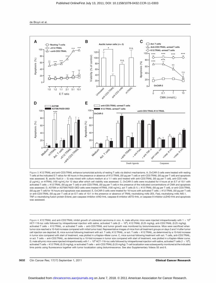
Anti-CD3:TRAIL comprises a T-cell–stimulating CD3antibody fragment. By virtue of this antibody fragment,anti-CD3:TRAIL might also be able to activate intrinsiccytotoxic effector mechanisms of T cells. In line with thishypothesis, resting T cells that lacked activity in cocultureexperiments with OvCAR-3 gained potent cytotoxic activityin the presence of anti-CD3:TRAIL (Fig. 3A, 100% apop-tosis at E:T ratio of 10:1). In contrast, resting T cellsremained ineffective in the presence of K12:TRAIL, withno cytotoxicity even at the highest E:T ratio tested (Fig. 3A).Furthermore, anti-CD3:TRAIL consistently reduced long-term outgrowth by more than 50% in ascitic fluid fromovarian cancer patients that contains both primary malig-nant cells and autologous T cells (Fig. 3B and Supplemen-tary Fig. S3A). Neither an anti-CD3 stimulatorymAb nor anuntargeted soluble rhTRAIL preparation alone inhibitedcancer cell outgrowth (Fig. 3B).
In line with these activating properties, anti-CD3:TRAILinduced a rapid calcium flux in T cells coculturedwith OvCAR-3 cells (Supplementary Fig. S3B) which wasfollowed in time by an increase in the number of T cellspositive for the activation marker CD69 (SupplementaryFig. S3C) and the release of proinflammatory T-cell cyto-kines (Supplementary Fig. S3D). Interestingly, preventingcellular contact between T cells and tumor cells abolishedT-cell activation induced by anti-CD3:TRAIL (Supplemen-tary Fig. S3E), as did the addition of TRAIL-neutralizing andTRAILR-blocking antibodies (Supplementary Fig. S3C). Incontrast, caspase inhibition did not block activation ofanti-CD3:TRAIL–armed T cells (Supplementary Fig. S3C)but did block induction of apoptosis by rhTRAIL (Supple-mentary Fig. S5A). Further analysis using T cells mechani-cally manipulated using a micropipette (SupplementaryFig. S4A for schematic representation) indicated that anti-CD3:TRAIL–mediated T-cell activation was likely becauseof shear stress upon simultaneous binding of anti-CD3:TRAIL to CD3 on T cells and TRAILR2 on tumor cells(Supplementary Fig. S4A–C).
K12:TRAIL and anti-CD3:TRAIL enhance tumoricidalactivity of resting T cells via distinct mechanisms
On the basis of these activating properties, T-cell cyto-toxicity induced by anti-CD3:TRAIL may, at least partly, beattributable to stimulation of intrinsic T-cell cytotoxicity.Indeed, the potentiating effect on T-cell activity by
Table 1. MFIs for TRAILR expression levelsdetermined on a panel of cancer cell lines
Figure 3. K12:TRAIL and anti-CD3:TRAIL enhance tumoricidal activity of resting T cells via distinct mechanisms. A, OvCAR-3 cells were treated with restingT cells at the indicated E:T ratios for 48 hours in the presence or absence of K12:TRAIL (50 pg per T cell) or anti-CD3:TRAIL (50 pg per T cell) and apoptosiswas assessed. B, ascitic fluid (n ¼ 5) was mixed with culture medium at a 1:1 ratio and treated with anti-CD3:TRAIL (50 pg per T cell), anti-CD3 mAb(2 mg/mL), or rhTRAIL (100 ng/mL) for 12 days after which cell viability was assessed. C, OvCAR-3 cells were incubated for 4 hours at an E:T of 10:1 withactivated T cells � K12:TRAIL (50 pg per T cell) or anti-CD3:TRAIL (50 pg per T cell) in the presence of the indicated concentrations of CMA and apoptosiswas assessed. D, A375M or A375M.FADD-DED cells were treated rhTRAIL (100 ng/mL), act T cells (5:1) � K12:TRAIL (50 pg per T cell), or anti-CD3:TRAIL(50 pg per T cell) for 16 hours and apoptosis was assessed. E, OvCAR-3 cells were treated for 16 hours with activated T cells � K12:TRAIL (50 pg per T cell)or anti-CD3:TRAIL (50 pg per T cell) at an E:T ratio of 10:1 in the presence or absence of TRAIL-neutralizing mAb 2E5, FasL-neutralizing mAb Alf2.1,TNF-a–neutralizing fusion protein Enbrel, pan-caspase inhibitor zVAD-fmk, caspase-8 inhibitor zIETD-fmk, or caspase-9 inhibitor zLEHD-fmk and apoptosiswas assessed.
Figure 4. K12:TRAIL and anti-CD3:TRAIL inhibit growth of colorectal carcinoma in vivo. A, male athymic mice were injected intraperitoneally with 1 � 106
HCT-116–luc cells followed by intraperitoneal injection with saline, activated T cells (5 � 106), K12:TRAIL (0.25 mg/kg), anti-CD3:TRAIL (0.25 mg/kg),activated T cells þ K12:TRAIL, or activated T cells þ anti-CD3:TRAIL and tumor growth was monitored by bioluminescence. Mice were sacrificed whentumor size reached a 10-fold increase compared with initial tumor load. Representative images of mice from all treatment groups on days 3 and 14 after tumorcell injection are depicted. B, mice survival following treatment with act. T cells, K12:TRAIL, or act. T cells þ K12:TRAIL, as determined by a 10-fold increasein tumor size compared with start of treatment, was plotted in a Kaplan–Meier curve. C, mice survival following treatment with act. T cells, anti-CD3:TRAIL,or act. T cells þ anti-CD3:TRAIL, as determined by a 10-fold increase in tumor size compared with start of treatment, was plotted in a Kaplan–Meier curve.D, male athymic mice were injected intraperitoneally with 1� 106 HCT-116–luc cells followed by intraperitoneal injection with saline, activated T cells (1� 106),activated T cellsþK12:TRAIL (0.25mg/kg), or activated T cellsþ anti-CD3:TRAIL (0.25mg/kg). T-cell localization was subsequently monitored at the indicatedtime points using fluorescence together with tumor localization using bioluminescence. See also Supplementary Videos S5 and 6.
de Bruyn et al.
Clin Cancer Res; 17(17) September 1, 2011 Clinical Cancer Research5632
anti-CD3:TRAIL, but not by K12:TRAIL, was dose depen-dently blocked by the granzyme/perforin release inhibitorCMA (Fig. 3C and Supplementary Fig. S5B). In addition,A375M cells overexpressing a mutant form of the TRAILRadaptor protein FADD (FADD-DED) that inhibits TRAILR-mediated caspase activation, remained sensitive to treat-ment with T cells þ anti-CD3:TRAIL but not to T cells þK12:TRAIL (Fig. 3D). Inhibition of other typical cytotoxicT-cell effectors, such as the tumoricidal proteins FasL andTNF-a, failed to inhibit apoptotic activity of anti-CD3:TRAIL/K12:TRAIL toward OvCAR-3 cells (Fig. 3E). Apop-totic activity of anti-CD3:TRAIL/K12:TRAIL–armed T cellswas abrogated by a pan-caspase inhibitor (Fig. 3E) and byinhibition of caspase-8 or caspase-9 (Fig. 3E). Of note, thepotentiating effect of anti-CD3:TRAIL on tumoricidal T-cellactivity closely resembled that of T cells redirected with thebispecific antibody Bis-1 (anti-CD3:anti-EpCAM), whichwas also abrogated by CMA (Supplementary Fig. S5C).Therefore, the mode of action of anti-CD3:TRAIL appearsto be largely dependent on granzyme/perforin-mediatedlysis, whereas K12:TRAIL functions via activation of initia-tor caspase-8/-9 and effector caspases. Furthermore,the inhibitory effect of caspase-9 blockade suggests thatmitochondrial amplification of the caspase-8 apoptoticsignal is required for effective tumoricidal activity.
K12:TRAIL and anti-CD3:TRAIL inhibit growth ofcolorectal carcinoma in vivo
The potent in vitro effects of K12:TRAIL and anti-CD3:TRAIL prompted us to evaluate activity against tumor cellsin vivo. To this end, we xenografted colorectal carcinomacell line HCT-116–luc intraperitoneally in mice as a modelfor advanced metastatic colon carcinoma. HCT-116–luccells rapidly established tumor nodules throughout theabdominal cavity, resulting in a median survival of only7 days (Fig. 4B and C). Importantly, growth of HCT-116–luc in vivowas significantly suppressed by cotreatment withK12:TRAIL and activated T cells, compared with treatmentwith either K12:TRAIL or T cells alone. Indeed, mediansurvival was increased 6-fold and 1 mouse survived for theduration of the experiment (70 days) with minimal resi-dual disease (Fig. 4A and B; 42 vs. 7 days). Of note,cotreatment with K12:TRAIL and T cells did not haveobvious off-target toxicity in these mice, with no apparentweight loss (data not shown). In addition, fibroblasts(NHDF-juv), normal kidney cells (HK-2), and endothelialcells (HUVEC) were resistant to cotreatment with K12:TRAIL and T cells (Supplementary Fig. S5D). Similarly,cotreatment of mice with T cells and anti-CD3:TRAILresulted in a striking increase in median survival with 4of 5 mice remaining tumor free for the duration of theexperiment (Fig. 4A and C). Treatment with anti-CD3:TRAIL alone had no effect on animal survival (Fig. 4C).Of note, while no deleterious effects on animal well beingwere observed during the course of the experiment,the in vitro treatment of NHDF-juv, HK-2, and HUVECswith T cells and anti-CD3:TRAIL did trigger an increase inthe percentage of apoptosis (Supplementary Fig. S5D). To
determine whether T cells localized to the intraperitoneallygrowing tumor cells, fluorescently labeled T cells werefollowed up to 5 days in an intraperitoneal tumor modelwith HCT-116–luc (Fig. 4D). Treatment with activated Tcells or activated T cells þ anti-CD3:TRAIL resulted incolocalization of bioluminescent (tumor) and fluorescent(T cell) signal in tumor nodes in the abdominal cavity after2 days but not after 5 days (Fig. 4D). Upon treatment withactivated T cells þ K12:TRAIL (Fig. 4D), the T cells werefound to colocalize with tumor signal until tumor cellswere eliminated (Fig. 4D). Taken together, K12:TRAIL andanti-CD3:TRAIL potently enhance antitumor activity of Tcells and inhibit growth of colorectal xenografts in vivo.
K12:TRAIL and anti-CD3:TRAIL are compatible withexperimental strategies to ameliorate GVHD andoptimize GVT
Earlier studies have shown that TRAIL expression on Tcells is required for optimal GVT activity during AHCT butdoes not exacerbate GVHD. Therefore, it was examinedwhether K12:TRAIL could be used in conjunction withtherapies currently being evaluated for optimizing AHCT.In particular, the so-called histone deacytelase inhibitors(HDACi) inhibit GVHD in preclinical settings (16, 17) butare also well known to synergize with the proapoptoticactivity of TRAIL (18, 19). Of note, HDACi do not potenti-ate the GVT effect. In line with this, pretreatment withHDACi VPA did not enhance the tumoricidal activity of Tcells alone (Fig. 5A). In contrast, pretreatment of OvCAR-3cells with VPA strongly optimized the potentiating effect ofK12:TRAIL and anti-CD3:TRAIL on tumoricidal T-cell activ-ity, with approximately 30% and 50% cell death in OvCAR-3 at E:T ratio of 1:50, respectively (Fig. 5A–C). Interestingly,VPA enhanced the antitumor effect of K12:TRAIL, but notanti-CD3:TRAIL, toward 2 patient-derived tumor samples(Fig. 5D).
Various other strategies have been developed to selec-tively enhance GVT or ameliorate GVHD without affectingGVT. These strategies include the selective depletion of apopulation of alloreactive CD69high T cells before infusion(20) or selectively transferring onlyCD4þ, CD8þ, or CD25þ
cells (21). K12:TRAIL and anti-CD3:TRAIL potentiated thetumoricidal activity of isolated CD69low, CD69medium, orCD69high T-cell populations to a similar degree (Fig. 5F).Similarly, K12:TRAIL and anti-CD3:TRAIL potentiated thetumoricidal activity of sorted CD4þ and CD8þ T cells to alevel similar to that observed in nonsorted T cells (Fig. 5G).Therefore, K12:TRAIL and anti-CD3:TRAIL appear suitablefor integration in existing strategies for optimizing AHCT.
Discussion
Here, we report on the preclinical evaluation of a newstrategy designed to deliver high levels of the proapoptoticeffector molecule TRAIL to the surface of T cells and therebysafely augment antitumor T-cell activity. Arming T cellswith anti-CD3:TRAIL or K12:TRAIL resulted in a more than500-fold increase in tumoricidal activity toward both
de Bruyn et al.
Clin Cancer Res; 17(17) September 1, 2011 Clinical Cancer Research5634
Figure 5. K12:TRAIL and anti-CD3:TRAIL are compatible with experimental strategies to ameliorate GVHD and optimize GVT. A, OvCAR-3 cells werepretreated with VPA (1 nmol/L) for 1 day and subsequently treated for 16 hours with activated T cells at the indicated E:T ratios. B, OvCAR-3 cells pretreatedwith VPA for 1 day were treated for 16 hours with K12:TRAIL-armed T cells (50 pg per T cell) at the indicated E:T ratios. C, OvCAR-3 cells pretreated withVPA for 1 day were treated for 16 hours with anti-CD3:TRAIL–armed T cells (50 pg per T cell) at the indicated E:T ratios. D, primary cancer cells (n ¼ 2)were pretreated with VPA (1 nmol/L) for 1 day and subsequently treated for another 16 hours with activated T cells or K12:TRAIL/anti-CD3:TRAIL–armedT cells (50 pg per T cell) at an E:T ratio of 5:1. E, activated T cells were double stained for CD3 and CD69, whereupon CD69low, CD69medium, and CD69high
populations were sorted from the CD3-positive cells. F, OvCAR-3 cells were treated with CD69low, CD69medium, and CD69high and corresponding K12:TRAIL/anti-CD3:TRAIL–armed T-cell populations (50 pg per T cell) for 16 hours at an E:T ratio of 1:1. G, activated T cells were separated into CD4 and CD8populations, whereupon OvCAR-3 cells were treated with the different T-cell populations and K12:TRAIL/anti-CD3:TRAIL–loaded populations (50 pgper T cell) for 16 hours at an E:T ratio of 5:1.
Enhancing T-cell Tumoricidal Activity with TRAIL
www.aacrjournals.org Clin Cancer Res; 17(17) September 1, 2011 5635
cancer cell lines and primary patient-derived malignantcells. In addition, cotreatment with anti-CD3:TRAIL orK12:TRAIL and T cells significantly prolonged survival timeof xenografted mice.
K12:TRAIL enhanced the tumoricidal activity of T cells byactivation of proapoptotic TRAIL receptor signaling intumor cells. Surprisingly, the potentiating activity ofanti-CD3:TRAIL was found to be predominantly becauseof granzyme/perforin signaling, analogous to the activity ofT-cell–redirecting bispecific antibodies. In line with thismechanism of redirected lysis, anti-CD3:TRAIL triggeredCD3-mediated calcium flux because of mechanical stressacting on the anti-CD3:TRAIL–CD3 complex. These find-ings corroborate with a recent report that suggests that CD3in the T-cell receptor complex functions as a mechanor-eceptor translating shear stresses into signaling (15, 22). Inthis way, the function of anti-CD3:TRAIL appears analo-gous to artificial antigen-presenting complexes or nativemajor histocompatibility complex molecules. In view ofthese activating properties, anti-CD3:TRAIL may be parti-cularly suited for ex vivo tumor cell purging of bone mar-row. In addition, anti-CD3:TRAIL may be used in patientsthat suffer from malignant disease that is restricted to ananatomically confined region. Of interest in this respect areperitonitis carcinomatosis patients that undergo heatedintraperitoneal chemotherapy (HIPEC), an operation inwhich anti-CD3:TRAIL may easily be incorporated.
Applicability of K12:TRAIL is likely not limited to aspecific kind of T-cell immunotherapy but may be appliedto ex vivo expanded tumor infiltrating lymphocytes, engi-neered T cells, T-cell bodies, as well as in GVT activityduring AHCT. Indeed, perhaps the most obvious applica-tion of T-cell–specific delivery of TRAIL using K12:TRAILwould be in the context of AHCT. Alloreactive donor T cellsare the primary mediators of GVT activity as well as GVHD.Earlier studies have shown that TRAIL expression on T cellswas required for optimal GVT, whereas TRAIL did notcontribute to GVHD (10). Although not investigated here,the data of the current study suggest that targeted deliveryof TRAIL to donor T cells using K12:TRAIL may be of valueto selectively enhance GVT. Interestingly, novel GVHDinhibitors, such as HDACi (16, 17) or rapamycin (23),are also known to sensitize cancer cells to TRAIL-inducedapoptosis (18, 19). In line with this, our experiments show
further optimization of the potentiating effect of K12:TRAIL by pretreatment of tumor cells with the HDACiVPA. Thus, rationally designed combinatorial strategiesthat include K12:TRAILmaywell yield optimal GVT activitywith no/minimal GVHD.
The here described immunotherapeutic approach tooptimize T-cell immunotherapy may be further exploredusing TRAIL fusion proteins that incorporate other immu-nomodulatory scFvs or ligands that would simultaneouslyprovide stimulatory signals to T cells and add TRAIL signal-ing to the T-cell armamentarium. Likely combinations tothis effect include stimulatory anti-CD28 scFvs (24) orinhibitory scFvs directed against cytotoxic T-lymphocyteantigen 4 (CTLA-4; ref. 25). In this respect, targeted activa-tion of CTLA-4 signaling using a fusion protein CTLA-4:TRAIL has been shown to limit T-cell activity and amelio-rate autoimmunity in preclinical models (26). Reversely,inhibition of CTLA-4 signaling may potentiate antitumorT-cell activity.
In conclusion, cell surface delivery of TRAIL to T cellspotently augments the antitumor activity of T cells in vitroand in vivo and might be widely applicable in current/experimental T-cell–based strategies to optimize anticancerefficacy.
Disclosure of Potential Conflicts of Interest
No potential conflicts of interest were disclosed.
Acknowledgments
The authors thank Joan Vos and Niels Kouprie for their technicalassistance.
Grant Support
This work was supported by the Dutch Cancer Society (grant numbersRUG 2009-4355 to E. Bremer and RUG2007-3784 to W. Helfrich), the Nether-lands Organization for Scientific Research (E. Bremer), the MelanomaResearch Alliance (E. Bremer), and the Alexander von Humboldt Foundation(E. Bremer).
The costs of publication of this article were defrayed in part by the paymentof page charges. This article must therefore be hereby marked advertisement inaccordance with 18 U.S.C. Section 1734 solely to indicate this fact.
Received February 11, 2011; revised June 10, 2011; accepted July 1, 2011;published OnlineFirst July 13, 2011.
References1. Chouaib S, Meslin F, Thiery J, Mami-Chouaib F. Tumor resistance to
specific lysis: a major hurdle for successful immunotherapy of cancer.Clin Immunol 2009;130:34–40.
2. Lee H, Timme T, Thompson T. Resistance to lysis by cytotoxic T cells:a dominant effect in metastatic mouse prostate cancer cells. CancerRes 2000;60:1927–33.
3. Abouzahr-Rifai S, HasmimM, Boukerche H, Hamelin J, Janji B, Jalil A,et al. Resistance of tumor cells to cytolytic T lymphocytes involvesRho-GTPases and focal adhesion kinase activation. J Biol Chem2008;283:31665–72.
4. Otten HG, van Ginkel WG, Hagenbeek A, Petersen EJ. Prevalence andclinical significance of resistance to perforin- and FAS-mediated celldeath in leukemia. Leukemia 2004;18:1401–5.
5. Hamai A, Meslin F, Benlalam H, Jalil A, Mehrpour M, Faure F, et al.ICAM-1 has a critical role in the regulation of metastatic melanomatumor susceptibility to CTL lysis by interfering with PI3K/AKT path-way. Cancer Res 2008;68:9854–64.
6. Suda T, Hashimoto H, Tanaka M, Ochi T, Nagata S. Membrane Fasligand kills human peripheral blood T lymphocytes, and soluble Fasligand blocks the killing. J Exp Med 1997;186:2045–50.
7. Hallermalm K, De Geer A, Kiessling R, Levitsky V, Levitskaya J.Autocrine secretion of Fas ligand shields tumor cells from Fas-mediated killing by cytotoxic lymphocytes. Cancer Res 2004;64:6775–82.
8. Takeda K, Hayakawa Y, Smyth MJ, Kayagaki N, Yamaguchi N,Kakuta S, et al. Involvement of tumor necrosis factor-related
de Bruyn et al.
Clin Cancer Res; 17(17) September 1, 2011 Clinical Cancer Research5636
apoptosis-inducing ligand in surveillance of tumor metastasis byliver natural killer cells. Nat Med 2001;7:94–100.
9. Cretney E, Takeda K, Yagita H, Glaccum M, Peschon JJ, SmythMJ. Increased susceptibility to tumor initiation and metastasis inTNF-related apoptosis-inducing ligand-deficient mice. J Immunol2002;168:1356–61.
10. Schmaltz C, Alpdogan O, Kappel BJ, Muriglan SJ, Rotolo JA, OngchinJ, et al. T cells require TRAIL for optimal graft-versus-tumor activity.Nat Med 2002;8:1433–7.
11. Kayagaki N, Yamaguchi N, NakayamaM, Eto H, Okumura K, Yagita H.Type I interferons (IFNs) regulate tumor necrosis factor-related apop-tosis-inducing ligand (TRAIL) expression on human T cells: a novelmechanism for the antitumor effects of type I IFNs. J Exp Med 1999;189:1451–60.
12. Tian JQ, Wang ZP, Rodriguez R, Fu JS, Lu JZ, Ma BL. In vitroenhanced cytotoxicity of tumor-infiltrating lymphocytes transfectedwith tumor necrosis factor-related apoptosis-inducing ligand and/orinterleukin-2 gene in human renal cell carcinoma. Urology 2006;67:1093–8.
13. Bremer E, de Bruyn M, Wajant H, Helfrich W. Targeted cancerimmunotherapy using ligands of the tumor necrosis factor super-family. Curr Drug Targets 2009;10:94–103.
14. Bremer E, Kuijlen J, Samplonius D, Walczak H, de Leij L, Helfrich W.Target cell-restricted and -enhanced apoptosis induction by a scFv:sTRAIL fusion protein with specificity for the pancarcinoma-asso-ciated antigen EGP21. Int J Cancer 2004;109:281–90.
15. Li YC, Chen BM, Wu PC, Cheng TL, Kao LS, Tao MH, et al. CuttingEdge: mechanical forces acting on T cells immobilized via the TCRcomplex can trigger TCR signaling. J Immunol 2010;184:5959–63.
16. Leng C, Gries M, Ziegler J, Lokshin A, Mascagni P, Lentzsch S, et al.Reduction of graft-versus-host disease by histone deacetylase inhi-bitor suberonylanilide hydroxamic acid is associated with modulation
of inflammatory cytokine milieu and involves inhibition of STAT1. ExpHematol 2006;34:776–87.
17. Reddy P, Sun Y, Toubai T, Duran-Struuck R, Clouthier SG,Weisiger E,et al. Histone deacetylase inhibition modulates indoleamine 2,3-diox-ygenase-dependent DC functions and regulates experimental graft-versus-host disease in mice. J Clin Invest 2008;118:2562–73.
18. Fulda S. Modulation of TRAIL-induced apoptosis by HDAC inhibitors.Curr Cancer Drug Targets 2008;8:132–40.
19. Panner A, Parsa AT, Pieper RO. Use of APO2L/TRAIL with mTORinhibitors in the treatment of glioblastoma multiforme. Expert RevAnticancer Ther 2006;6:1313–22.
20. Hartwig UF, Nonn M, Khan S, Meyer RG, Huber C, Herr W. Depletionof alloreactive T cells via CD69: implications on antiviral, antileukemicand immunoregulatory T lymphocytes. Bone Marrow Transplant2005;37:297–305.
22. Kim ST, Takeuchi K, Sun ZY, ToumaM, Castro CE, Fahmy A, et al. Thealphabeta T cell receptor is an anisotropic mechanosensor. J BiolChem 2009;284:31028–37.
2011;17:5626-5637. Published OnlineFirst July 13, 2011.Clin Cancer Res Marco de Bruyn, Yunwei Wei, Valerie R. Wiersma, et al. Activity of T CellsCell Surface Delivery of TRAIL Strongly Augments the Tumoricidal
Updated version
10.1158/1078-0432.CCR-11-0303doi:
Access the most recent version of this article at: