IntroductionThe outer nuclear membrane (ONM) of the nuclear envelope (NE)is continuous with the rough endoplasmic reticulum and ischaracterized by the presence of members of the nesprin proteinfamily. To date, four members have been identified in mammals,nesprin-1 (Padmakumar et al., 2004; Zhang et al., 2001), nesprin-2 (Zhang et al., 2001; Zhen et al., 2002), nesprin-3 (Wilhelmsen etal., 2005) and the recently identified nesprin-4 (Roux et al., 2009).All these proteins are characterized by multiple spectrin repeats(SRs) and a C-terminal Klarsicht/ANC-1/Syne homology (KASH)domain (Wilhelmsen et al., 2006; Zhang et al., 2002). The KASHdomain comprises a transmembrane region followed by a shortluminal domain that extends into the periplasmic space, where itinteracts with the inner nuclear membrane (INM) proteins SUN1and SUN2 (Crisp et al., 2006; Haque et al., 2006; Padmakumar etal., 2005). The SUN–nesprin interaction is essential for retainingnesprins at the ONM and for maintaining a proper NE architecture(Crisp et al., 2006). Inside the nucleus, SUN proteins interactthrough their N-terminal domain with the nuclear lamina (Crisp etal., 2006; Haque et al., 2006).
The nesprin family members differ from each other in thenumber of SRs and in their N-terminal domain. The giant isoformsof nesprin-1 and nesprin-2 have an N-terminal actin-binding domain(ABD) through which they connect the NE to the actin cytoskeleton(Padmakumar et al., 2004; Zhen et al., 2002). Nesprin-3 andnesprin-4 are much smaller in size and lack an N-terminal ABD.However, these proteins can link the cytoskeleton to the NE in anindirect manner. Nesprin-3 binds the cytoskeletal crosslinker protein
plectin, by which it can anchor the intermediate filaments (IFs) tothe ONM (Wilhelmsen et al., 2005). Furthermore, nesprin-4 bindsto the microtubule motor protein kinesin-1 (Roux et al., 2009),thereby establishing a link between the nucleus and microtubules.The nesprin–SUN complexes thus establish a connection betweenthe cytoskeleton and the nuclear lamina; a complex referred to asthe LINC complex (linker of nucleoskeleton and cytoskeleton)(Crisp et al., 2006; Stewart-Hutchinson et al., 2008).
The connection of the NE with the cytoskeleton suggests thatnesprins have important functions in nuclear positioning andanchorage. Indeed, studies in Caenorhabditis elegans andDrosophila melanogaster showed nuclear anchorage defects uponloss of the nesprin-1 and nesprin-2 homologs ANC-1 and MSP-300, respectively (Starr and Han, 2002; Wilhelmsen et al., 2006;Yu et al., 2006). Furthermore, various nesprin-1 mutant mice arecharacterized by an abnormal clustering of skeletal muscle nucleiand the absence of synaptic nuclei from the neuromuscular junction(Puckelwartz et al., 2009; Zhang et al., 2010; Zhang, X. et al.,2007). Although nesprin-2 mutant mice have no overt abnormalphenotype (Lüke et al., 2008; Zhang, X. et al., 2007), mice inwhich the KASH domains of both nesprin-1 and nesprin-2 aredeleted die shortly after birth due to respiratory failure (Zhang, X.et al., 2007), indicating a redundancy of their function.
Nesprin-3 is the only family member currently known to anchorthe IF system to the NE. We have previously described the presenceof two nesprin-3 isoforms in mice, nesprin-3a and nesprin-3b, thelatter not binding to plectin. These isoforms are produced as aresult of alternative splicing (Wilhelmsen et al., 2005). Nesprin-3a
SummaryThe outer nuclear membrane protein nesprin-3 binds the cytoskeletal linker protein plectin, which are proposed to anchor theintermediate filaments to the nuclear envelope. To investigate the function of nesprin-3 in vivo, we used the zebrafish as a vertebratemodel system. Zebrafish nesprin-3 is expressed at the nuclear envelope of epidermal and skeletal muscle cells during development.Unexpectedly, loss of nesprin-3 did not affect embryonic development, viability or fertility. However, nesprin-3-deficient zebrafishembryos showed a reduced concentration of intermediate filaments around the nucleus. Additional analysis revealed the presence oftwo nesprin-3 isoforms in zebrafish, nesprin-3a and nesprin-3b. Nesprin-3b is only expressed during early development and lacksseven amino acids in its first spectrin repeat that are crucial for plectin binding and recruitment to the nuclear envelope. These sevenamino acids are highly conserved and we showed that residues R43 and L44 within this motif are required for plectin binding.Furthermore, several residues in the actin-binding domain of plectin that are crucial for binding to the integrin b4 subunit are alsoimportant for the binding to nesprin-3a, indicating partial overlapping binding sequences for nesprin-3a and integrin b4. All this showsthat nesprin-3 is dispensable for normal development in zebrafish, but important for mediating the association of the intermediatefilament system with the nucleus in vivo.
Nesprin-3 augments peripheral nuclear localization ofintermediate filaments in zebrafishRuben Postel1,*, Mirjam Ketema1,*, Ingrid Kuikman1, José M. de Pereda2 and Arnoud Sonnenberg1,‡
1Division of Cell Biology, The Netherlands Cancer Institute, Plesmanlaan 121, 1066 CX Amsterdam, The Netherlands2Instituto de Biología Molecular y Celular del Cáncer, Consejo Superior de Investigaciones Científicas, Universidad de Salamanca, CampusUnamuno, 37007 Salamanca, Spain*These authors contributed equally to this work‡Author for correspondence ([email protected])
Jour
nal o
f Cel
l Sci
ence
contains eight SRs and a C-terminal KASH domain, and thenesprin-3b isoform differs only in that it lacks the first SR. Becausethe first SR is essential for plectin binding, only nesprin-3a canestablish a connection with the IF system (Wilhelmsen et al.,2005). However, the function of nesprin-3a in vivo is currentlyunknown.
Plectin is a cytoskeletal crosslinker protein of the plakin familythat is highly versatile in its binding properties (Wiche, 1998).Similarly to other family members, plectin interconnects differentcomponents of the cytoskeleton and attaches them to junctionalcomplexes at the plasma membrane (McLean et al., 1996;Sonnenberg and Liem, 2007). For example, plectin crosslinks theactin cytoskeleton with the IF system via its N-terminal ABD andC-terminal plakin repeats, respectively (Andrä et al., 1998; Garcia-Alvarez et al., 2003; Nikolic et al., 1996). In addition to itsinteraction with F-actin, the plectin ABD also mediates bindingto the integrin b4 subunit and nesprin-3a, thereby connecting theIF system to both the plasma membrane and the NE (Geerts etal., 1999; Litjens et al., 2003; Wilhelmsen et al., 2005). Moreover,we have previously demonstrated that both nesprin-3a andintegrin b4 compete with F-actin for plectin binding (Geerts etal., 1999; Ketema et al., 2007; Wilhelmsen et al., 2005).Deficiency of plectin in both patients and mice leads to skinfragility and muscular dystrophy, illustrating its essential role inmaintaining cell integrity (Andrä et al., 1997; McLean et al.,1996).
In the present study, we have investigated the function of nesprin-3 in vivo. Because nesprin-1, nesprin-2 and nesprin-3 are conservedin zebrafish (Simpson and Roberts, 2008; Tsujikawa et al., 2007),we have used this organism as a vertebrate model system to studythe distribution of nesprin-3 during embryonic development. Inaddition, we have generated and analyzed nesprin-3-deficientzebrafish using TILLING (targeted induced local lesions ingenomes), an approach for the identification of null mutationsfrom a randomly N-ethyl-N-nitrosourea (ENU)-mutagenizedzebrafish library (Wienholds and Plasterk, 2004).
ResultsNesprin-3 is expressed at the NE of epidermal and skeletalmuscle cells during zebrafish embryonic developmentNesprins are expressed in various tissues such as brain, skin,skeletal muscle and heart (Lüke et al., 2008; Wilhelmsen et al.,2005; Zhang et al., 2001). However, the expression profile andthe function of nesprin-3 are still largely unknown. Becausenesprin-3 is conserved in zebrafish, we decided to use zebrafishto study the function of nesprin-3 (Simpson and Roberts, 2008).To investigate the mRNA expression of nesprin-3 during zebrafishembryonic development, we performed whole-mount in situhybridization (ISH) with a digoxigenin-labeled antisense nesprin-3 probe on wild-type embryos of various developmental stages.Maternal nesprin-3 mRNA expression was observed during initialcellular division, before the start of zygotic transcription (Fig.1A). At 24 hours post-fertilization (hpf), nesprin-3 mRNAappeared to be expressed ubiquitously (Fig. 1B). Strongestnesprin-3 expression was detected in skeletal muscle and theepidermis (Fig. 1C,D).
To study the distribution of nesprin-3 protein in zebrafish, weproduced a rabbit polyclonal antibody directed against SR7 ofzebrafish nesprin-3. Whole-mount immunohistochemistry on 3-day-old embryos revealed localization of nesprin-3 protein at theNE in epidermal (Fig. 1E) and skeletal muscle cells (Fig. 1Einset). In addition to the NE staining, we also observed stainingat the myotendinous junctions and in the cytoplasm of skeletalmuscle cells. However, because a similar staining pattern wasfound with the pre-immune serum (data not shown), we regardedthis reaction as nonspecific. This nonspecific background staininghindered the study of nesprin-3 expression in skeletal musclecells at most stages of development. Unlike in skeletal musclecells, nesprin-3 expression at the NE of epidermal cells wasobserved during all stages of development [1–6 days postfertilization (dpf)]. The localization of nesprin-3 at the NE inepidermal cells was confirmed by double labeling with the basalepidermal marker p63 (Fig. 1F-H) (Lee and Kimelman, 2002).
756 Journal of Cell Science 124 (5)
Fig. 1. Nesprin-3 is expressed at the NE of epidermal and skeletal muscle cells during zebrafish embryonic development. (A,B)Whole-mount ISH fornesprin-3 mRNA expression at the 16-cell stage (A) and 1 dpf (B). (C,D)Semi-thin sections of 1-day-old embryos after ISH at the level of the trunk (C) and at theend of the tail (D). Asterisks indicate the nesprin-3 mRNA expression in the skeletal muscle, and black arrowheads indicate the nesprin-3 mRNA expression in theepidermis. Sections were counterstained with neutral red. (E)Whole-mount immunostaining with anti-nesprin-3 antibody on 3-day-old wild-type embryos in thetail-fin and skeletal muscle (inset). The arrow marks nonspecific (pre-immune) background staining at the myotendinous junctions. Scale bars: 50mm. (F–H) Co-staining with anti-p63 antibody (green, F) and anti-nesprin-3 antibody (red, G) in 3-day-old wild-type embryos shows nesprin-3 localization at the NE of basalepidermal cells and peridermal cells (H). White arrows indicate nesprin-3 localization at the NE of peridermal cells. The inset shows an xz-projection of two basalepidermal cells (be) with p63 staining in the nucleus (green) and nesprin-3 staining at the NE (red), whereas in the peridermal cell (pe) only nesprin-3 staining isobserved at the NE (red). Scale bar: 20mm.
Jour
nal o
f Cel
l Sci
ence
However, a layer of nesprin-3-positive cells overlying the cellsdouble-labeled with p63 and nesprin-3 was also observed (Fig.1H inset). Because the zebrafish embryonic epidermis contains abasal and a peridermal cell layer at this stage of development (LeGuellec et al., 2004), we conclude that nesprin-3 is expressed inboth epidermal cell layers. Furthermore, in a heterogeneousepidermal cell population derived from 21-day-old larvae, weobserved nesprin-3 expression at the NE (data not shown). Thissuggests that nesprin-3 also resides at the NE of epidermal cellsduring later stages of development.
Complete loss of nesprin-3 does not affect embryonicdevelopment, viability or fertility in zebrafishTo study the function of nesprin-3 in vivo, we screened for mutantalleles in zebrafish. From a library of ENU-mutagenized zebrafishwe isolated a nesprin-3 mutant allele, hu6448. The hu6448 allele
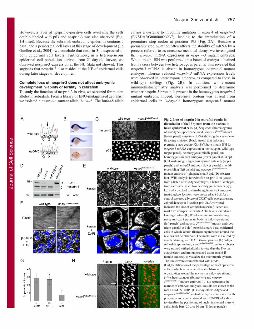
carries a cysteine to threonine mutation in exon 4 of nesprin-3(ENSDARG00000023237), leading to the introduction of apremature stop codon at position 195 (Fig. 2A). Because apremature stop mutation often affects the stability of mRNA by aprocess referred to as nonsense-mediated decay, we investigatedthe nesprin-3 mRNA expression in nesprin-3 mutant embryos.Whole-mount ISH was performed on a batch of embryos obtainedfrom a cross between two heterozygous parents. This revealed thatnesprin-3 mRNA is absent in homozygous nesprin-3 mutantembryos, whereas reduced nesprin-3 mRNA expression levelswere observed in heterozygous embryos as compared to those inwild-type siblings (Fig. 2B). In addition, whole-mountimmunohistochemistry analysis was performed to determinewhether nesprin-3 protein is present in the homozygous nesprin-3mutant embryos. Indeed, nesprin-3 protein was absent fromepidermal cells in 3-day-old homozygous nesprin-3 mutant
757Nesprin-3 in zebrafish
Fig. 2. Loss of nesprin-3 in zebrafish results indissociation of the IF system from the nucleus inbasal epidermal cells. (A)Sequence chromatogramsof wild-type (upper panel) and nesprin-3hu6448 mutant(lower panel) nesprin-3 cDNA showing the cysteine tothreonine mutation (black arrow) that induces apremature stop codon (X). (B)Whole-mount ISH fornesprin-3 mRNA expression in homozygous wild-type(upper panel), heterozygous (middle panel) andhomozygous mutant embryos (lower panel) at 24 hpf.(C)Co-staining using anti-nesprin-3 antibody (upperpanels) and anti-p63 antibody (lower panels) in wild-type sibling (left panels) and nesprin-3hu6448/hu6448
mutant embryos (right panels) at 3 dpf. (D)Westernblot (WB) analysis for zebrafish nesprin-3 on lysatesfrom a batch of wild-type embryos, a batch of embryosfrom a cross between two heterozygous carriers (zygko) and a batch of maternal-zygotic mutant embryos(mat-zyg ko). Lysates were prepared at 8 hpf. As acontrol we used a lysate of COS7 cells overexpressingzebrafish nesprin-3a (zfnesprin-3). Arrowheadindicates the size of zebrafish nesprin-3. Asterisksmark two nonspecific bands. Actin levels served as aloading control. (E)Whole-mount immunostainingusing anti-pan keratin antibody in wild-type sibling(left panels) and nesprin-3hu6448/hu6448 mutant embryos(right panels) at 5 dpf. Asterisks mark basal epidermalcells in which keratin filament organization around thenucleus can be observed. The nuclei were visualized bycounterstaining with DAPI (lower panels). (F)5-day-old wild-type and nesprin-3hu6448hu6448 mutant embryoswere stained with phalloidin to visualize the F-actincytoskeleton and immunostained using an anti-b-tubulin antibody to visualize the microtubule system.The nuclei were counterstained with DAPI.(G)Quantification of the percentage of basal epidermalcells in which we observed keratin filamentorganization around the nucleus in wild-type sibling(+/+), heterozygous sibling (+/–) and nesprin-3hu6448/hu6448 mutant embryos (–/–). n represents thenumber of embryos analyzed. Results are shown as themean ± s.d. *P<0.05. (H)3-day-old wild-type andnesprin-3hu6448/hu6448 mutant embryos were stained withphalloidin and counterstained with TO-PRO-3 iodideto visualize the positioning of nuclei in skeletal musclecells. Scale bars: 20mm; 10mm (E, lower panels).
Jour
nal o
f Cel
l Sci
ence
embryos (Fig. 2C). Together, this shows that the hu6448 allele isa complete nesprin-3 null mutant.
The homozygous nesprin-3 mutant embryos showed no obviousdefects during embryonic development and were fertile. Becausenesprin-3 mRNA is expressed maternally (Fig. 1A), it is possiblethat the amount of nesprin-3 protein produced from this mRNA issufficient to carry the embryo through early stages of development.To investigate the contribution of maternal nesprin-3 mRNA, wegenerated maternal-zygotic nesprin-3 mutant embryos from a crossbetween two homozygous nesprin-3 mutant parents. Western blotanalysis demonstrated that the nesprin-3 protein was absent in thematernal-zygotic mutant embryos, whereas a 50% reduction innesprin-3 protein level, compared to homozygous wild-type, wasobserved for a batch derived from two heterozygous parents (Fig.2D). The maternal-zygotic nesprin-3 mutant embryos alsodeveloped normally and were fertile, thereby ruling out thepossibility that maternally derived nesprin-3 compensates for alack of nespin-3 in the developing knockout embryos.
Association of the IF system with the NE is reduced innesprin-3-deficient zebrafish embryosPrevious in vitro studies have suggested that nesprin-3a, by bindingto plectin, connects the IF system with the NE (Wilhelmsen et al.,2005). We predicted that, in epidermal cells of nesprin-3 mutantembryos, the connection between the nucleus and IF system wasdisrupted, which could lead to potential disturbances in theorganization of the IF system in the proximity of the nucleus.Because nesprin-3 is present at the NE of epidermal cells duringzebrafish development, we studied the structure of keratin filamentsin basal epidermal cells of 5-day-old wild-type sibling and nesprin-3 mutant embryos. Using whole-mount immunohistochemistrywith an anti-pan keratin antibody, we observed cytoplasmic keratinfilaments in all epidermal cells of wild-type embryos. Furthermore,in many cells a dense filamentous keratin network around thenucleus could be distinguished (Fig. 2E). In nesprin-3 mutantembryos, this organization of keratin filaments around the nucleuswas reduced, whereas the cytoplasmic keratin organization appearedunaffected (Fig. 2E). Organization of other cytoskeletal componentssuch as actin filaments and microtubules was unaffected by theloss of nesprin-3 in epidermal cells (Fig. 2F). We quantified thepercentage of epidermal cells in which a dense filamentous keratinnetwork was observed around the nucleus in homozygous wild-type, heterozygous and homozygous nesprin-3 mutant embryos. Inhomozygous wild-type and heterozygous embryos, keratinfilaments were found around the nucleus in approximately 29%and 27% of the cells, respectively. By contrast, the percentage ofepidermal cells with perinuclear keratin was only 7% in nesprin-3mutant embryos (P<0.05) (Fig. 2G). Hence, nesprin-3 has no effecton the overall organization of the IF system, but is important forthe association of the IF cytoskeleton with the nucleus in epidermalcells in vivo.
It has previously been demonstrated that nesprin-1 is involvedin nuclear positioning and anchorage in skeletal muscle cells(Puckelwartz et al., 2009; Zhang et al., 2010; Zhang, X. et al.,2007). Because nesprin-3 is expressed in skeletal muscle andbecause loss of this protein affects the connection between thenucleus and the IF system in epidermal cells, we wondered whetherloss of nesprin-3 influences the positioning and anchorage of nucleiin multinucleated skeletal muscle cells. To investigate this, westained 3-day-old embryos from a cross between two heterozygousparents for F-actin and the nuclear marker TO-PRO-3 and examined
the position of the nuclei by confocal microscopy. This revealedno significant alterations in the positioning of nuclei in skeletalmuscle cells of nesprin-3 mutant embryos as compared with wild-type siblings (Fig. 2H). Furthermore, we observed no motilitydefects in nesprin-3 mutant embryos or adult zebrafish. In summary,the data presented here indicate that nesprin-3 is dispensable fornormal embryonic development in zebrafish, but is important forthe efficient localization of IFs at the nuclear periphery in epidermalcells.
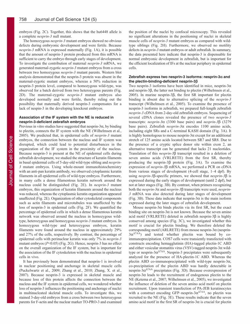
Zebrafish express two nesprin-3 isoforms: nesprin-3a andthe plectin-binding-deficient nesprin-3bTwo nesprin-3 isoforms have been identified in mice, nesprin-3aand nesprin-3b, the latter not binding to plectin (Wilhelmsen et al.,2005). In murine nesprin-3b, the first SR important for plectinbinding is absent due to alternative splicing of the nesprin-3transcript (Wilhelmsen et al., 2005). To examine the presence ofnesprin-3 isoforms in zebrafish, we prepared full-length zebrafishnesprin-3 cDNA from 2-day-old zebrafish embryos. Sequencing ofseveral cDNA clones revealed the presence of two nesprin-3transcripts: nesprin-3a (3300 base pairs) and nesprin-3b (3279base pairs). Zebrafish nesprin-3a encodes 1100 amino acids,including eight SRs and a C-terminal KASH domain (Fig. 3A). Itis highly homologous to mouse nesprin-3a except for an additionalstretch of 125 amino acids located between SR7 and SR8. Due tothe presence of a cryptic splice donor site within exon 2, analternative transcript can be generated that lacks 21 nucleotides.Translation of this nesprin-3b transcript resulted in a deletion ofseven amino acids (VRLRETE) from the first SR, therebyproducing the nesprin-3b protein (Fig. 3A). To examine theexpression of nesprin-3b mRNA, we performed a PCR on cDNAfrom various stages of development (4-cell stage, 1–4 dpf). Byusing nesprin-3b-specific primers, we showed that nesprin-3b isexpressed during the early stages of development (until 2 dpf), butnot at later stages (Fig. 3B). By contrast, when primers recognizingboth the nesprin-3a and nesprin-3b transcripts were used, nesprin-3 mRNA was also detected during later stages of development(Fig. 3B). These data indicate that nesprin-3a is the main isoformexpressed during the later stages of zebrafish development.
Mouse nesprin-3a binds plectin via its first SR, but the exactbinding site on nesprin-3a is not known. Because the seven aminoacid motif (VRLRETE) deleted in zebrafish nesprin-3b is highlyconserved among species (Fig. 3C), we investigated whether thismotif is crucial for plectin binding. We therefore deleted thecorresponding motif (ARLRETE) from mouse nesprin-3a (nesprin-3aD7AA) and tested whether plectin was bound by co-immunoprecipitation. COS7 cells were transiently transfected withconstructs encoding hemagglutinin (HA)-tagged plectin-1C ABDand either vesicular stomatitis virus (VSV)-tagged nesprin-3a wild-type or nesprin-3aD7AA. Nesprin-3 precipitates were subsequentlyanalyzed for the presence of HA-plectin-1C ABD. Whereas theplectin ABD co-immunoprecipitated with wild-type nesprin-3a,co-precipitation of the plectin ABD was hardly detectable innesprin-3aD7AA precipitates (Fig. 3D). Because overexpression ofnesprin-3a leads to the recruitment of endogenous plectin to theNE (Ketema et al., 2007; Wilhelmsen et al., 2005), we investigatedthe influence of deletion of the seven amino acid motif on plectinrecruitment. Upon transient transfection of PA-JEB keratinocyteswith a construct encoding GFP–nesprin-3aD7AA, no plectin wasrecruited to the NE (Fig. 3E). These results indicate that the sevenamino acid motif in the first SR of nesprin-3a is crucial for plectin
758 Journal of Cell Science 124 (5)
Jour
nal o
f Cel
l Sci
ence
759Nesprin-3 in zebrafish
Fig. 3. R43 and L44 in the first SR of nesprin-3 are essential for plectin binding. (A)Representation of the zebrafish nesprin-3a and nesprin-3b isoforms. Thezebrafish nesprin-3b isoform misses seven amino acids in the first SR. TM, transmembrane domain. (B)As a control for their specificity, nesprin-3b primers weretested on a nesprin-3a cDNA construct (3a) and a nesprin-3b cDNA construct (3b). RT-PCR on cDNA from wild-type embryos of various stages of development: 4-cell (4c), 1 dpf (1d), 2 dpf (2d), 3 dpf (3d) and 4 dpf (4d) with the nesprin-3b-specific primers, primers for total nesprin-3 and primers for ef1a. (C)Alignment of thenesprin-3 protein sequence containing the seven amino acid motif in several species. Residues indicated in green are conserved in all species. The amino acids presentin the seven amino acid motif are enclosed by a red box. (D)COS7 cells were transiently transfected with constructs encoding HA–plectin-1C ABD, VSV–mouse-nesprin-3aWT, VSV–mouse-nesprin-3aD7AA or point mutants of VSV–mouse-nesprin-3a. Cells were lysed and both input samples and nesprin-3 precipitates (IP) wereprobed for VSV glycoprotein and HA. WB, western blot; WT, wild-type. (E)PA-JEB keratinocytes were transiently transfected with GFP–mouse-nesprin-3aWT, GFP–mouse-nesprin-3aD7AA or point mutants of GFP–mouse-nesprin-3a. Cells were fixed in PFA and stained for endogenous plectin. Merged images also showcounterstaining of the nuclei with TO-PRO-3 iodide. Arrows indicate the recruitment of endogenous plectin to the NE in cells expressing GFP–mouse-nesprin-3aL44A.Scale bar: 10mm.
Jour
nal o
f Cel
l Sci
ence
binding. Moreover, they also suggest that the nesprin-3b isoformof zebrafish does not bind plectin.
Arginine 43 and leucine 44 of mouse nesprin-3a areessential for plectin binding and recruitment to the NETo identify the actual residues involved in plectin binding, theamino acids comprising the seven amino acid motif in mousenesprin-3a were mutated to alanine. These point mutants of nesprin-3a were subsequently tested for their ability to bind the plectinABD in co-immunoprecipitation experiments. Upon co-transfectionin COS7 cells, lesser amounts of the plectin ABD were co-precipitated with nesprin-3aR43A and nesprin-3aL44A than withthe other point mutants of the seven amino acid motif (Fig. 3D).When the R43A and L44A mutations were combined, co-immunoprecipitation of the plectin ABD was reduced to the sameextent as observed for the nesprin-3aD7AA mutant (Fig. 3D). Toconfirm these observations, the mutants were subsequently testedfor their ability to recruit endogenous plectin to the NE. Upontransfection of the different GFP-tagged nesprin-3a mutants inPA-JEB keratinocytes, plectin recruitment to the NE was abrogatedfor the R43A and the R43A-L44A double mutant of nesprin-3a(Fig. 3E). The L44A mutation severely diminished plectinrecruitment, but did not completely abrogate it. By contrast, othermutations in the seven amino acid motif had no effect on therecruitment of endogenous plectin to the NE (Fig. 3E; data notshown).
All this shows that residues R43 and L44 present in the first SRof mouse nesprin-3a are crucial for plectin binding and recruitmentto the NE, thereby establishing an indirect link between the nucleusand the IF system.
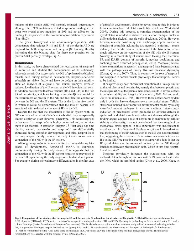
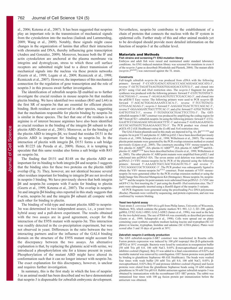
Binding sites on the plectin ABD for nesprin-3a and theintegrin b4 subunit partially overlapBesides its interaction with nesprin-3a, the plectin ABD can alsointeract with F-actin and the integrin b4 subunit (Geerts et al.,1999; Litjens et al., 2003). The crucial residues in the ABD thatmediate the interaction with integrin b4 were previously identifiedand found to overlap with those important for binding to actin (dePereda et al., 2009; Litjens et al., 2003; Litjens et al., 2005).Moreover, two arginine residues in integrin b4, R1225 and R1281,are of crucial importance for the interaction with the plectin ABD(Koster et al., 2001). Because binding of nesprin-3a to the plectinABD is also mediated via an arginine residue, we determinedwhether the binding sites for nesprin-3a and integrin b4 on theplectin ABD overlap with each other.
We initially used a yeast two-hybrid assay to test the interactionof several point mutants of the plectin-1C ABD with either nesprin-3a or part of the cytoplasmic domain of the integrin b4 subunit(b41115-1436). The point mutants were selected on the basis of theirreported effects on the interaction with integrin b4 (de Pereda etal., 2009; Litjens et al., 2005). Most of these mutations abolishedintegrin b4 binding; only the E200R and D208L mutants did not.As shown in Fig. 4A, the binding profiles of nesprin-3a andintegrin b4 partially overlap. Both nesprin-3a and integrin b4bound wild-type plectin ABD and the E200R and D208L pointmutants (Fig. 4A). Binding was not detected when residues E95,R148 and D151 of the plectin ABD had been mutated (Fig. 4A).However, there were also differences in plectin binding betweenintegrin b4 and nesprin-3a. Whereas R98Q, R121W and R123Wreduced binding to integrin b4, they did not influence the interactionwith nesprin-3a (Fig. 4A).
To confirm the role of these residues in the binding of theplectin ABD to nesprin-3a, the point mutants were tested for theirinteraction with nesprin-3a in a co-immunoprecipitationexperiment. VSV-tagged nesprin-3a was coexpressed in COS7cells together with HA-tagged wild-type plectin ABD or its pointmutants. As expected, wild-type plectin ABD as well as its R98Q,R121W, R123W, E200R and D208L point mutants co-precipitatedwith nesprin-3a (Fig. 4B,C). Similar to the results of the yeasttwo-hybrid assay, co-precipitation of the R148A and D151A
760 Journal of Cell Science 124 (5)
Fig. 4. Partial overlapping binding sites for nesprin-3a and the integrin b4subunit on the plectin ABD. (A)Binding of plectin-1C ABD point mutants tob41115-1436 and nesprin-3a in yeast two-hybrid assays. Plating efficiencies onselective SC-LTHA plates are expressed relative to those on nonselective SC-LT plates of the same transformation. ++, >40%; +, 10–40%; and –, <10%.Plates were scored after 5 and 10 days of growth. (B)COS7 cells weretransiently transfected with constructs encoding VSV-mouse-nesprin-3a, wild-type HA-plectin-1C ABD or point mutants of HA-plectin-1C ABD. Cells werelysed and both input samples and nesprin-3 precipitates (IP) were probed forVSV glycoprotein and HA. WB, Western blot; WT, wild-type. (C)Quantitativeanalysis of the effect demonstrated in (B). Intensities are corrected for nesprin-3 precipitation and normalized to the value for wild-type HA-plectin-1C ABD.Results are shown as the mean ± s.d. of three different experiments per mutant.
Jour
nal o
f Cel
l Sci
ence
mutants of the plectin ABD was strongly reduced. Interestingly,although the E95S mutation affected nesprin-3a binding in theyeast two-hybrid assay, mutation of E95 had no effect on thebinding to nesprin-3a in the co-immunoprecipitation experiment(Fig. 4B,C).
The yeast two-hybrid and co-immunoprecipitation datademonstrate that residues R148 and D151 of the plectin ABD arerequired for both nesprin-3a and integrin b4 binding, therebyindicating that the binding sites for these two proteins on theplectin ABD partially overlap (Fig. 5).
DiscussionIn this study, we have characterized the localization of nesprin-3in zebrafish and studied the consequences of its deficiency.Although nesprin-3 is expressed at the NE of epidermal and skeletalmuscle cells during zebrafish development, nesprin-3-deficientzebrafish are viable, fertile and have no defects in their motility.Detailed analyses of nesprin-3 null mutant embryos revealedreduced localization of the IF system at the NE in epidermal cells.In addition, we showed that two residues (R43 and L44) in the firstSR of nesprin-3a, which are lacking in nesprin-3b, are crucial forthe recruitment of plectin to the NE and facilitate the connectionbetween the NE and the IF system. This is the first in vivo modelin which it could be demonstrated that the loss of nesprin-3 isassociated with reduced anchorage of IFs to the NE.
Despite the fact that the association of the IF system with theNE was reduced in nesprin-3-deficient zebrafish, they unexpectedlydid not display an overt abnormal phenotype. This result surprisedus because: first, nesprin-3a is highly conserved and, as in mice,there is an isoform in zebrafish (nesprin-3b) that does not bindplectin; second, nesprin-3a and nesprin-3b are differentiallyexpressed during zebrafish development; and third, nesprin-3a isthe only nesprin family member currently known to mediate aconnection of the NE with the IF system.
Although nesprin-3a is the main isoform expressed during laterstages of development, nesprin-3b mRNA is expressedpredominantly during the early stages. This suggests that theconnection of the NE with the IF system needs to be prevented incertain cell types during the early stages of zebrafish development.For example, during skeletal muscle differentiation in the first days
of zebrafish development, single myocytes need to fuse in order toform a multinucleated skeletal muscle fiber (Ochi and Westerfield,2007). During this process, a complex reorganization of thecytoskeleton is needed to stabilize and anchor multiple nuclei indifferentiating skeletal muscle cells (Peckham, 2008). However,because nuclear positioning and anchorage appear normal inmuscles of zebrafish lacking the two nesprin-3 isoforms, it seemsunlikely that the differential expression of the two isoforms hasmuch influence on the connection of the NE with the IF system.Notably, in a recent study of mutant mice lacking the C-terminalSR and KASH domain of nesprin-1, nuclear positioning andanchorage were disturbed (Zhang et al., 2010). Moreover, severalmissense mutations in nesprin-1 and nesprin-2 have been associatedwith the pathogenesis of Emery–Dreifuss muscular dystrophy(Zhang, Q. et al., 2007). Thus, in contrast to the role of nesprin-1and nesprin-2 in normal muscle physiology, that of nesprin-3 seemsto be limited.
It has previously been shown that disruption of a linkage similarto that of plectin and nesprin-3a, namely that between plectin andthe integrin a6b4 at the plasma membrane, results in severe defectsin cellular stability and integrity (Koster et al., 2001; Nakano et al.,2001; Pulkkinen et al., 1998). However, these defects were evidentonly in cells that have undergone severe mechanical stress. Cellularstress was induced in our zebrafish developmental model by raisingnesprin-3 mutant embryos in viscous medium. Interestingly,induction of mechanical stress produced no obvious defects inepidermal or skeletal muscle cells (data not shown). Although thisfinding argues against a role of neprin-3a in maintaining cellularstability and integrity, it cannot be excluded that the strength of themechanical stress applied in this experiment was insufficient toreveal such a role of nesprin-3. Furthermore, it should be understoodthat the binding of the IF cytoskeleton to the NE was not completelylost, suggesting the existence of alternative mechanisms to anchorIFs to the NE. One possible example to support this idea is that theIF cytoskeleton can be connected indirectly to the NE throughinteractions between plectin and F-actin, which in turn bind nesprin-1 and nesprin-2.
Nesprins physically integrate the cytoskeleton with thenucleoskeleton through interactions with SUN proteins localized atthe INM, which in turn bind lamins (Crisp et al., 2006; Haque et
761Nesprin-3 in zebrafish
Fig. 5. Comparison of the binding sites for nesprin-3a and the integrin b4 subunit on the structure of the plectin ABD. (A)Surface representation of theABD of plectin (PDB code 3F7P), which consists of two calponin homology domains (CH1 and CH2). The integrin b4-binding surface is located in the CH1 and iscolored in orange (darker for residues at the core of the interface). The labels indicate the point mutations that were analyzed and are colored indicating whetherthey compromised binding to nesprin-3a (red) or not (green). R148 and D151 lay adjacent in the 3D structure and form part of the integrin b4-binding site.(B)Ribbon representation of the ABD in the same orientation as in A. For clarity, only the side chains of the residues analyzed are shown. The molecularrepresentations were created with the program Pymol (DeLano, 2002).
Jour
nal o
f Cel
l Sci
ence
al., 2006; Ketema et al., 2007). It has been suggested that nesprinsplay an important role in the transmission of mechanical signalsfrom the cytoskeleton into the nucleus (Jaalouk and Lammerding,2009; Wang et al., 2009). Notably, these signals might causechanges in the organization of lamins that affect their interactionwith chromatin and DNA, thereby influencing gene transcription(Andres and Gonzalez, 2009). Moreover, because both the IF andactin cytoskeleton are anchored at the plasma membrane viaintegrins and dystroglycan, stress to which these cell surfacereceptors are submitted might lead to a direct transmission ofmechanical signals into the nucleus via these protein networks(Geerts et al., 1999; Legate et al., 2009; Rezniczek et al., 1998;Rezniczek et al., 2007). However, the importance of this mechanicalconnection for the regulation of gene transcription and the role ofnesprin-3 in this process await further investigation.
The identification of zebrafish nesprin-3b enabled us to furtherinvestigate the crucial residues in mouse nesprin-3a that mediateplectin binding. We have identified two residues (R43 and L44) inthe first SR of nesprin-3a that are essential for efficient plectinbinding. Both residues are conserved in other species, suggestingthat the mechanism responsible for plectin binding by nesprin-3ais similar in these species. The fact that one of the residues is anarginine is of interest because arginines have also been identifiedas crucial residues in the binding of the integrin b4 subunit to theplectin ABD (Koster et al., 2001). Moreover, as for the binding ofthe plectin ABD to integrin b4, we found that residue D151 in theplectin ABD is important for binding to nesprin-3a. In theinteraction of plectin with integrin b4, D151 forms a salt bridgewith R1225 (de Pereda et al., 2009). Hence, it is tempting tospeculate that this same residue also forms a salt bridge with R43on nesprin-3a.
The finding that D151 and R148 on the plectin ABD areimportant for its binding to both integrin b4 and nesprin-3 suggeststhat the binding sites for these two proteins on the plectin ABDoverlap (Fig. 5). They, however, are not identical because severalother residues important for binding to integrin b4 are not involvedin nesprin-3 binding. We have previously shown that both integrinb4 and nesprin-3a compete with F-actin for binding to plectin(Geerts et al., 1999; Ketema et al., 2007). The overlap in nesprin-3a and integrin b4 binding sites reported in this study suggests thatF-actin, nesprin-3a and the integrin b4 subunit all compete witheach other for binding to plectin.
The binding of wild-type and mutant plectin ABD to nesprin-3a was determined in two independent assays, i.e., a yeast two-hybrid assay and a pull-down experiment. The results obtainedwith the two assays are in good agreement, except for theinteraction of the E95S mutant with nesprin-3a. This interactionwas readily detected in the co-precipitation experiment, but wasnot observed in yeast. Differences in the ratio between the twointeracting partners and/or the influence of the GAL4 bindingdomain on the structure of the E95S mutant might account forthe discrepancy between the two assays. An alternativeexplanation is that, by replacing the glutamic acid with serine, weintroduced a phosphorylation site for a kinase present in yeast.Phosphorylation of the mutant ABD might have altered itsconformation such that it can no longer interact with nesprin-3a.The exact explanation for this discrepancy, however, is unclearand requires further investigation.
In summary, this is the first study in which the loss of nesprin-3 in an animal model has been described and we have demonstratedthat nesprin-3 is dispensable for zebrafish embryonic development.
Nevertheless, nesprin-3a contributes to the establishment of achain of proteins that connects the nucleus with the IF system inepidermal cells. Further study of this and other animal models yetto be developed should provide more detailed information on thefunction of nesprin-3 at the cellular level.
Materials and MethodsFish strains and screening F1 ENU-mutation libraryEmbryos and adult fish were raised and maintained under standard laboratoryconditions. An ENU-induced mutation library was screened for mutations in exon 4of the nesprin-3 gene as described (Wienholds and Plasterk, 2004). The mutant allele(nesprin-3hu6448) was outcrossed against the TL strain.
ConstructsFull-length zebrafish nesprin-3a was produced from cDNA with the followingprimers: forward 5�-CCATCGATACCATGACCCAGCAGGAGCAGCATG-3�,reverse 5�-GCTCTAGATTAAGTGGGTGGAGGACCGTCG-3�, and cloned intopCS2+ using ClaI and XbaI restriction sites. The nesprin-3 fragment for probesyntheses was produced with the following primers: forward 5�-CGTCTGTATGCT-GACTGGAG-3�, reverse 5�-AGAGAAGTGTCCTCGACCAG-3�, and cloned intothe pGEM-T Easy vector. The following primers were used to PCR nesprin-3b:forward 5�-AGCACTGGAGAAAATCCACA-3�, reverse 5�-TCCTGTACG-GTTGAACAGAG-3�; nesprin-3: forward 5�-AAGAGCTGACTCTCCAGCAC-3�,reverse 5�-GGAAGGATCTGCCTTTCAC-3�; and ef1a: forward 5�-GTGCTGTGCT-GATTGTTGCT-3�, reverse 5�-TGTATGCGCTGACTTCCTTG-3�. The GST-zebrafish-nesprin-3-SR7 construct was produced by amplifying the coding region forSR7 from pCS2+-zebrafish-nesprin-3a using the following primers: forward 5�-CCG-GAATTCCCGGAGAGCATGAGAGCTTTC-3� and reverse 5�-GATAAGAATGCG-GCCGCAGTGGTCTGAAGAAGCCTG-3�. The PCR product was cloned intopGEX-4T-2 (GE Healthcare) using the EcoRI and NotI restriction sites.
The GAL4 fusion plasmids used in this study are depicted in Fig. 4A. b41115-1436 andnesprin-3a in pACT2 and plectin-1C ABD in pAS2.1 have been described previously(Geerts et al., 1999; Schaapveld et al., 1998; Wilhelmsen et al., 2005). The variousplectin point mutants were generated by the PCR overlap extension method or createdpreviously (Litjens et al., 2005). The constructs encoding VSV–mouse-nesprin-3a,HA–plectin-1C ABDWT, HA–plectin-1C ABDE95S, HA–plectin-1C ABDR98Q and HA–plectin-1C ABDR123W have been described before (Geerts et al., 1999; Wilhelmsen etal., 2005). The other plectin-1C ABD point mutants were amplified from pAS2.1 andsubcloned into pcDNA3–HA. The seven amino acid deletion was introduced intopcDNA3–2�VSV–mouse-nesprin-3a by PCR of the plasmid using the followingprimers: forward 5�-AAAATCTGCCAGCTGGAGTCTG-3� and reverse 5�-CTCCAGGGCCGCTCGGGGC-3�. The PCR product was subsequently ligated toobtain the VSV–mouse-nesprin-3aD7AA construct. Point mutants of VSV–mouse-nesprin-3a were generated either by the PCR overlap extension method or using theQuikChange Site-Directed Mutagenesis Kit (Stratagene). Mouse nesprin-3a, nesprin-3aD7AA and the nesprin-3a point mutants were subcloned from pcDNA3–2�VSV intomGFP-C1 by first inserting the 3� part using the BamHI–XbaI restriction sites. The 5�parts were subsequently inserted using a BamHI digest of the nesprin-3 variants.
All PCR fragments were generated using the proofreading Pwo DNA polymerase(Roche). Plasmids were verified by sequencing, and protein expression, and size wasconfirmed by western blotting.
Yeast two-hybrid assayYeast strain S. cerevisiae PJ69-4A (a gift from Philip James, University of Wisconsin,Madison, WI), which contains the genetic markers W1–901, L2–3, H3–200, gal4D,gal80D, LYS2::GAL1-HIS3, GAL2-ADE2 (James et al., 1996), was used as the hostfor the two-hybrid assay. The use of PJ69-4A was essentially as described previously(Geerts et al., 1999; Schaapveld et al., 1998). Cells were spread out on platescontaining yeast synthetic complete medium lacking leucine and tryptophan (SC-LTplates) or leucine, tryptophan, histidine and adenine (SC-LTHA plates). Plates werescored after 5 and 10 days of growth at 30°C.
Zebrafish nesprin-3 antibody productionThe GST–zebrafish-nesprin-3-SR7 construct was transformed in Rosetta cells.Fusion protein expression was induced by 100 mM isopropyl thio b-D-galactoside(IPTG) at 18°C overnight. Bacteria were lysed by sonication in resuspension buffer[50 mM Tris pH 8.0, 100 mM NaCl, 0.05% b-mercaptoethanol and proteaseinhibitor cocktail (Sigma)]. Brij-35 was added to a final concentration of 0.02% andinsoluble material was removed by centrifugation. GST fusion proteins were purifiedby binding to glutathione Sepharose 4B (GE Healthcare). The beads were washedfour times with wash buffer [50 mM Tris pH 8.0, 100 mM NaCl, 0.05% b-mercaptoethanol, 0.02% Brij-35 and protease inhibitor cocktail (Sigma)] and fusionproteins were eluted from the glutathione Sepharose beads using 10 mM reducedglutathione in 50 mM Tris pH 8.0. Rabbit antiserum against zebrafish nesprin-3 wasobtained by immunization with the recombinant GST–SR7 protein. The rabbit wasimmunized four times with 100 mg fusion protein per immunization before theantiserum was obtained.
762 Journal of Cell Science 124 (5)
Jour
nal o
f Cel
l Sci
ence
AntibodiesThe rabbit polyclonal antibody (pAb) against mouse nesprin-3 has been describedpreviously (Wilhelmsen et al., 2005). The following other primary antibodies wereused: anti-VSV glycoprotein monoclonal antibody (mAb) (P5D4; Sigma), anti-HAmAb (12CA5; Santa Cruz Biotechnology), anti-plectin mAb (clone 31; BDTransduction Laboratories), anti-actin mAb (Chemicon International), anti-p63 mAb(1:100; 4A4; Santa Cruz Biotechnology), anti-b-tubulin mAb (1:100; N357;Amersham Life Science) and Ks pan 1–8 mAb (1:10; Progen Biotechnik). Thefollowing secondary antibodies were used: goat-anti-mouse FITC (RocklandImmunochemicals), goat-anti-mouse Texas Red (Invitrogen), goat-anti-rabbit TexasRed (Invitrogen), goat-anti-mouse horseradish peroxidase (GE Healthcare) anddonkey-anti-rabbit horseradish peroxidase (GE Healthcare). Alexa Fluor 488phalloidin and TO-PRO-3 iodide 642/661 were obtained from Invitrogen, and DAPIwas purchased from Sigma-Aldrich.
In situ hybridizationEmbryos were fixed overnight in 4% paraformaldehyde (PFA) in PBS at 4°C,washed in PBS and stored in methanol at –20°C. Whole mount ISH was performedas described previously (Thisse et al., 1993). The RNA probe was produced accordingto standard protocols.
Plastic sectioningEmbryos stained by whole-mount ISH were transferred from benzyl benzoate/benzylalcohol to 100% methanol, incubated for 10 minutes, washed twice with 100%ethanol for 10 minutes and incubated overnight in 100% Technovit 8100 infiltrationsolution (Kulzer) at 4°C. Next, embryos were embedded overnight in Technovit8100 embedding medium (Kulzer) at 4°C. Sections of 7 mm thickness were cut witha Reichert-Jung 2050 microtome, stretched on water and mounted on glass slides.Sections were dried overnight. Counterstaining was done using 0.05% neutral redfor 12 seconds, followed by extensive washing with water. Sections were preservedwith Pertex and mounted under a coverslip.
Cell culture and transfectionCOS7 cells were grown in DMEM (GIBCO Life Technologies) supplemented with10% fetal calf serum, 100 U/ml penicillin and 100 U/ml streptomycin. PA-JEBkeratinocytes were grown in keratinocyte serum-free medium (GIBCO LifeTechnologies) supplemented with 50 mg/ml bovine pituitary extract, 5 ng/ml EGF,100 U/ml penicillin and 100 U/ml streptomycin. PA-JEB cells were transientlytransfected with Lipofectin (Invitrogen) according to the instructions provided by themanufacturer. Transient transfection of COS7 cells was achieved using the DEAE–dextran method. All cells were analyzed 24–48 hours after transfection.
ImmunofluorescenceCells grown on glass coverslips were fixed in PBS containing 1% PFA for 15minutes and permeabilized with 0.5% Triton X-100 in PBS for 5 minutes. Afterblocking with 2% BSA in PBS, the cells were incubated with the primary antibodyfor 1.5 hours at room temperature. Cells were washed three times with PBS andincubated for 1 hour at room temperature with the secondary antibody. After threewashes with PBS, coverslips were mounted in Mowiol-DAPCO. Embryos werefixed overnight in PBS containing 2% PFA at 4°C, washed with 0.3% Triton X-100in PBS, blocked with 1% BSA and 0.3% Triton X-100 in PBS, and incubated withthe primary antibody. After overnight incubation at 4°C, embryos were washed for3 hours with 0.3% Triton X-100 in PBS, blocked with 1% BSA and 0.3% Triton X-100 in PBS for 1 hour, and incubated with the secondary antibody for 3 hours.Embryos stained for keratin or b-tubulin were fixed in Dent’s fixative (80% methanol,20% DMSO). Embryos and cells were viewed under a confocal laser scanningmicroscope (model SP-2 AOBS; Leica).
Lysis and immunoprecipitationProtein extracts were prepared from 100 deyolked embryos (8 hpf) inradioimmunoprecipitation (RIPA) buffer (2 ml/embryo). RIPA lysis buffer contained10 mM sodium phosphate pH 7, 150 mM NaCl, 1% Nonidet P40 (NP40), 1%sodium deoxycholate, 0.1% SDS, 2 mM EDTA, 50 mM NaF, 100 mM sodiumvanadate and protease inhibitor cocktail (Sigma). COS7 cells grown to confluencyin 10-cm tissue culture dishes were lysed in 1 ml lysis buffer consisting of M-PERMammalian Protein Extraction Reagent (Thermo Scientific) and NP40 lysis buffer[1% NP40, 150 mM NaCl, 50 mM Tris pH 8.0 and protease inhibitor cocktail(Sigma)] in a ratio of 9:1. Lysates were cleared by centrifugation at 20,000 g in amicrocentrifuge at 4°C for 60 minutes. Nesprin-3 proteins were precipitated byincubation of the cell lysates with 1.5 ml rabbit-anti-mouse-nesprin-3 pAb for 1 houron ice, followed by a subsequent incubation with 100 ml of a 10% slurry of ProteinA Sepharose (Amersham Biosciences) for 2.5 hours at 4°C while being steadilyinverted. Immunoprecipitates were washed four times with Tris-buffered saline,boiled for 5 minutes in SDS sample buffer (50 mM Tris pH 6.8, 10% glycerol, 1%SDS, 0.5% b-mercaptoethanol and 0.025% bromophenol blue) and resolved bySDS-PAGE.
Quantification and statisticsPerinuclear keratin was quantified by determining the percentage of cells with NE-associated keratin staining per embryo. A minimum of 36 cells per embryo in at leasttwo microscopic fields were analyzed in a blind fashion. Western blot quantificationwas performed with Adobe Photoshop software. All data are represented as the mean± s.d. Comparisons were carried out with the one-way Anova and statisticalsignificance was assumed for P<0.05.
Identification of the zebrafish nesprin-3 knockout was made possibleas part of the ZF-MODELS Integrated Project in the 6th FrameworkProgramme (Contract No. LSHG-CT-2003-503496) funded by theEuropean Commission. We thank Henk van Roekel, Ewart de Bruijnand Edwin Cuppen from the Hubrecht Institute in Utrecht for screeningthe zebrafish knock-out TILLING library, Philip James for providingthe yeast strain PJ69-4A, and Kevin Wilhelmsen and Michael Ports forcritical reading of the manuscript. We acknowledge Maaike Kreft,Simon Samwel and Rabab Charafeddine for technical assistance. Thiswork was supported by a grant from the Netherlands ScienceOrganization (NWO/ALW). Research in the J.M.d.P. laboratory wassupported by the Spanish Ministry of Science and Innovation and theEuropean Regional Development Found (BFU2009-08389).
ReferencesAndrä, K., Lassmann, H., Bittner, R., Shorny, S., Fassler, R., Propst, F. and Wiche,
G. (1997). Targeted inactivation of plectin reveals essential function in maintaining theintegrity of skin, muscle, and heart cytoarchitecture. Genes Dev. 11, 3143-3156.
Andrä, K., Nikolic, B., Stocher, M., Drenckhahn, D. and Wiche, G. (1998). Not justscaffolding: plectin regulates actin dynamics in cultured cells. Genes Dev. 12, 3442-3451.
Andres, V. and Gonzalez, J. M. (2009). Role of A-type lamins in signaling, transcription,and chromatin organization. J. Cell Biol. 187, 945-957.
Crisp, M., Liu, Q., Roux, K., Rattner, J. B., Shanahan, C., Burke, B., Stahl, P. D. andHodzic, D. (2006). Coupling of the nucleus and cytoplasm: role of the LINC complex.J. Cell Biol. 172, 41-53.
de Pereda, J. M., Lillo, M. P. and Sonnenberg, A. (2009). Structural basis of theinteraction between integrin a6b4 and plectin at the hemidesmosomes. EMBO J. 28,1180-1190.
DeLano, W. L. (2002). The PyMOL Molecular Graphics System. San Carlos, CA: DeLanoScientific.
Garcia-Alvarez, B., Bobkov, A., Sonnenberg, A. and de Pereda, J. M. (2003). Structuraland functional analysis of the actin binding domain of plectin suggests alternativemechanisms for binding to F-actin and integrin b4. Structure 11, 615-625.
Geerts, D., Fontao, L., Nievers, M. G., Schaapveld, R. Q., Purkis, P. E., Wheeler, G.N., Lane, E. B., Leigh, I. M. and Sonnenberg, A. (1999). Binding of integrin a6b4to plectin prevents plectin association with F-actin but does not interfere withintermediate filament binding. J. Cell Biol. 147, 417-434.
Haque, F., Lloyd, D. J., Smallwood, D. T., Dent, C. L., Shanahan, C. M., Fry, A. M.,Trembath, R. C. and Shackleton, S. (2006). SUN1 interacts with nuclear lamin A andcytoplasmic nesprins to provide a physical connection between the nuclear lamina andthe cytoskeleton. Mol. Cell. Biol. 26, 3738-3751.
Jaalouk, D. E. and Lammerding, J. (2009). Mechanotransduction gone awry. Nat. RevMol. Cell Biol. 10, 63-73.
James, P., Halladay, J. and Craig, E. A. (1996). Genomic libraries and a host straindesigned for highly efficient two-hybrid selection in yeast. Genetics 144, 1425-1436.
Ketema, M., Wilhelmsen, K., Kuikman, I., Janssen, H., Hodzic, D. and Sonnenberg,A. (2007). Requirements for the localization of nesprin-3 at the nuclear envelope andits interaction with plectin. J. Cell Sci. 120, 3384-3394.
Koster, J., Kuikman, I., Kreft, M. and Sonnenberg, A. (2001). Two different mutationsin the cytoplasmic domain of the integrin b4 subunit in nonlethal forms of epidermolysisbullosa prevent interaction of b4 with plectin. J. Invest. Dermatol. 117, 1405-1411.
Le Guellec, D., Morvan-Dubois, G. and Sire, J. Y. (2004). Skin development in bonyfish with particular emphasis on collagen deposition in the dermis of the zebrafish(Danio rerio). Int. J. Dev. Biol. 48, 217-231.
Lee, H. and Kimelman, D. (2002). A dominant-negative form of p63 is required forepidermal proliferation in zebrafish. Dev. Cell 2, 607-616.
Legate, K. R., Wickstrom, S. A. and Fassler, R. (2009). Genetic and cell biologicalanalysis of integrin outside-in signaling. Genes Dev. 23, 397-418.
Litjens, S. H., Koster, J., Kuikman, I., van Wilpe, S., de Pereda, J. M. and Sonnenberg,A. (2003). Specificity of binding of the plectin actin-binding domain to b4 integrin.Mol. Biol. Cell 14, 4039-4050.
Litjens, S. H., Wilhelmsen, K., de Pereda, J. M., Perrakis, A. and Sonnenberg, A.(2005). Modeling and experimental validation of the binary complex of the plectinactin-binding domain and the first pair of fibronectin type III (FNIII) domains of the b4integrin. J. Biol. Chem. 280, 22270-22277.
Lüke, Y., Zaim, H., Karakesisoglou, I., Jaeger, V. M., Sellin, L., Lu, W., Schneider,M., Neumann, S., Beijer, A., Munck, M. et al. (2008). Nesprin-2 Giant (NUANCE)maintains nuclear envelope architecture and composition in skin. J. Cell Sci. 121, 1887-1898.
763Nesprin-3 in zebrafish
Jour
nal o
f Cel
l Sci
ence
McLean, W. H., Pulkkinen, L., Smith, F. J., Rugg, E. L., Lane, E. B., Bullrich, F.,Burgeson, R. E., Amano, S., Hudson, D. L., Owaribe, K. et al. (1996). Loss ofplectin causes epidermolysis bullosa with muscular dystrophy: cDNA cloning andgenomic organization. Genes Dev. 10, 1724-1735.
Nakano, A., Pulkkinen, L., Murrell, D., Rico, J., Lucky, A. W., Garzon, M., Stevens,C. A., Robertson, S., Pfendner, E. and Uitto, J. (2001). Epidermolysis bullosa withcongenital pyloric atresia: novel mutations in the b4 integrin gene (ITGB4) andgenotype/phenotype correlations. Pediatr. Res. 49, 618-626.
Nikolic, B., Mac Nulty, E., Mir, B. and Wiche, G. (1996). Basic amino acid residuecluster within nuclear targeting sequence motif is essential for cytoplasmic plectin-vimentin network junctions. J. Cell Biol. 134, 1455-1467.
Ochi, H. and Westerfield, M. (2007). Signaling networks that regulate muscledevelopment: lessons from zebrafish. Dev. Growth Differ. 49, 1-11.
Padmakumar, V. C., Abraham, S., Braune, S., Noegel, A. A., Tunggal, B.,Karakesisoglou, I. and Korenbaum, E. (2004). Enaptin, a giant actin-binding protein,is an element of the nuclear membrane and the actin cytoskeleton. Exp. Cell Res. 295,330-339.
Padmakumar, V. C., Libotte, T., Lu, W., Zaim, H., Abraham, S., Noegel, A. A.,Gotzmann, J., Foisner, R. and Karakesisoglou, I. (2005). The inner nuclear membraneprotein Sun1 mediates the anchorage of Nesprin-2 to the nuclear envelope. J. Cell Sci.118, 3419-3430.
Peckham, M. (2008). Engineering a multi-nucleated myotube, the role of the actincytoskeleton. J. Microsc. 231, 486-493.
Puckelwartz, M. J., Kessler, E., Zhang, Y., Hodzic, D., Randles, K. N., Morris, G.,Earley, J. U., Hadhazy, M., Holaska, J. M., Mewborn, S. K. et al. (2009). Disruptionof nesprin-1 produces an Emery Dreifuss muscular dystrophy-like phenotype in mice.Hum. Mol. Genet. 18, 607-620.
Pulkkinen, L., Rouan, F., Bruckner-Tuderman, L., Wallerstein, R., Garzon, M.,Brown, T., Smith, L., Carter, W. and Uitto, J. (1998). Novel ITGB4 mutations inlethal and nonlethal variants of epidermolysis bullosa with pyloric atresia: missenseversus nonsense. Am. J. Hum. Genet. 63, 1376-1387.
Rezniczek, G. A., de Pereda, J. M., Reipert, S. and Wiche, G. (1998). Linking integrina6b4-based cell adhesion to the intermediate filament cytoskeleton: direct interactionbetween the b4 subunit and plectin at multiple molecular sites. J. Cell Biol. 141, 209-225.
Rezniczek, G. A., Konieczny, P., Nikolic, B., Reipert, S., Schneller, D., Abrahamsberg,C., Davies, K. E., Winder, S. J. and Wiche, G. (2007). Plectin 1f scaffolding at thesarcolemma of dystrophic (mdx) muscle fibers through multiple interactions with beta-dystroglycan. J. Cell Biol. 176, 965-977.
Roux, K. J., Crisp, M. L., Liu, Q., Kim, D., Kozlov, S., Stewart, C. L. and Burke, B.(2009). Nesprin 4 is an outer nuclear membrane protein that can induce kinesin-mediated cell polarization. Proc. Natl. Acad. Sci. USA 106, 2194-2199.
Schaapveld, R. Q., Borradori, L., Geerts, D., van Leusden, M. R., Kuikman, I.,Nievers, M. G., Niessen, C. M., Steenbergen, R. D., Snijders, P. J. and Sonnenberg,A. (1998). Hemidesmosome formation is initiated by the b4 integrin subunit, requirescomplex formation of b4 and HD1/plectin, and involves a direct interaction between b4and the bullous pemphigoid antigen 180. J. Cell Biol. 142, 271-284.
Simpson, J. G. and Roberts, R. G. (2008). Patterns of evolutionary conservation in thenesprin genes highlight probable functionally important protein domains and isoforms.Biochem. Soc. Trans. 36, 1359-1367.
Sonnenberg, A. and Liem, R. K. (2007). Plakins in development and disease. Exp. CellRes. 313, 2189-2203.
Starr, D. A. and Han, M. (2002). Role of ANC-1 in tethering nuclei to the actincytoskeleton. Science 298, 406-409.
Stewart-Hutchinson, P. J., Hale, C. M., Wirtz, D. and Hodzic, D. (2008). Structuralrequirements for the assembly of LINC complexes and their function in cellularmechanical stiffness. Exp. Cell Res. 314, 1892-1905.
Thisse, C., Thisse, B., Schilling, T. F. and Postlethwait, J. H. (1993). Structure of thezebrafish snail1 gene and its expression in wild-type, spadetail and no tail mutantembryos. Development 119, 1203-1215.
Tsujikawa, M., Omori, Y., Biyanwila, J. and Malicki, J. (2007). Mechanism ofpositioning the cell nucleus in vertebrate photoreceptors. Proc. Natl. Acad. Sci. USA104, 14819-14824.
Wang, N., Tytell, J. D. and Ingber, D. E. (2009). Mechanotransduction at a distance:mechanically coupling the extracellular matrix with the nucleus. Nat. Rev Mol. CellBiol. 10, 75-82.
Wiche, G. (1998). Role of plectin in cytoskeleton organization and dynamics. J. Cell Sci.111, 2477-2486.
Wienholds, E. and Plasterk, R. H. (2004). Target-selected gene inactivation in zebrafish.Methods Cell Biol. 77, 69-90.
Wilhelmsen, K., Litjens, S. H., Kuikman, I., Tshimbalanga, N., Janssen, H., van denBout, I., Raymond, K. and Sonnenberg, A. (2005). Nesprin-3, a novel outer nuclearmembrane protein, associates with the cytoskeletal linker protein plectin. J. Cell Biol.171, 799-810.
Wilhelmsen, K., Ketema, M., Truong, H. and Sonnenberg, A. (2006). KASH-domainproteins in nuclear migration, anchorage and other processes. J. Cell Sci. 119, 5021-5029.
Yu, J., Starr, D. A., Wu, X., Parkhurst, S. M., Zhuang, Y., Xu, T., Xu, R. and Han,M. (2006). The KASH domain protein MSP-300 plays an essential role in nuclearanchoring during Drosophila oogenesis. Dev. Biol. 289, 336-345.
Zhang, J., Felder, A., Liu, Y., Guo, L. T., Lange, S., Dalton, N. D., Gu, Y., Peterson,K. L., Mizisin, A. P., Shelton, G. D. et al. (2010). Nesprin 1 is critical for nuclearpositioning and anchorage. Hum. Mol. Genet. 19, 329-341.
Zhang, Q., Skepper, J. N., Yang, F., Davies, J. D., Hegyi, L., Roberts, R. G., Weissberg,P. L., Ellis, J. A. and Shanahan, C. M. (2001). Nesprins: a novel family of spectrin-repeat-containing proteins that localize to the nuclear membrane in multiple tissues. J.Cell Sci. 114, 4485-4498.
Zhang, Q., Ragnauth, C., Greener, M. J., Shanahan, C. M. and Roberts, R. G. (2002).The nesprins are giant actin-binding proteins, orthologous to Drosophila melanogastermuscle protein MSP-300. Genomics 80, 473-481.
Zhang, Q., Bethmann, C., Worth, N. F., Davies, J. D., Wasner, C., Feuer, A., Ragnauth,C. D., Yi, Q., Mellad, J. A., Warren, D. T. et al. (2007). Nesprin-1 and -2 are involvedin the pathogenesis of Emery Dreifuss muscular dystrophy and are critical for nuclearenvelope integrity. Hum. Mol. Genet. 16, 2816-2833.
Zhang, X., Xu, R., Zhu, B., Yang, X., Ding, X., Duan, S., Xu, T., Zhuang, Y. and Han,M. (2007). Syne-1 and Syne-2 play crucial roles in myonuclear anchorage and motorneuron innervation. Development 134, 901-908.
Zhen, Y. Y., Libotte, T., Munck, M., Noegel, A. A. and Korenbaum, E. (2002).NUANCE, a giant protein connecting the nucleus and actin cytoskeleton. J. Cell Sci.115, 3207-3222.