Prelab Exercise 1 – CELLS, EPITHELIA, GLANDS, CONNECTIVE TISSUE 1 Prelab Exercise 1 CELLS, EPITHELIA, GLANDS AND CONNECTIVE TISSUE Prelab guide: this will function as a review of material that will be important to your performance of the lab. The first lab will examine cell structure (in the light microscope), epithelia and glands and connective tissue. There will be a checklist for your understanding of cells, one for glands and a final one for connective tissue. Cell structure The objectives of this lab exercise are: to acquaint you with general cell structure and appearance under the light and electron microscope; to familiarize yourself with the basic organization of cells into epithelia and glands and to study the components of loose and dense connective tissue. A question that arises every year is why many of the histology slides are from animal tissues. There are 4 basic reasons for this: 1. There are very few differences in the structure of mammalian tissues at the microscopic level. These few difference will be pointed out in class. 2. Cadaver tissues are poorly suited for histological investigation. The cells begin to break down quite quickly at the time of death and, even though the overall shape and size of an organ or tissue hasn't changed very much, by the time of embalming, the microscopic structure of cells is not very well preserved. 3. The source of most pathological specimens is the operating room, where biopsies and surgical specimens are harvested "fresh," and are frozen or fixed rapidly. However, most surgical pathological tissues are diseased and don't represent the normal structure of the organ. Exceptions include organs that may be removed even though they are not structurally diseased (such as the uterine tubes or vas deferens, sections of which are removed during sterilization procedures). Otherwise, it is not easy to find normal human tissue since this is not customarily removed surgically. 4. The examination of small animal tissue is often better than examining a small section of a large human organ. For example, one can see the entire organization of a portion of the rodent gastrointestinal tract, where only a tiny part of the wall of the human organ would fit on a slide. This permits a better understanding of the relationship between portions of the organ. GENERAL CELL FORM AND ORGANIZATION When cells are part of an organized tissue, they possess a distinctive form, which is associated with special functions and assume a shape, which is an adaptation to the external pressures and tensions applied to them by contiguous cells and intercellular substances. NUCLEI Nuclei of different cell types vary appreciably in size, shape, amount of chromatin, degree of aggregation of chromatin, and number and size of the nucleoli. Therefore, it is important to train yourself to recognize the differences in structure of the nuclei of various cells. Differentiate the nuclear membrane, nucleolus, chromatin and nucleoplasm. The appearance of the nucleus can tell you something about the metabolic activity of the cell. DNA that is not being transcribed tends to clump together. The part that will never be transcribed clumps together in the periphery of the nucleus, where it is called heterochromatin. This stains dark blue with hematoxylin. A cell that is not making much protein or is only making a few types of protein tends to have a very dark nucleus (heterchromatic). DNA that is less clumped and less coiled does not stain darkly (euchromatin). It is
CELLS, EPITHELIA, GLANDS AND CONNECTIVE TISSUE Prelab guide: this will function as a review of material that will be important to your performance of
the lab. The first lab will examine cell structure (in the light microscope), epithelia and glands and connective tissue. There will be a checklist for your understanding of cells, one for glands and a final
one for connective tissue.
Cell structure The objectives of this lab exercise are: to acquaint you with general cell structure and appearance under the light and electron microscope; to familiarize yourself with the basic organization of cells into epithelia and glands and to study the components of loose and dense connective tissue. A question that arises every year is why many of the histology slides are from animal tissues. There are 4 basic reasons for this: 1. There are very few differences in the structure of mammalian tissues at the microscopic level. These few difference will be pointed out in class. 2. Cadaver tissues are poorly suited for histological investigation. The cells begin to break down quite quickly at the time of death and, even though the overall shape and size of an organ or tissue hasn't changed very much, by the time of embalming, the microscopic structure of cells is not very well preserved. 3. The source of most pathological specimens is the operating room, where biopsies and surgical specimens are harvested "fresh," and are frozen or fixed rapidly. However, most surgical pathological tissues are diseased and don't represent the normal structure of the organ. Exceptions include organs that may be removed even though they are not structurally diseased (such as the uterine tubes or vas deferens, sections of which are removed during sterilization procedures). Otherwise, it is not easy to find normal human tissue since this is not customarily removed surgically. 4. The examination of small animal tissue is often better than examining a small section of a large human organ. For example, one can see the entire organization of a portion of the rodent gastrointestinal tract, where only a tiny part of the wall of the human organ would fit on a slide. This permits a better understanding of the relationship between portions of the organ. GENERAL CELL FORM AND ORGANIZATION
When cells are part of an organized tissue, they possess a distinctive form, which is associated with special functions and assume a shape, which is an adaptation to the external pressures and tensions applied to them by contiguous cells and intercellular substances. NUCLEI Nuclei of different cell types vary appreciably in size, shape, amount of chromatin, degree of aggregation of chromatin, and number and size of the nucleoli. Therefore, it is important to train yourself to recognize the differences in structure of the nuclei of various cells. Differentiate the nuclear membrane, nucleolus, chromatin and nucleoplasm. The appearance of the nucleus can tell you something about the metabolic activity of the cell. DNA that is not being transcribed tends to clump together. The part that will never be transcribed clumps together in the periphery of the nucleus, where it is called heterochromatin. This stains dark blue with hematoxylin. A cell that is not making much protein or is only making a few types of protein tends to have a very dark nucleus (heterchromatic). DNA that is less clumped and less coiled does not stain darkly (euchromatin). It is
available for transcription and gives a clear appearance to the nucleus (euchromatic). The nucleolus, which is a site of ribosome production, is prominent in a cell that is manufacturing proteins. Review the ultrastructure of the nucleus from your textbook and figs 3 & 4 of the “EM of organelles” module of virtual histology, particularly the nuclear envelope, the nucleolus, and chromatin. THE CYTOPLASM (CYTOSOL) The cytoplasm of living cells appears relatively structureless at magnifications and resolutions possible with the light microscope. In the fixed cell, cytoplasm assumes more definite structure for a number of reasons: (a) proteins and, depending on the type of fixative, other substances are retained or precipitated; (b) the ultrastructure of the cell may be distorted; (c) special components (e.g., fibrils) are made more visible; and (d) some components (e.g., carbohydrates and fats) may be dissolved out. Thus, the cytoplasmic structure which one sees is dependent on the type of cell under observation, its functional state when fixed, and the technical procedures employed in making the preparation.
ORGANELLES Cytoplasmic structures called “organelles” (little organs) are essential for the proper functioning of the cell containing them. For the most part, special methods of fixation and staining must be utilized in order to demonstrate their presence with a light microscope. Also, these structures are small in size, making them difficult to visualize. For these reasons, you will not be able to identify distinct organelles on most of your slides.
Much of our understanding of the structure and function of cell organelles comes from electron microscopic examination together with biochemical and genetic analyses. Study carefully the ultrastructure of cell organelles in the electron micrographs contained in the textbook and in the “EM of organelles” module associated with the online version of this lab. Be prepared to give an account of the functions of these different organelles. The following is a list of organelles to study: Mitochondria Mitochondria are present in all cells, but are not readily visible in most routine H&E stained sections. Examine mitochondria on TEM from your book and figs. 12 & 13 of the “EM of organelles” module of virtual histology. These widely vary in shape and size, but they contain a double layer of membrane and an internal architecture characterized by infoldings of the inner mitochondrial membrane (christae). Generally, the more mitochondria a cell contains and the denser the christae of the existing mitochondria, the more metabolically active the cell.
Rough endoplasmic reticulum (RER) Examine the EM of rough ER (fig. 8) and polyribosomes (fig. 9) in the “EM of organelles” module of the virtual histology site. Rough endoplasmic reticulum (RER) and/or the free ribosomes as seen with the TEM (figs 7 & 8 of the “EM of organelles” module of virtual histology) coincide with strongly basophilic regions of the cytoplasm of many cells as seen with the light microscope. Ribosomes are composed of RNA plus associated protein, the former accounting for the strong basophilia. Smooth endoplasmic reticulum (SER) The presence of this organelle is not usually visible in stained sections with conventional light microscopy. Large amounts of smooth ER would result in a clear or “foamy” cytoplasm since there are no ribosomes on the SER membranes to take up the stain. By electron microscopy (an example is in fig. 10 of the “EM of organelles” module of virtual histology), it can be seen that this is the predominant type of ER present in striated muscle cells, and cells that secrete steroid hormones (testicular interstitial cells, and adrenal cortical cells, to be studied later). The SER is involved in folding and transport of proteins to be used in the cell membrane or to be secreted (exocytosed) from the cell (e.g. digestive enzymes). In muscle (and other cells) it sequesters calcium; and it is involved in production and storage of steroids, glycogen, and other macromolecules.
The Golgi complex (aka Golgi apparatus, Golgi body). This organelle does not bind the routine histological dyes used for light microscopy but can be positively stained using special preparations such as heavy metal (osmium or silver) impregnation or, of course, by immunochemical methods using antibodies against proteins that are resident on the Golgi membranes.
The failure of H&E to stain the Golgi does, however, permit its discrimination in some cell types since the Golgi will appear as an unstained or somewhat clear area of the cell surrounded by the typical eosinophilia or basophilia of the rest of the cytoplasm. We will defer identification of this pale Golgi zone until a later lab session. Review the characteristic appearance of the Golgi complex from the electron micrographs in your textbooks (also fig. 11 of the “organelle” module of virtual histology). The Golgi complex has varying appearances and locations in the cytoplasm, depending on the cell type and its functional state. What are the major functions of the Golgi complex? Lysosomes In most instances these enzyme-rich organelles require specific histochemical or immunochemical techniques to demonstrate their distribution in the cell. In long-lived cells (e.g., muscle cells, neurons), the end products of lysosomal digestion are stored in defunct lysosomal compartments known as residual bodies. At the light microscopic level, collections of these residual bodies can be seen and are termed lipofuchsin pigment or “wear & tear” pigment. Study the varied fine structural appearances of lysosomes in your textbooks and (fig. 14 of the “EM of organelles” module of virtual histology). Be familiar with the role of lysosomes in intracellular digestive and autolytic processes. THE CYTOSKELETON (actin thin filaments, intermediate filaments, microtubules). The original identification and our present understanding of the distribution and functional importance of this system was largely the result of electron microscopic and biochemical studies. The dimensions of the individual filamentous and tubular components of this system are well below the resolution capabilities of the LM unless specific, fluorescent-tagged antibody methods are used. Review the figures in your book and the 12 figures of the “cytoskeleton” module of virtual histology.
In studying mitosis you will examine the mitotic apparatus, comprised largely of microtubules and associated proteins. Striated muscle contains an elaborate array of actin, myosin and other cytoskeletal-associated proteins. PLASMA MEMBRANE (cell membrane; plasmalemma). In most cells, the plasma membrane is not visible with the light microscope, making it difficult to determine the boundaries of the cells. In some instances, it appears that you can see the plasma membrane despite the fact that the thickness of the membrane, ≈ 10nm, is below the resolving power of the light microscope, as in of the liver. The reason for this illusion is because there is significant interdigitation of the membrane between neighboring cells. You may be able to “see” the membrane if it is cut obliquely (as in “B”, below). Some cytoskeletal elements (e.g., actin) and cell surface coats (e.g., glycocalyx) may also line up along the plasma membrane. If these are stained, it will facilitate visualization of the location of the cell membrane.
What is the appearance of the plasma membrane when viewed by the electron microscope in conventional thin sections?
Intracellularly Products of Cellular Function (INCLUSIONS). Numerous products of cellular activity are visible microscopically within the cytoplasm, but they vary in quantity depending upon the normal functional state of the cell or upon pathological processes. However, the general vitality of the cell is usually not dependent upon the presence or absence of these products. Some of the products require special staining procedures for their demonstration. Inclusions are of several types, such as secretory granules (the contents of which will be released from the cell), mucin, glycogen (an energy storage compound), melanin (a pigment) or lipids, which may appear as a single, giant fat droplet within the cytoplasm of an adipose cell.
MITOSIS Keep in mind as you study the following slides that the classically described phases of mitosis (prophase, metaphase, anaphase, telophase) represent only a relatively brief final stage of the cell cycle. Before these microscopically visible climactic events could take place, numerous vital synthetic processes at the molecular level had to be completed in the cells during the preceding, usually much longer, interphase stage. What components of the mitotic apparatus can you see in these cells with the light microscope?
• Find a cell in each of the following mitotic stages: prophase, metaphase, anaphase and
telophase. • Summarize the morphological changes taking place in the cell during each of these stages. • Realize that these stages are not achieved abruptly in the living cell. Rather, there is a gradual
transition from one stage to the next. Examine images 1 & 2 from the “Organelle” EM module.
CHECK LIST FOR CELLS
Identify each of the components of cells at the light (where appropriate) and the electron microscope level.
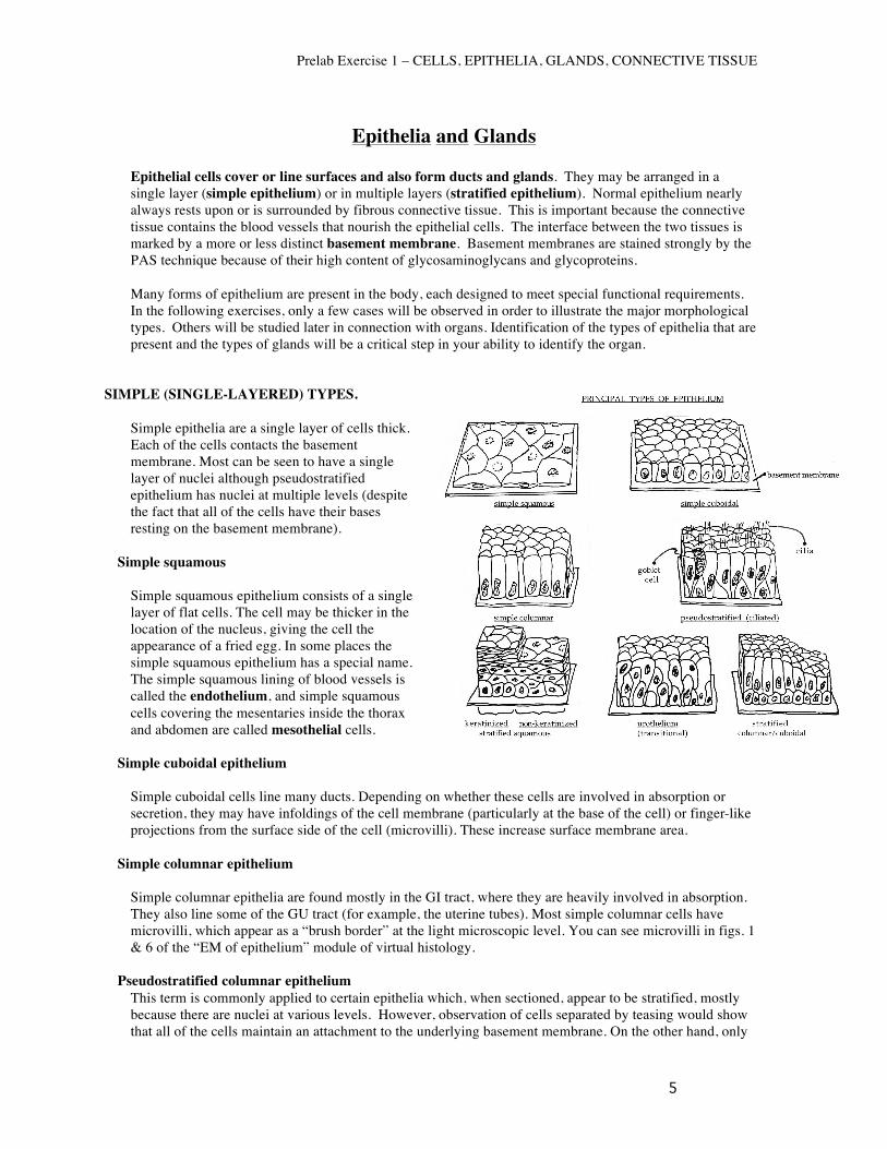
Epithelia and Glands Epithelial cells cover or line surfaces and also form ducts and glands. They may be arranged in a single layer (simple epithelium) or in multiple layers (stratified epithelium). Normal epithelium nearly always rests upon or is surrounded by fibrous connective tissue. This is important because the connective tissue contains the blood vessels that nourish the epithelial cells. The interface between the two tissues is marked by a more or less distinct basement membrane. Basement membranes are stained strongly by the PAS technique because of their high content of glycosaminoglycans and glycoproteins. Many forms of epithelium are present in the body, each designed to meet special functional requirements. In the following exercises, only a few cases will be observed in order to illustrate the major morphological types. Others will be studied later in connection with organs. Identification of the types of epithelia that are present and the types of glands will be a critical step in your ability to identify the organ.
SIMPLE (SINGLE-LAYERED) TYPES.
Simple epithelia are a single layer of cells thick. Each of the cells contacts the basement membrane. Most can be seen to have a single layer of nuclei although pseudostratified epithelium has nuclei at multiple levels (despite the fact that all of the cells have their bases resting on the basement membrane).
Simple squamous
Simple squamous epithelium consists of a single layer of flat cells. The cell may be thicker in the location of the nucleus, giving the cell the appearance of a fried egg. In some places the simple squamous epithelium has a special name. The simple squamous lining of blood vessels is called the endothelium, and simple squamous cells covering the mesentaries inside the thorax and abdomen are called mesothelial cells.
Simple cuboidal epithelium
Simple cuboidal cells line many ducts. Depending on whether these cells are involved in absorption or secretion, they may have infoldings of the cell membrane (particularly at the base of the cell) or finger-like projections from the surface side of the cell (microvilli). These increase surface membrane area.
Simple columnar epithelium
Simple columnar epithelia are found mostly in the GI tract, where they are heavily involved in absorption. They also line some of the GU tract (for example, the uterine tubes). Most simple columnar cells have microvilli, which appear as a “brush border” at the light microscopic level. You can see microvilli in figs. 1 & 6 of the “EM of epithelium” module of virtual histology.
Pseudostratified columnar epithelium
This term is commonly applied to certain epithelia which, when sectioned, appear to be stratified, mostly because there are nuclei at various levels. However, observation of cells separated by teasing would show that all of the cells maintain an attachment to the underlying basement membrane. On the other hand, only
some reach the free surface of the epithelium. This kind of epithelium is most commonly found lining the passages of the respiratory system (e.g., trachea and bronchi). It is also found in some ducts in the male reproductive system that we will study later in the course.
STRATIFIED (MULTI-LAYERED) EPITHELIA
It is imperative to understand that the different types of stratified epithelia are identified on the basis of the morphology of the most superficial layer of cells (i.e. cells of the luminal or free surface). Deeper lying cells in these epithelia may have a different appearance. Stratified squamous epithelia covers internal and external surfaces (moist or dry) that are prone to abrasion and need the added protection that this type of epithelium affords. Areas lined with this type of epithelium include the skin, oral cavity, esophagus, and vagina among others. Stratified squamous epithelia are characterized by several layers (strata) of cells. The surface cells have one of two appearances that define the stratified squamous epithelium as being either kertinized or non-kerainized. In mostly dry areas, such as the skin, the nuclei of the surface layers shrink and become pyknotic (and in some cases disappear altogether). In addition, the cytoplasm undergoes varying degrees of keratinization (sometimes referred to as cornification) during which keratin and associated matrix proteins increase in abundance in the cytoplasm, eventually filling the cell. Keratin is also the protein that comprises finger and toe-nails. Toward the surface of the skin, cells undergo transformation from living cells to dead scales, which are then sloughed from the epithelium. In moist areas (such as the mouth, esophagus and vagina) non-keratinized epithelial cells retain their nuclei all the way to the surface. This epithelium is kept moist by the presence of mucus or other fluids.
Consider the structural changes that occur in the nucleus and cytoplasm as living cells move from the basal layer to the free surface during their conversion to a dead scale. Observe pyknosis, which is the shrinkage of the nucleus to form a small, irregularly shaped, compact mass. Loss of normal nuclear structure is one of the most useful criteria for determination of loss of cellular vitality due to trauma, disease, or as in this case, natural processes. This criterion for evaluation of cellular vitality will be used repeatedly in CTO and in pathology.
Stratified cuboidal or columnar epithelium. These types of stratified epithelia are uncommon and have a very limited distribution in the body. When present, they usually occur in large glandular ducts (e.g. pancreatic duct, salivary ducts). They occur occasionally in zones of transition between two different varieties of epithelium (e.g., transition between simple columnar and stratified squamous).
Transitional epithelium (urothelium). The distribution of this epithelium is limited to lining the different parts of the urinary tract (renal pelvis, ureter, urinary bladder). This epithelium is specifically adapted for stretching and its morphology may correspondingly change. For example, in the distended urinary bladder, the epithelium appears only two or three layers thick, whereas, in the contracted bladder, it may be four to six or even more layers thick. This type of multilayered epithelium (when not under tension) is characterized by its surface layer of dome, pillow-like, or umbrella-like cells. Subjacent layers contain cells that may be cuboidal, columnar or polyhedral in shape.
SURFACE SPECIALIZATIONS OF EPITHELIAL CELLS While these structures may be discerned by light microscopy, their detailed morphology can only be appreciated by electron microscopy. You should correlate your observations here with the electron micrograph illustrations in your textbooks. Also, keep in mind that similar surface specializations may occur in the cells of other tissues besides epithelium.
Microvilli
As described above microvilli are projections of the surface membrane of columnar and some cuboidal cells that are designed to increase surface area. The actin filament cores of the microvilli extend down
into the apical cytoplasm of the cell where they are crosslinked by myosin and other actin-associated proteins. This dense meshwork of filaments creates the terminal web and can be see as a denser, eosinophilic line immediately beneath the microvilli. Also observe microvilli in figs 1 & 6 of the “epithelium” module of virtual histology. Observe the columnar surface epithelial cells in Slide #37 [small intestine, monkey, P.A.S. + H]. The brush borders of these cells are distinctly “P.A.S. positive” (stain red). Why?
Cilia Cilia are long, motile projections from the surface of the epithelium. They are anchored to ciliary basal bodies, which are located in a row beneath the apical epithelium. Review the ultrastructure of cilia from the illustrations in your textbook and from figs. 3 & 4 of the “EM of epithelium” module of virtual histology.
Stereocilia Stereocilia are immotile structures and basically represent very long, somewhat less-rigid microvilli.
Intercellular Junctions Generally, junctional specializations of the opposed lateral surfaces of epithelial cells (e.g., tight junctions) are below the resolution of the light microscope. However, pre-EM light microscopists found some evidence of their existence in the so-called “terminal bars” located apically between contiguous epithelial cells. Examine these specializations in figs. 1, 6, 7 & 8 of the “EM of epithelium” module of virtual histology. Figure 6 shows the entire junctional complex of zonula occludens (tight junction), zonula adherens (intermediate junction) and macua adherens (desmosome).
EPITHELIAL GLANDS
Glands may be classified in a variety of ways. Perhaps the most basic is classifying glands as either “exocrine” (secretion into a lumen or onto a surface) or “endocrine” (secretion into the circulation via capillaries). Although these glands all originate as an epithelium, the exocrine glands maintain a connection with the surface via a duct, and the endocrine glands lose this connection. You will examine exocrine glands in this lab while endocrine glands will be studied later in the course. There are three methods for classifying exocrine glands: 1. According to structural organization; 2. Based on type of secretion; 3. Based on their mode of secretion.
EXOCRINE GLANDS - Classification based on the STRUCTURAL organization of cells
Unicellular glands: The goblet cells are examples of unicellular (single cell) glands. They secrete mucin onto the surface of the epithelium.
Multicellular glands: Simple glands: Such a gland consists of a secretory unit (collection of secretory cells) connected to the
surface epithelium directly or by a single unbranched duct.
Compound glands: consist of a varying number of simple glands whose small excretory ducts join to form progressively larger and larger ducts which carry the secretion onto an epithelial surface. The pattern of the structure and arrangement of the ducts is often characteristic in a specific compound gland. Large compound glands, such as salivary glands (compound tubuloacinar) and the pancreas (compound acinar) will be studied in detail later in conjunction with the specific systems to which they belong. (A) compound tubular (B) compound acinar (C) compound tubuloacinar
EXOCRINE GLANDS - Classification based on the TYPE OF SECRETION
This classification cannot be applied to all the glands of the body. It is applicable primarily to the glands that pour their secretions into the oral cavity and other parts of the digestive system and to glands found in the respiratory system.
3 types of glandular secretions are found: • Mucous glands: cells secrete a viscous substance that contains
proteoglycans. Since secretions are water soluble, they are lost during tissue preparation and the cytoplasm of these cells appears empty.
• Serous glands: cells secrete a watery fluid, often containing enzymes or other proteins. Within the cell the secretory product is usually in the form of secretory granules.
• Mixed (seromucous) glands: contain both mucous and serous secreting cells. Most glands are of this type. Serous cells often form a crescent shape cluster around the mucous cells (a “demilune”).
EXOCRINE GLANDS - Classification based on the MODE OF SECRETION (i.e., by the method their
cells use for secretion).
In merocrine glands, the glandular cells remain intact and the products of their synthetic activity are secreted in vesicles via exocytosis. Serous gland cells use this mode of secretion, such as those in the pancreas. This is the most common mechanism of secretion.
In apocrine glands, portions of the apical surface of glandular cells form part of the secretion. One
example of this type of secretion is seen in the lactating breast, specifically for the lipid component of breast milk.
In holocrine glands, the entire glandular cell breaks down and the products of this disintegration are
discharged as the secretion. Sebaceous glands are the main example of glands that use the holocrine method of secretion.
Be able to identify the seven listed “principal types of epithelium”: -simple squamous, -simple cuboidal, -simple columnar, -pseudostratified, -keratinized and non-keratinized stratified squamous -urothelium. -stratified columnar/cuboidal
Connective Tissue The essential components of all types of connective tissue, including bone and cartilage, are cells, fibers, and ground substance (the material in between the fibers and cells). In the different types of connective tissues these components vary with respect to kind, amount and arrangement to meet the functional demands of the particular site in the body. You must examine each of these three basic components when studying connective tissue, assessing them in terms of their functional significance. Note that the end of this lab includes a study of plasma cells and the mononuclear phagocyte system (MPS). Although not strictly connective tissue cells, they are “transient cells” of connective tissue throughout the body. Fibroblasts and adipocytes (fat cells) are the principal “resident cells” in most connective tissue. The fibroblasts are responsible for manufacturing the fibers and much of the ground substance.
EMBRYONIC CONNECTIVE TISSUE
Primitive Mesenchyme
Primitive mesenchyme is the embryological tissue from which all types of supporting connective tissue, including the skeleton, are derived. It contains fusiform (spindle) or stellate (star) shaped undifferentiated cells. For the most part, these cells will differentiate into fibroblasts, though they can differentiate along other lines. They have a high ratio of nuclear to cytoplasmic volume. The intercellular material that surrounds these cells consists of sparse, delicate collagen fibrils and a viscous, amorphous ground substance, neither of which can be seen well in this routine type of preparation.
Mucoid Connective Tissue Mucoid (aka mucous, mucin or mucinogen – the carbohydrate-rich glycoproteins found in glandular tissue in the adult) connective tissue can be found in the umbilical cord. It consists of a specialized extracellular matrix whose ground substance is called Wharton’s Jelly. This contains Type I collagen and a mucoid ground substance containing much chondroitin sulfate and hyaluronic acid (it appears vaguely eosinophilic, and darker near the fibroblasts).
FIBROUS CONNECTIVE TISSUE.
Fibrous connective tissue is usually classified into loose and dense based on the density of the fibrous component of the connective tissue. Additionally, dense connective tissue is usually subdivided into regular and irregular connective tissue based on the degree to which fibers are organized in parallel arrays. It is important to realize that there is a continuous spectrum of degrees of density and orientation of fibers. Some tissues, such as the mesentery and the subcutaneous connective tissue, have a very loose arrangement; others, like the lamina propria of many of the hollow organs, are intermediate; while others, such as tendons and ligaments, can be definitely classified as dense fibrous connective tissue. Finally, some connective tissues are labeled as collagenous (fibrous), reticular or elastic based on the dominant extracellular fiber types.
Loose CT (aka areolar CT). This connective tissue contains cells and fibers. The most common cell type is fibroblasts. These cells have oval nuclei. There may be a few macrophages, which are wandering, phagocytic cells. Often surrounding the dense, irregular nuclei of the macrophages are dark particles which the macrophages have phagocytosed and which lie in their cytoplasm. A number of mast cells are also present. Their cytoplasm is usually so filled with metachromatic (purplish) granules that their rather small, round nuclei are often not visible.
There are collagen fibers (Type I) extending throughout the tissue. Elastic fibers are much thinner, branching strands.
Dense, irregular fibrous CT. This is a common connective tissue, most prominently located beneath the epithelium of many organs. Both collagen and elastic fibers are present in abundance, but only the former can be seen in routine H&E preparations such as this (elastic fibers don’t stain with H&E). The most prominent cell type is the fibroblast, which manufactures and maintains the fibers that comprise the bulk of the tissue.
Dense, regular fibrous CT. Ligaments, tendons and aponeuroses (a sheet or band of fibrous connective tissue separating or binding together muscles) have a similar appearance. There are longitudinally-oriented bundles of acidophilic collagen fibers (which have a slightly wavy appearance). There are fibroblasts in between the collagen bundles. This type of connective tissue has a paucity of blood vessels.
SPECIAL CONNECTIVE TISSUE
Elastic connective tissue. In this connective tissue, elastic rather than collagenous fibers predominate, although some collagenous fibers are also present. It is rather restricted in its distribution, being found primarily in the walls of the large arteries and in the alveolar walls of the lung. How do these fibers differ from collagen fibers?
Reticular connective tissue. Reticular connective tissue is so named for the predominance of reticular fibers (Type III collagen) in it. Such reticular tissue is found primarily in the blood forming organs and in glands (to be studied in more detail later) where it forms a supportive framework around the gland cells and blood vessels. Silver impregnations are commonly used to demonstrate the fine reticular (argyrophilic. e.g., “silver-loving”) fibers. They are not visible in H&E preparations.
Adipose (fat) tissue. In this specialized type of connective tissue, energy-storing fat cells (adipocytes) predominate. The distribution and amount of this tissue in the human body varies with the age, sex, and physical condition of the individual. There are two basic types of adipose tissue: white (unilocular) and brown multilocular).
White (unilocular) fat. The appearance of the fat cells
forming this tissue change with the degree of their lipid storage. In the "fully loaded" cell, neutral lipid is concentrated in a large central vacuole surrounded by a thin rim of cytoplasm. Unless prepared by solvent-free histological procedures (e.g., frozen sections), all of the lipid is dissolved away in slide preparation leaving a group of empty, circular cells of the so-called “signet ring” configuration. (diagram below). These adipose cells are held in place by collagen fibers (with a few fibroblasts), and there are numerous blood vessels.
Brown (multilocular) fat. This specialized thermogenic adipose tissue is more common in lower mammals (e.g., rodents) than in humans. (In humans, it is more evident in the newborn and infant and is more variable in adulthood) It differs considerably from white adipose tissue both in morphology and function. Review the mechanism of thermogenesis in brown adipose tissue.
PLASMA CELLS These cells originate from white blood cells (lymphocytes) that migrate into connective tissue. (Their life history will be covered later.) Although not truly a connective tissue cell, they are common in this tissue (particularly in part of the respiratory and digestive tracts) and are often encountered at this time. It is important that you learn to identify these cells in tissue sections. Their presence in large numbers is an important diagnostic feature of chronic inflammation. What are the characteristic cytological features of the plasma cell as seen: 1) by light microscopy; 2) by electron microscopy? What is the major function of these cells?
This diffuse system of free and fixed phagocytic cells (macrophages) replaces the outdated “reticuloendothelial system” concept (a term that you will still occasionally encounter). The criteria for including a phagocytic cell in this system are: 1) origin from bone marrow precursor cells; 2) high level of phagocytic activity mediated by immunoglobulin (antibodies) and components of the serum complement system.