33
| Date post: | 28-Dec-2015 |
| Category: |
Documents |
| Upload: | angel-morgan |
| View: | 241 times |
| Download: | 1 times |


Central Nervous System (CNS)
• CNS – brain – spinal cord
The Brain
• Composed of wrinkled, pinkish gray tissue
• Surface anatomy includes – cerebral hemispheres, – cerebellum, – brain stem
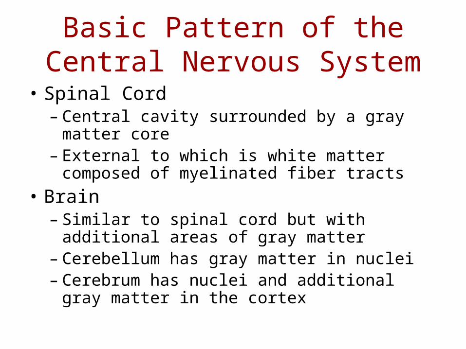
Basic Pattern of the Central Nervous System
• Spinal Cord – Central cavity surrounded by a gray matter core – External to which is white matter composed of
myelinated fiber tracts
• Brain– Similar to spinal cord but with additional areas
of gray matter– Cerebellum has gray matter in nuclei– Cerebrum has nuclei and additional gray matter
in the cortex

Basic Pattern of the Central Nervous System
Figure 12.4

Ventricles of the Brain
• Arise from expansion of the lumen of the neural tube
• The ventricles are:– The paired C-shaped lateral ventricles
separated by thin septum pellucidum– The third ventricle found in the diencephalon– The fourth ventricle found in the hindbrain
dorsal to the pons• Paired lateral apertures• Median aperture• Connect ventricles to subarachnoid fluid-filled
space.
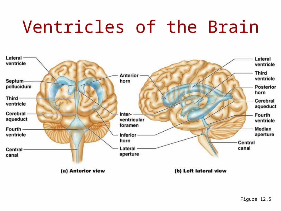
Ventricles of the Brain
Figure 12.5
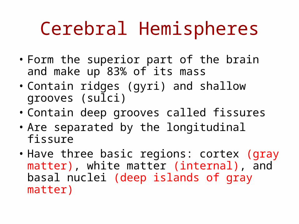
Cerebral Hemispheres
• Form the superior part of the brain and make up 83% of its mass
• Contain ridges (gyri) and shallow grooves (sulci)
• Contain deep grooves called fissures• Are separated by the longitudinal fissure• Have three basic regions: cortex (gray
matter), white matter (internal), and basal nuclei (deep islands of gray matter)

• Deep sulci divide the hemispheres into five lobes:– Frontal, parietal, temporal, occipital, and
insula (deep within the lateral sulcus forms part of cerebral floor.
• Central sulcus – separates the frontal and parietal lobes
Major Lobes, Gyri, and Sulci of the Cerebral Hemisphere
• Parieto-occipital sulcus – separates the parietal and occipital lobes
• Lateral sulcus – separates the parietal and temporal lobes
• The precentral and postcentral gyri border the central sulcus
Major Lobes, Gyri, and Sulci of the Cerebral Hemisphere

Cerebral Cortex• The cortex – superficial gray matter;
accounts for 40% of the mass of the brain• It enables sensation, communication,
memory, understanding, and voluntary movements
• Each hemisphere acts contralaterally (controls the opposite side of the body)
• Hemispheres are not equal in function• No functional area acts alone; conscious
behavior involves the entire cortex

• The three types of functional areas are:– Motor areas – control voluntary movement– Sensory areas – conscious awareness of
sensation– Association areas – integrate diverse
information
Functional (Brodmann) Areas of the Cerebral Cortex
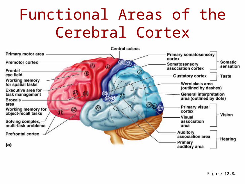
Functional Areas of the Cerebral Cortex
Figure 12.8a

Functional Areas of the Cerebral Cortex
Figure 12.8b
Cerebral Cortex: Motor Areas
• Primary (somatic) motor cortex
• Premotor cortex
• Broca’s area
• Frontal eye field
• Located in the precentral gyrus• Composed of pyramidal cells whose axons
make up the (pyramidal) corticospinal tracts
• Allows conscious control of precise, skilled, voluntary movements
• Motor homunculus – caricature of relative amounts of cortical tissue devoted to each motor function
(Motor areas) Primary (Somatic) Motor Cortex
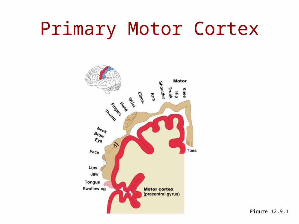
Primary Motor Cortex
Figure 12.9.1

(Motor areas) Premotor Cortex
• Located anterior to the precentral gyrus
• Controls learned, repetitious, or patterned motor skills
• Coordinates simultaneous or sequential actions
• Supplies 15% of pyramidal tract fibers.
• Memory bank for skilled motor activities
• Involved in the planning of movements
(Motor areas) Broca’s Area
• Broca’s area– Located anterior to the inferior region of the
premotor area– Present in one hemisphere only (usually the
left)– A motor speech area that directs muscles of
the tongue– Is active as one prepares to speak
(Motor areas) Frontal Eye Field
• Frontal eye field– Located anterior to the premotor cortex and
superior to Broca’s area– Controls voluntary eye movement

Sensory Areas
• Primary somatosensory cortex
• Somatosensory association cortex
• Visual and auditory areas
• Olfactory, gustatory, and vestibular cortices
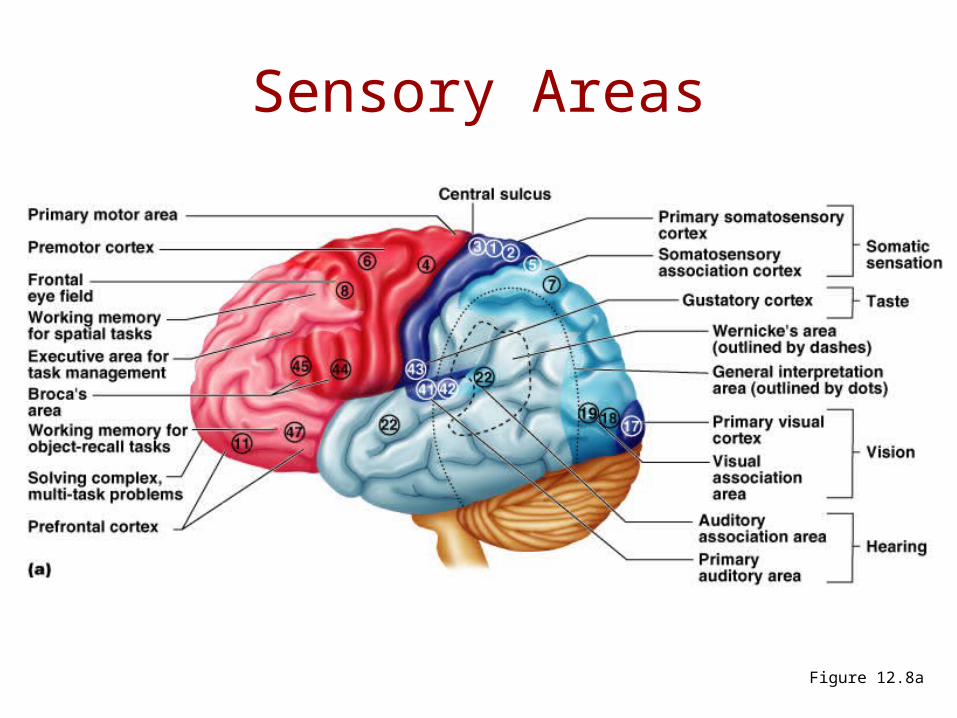
Sensory Areas
Figure 12.8a
(Senosry areas) Primary Somatosensory Cortex
• Located in the postcentral gyrus, this area:– Receives information from the skin and
skeletal muscles– Exhibits spatial discrimination (identifies areas
being stimulated)
• Somatosensory homunculus – caricature of relative amounts of cortical tissue devoted to each sensory function
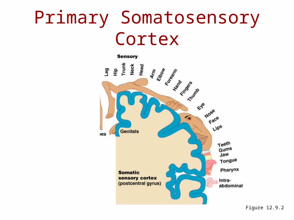
Primary Somatosensory Cortex
Figure 12.9.2

(Sensory area) Somatosensory Association Cortex
• Located posterior to the primary somatosensory cortex
• Integrates sensory information
• Forms comprehensive understanding of the stimulus
• Determines size, texture, and relationship of parts
(Sensory areas) Visual Areas
• Primary visual (striate) cortex– Seen on the extreme posterior tip of the
occipital lobe– Most of it is buried in the calcarine sulcus
– Receives visual information from the retinas
• Visual association area– Surrounds the primary visual cortex– Interprets visual stimuli (e.g., color, form, and
movement)
(Sensory areas) Auditory Areas• Primary auditory cortex
– Located at the superior margin of the temporal lobe– Receives information related to pitch, rhythm, and
loudness
• Auditory association area– Located posterior to the primary auditory cortex– Stores memories of sounds and permits perception of
sounds– Wernicke’s area sounding out unfamiliar words
(Sensory areas) Olfactory Cortex
• Small area of frontal lobe just above the orbit
• Conscious awareness of odor
• Formed from primitive rhinencephalon– Olfactory bulb– Olfactory tract– Olfactory cortices
(Sensory areas) Gustatory Cortex
• Located in the parietal lobe
• Perception of taste stimuli
(Sensory areas) Vestibular Cortex
• Posterior part of the insula
• Conscious awareness of balance (position of head in space.
Association Areas
• Prefrontal cortex
• Language areas
• General (common) interpretation area
• Visceral association area

Association Areas
Figure 12.8a
(Association areas) Prefrontal Cortex
• Located in the anterior portion of the frontal lobe
• Involved with intellect, cognition, recall, and personality
• Necessary for judgment, reasoning, persistence, and conscience
• Closely linked to the limbic system (emotional part of the brain)


