18
Ch 27: Reproductive System • General organization • Anatomy of male reproductive system • Anatomy of female reproductive system
Ch 27: Reproductive System
• General organization
• Anatomy of male reproductive system
• Anatomy of female reproductive system
General Organization
• Gonads gametes & hormones
• Ducts transport of . . . ?
• Glands secrete fluid
• Perineal structures = external genitalia
Anatomy of Male Repro System
• Primary reproductive organs produce gametes
• Secondary reproductive organs . . . .
• Male reproductive and urinary tracts are partially shared
Fig 27-1

Testes (paired glands)
• Develop adjacent to kidneys
• Descend into scrotum through inguinal canal
• Peritoneal lining is carried along lining of scrotum
• Cryptorchidism (in 3% of full-term and 30% of premature deliveries) – Significance?– Treatment?
Fig 27-2
4 month

ScrotumFunction: supports and protects testes
Structure: Skin & underlying superficial fascia– Dartos muscle in subcutis– Cremaster muscle deep to dermis (continuation
of ___________)
Involuntary contraction (cremasteric reflex) in response to ________
Scrotal sac forms 2 separate chambers
Cremaster muscle
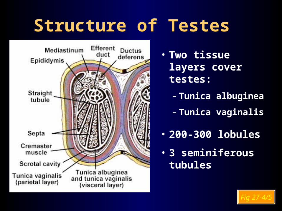
Structure of Testes• Two tissue layers
cover testes:– Tunica albuginea
– Tunica vaginalis
• 200-300 lobules
• 3 seminiferous tubules
Fig 27-4/5

From Spermatocyte to Spermatozoon
• Spermatogenesis: Meiosis of primary spermatocytes spermatids
• Spermiogenesis: Spermatid maturation into spermatozoa within Sertoli cells
• Spermiation: Spermatozoon released into lumen

Sustentacular (Sertoli) Cells• Maintenance of blood testis barrier
» special lumen fluid» sperm specific ag
• Support of spermatogenesis» FSH and Testosterone work via Sertoli cells
• Support of spermiogenesis• Secretion of inhibin• Secretion of androgen-binding protein (ABP)
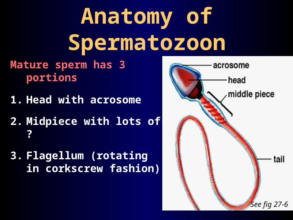
Anatomy of SpermatozoonMature sperm has 3
portions
1. Head with acrosome
2. Midpiece with lots of ?
3. Flagellum (rotating in corkscrew fashion)
See fig 27-6

Epididymis
~ 7 m long
1. Sperm-maturation
2. Recycling of damaged spermatozoa
3. Adjusting composition of tubular fluid (stereocilia!!)
Functions:

Path of Spermatozoa from tail of epididymis:
ductus (vas) deferens
ampulla
ejaculatory duct
urethra

CapacitationActivation of spermatozoa
Occurs after spermatozoa leave epididymis and come in contact with seminal fluid. Seminal fluid + Sperm = Semen
Final capacitation when exposed to conditions inside female reproductive tract
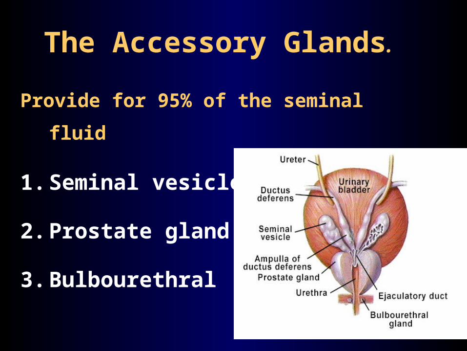
The Accessory Glands.
Provide for 95% of the seminal fluid
1. Seminal vesicles
2. Prostate gland
3. Bulbourethral glands

Seminal Vesicles
Produce 60% of seminal fluid
Tubular glands (~ 15 cm)
Secretionis rich in fructose
leads to sperm motility
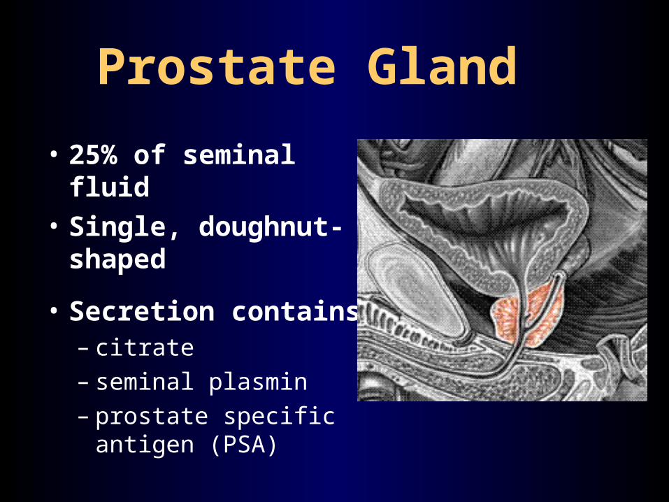
Prostate Gland• 25% of seminal fluid • Single, doughnut-
shaped
• Secretion contains:– citrate – seminal plasmin– prostate specific antigen
(PSA)

Bulbourethral glands (Cowper’s glands)
Pea size
Alkaline secretion containing lots of mucus. function??
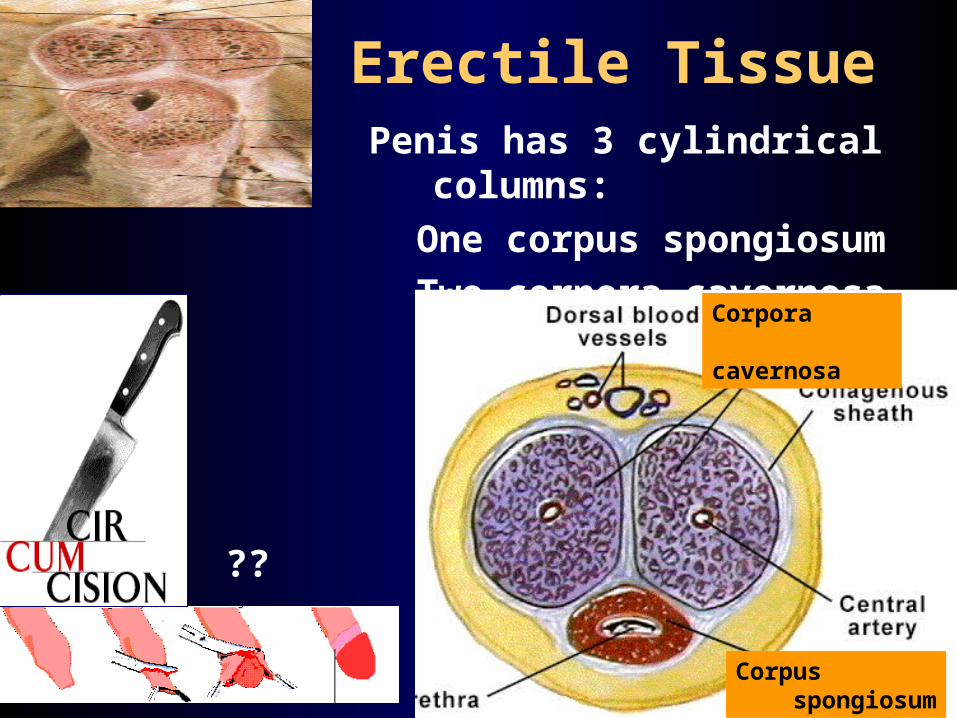
Erectile TissuePenis has 3 cylindrical columns:
One corpus spongiosumTwo corpora cavernosa
Corpora cavernosa
Corpus spongiosum
??