Conjugative Mapping of Pyruvate, 2-Ketoglutarate, and Branched-Chain Keto Acid Dehydrogenase Genes in Pseudomonas putida
MutantsPAMELA J. SYKES,1 JOAN MENARD,2 VICKI McCULLY,' AND JOHN R. SOKATCHl*
Departments of Biochemistry and Molecular Biologyl and Microbiology and Immunology,2 The University of OklahomaHealth Sciences Center, Oklahoma City, Oklahoma 73190
Received 31 August 1984/Accepted 15 January 1985
Branched-chain keto acid dehydrogenase, an enzyme in the common pathway of branched-chain amino acidcatabolism of Pseudomonas putida, is a multienzyme complex which catalyzes the oxidative decarboxylation ofbranched-chain keto acids. The objective of the present study was to isolate strains with mutations of this andother keto acid dehydrogenases and to map the location of the mutations on the chromosome of P. putida.Several strains with mutations of branched-chain keto acid dehydrogenase, two pyruvate and two 2-ketoglu-tarate dehydrogenase, were isolated, and the defective subunits were identified by biochemical analysis. Byusing a recombinant XYL-K plasmid to mediate conjugation, these mutations were mapped in relation to aseries of auxotrophic and other catabolic mutations. The last time of entry recorded was at approximately 35min, and the data were consistent with a single point of entry. Branched-chain keto acid dehydrogenasemutations affecting El, El plus E2, and E3 subunits mapped at approximately 35 min. One other strainaffected in the common pathway was deficient in branched-chain amino acid transaminase, and the mutationwas mapped at 16 min. The mutations in the two pyruvate dehydrogenase mutants, one deficient in El and theother deficient in El plus E2, mapped at 22 minutes. The 2-ketoglutarate dehydrogenase mutation affecting theEl subunit mapped at 12 minutes. A 2-ketoglutarate dehydrogenase mutant deficient in E3 was isolated, butthe mutation proved too leaky to map.
Branched-chain keto acid dehydrogenase ofPseudomonasputida is a multienzyme complex in the common pathway ofbranched-chain amino acid metabolism. The complex iscomposed of three subunits: El, the dehydrogenase, E2, thetransacylase, and E3, lipoamide dehydrogenase. In mam-mals, only conformational isomers of lipoamide dehydroge-nase have been identified (24). However, two structurallyand functionally different lipoamide dehydrogenases havebeen identified in P. putida (17). The higher-molecular-weight lipoamide dehydrogenase, LPD-glc, is the specific E3subunit of 2-ketoglutarate dehydrogenase (17) and is re-quired for glycine oxidation (16). The smaller lipoamidedehydrogenase, LPD-val, is the specific E3 subunit ofbranched-chain keto acid dehydrogenase. It is of interest tomap the genes encoding LPD-val and LPD-glc in relation togenes coding for other enzyme subunits of branched-chainketo acid dehydrogenase to gain insight into the regulation ofketo acid metabolism and into the evolutionary relationshipof LPD-glc and LPD-val.
Despite its metabolic and genetic diversity, little is knownabout the arrangement of genes in P. putida compared with,for example, Escherichia coli and even Pseudomonas aerugi-nosa. One reason for this has been the lack of a suitablerange of conjugative vectors in P. putida. A number of geneshave been mapped in P. putida by using the K sex factor(12), and recently, the P. putida chromosome was shown tobe circular and approximately 103 min in length by using a P.aeruginosa plasmid loaded with transposon TnS01 (2).
In the present study, we isolated a number of mutantsinvolved in keto acid metabolism and identified the enzymesubunits affected. The mutant genes were then mapped onthe P. putida chromosome by using a recombinant XYL-K
* Corresponding author.
sex factor (12) to mediate conjugation. No genes in keto acidmetabolism have previously been mapped in P. putida, andthis is the first report to map genes encoding branched-chainketo acid dehydrogenase in any organism.
MATERIALS AND METHODS
Organisms and growth conditions. Both P. putida PpG2and the donor for the interrupted matings, strain AC143 (12),were obtained from I. C. Gunsalus. E. coli NECO 100, usedfor transposon mutagenesis, was obtained from DavidGibson. The mutants and their genotypes are listed in Table1.The growth conditions and many of the media were
described in earlier publications (9, 18). Valine-isoleucineagar contained 0.3% L-valine and 0.1% L-isoleucine in thebasal medium described earlier (9). Pyruvate dehydrogenasemutants were grown in medium with 60 mM acetate, and the2-ketoglutarate dehydrogenase mutants were grown in GASmedium which contained 10 mM glucose supplemented with2 mM acetate and 2 mM succinate (18). When antibioticsupplements were added, the final concentrations were(micrograms per milliliter): streptomycin, 100; kanamycin,90; and chloramphenicol, 50. The final concentration ofamino acid supplements was 20 ,ug/ml. E. coli NECO 100was grown overnight in L broth supplemented with kana-mycin. When strain PpG2 was used in transposon mutagen-esis experiments, it was grown overnight in L broth withchloramphenicol. Transposon-induced auxotrophs weregrown in L broth supplemented with kanamycin and chlor-amphenicol.Immunological methods. The preparation of antisera
against LPD-val and LPD-glc and double-diffusion experi-ments have been previously described (18).
203
Dow
nloa
ded
from
http
s://j
ourn
als.
asm
.org
/jour
nal/j
b on
26
Dec
embe
r 20
21 b
y 19
1.53
.193
.188
.
204 SYKES ET AL.
TABLE 1. List of P. putida strains used in this study
Strain Genotypea Source orreference
AC143 XYL-K, met-i 12PpG2 Wild type I. C. GunsalusJS112 bkdAB This studyJS113 bkdA This studyJS116 bct This studyJS161 bkdA This studyJS287 lpdV 18JS326 bkdAB This studyJS329 trp This studyJS330 met This studyJS332 leu This studyJS334 put This studyJS335 hut This studyJS336 arc This studyJS342 pdhAB This studyJS343 pdhA This studyJS347 kgdA This studyJS348 IpdG This studyJS401 trp::TnS This studyJS402 his::TnS This studyJS404 met::Tn5 This studyJS411 ilv-2::TnS This studyJS412 ilv-3::TnS This studyJS419 leu::Tn5 This studyJS420 leu::TnS This studyJS422 ilv-19::TnS This study
a Gene designations of catabolic markers used in this study are: arc,arginine catabolism; bct, branched-chain amino acid transaminase; bkdA, Elof branched-chain keto acid dehydrogenase; bkdB, E2 of branched-chain ketoacid dehydrogenase; hut, histidine catabolism; kgdA, El of 2-ketoglutaratedehydrogenase; lpdG, LPD-glc, IpdV, LPD-val; pdhA, El of pyruvate dehy-drogenase; pdhB, E2 of pyruvate dehydrogenase; put, proline catabolism.Conventional gene designations were used for auxotrophic markers.
Enzyme assays. The assays for pyruvate, 2-ketoglutarate,branched-chain keto acid, and lipoamide dehydrogenaseshave been described previously (17, 18). Branched-chainamino acid transaminase was assayed by the method ofDuggan and Wechsler (4) with valine and 2-ketoglutarate.The mixture for the El assay contained 100 mM potassiumphosphate buffer (pH 7.0), 4 mM magnesium chloride, 0.2mM thiamine pyrophosphate, 5 mM L-valine, 300 ,ug ofphenazine methosulfate per ml, 1.2 mM dichlorophenol-in-dophenol, enough El to yield 0.02 to 0.03 ,umol of CO2 per15 min, and 1 mM [1-_4C]pyruvate, [1-14C]2-ketoglutarate,or [1-14C]2-ketoisovalerate (specific activity, 30,000 cpm/,umol). The reaction was started by the addition of radioac-tive substrate. The reaction mixture was incubated in 10-mlflasks (Kontes K-882300) fitted with a rubber serum stopperand plastic center wells (Kontes K-882320); the center wellcontained a strip of filter paper moistened with 40 ,ul ofProtosol (New England Nuclear Corp.). The reaction mix-ture was incubated for 15 min at 30°C with shaking and thenstopped with 0.1 ml of 10 N sulfuric acid injected through thestopper. The flask was shaken for an additional 5 min atwhich time the filter paper strip was placed in a vial with 10ml of Econofluor (New England Nuclear Corp.) containing10% methanol and counted. Specific activity is defined asmicromoles of CO2 produced per 15 min/mg of protein.[1 -14C]pyruvate and [1-_4C]2-ketoglutarate were incubatedfor 1 h at 30°C with shaking in a stoppered 10-ml flask asdescribed above to remove 14CO2 produced by chemicaldecarboxylation.The principle of the E2 assay is that E2 catalyzes trans-
acylation between coenzyme A (CoA) and dihydro-
lipoamide: Acyl-CoA plus dihydrolipoamide acyl-dihydro-lipoamide plus CoA. Acetyl and succinyl CoA were pre-pared from the corresponding anhydrides and CoA as de-scribed by Stadtman (20). The concentration of acyl-CoAwas determined by the hydroxamic acid assay (20). Thereaction mixture for the E2 assay contained (in 1.0 ml) 0.1 Mpotassium phosphate buffer (pH 7.0), 10 mM dihydrolipoa-mide prepared as described by Reed et al. (15), 0.03 to 0.13units of enzyme, and 1 mM acyl-CoA. The reaction wasstarted with acyl-CoA, incubated for 5 min at 30°C at whichtime the reaction was stopped with 1 ,l of redistilleddiketene, which converts CoA to acetoacetyl-CoA, mixed,and placed in an ice bath for 3 min. The reaction mixture (0.9ml) was transferred to a spectrophotometer cell, and 0.3,umol of NADH in 1% sodium bicarbonate was added. Theabsorbance was read at 340 nm, and then 4.5 ,u1 of hydroxy-acyl-CoA dehydrogenase (Sigma type III from pig heart; 2mg/ml) was added in a final volume of 1 ml. The absorbancewas read again, and the amount of NADH oxidized wascalculated from the difference. The specific activity of thisassay is defined as micromoles of NADH oxidized per 5min/mg of protein.
Isolation of mutants. The conditions for nitrosoguanidinemutagenesis and penicillin enrichment were described pre-viously (17). The method of Ornston (13) was used to isolatecatabolic mutants. Pyruvate dehydrogenase mutants wereobtained by selection after penicillin enrichment in 20 mMlactate broth for growth on 60 mM acetate but not on 20 mMlactate. 2-Ketoglutarate dehydrogenase and LPD-glc-negative mutants were isolated after penicillin enrichment in10 mM 2-ketoglutarate on the basis of their ability to grow inGAS medium, but not with 10 mM 2-ketoglutarate as thesole carbon source.Mutations induced by TnS were obtained by the use of
pRKTV14, a suicide plasmid in P. putida but stable in E. coliNECO 100. The plasmid was obtained from David Gibsonand was used to isolate auxotrophic mutants of P. putida (B.A. Finette and D. T. Gibson, Abstr. Annu. Meet. Am. Soc.Microbiol. 1983, H127, p. 127). A log-phase culture (1 ml) ofE. coli NECO 100 in L broth plus kanamycin was mixed with2 ml of an overnight culture of P. putida in L broth. Themixture was filtered onto a 0.45-,um membrane filter, and thefilter was left overnight at 32°C on an L agar plate with a softagar overlay. The cells were dislodged from the filter into 5ml of L broth, and 0.1 ml of this culture or of a 10-1 dilutionwas plated onto L agar plus kanamycin and chloramphenicoland incubated at 32°C. Chloramphenicol was used to preventthe growth of E. coli. The resulting Km' Cm' recombinantcolonies were replica plated onto 10 mM glucose agarcontaining amino acid pools plus kanamycin and chloram-phenicol to characterize the auxotrophic mutants. The per-centage of kanamycin resistant colonies which were identi-fied as auxotrophs varied from 0.1 to 0.2% in individualexperiments.
Conjugations. The conjugation procedure was that ofMylroie et al. (12). Counterselection against strain AC143was accomplished by the use of phage pfl6, to which thedonor was sensitive, and media lacking methionine. Themixture was vortexed vigorously for 30 s to separate matingpairs. The time of entry for individual gene markers wasestimated from a plot of number of recombinants per 108donor cells versus time by using data from at least threeconjugation experiments for each gene marker. The muta-tions of auxotrophic strains were mapped by using 10 mMglucose to select for recombinant colonies. The selectionsystem for recombinants of pyruvate genes was 20 mM
J. BACTERIOL.
Dow
nloa
ded
from
http
s://j
ourn
als.
asm
.org
/jour
nal/j
b on
26
Dec
embe
r 20
21 b
y 19
1.53
.193
.188
.
CONJUGATIVE MAPPING IN P. PUTIDA MUTANTS 205
lactate. Structural genes for branched-chain keto acid dehy-drogenase, branched-chain amino acid transaminase, and2-ketoglutarate dehydrogenase were mapped with valine-iso-leucine agar as the selective medium.
RESULTSNutritional phenotypes of keto acid dehydrogenase mutants.
Pyruvate dehydrogenase mutants JS342 and JS343, selectedfor their lack of ability to grow on lactate agar, also wereunable to grow on pyruvate agar (Table 2). Pyruvate dehy-drogenase mutants grew on GAS medium which is supple-mented with 2 mM acetate (18). Presumably this reflects arequirement for acetyl-CoA which can no longer be suppliedby pyruvate dehydrogenase (5). It may be significant thatneither of the pyruvate dehydrogenase mutants grew onsuccinate, suggesting that succinate is metabolized via pyruv-ate. Strain JS347, the mutant lacking 2-ketoglutarate dehy-drogenase, failed to grow or grew slowly on a number ofcompounds thought to be metabolized via the tricarboxylicacid cycle: acetate, pyruvate, lactate, arabinose, and severalof the tricarboxylic acid cycle intermediates. In contrast,strain JS348, the lpdG mutant, grew well on most com-pounds tested with the exception of acetate but grew slowlyon 2-ketoglutarate.Keto acid dehydrogenase and branched-chain amino acid
transaminase content of mutants. Since mutants defective inbranched-chain keto acid dehydrogenase do not grow withvaline as the sole carbon source, it was necessary to useGASV medium which allows growth and causes induction ofbranched-chain keto acid dehydrogenase in these mutants(18). GASV medium contains valine in addition to glucose,acetate, and succinate, and valine is deaminated to 2-ke-toisovalerate, the inducer of branched-chain keto acid dehy-drogenase (10). The data (Table 3) show that strains JS112,JS113, JS161, and JS326 lacked branched-chain keto aciddehydrogenase even when the assay was supplemented withpurified LPD-val. In comparison, strain JS287, a mutantwhich fails to produce LPD-val, regained full activity whenthe assay was supplemented with purified LPD-val. These
TABLE 2. Growth characteristics of pyruvate and 2-ketoglutaratedehydrogenase mutants
Growth characteristicsa of P. putida:Carbon source' PpG2 JS342 JS343 JS347 JS348
a Values for branched-chain keto acid dehydrogenase are measured innanomoles ofNADH produced per minute per milligram of protein; values forlipoamide dehydrogenase are measured in micromoles ofNADH oxidized perminute per milligram of protein. Other units are described in the text.
results suggested that the lesions in strains JS112, JS113,JS161, and JS326 were in either El or E2 of the complex.Strain JS116, which also fails to grow on valine agar, hadonly 3% of the branched-chain amino acid transaminase ofstrain PpG2. All of the mutants listed in Table 3 had normallevels of pyruvate and 2-ketoglutarate dehydrogenases.Data are shown for assays of El and E2 subunits of
branched-chain keto acid dehydrogenase as well as totallipoamide dehydrogenase (LPD-glc and LPD-val) in mutantsof P. putida grown in GASV medium (Table 3). For com-parative purposes, there was approximately five times asmuch branched-chain keto acid dehydrogenase in wild-typePpG2 grown in GASV medium compared with strain PpG2grown in glucose-mineral salts medium. Strains JS112, JS113,JS161, and JS326 all lacked El and contained variousamounts of E2. It was not possible to demonstrate a clear-cut deficiency of lipoamide dehydrogenase, even in strainJS287 which is known to lack LPD-val. None of the mutantsso far characterized were deficient only in E2.
It was possible that the failure to produce branched-chainketo acid dehydrogenase was due to a failure to metabolizevaline to the inducer, 2-ketoisovalerate. To test this idea, weattempted to induce El and E2 in these mutants by thefollowing procedure. Strains PpG2, JS112, and JS326 weregrown in flasks of GAS medium, harvested, and then trans-ferred to the basal medium with 5 mM 2-ketoisovalerate, theinducer of branched-chain keto acid dehydrogenase (10).The change in the concentration of 2-ketoisovalerate wasfollowed for 5 h, at which time it had disappeared from theflask with strain PpG2 but had changed little in flaskscontaining the mutants. No El was detected in either of themutants, but the amount of E2 in strain JS326 increased to44% of that found in strain PpG2. We conclude from thesedata that there were mutations in El and E2 subunits ofbranched-chain keto acid dehydrogenase.
Pyruvate and 2-ketoglutarate dehydrogenase content ofmutants. Enzyme activities of mutants in pyruvate dehydro-
TABLE 4. Subunit activities of pyruvate dehydrogenase mutantsof P. putida
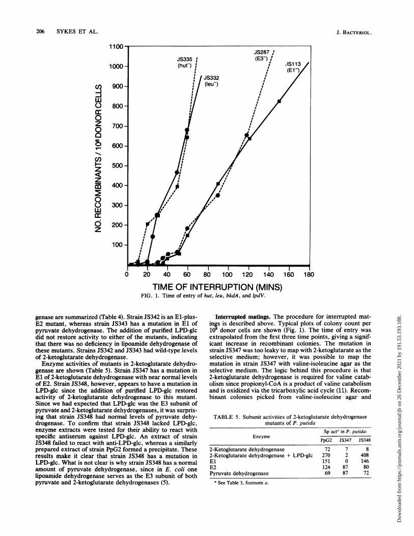
TIME OF INTERRUPTION (MINS)FIG. 1. Time of entry of hut, leu, bkdA, and lpdV.
genase are summarized (Table 4). Strain JS342 is an El-plus-E2 mutant, whereas strain JS343 has a mutation in El ofpyruvate dehydrogenase. The addition of purified LPD-glcdid not restore activity to either of the mutants, indicatingthat there was no deficiency in lipoamide dehydrogenase ofthese mutants. Strains JS342 and JS343 had wild-type levelsof 2-ketoglutarate dehydrogenase.Enzyme activities of mutants in 2-ketoglutarate dehydro-
genase are shown (Table 5). Strain JS347 has a mutation inEl of 2-ketoglutarate dehydrogenase with near normal levelsof E2. Strain JS348, however, appears to have a mutation inLPD-glc since the addition of purified LPD-glc restoredactivity of 2-ketoglutarate dehydrogenase to this mutant.Since we had expected that LPD-glc was the E3 subunit ofpyruvate and 2-ketoglutarate dehydrogenases, it was surpris-ing that strain JS348 had normal levels of pyruvate dehy-drogenase. To confirm that strain JS348 lacked LPD-glc,enzyme extracts were tested for their ability to react withspecific antiserum against LPD-glc. An extract of strainJS348 failed to react with anti-LPD-glc, whereas a similarlyprepared extract of strain PpG2 formed a precipitate. Theseresults make it clear that strain JS348 has a mutation inLPD-glc. What is not clear is why strain JS348 has a normalamount of pyruvate dehydrogenase, since in E. coli onelipoamide dehydrogenase serves as the E3 subunit of bothpyruvate and 2-ketoglutarate dehydrogenases (5).
Interrupted matings. The procedure for interrupted mat-ings is described above. Typical plots of colony count per108 donor cells are shown (Fig. 1). The time of entry wasextrapolated from the first three time points, giving a signif-icant increase in recombinant colonies. The mutation instrain JS347 was too leaky to map with 2-ketoglutarate as theselective medium; however, it was possible to map themutation in strain JS347 with valine-isoleucine agar as theselective medium. The logic behind this procedure is that2-ketoglutarate dehydrogenase is required for valine catab-olism since propionyl-CoA is a product of valine catabolismand is oxidized via the tricarboxylic acid cycle (11). Recom-binant colonies picked from valine-isoleucine agar and
TABLE 5. Subunit activities of 2-ketoglutarate dehydrogenasemutants of P. putida
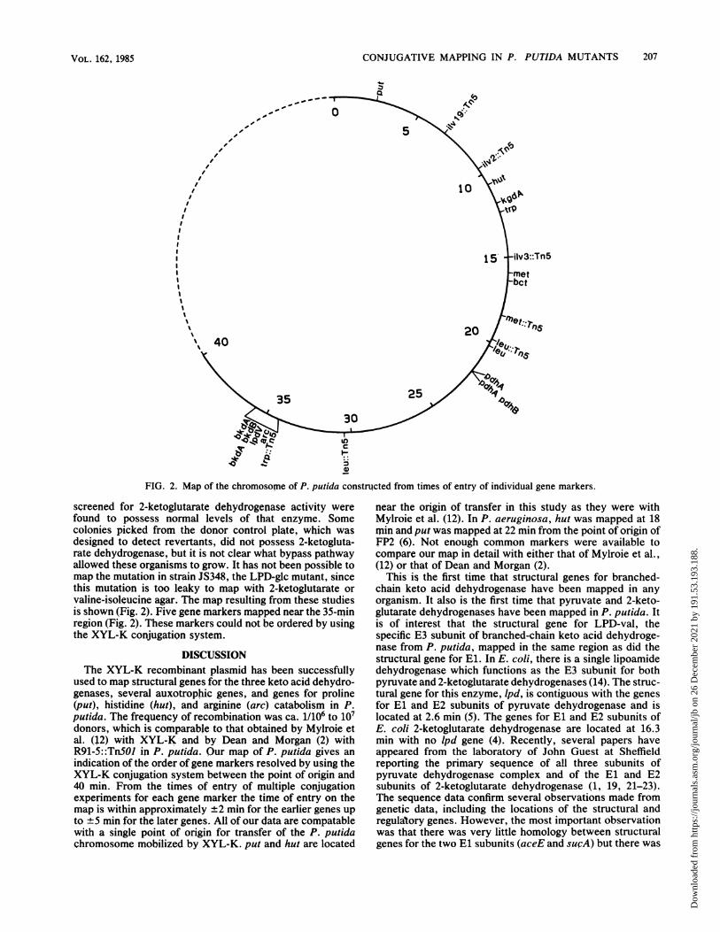
FIG. 2. Map of the chromosome of P. putida constructed from times of entry of individual gene markers.
screened for 2-ketoglutarate dehydrogenlase activity werefound to possess normal levels of that enzyme. Somecolonies picked from the donor control plate, which wasdesigned to detect revertants, did not possess 2-ketogluta-rate dehydrogenase, but it is not clear what bypass pathwayallowed these organisms to grow. It has not been possible tomap the mutation in strain JS348, the LPD-glc mutant, sincethis mutation is too leaky to map with 2-ketoglutarate orvaline-isoleucine agar. The map resulting from these studiesis shown (Fig. 2). Five gene markers mapped near the 35-minregion (Fig. 2). These markers could not be ordered by usingthe XYL-K conjugation system.
DISCUSSIONThe XYL-K recombinant plasmid has been successfully
used to map structural genes for the three keto acid dehydro-genases, several auxotrophic genes, and genes for proline(put), histidine (hut), and arginine (arc) catabolism in P.putida. The frequency of recombination was ca. 1/106 to 107donors, which is comparable to that obtained by Mylroie etal. (12) with XYL-K and by Dean and Morgan (2) withR91-5::TnS01 in P. putida. Our map of P. putida gives anindication of the order of gene markers resolved by using theXYL-K conjugation system between the point of origin and40 min. From the times of entry of multiple conjugationexperiments for each gene marker the time of entry on themap is within approximately +2 mmn for the earlier genes upto ±5 min for the later genes. All of our data are compatablewith a single point of origin for transfer of the P. putidachromosome mobilized by XYL-K. put and hut are located
near the origin of transfer in this study as they were withMylroie et al. (12). In P. aeruginosa, hut was mapped at 18rmin and put was mapped at 22 min from the point of origin ofFP2 (6). Not enough common markers were available tocompare our map in detail with either that of Mylroie et al.,(12) or that of Dean and Morgan (2).
This is the first time that structural genes for branched-chain keto acid dehydrogenase have been mapped in anyorganism. It also is the first time that pyruvate and 2-keto-glutarate dehydrogenases have been mapped in P. putida. Itis of interest that the structural gene for LPD-val, thespecific E3 subunit of branched-chain keto acid dehydroge-nase from P. putida, mapped in the same region as did thestructural gene for El. In E. coli, there is a single lipoamidedehydrogenase which functions as the E3 subunit for bothpyruvate and 2-ketoglutarate dehydrogenases (14). The struc-tural gene for this enzyme, lpd, is contiguous with the genesfor El and E2 subunits of pyruvate dehydrogenase and islocated at 2.6 min (5). The genes for El and E2 subunits ofE. coli 2-ketoglutarate dehydrogenase are located at 16.3min with no lpd gene (4). Recently, several papers haveappeared from the laboratory of John Guest at Sheffieldreporting the primary sequence of all three subunits ofpyruvate dehydrogenase complex and of the El and E2subunits of 2-ketoglutarate dehydrogenase (1, 19, 21-23).The sequence data confirm several observations made fromgenetic data, including the locations of the structural andregulalory genes. However, the most important observationwas that there was very little homology between structuralgenes for the two El subunits (aceE and suCA) but there was
VOL. 162, 1985
#I.e
.0op
Dow
nloa
ded
from
http
s://j
ourn
als.
asm
.org
/jour
nal/j
b on
26
Dec
embe
r 20
21 b
y 19
1.53
.193
.188
.
208 SYKES ET AL.
homology between structural genes for the two E2 subunits(aceF and sucB). These findings suggest that the two E2subunits had a common ancestor but that the El subunitswere evolved independently. Recently Lowe et al. (8) pre-sented biochemical and genetic evidence that pyruvate de-hydrogenase of Bacillus subtilis was actually a dual functioncomplex also responsible for branched-chain keto acid de-hydrogepase activity. The findings of Lowe et al. (8) suggestthat the evolutionary origin of branched-chain keto aciddehydrogenase might have been pyruvate dehydrogenase. Itis unfortunate that so far we have been unable to map thelocation of LPD-glc since there are a number of interestingquestions about the origins of LPD-glc apd LPD-val. Peptidemaps, antigenic re4ctivity, genetic data, and amino acidcomposition all show that LPD-glc and LPD-val are notclosely related. In fact, LPD-glc appears to be more closelyrelated to E. coli and pig heart lipoamide dehydrogenasesthan to LPD-val (3). It would have been informative todetermine whether lpdG mapped with pyruvate or 2-keto-glutarate dehydrogenase. This would have been particularlyimportant since strain JS348, the lpdG mutant, had normalpyruvate dehydrogenase (Table 5), whereas lpd mutants ofE. coli lacked both pyruvate and 2-ketoglutarate dehydroge-nases.
It is worth commenting on the nutritional phenotypes ofpyruvate and 2-ketoglutarate dehydrogerlase mutants sincethese mutants have not been widely studied in Pseudomonasspp. Strain JS343, which is a pdhA mutant, had a phenotypesimilar to P. aeruginosa PA02851 (7) and failed to grow onlactate, pyruvate, unsupplemented glucose medium, andsuccinate agar. The latter observation is interesting since itsuggests that pyruvate is an intermediate in succinate catab-olismn in pseudomonads. It is curious that pyruvate dehy-drogenase mutants failed to grow on citrate agar, sincepyruvate dehydrogenase should not be required for citrateoxidation. Strain JS347, which is a kgdA mutant, was unableto grow on unsupplemented media containing carbon sourcesexpected to be metabolized via the tricarboxylic acid cycle,including glucose, arabinose, glutamate, and valine. StrainJS347 did not grow well on glucose agar but did grow onGAS agar which is supplemented with acetate and succinate.The nutritional phenotype of strain JS348, the lpdG mutant,was unexpected since it failed to grow only on acetate,although it grew slowly on 2-ketoglutarate and isocitrate.The reason for this nutritional phenotype is not apparent.
ACKNOWLEDGMENTSThis work was supported by Public Health Service grants AM
21737 and GM 30428 from the National Institutes of Health.
LITERATURE CITED1. Darlison, M. G., M. E. Spencer, and J. R. Guest. 1984. Nucleo-
tide sequence of the sucA gene encoding the 2-oxoglutaratedehydrogenase of Escherichia coli K12. Eur. J. Biochem.141:351-359.
2. Dean, H. F., and A. F. Morgan. 1983. Integration of R91-5::TnS01 into the Pseudomonas putida PPN chromosome andgenetic circularity of the chromosomal map. J. Bacteriol.153:485-497.
3. Delaney, R., G. Burps, and J. R. Sokatch. 1984. Relationship oflipoamide dehydrogenases from Pseudomonas putida to otherFAD-linked dehydrogenases. FEBS Letts. 168:265-270.
4. Duggan, D. E., and J. A. Wechsler. 1973. An assay for transami-
nase B enzyme activity in Escherichia coli K-12. Anal. Bio-chem. 51:67-79.
5. Guest, J. R. 1978. Aspects of the molecular biology of lipoamidedehydrogenase. Adv. Neurol. 21:219-244.
6. Holloway, B. W., V. Krishnaplllai, and A. F. Morgan. 1979.Chromosomal genetics of Pseudomonas. Microbiol. Rev.43:73-102.
7. Jeyaseelan, K., and J. R. Guest. 1980. Isolation and properties ofpyruvate dehydrogenase complex mutants of Pseudomonasaerqginosa PAO. J. Gen. Microbiol. 120:385-392.
8. Lowe, P. N., J. A. Hodgson, and R. N. Perham. 1983. Dual roleof a single multienzyme complex in the oxidation decarboxyla-tion of pyruvate and branched chain 2-oxoacids in Bacillussubtilis. Biochem. J. 215:133-140.
9. Marshall, V. 1?., and J. R. Sokatch. 1972. Regulation of valinecatabolism in Pseudomonas putida. J. Bacteriol. 110:1073-1081.
10. Martin, R. R., V. P. Marshall, J. R. Sokatch, and L. Unger.1973. Common enzymes of branched-chain amino acid catabo-lism in Pseudomonas putida. J. Bacteriol. 115:198-204.
11. Massey, L. K., J. R. Sokatch, and R. S. Conrad. 1976. Branched-chain amino acid catabolism in bacteria. Bacteriol. Rev.40:42-54.
12. Myiroie, J. R., D. A. Friello, T. V. Siemens, and A. M.Chakrabarty. 1977. Mapping of Pseudomonas putida chromo-somal genes with a recombinant sex-factor plasmid. Mol. Gen.Genet. 157:231-237.
13. Ornston, L. N. 1966. The conversion of catechol and protocat-echuate to 3-ketoadipate by Pseudomonas putida. IV. Regula-tion. J. Biol. Chem. 241:3800-3810.
14. Pettit, F. H., and L. J. Reed. 1967. a-Keto acid dehydrogenasecomplexes. VII. Comparison of dihydrolipoyl dehydrogenasesfrom pyruvate and a-ketoglutarate dehydrogenase complexes ofEscherichia coli. Proc. Natl. Acad. Sci. U.S.A. 58:1126-1130.
15. Reed, L. J., M. Koike, M. E. Levitch, and F. R. Leach. 1958.Studies on the nature and reactions of protein-bound lipoic acid.J. Biol. Chem. 232:143-158.
16. Sokatch, J. R., and G. Burns. 1984. Oxidation of glycine byPseudomonas putida requires a specific lipoamide dehydroge-nase. Arch. Biochem. Biophys. 228:660-666.
17. Sokatch, J. R., V. McCully, J. Gebrosky, and D. J. Sokatch.1981. Isolation of a specific lipoamide dehydrogenase for abranched-chain keto acid dehydrogenase from Pseudomonasputida. J. Bacteriol. 148:639-646.
18. Sokatch, J. R., V. McCufly, J. G. Sahm, and M. Reyes-Maguire.1983. Mutations affecting lipoamide dehydrogenases of Pseu-domonas putida. J. Bacteriol. 153:96-975.
19. Spencer, M. E., M. G. Darlison, P. E. Stephens, I. K. Dukenfield,an4 J. R. Guest. 1984. Nucleotide sequence of the sucB geneencoding the dihydrolipoamide succinyltransferase of Escheri-chia coli K12 and homology with the corresponding acetyltrans-ferase. Eur. J. Biochem. 141:361-374.
20. Stadtman, E. R. 1957. Preparation and assay of acyl coenzymeA and other thiol esters; use of hydroxylamine. MethodsEnzymol. 3:931-941.
21. Stephens, P. E., M. G. Darlison, H. M. Lewis, and J. R. Guest.1983. The pyruvate dehydrogenase complex of Escherichia coliK12. Nucleotide sequence encoding the pyruvate dehydroge-nase component. Eur. J. Biochem. 133:155-162.
22. Stephens, P. E., M G. Darlison, H. M. Lewis, and J. R. Guest.1983. The pyruvate dehydrogenase complex of Escherichia coliK12. Nucleotide sequence encoding the dihydrolipoamide ace-tyltransferase component. Eur. J. Biochem. 133:481-489.
23. Stephens, P. E., H. M. Lewis, M. G. Darlison, and J. R. Guest.1983. Nucleotide sequence of the lipoamide dehydrogenasegene of Escherichia coli K12. Eur. J. Biochem. 135:519-527.
24. Williams, C. H. 1976. Flavin-containing dehydrogenases, p.89-173. In P. D. Boyer (ed.), The enzymes, vol. 13. AcademicPress, Inc., New York.