1 Chapter 6 The Integumentary System 2 INTRODUCTION • The integumentary system consists of: – the skin – accessory structures (hair, glands, nails) • The integumentary system functions to: – guard the body’s physical and biochemical integrity – maintain a constant body temperature – provide sensory information about the surrounding environment 3 Chapter Overview • Skin and its accessory structures – structure – function – growth and repair – development – aging – disorders 4 General Anatomy • A large organ composed of all 4 tissue types • 22 square feet • 1-2 mm thick • Weight 10 lbs.
Transcript
1
Chapter 6
The Integumentary System
2
INTRODUCTION
• The integumentary system consists of:
– the skin
– accessory structures (hair, glands, nails)
• The integumentary system functions to:
– guard the body’s physical and biochemical integrity
– maintain a constant body temperature
– provide sensory information about the surrounding
environment
3
Chapter Overview
• Skin and its accessory structures
– structure
– function
– growth and repair
– development
– aging
– disorders
4
General Anatomy
• A large organ composed of all 4
tissue types
• 22 square feet
• 1-2 mm thick
• Weight 10 lbs.
5 6
Structure of the Skin
• The superficial portion of the skin is the epidermis
and is composed of epithelial tissue.
• The deeper layer of the skin is the dermis and is
primarily composed of connective tissue.
• Deep to the dermis is the subcutaneous layer or
hypodermis (not a part of the skin).
– it consists of areolar and adipose tissue.
– fat storage, an area for blood vessel passage, and an
area of pressure-sensing nerve endings.
7
Overview of Epidermis
• Stratified squamous epithelium
– avascular (contains no blood vessels)
– 4 types of cells
– 4 or 5 distinct strata (layers) of cells8
Four Principle Cells of the Epidermis
• keratinocytes
– produce the protein keratin, which helps protect the
skin and underlying tissue from heat, microbes, and
chemicals, and lamellar granules, which release a
waterproof sealant
• melanocytes
– produce the pigment melanin which contributes to skin
color and absorbs damaging ultraviolet (UV) light
• Langerhans cells
– participate in immune response
• Merkel cells
– contact a sensory structure called a tactile (Merkel) disc
and function in the sensation of touch
9
Layers of the Epidermis
• There are four or five layers of the epidermis
• From deepest to most superficial the layers are
– stratum basale (stratum germinativum)
– stratum spinosum
– stratum granulosum
– stratum lucidum (only in palms and soles)
– stratum corneum
10
Layers (Strata) of the Epidermis
11
Stratum Basale (stratum germinativum)
• Deepest single layer of
epidermis
– Merkel cells, melanocytes,
keratinocytes & stem cells
that divide repeatedly
– keratinocytes have a
cytoskeleton of tonofilaments
– Cells attached to each other &
to basement membrane by
desmosomes & hemi-
desmosomes
12
Stratum Spinosum
• provides strength and
flexibility to the skin
– 8 to 10 cell layers are held
together by desmosomes.
– During slide preparation,
cells shrink and appear spiny
(where attached to other cells
by desmosomes)
• Melanin is taken in by
keratinocytes (by
phagocytosis) from nearby
melanocytes
13
Stratum Granulosum
• Transition between thedeeper, metabolically activestrata and the dead cells ofthe more superficial strata
• 3-5 layers of flat dying cellsthat show nucleardegeneration– example of apoptosis
• Contain lamellar granulesthat release lipid that repelswater
• 25 to 30 layers of flat deadcells filled with keratin andsurrounded by lipids
– continuously shed
• Barrier to light, heat, water,chemicals & bacteria
• Lamellar granules in thislayer make it water-repellent
• Constant exposure tofriction will cause this layerto increase in depth with theformation of a callus, anabnormal thickening of theepidermis
16
Keratinization and Growth of the Epidermis
• Stem cells divide to produce keratinocytes
• As keratinocytes are pushed up towards the
surface, they fill with keratin
– Keratinization is replacement of cell contents with the
protein keratin; occurs as cells move to the skin surface
over 2-4 weeks.
• Epidermal growth factor (EGF) and other hormone-
like proteins play a role in epidermal growth.
17
Clinical Application
• Psoriasis is a chronic skin disorder characterizedby a more rapid division and movement ofkeratinocytes through the epidermal strata– cells shed in 7 to 10 days as flaky silvery scales
– abnormal keratin produced
• Skin Grafts– new skin can not regenerate if stratum basale and its
stem cells are destroyed
– autograft: covering of wound with piece of healthy skinfrom self
– isograft is from twin
– autologous skin graft
• transplantation of patient’s skin after it has grown inculture
18
Dermis
• Connective tissue layer deep to the epidermis
• Contains hair follicles, glands, nerves & blood
vessels
• Two major regions of dermis
– papillary region
– reticular region
19
Dermis - Papillary Region
• Top 20% of dermis
• Areolar connective tissue
– collagen and elastic fibers provide strength, extensibility
(ability to stretch), and elasticity (ability to return to
original shape after stretching) to skin.
• Finger like projections are called dermal papillae
endings for sensations of heat, cold, pain, tickle, and
itch
20
Dermis - Reticular Region
• Dense irregular connective tissue
• Contains interlacing collagen and elastic fibers
• Provides strength, extensibility & elasticity to skin
– stretch marks are dermal tears from extreme stretching
21
Dermis -- Structure
• Epidermal ridges form in fetus as epidermis
conforms to dermal papillae
– increase friction for better grasping ability
– provide the basis for fingerprints and footprints
– fingerprints are left by sweat glands open on ridges
22
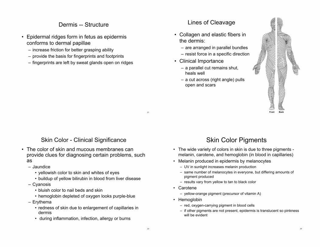
Lines of Cleavage
• Collagen and elastic fibers in
the dermis:
– are arranged in parallel bundles
– resist force in a specific direction
• Clinical Importance
– a parallel cut remains shut,
heals well
– a cut across (right angle) pulls
open and scars
23
Skin Color - Clinical Significance
• The color of skin and mucous membranes canprovide clues for diagnosing certain problems, suchas– Jaundice
• yellowish color to skin and whites of eyes
• buildup of yellow bilirubin in blood from liver disease
– Cyanosis
• bluish color to nail beds and skin
• hemoglobin depleted of oxygen looks purple-blue
– Erythema
• redness of skin due to enlargement of capillaries indermis
• during inflammation, infection, allergy or burns
24
Skin Color Pigments
• The wide variety of colors in skin is due to three pigments -
melanin, carotene, and hemoglobin (in blood in capillaries)
• Melanin produced in epidermis by melanocytes
– UV in sunlight increases melanin production
– same number of melanocytes in everyone, but differing amounts of
pigment produced
– results vary from yellow to tan to black color
• Carotene
– yellow-orange pigment (precursor of vitamin A)
• Hemoglobin
– red, oxygen-carrying pigment in blood cells
– if other pigments are not present, epidermis is translucent so pinkness
will be evident
25
Skin Color Pigments
• Clinical observations
– freckles or liver spots = overproduction of melanin
– Nevus or mole = overgrowth of melanocytes in a patch
– albinism = inherited lack of tyrosinase; no pigment
– vitiligo = autoimmune loss of melanocytes in areas of
the skin produces white patches
26
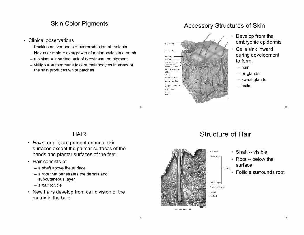
Accessory Structures of Skin
• Develop from the
embryonic epidermis
• Cells sink inward
during development
to form:
– hair
– oil glands
– sweat glands
– nails
27
HAIR
• Hairs, or pili, are present on most skin
surfaces except the palmar surfaces of the
hands and plantar surfaces of the feet
• Hair consists of
– a shaft above the surface
– a root that penetrates the dermis and
subcutaneous layer
– a hair follicle
• New hairs develop from cell division of the
matrix in the bulb
28
Structure of Hair
• Shaft -- visible
• Root -- below the
surface
• Follicle surrounds root
29
Structure of Hair
• Shaft -- visible
– medulla, cortex & cuticle
– cross-section round in
straight hair
– cross-section oval in wavy
hair
• Root -- below the surface
• Follicle surrounds root
– base of follicle is bulb
• blood vessels
• germinal cell layer
30
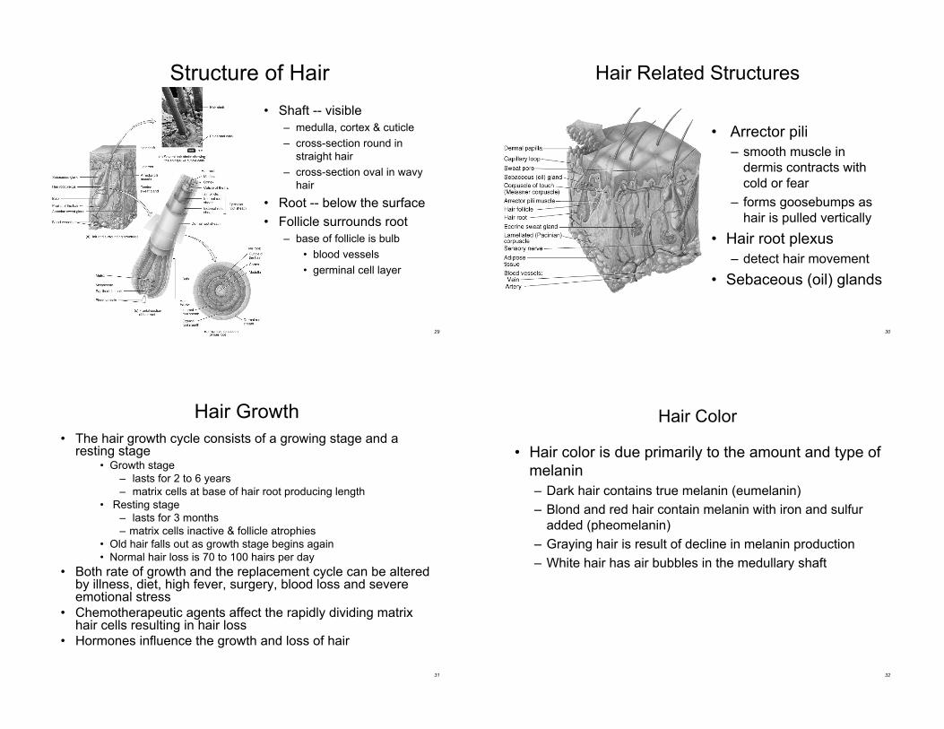
Hair Related Structures
• Arrector pili
– smooth muscle in
dermis contracts with
cold or fear
– forms goosebumps as
hair is pulled vertically
• Hair root plexus
– detect hair movement
• Sebaceous (oil) glands
31
Hair Growth
• The hair growth cycle consists of a growing stage and aresting stage
• Growth stage
– lasts for 2 to 6 years
– matrix cells at base of hair root producing length
• Resting stage
– lasts for 3 months
– matrix cells inactive & follicle atrophies
• Old hair falls out as growth stage begins again
• Normal hair loss is 70 to 100 hairs per day
• Both rate of growth and the replacement cycle can be alteredby illness, diet, high fever, surgery, blood loss and severeemotional stress
• Chemotherapeutic agents affect the rapidly dividing matrixhair cells resulting in hair loss
• Hormones influence the growth and loss of hair
32
Hair Color
• Hair color is due primarily to the amount and type of
melanin
– Dark hair contains true melanin (eumelanin)
– Blond and red hair contain melanin with iron and sulfur
added (pheomelanin)
– Graying hair is result of decline in melanin production
– White hair has air bubbles in the medullary shaft
33
Hair Texture
• Texture – related to differences in cross-sectional
shape
– straight hair is round
– wavy hair is oval
– curly hair is relatively flat
34
Hair Color and Texture
Blond, straight
Black, straight Red, wavy
Gray, wavy
Cuticle
Cortex
Medulla
Eumelanin
Pheomelanin
Eumelanin
Pheomelanin
Air
space
35
Functions of Hair
• Prevents heat loss
• Decreases sunburn
• Eyelashes help protect eyes
• Touch receptors (hair root
plexus) senses light touch
36
Glands of the Skin
Specialized exocrine glands found in dermis:
• Sebaceous (oil) glands
• Sudoriferous (sweat) glands
• Ceruminous (wax) glands
• Mammary (milk) glands
37
Sebaceous (oil) glands
• Sebaceous (oil) glands are usually connectedto hair follicles; they are absent in the palmsand soles
• Secretory portion of gland is located in thedermis– produce sebum
• contains cholesterol, proteins, fats & salts
• moistens hairs
• waterproofs and softens the skin
• inhibits growth of bacteria & fungi (ringworm)
• Acne– bacterial inflammation of glands
– secretions are stimulated by hormones at puberty
38
Sudoriferous (sweat) glands
• Eccrine sweat glands have an extensivedistribution most areas of skin– ducts terminate at pores at the surface of the epidermis
– regulate body temperature through evaporation(perspiration)
– help eliminate wastes such as urea
• Apocrine sweat glands are limited in distribution tothe skin of the axilla, pubis, and areolae– start funtioning at puberty
– duct that opens onto hair follicle
– secretions are more viscous
Both sweat glands secrete by a merocrinemechanism!
39
Ceruminous Glands
• Ceruminous glands are modified sudoriferous
glands that produce a waxy substance called
cerumen (ear wax)
– found in the external auditory canal
– barrier for entrance of foreign bodies
• An abnormal amount of cerumen can prevent
sound waves from reaching the ear drum
40
Structure of Nails
• Tightly packed keratinized cells
• Nail body
– visible portion pink due to underlying
capillaries
– free edge appears white
• Nail root
– buried under skin layers
– lunula is white due to thickened
stratum basale
• Eponychium (cuticle)
– stratum corneum layer
41
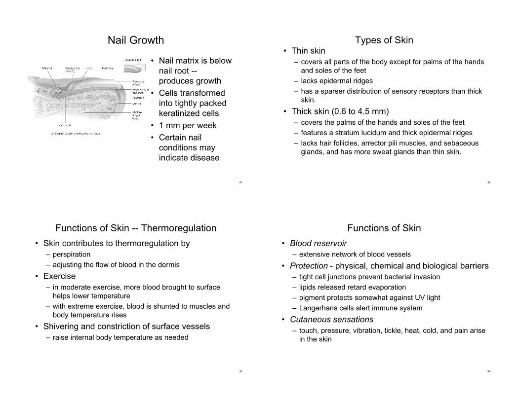
Nail Growth
• Nail matrix is below
nail root --
produces growth
• Cells transformed
into tightly packed
keratinized cells
• 1 mm per week
• Certain nail
conditions may
indicate disease
42
Types of Skin• Thin skin
– covers all parts of the body except for palms of the hands
and soles of the feet
– lacks epidermal ridges
– has a sparser distribution of sensory receptors than thick
skin.
• Thick skin (0.6 to 4.5 mm)
– covers the palms of the hands and soles of the feet
– features a stratum lucidum and thick epidermal ridges
– lacks hair follicles, arrector pili muscles, and sebaceous
glands, and has more sweat glands than thin skin.
43
Functions of Skin -- Thermoregulation
• Skin contributes to thermoregulation by
– perspiration
– adjusting the flow of blood in the dermis
• Exercise
– in moderate exercise, more blood brought to surface
helps lower temperature
– with extreme exercise, blood is shunted to muscles and
body temperature rises
• Shivering and constriction of surface vessels
– raise internal body temperature as needed
44
Functions of Skin
• Blood reservoir
– extensive network of blood vessels
• Protection - physical, chemical and biological barriers
– tight cell junctions prevent bacterial invasion
– lipids released retard evaporation
– pigment protects somewhat against UV light
– Langerhans cells alert immune system
• Cutaneous sensations
– touch, pressure, vibration, tickle, heat, cold, and pain arise
in the skin
45
Functions of Skin
• Synthesis of Vitamin D
– activation of a precursor molecule in the skin by UV light
– necessary vitamin for absorption of calcium from food in
the gastrointestinal tract
• Excretion
– 400 mL of water/day, small amounts salt, CO2, ammonia
and urea
46
Transdermal Drug Administration
• Method of drug passage across the epidermis and
into the blood vessels of the dermis
– drug absorption is most rapid in areas where skin is thin
(scrotum, face and scalp)
• Examples:
– nitroglycerin (prevention of chest pain from coronary
artery disease)
– scopolamine (motion sickness)
– estradiol (estrogen replacement therapy)
– nicotine (stop smoking alternative)
47
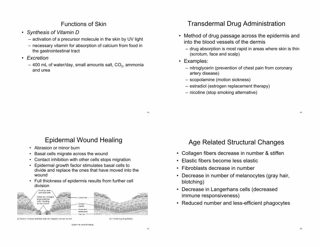
Epidermal Wound Healing• Abrasion or minor burn
• Basal cells migrate across the wound
• Contact inhibition with other cells stops migration
• Epidermal growth factor stimulates basal cells todivide and replace the ones that have moved into thewound
• Full thickness of epidermis results from further celldivision
48
Age Related Structural Changes
• Collagen fibers decrease in number & stiffen
• Elastic fibers become less elastic
• Fibroblasts decrease in number
• Decrease in number of melanocytes (gray hair,
blotching)
• Decrease in Langerhans cells (decreased
immune responsiveness)
• Reduced number and less-efficient phagocytes
49
Photodamage
• Ultraviolet light (UVA and UVB) both damage the
skin
• Acute overexposure causes sunburn
• DNA damage in epidermal cells can lead to skin
cancer
• UVA produces oxygen free radicals that damage
collagen and elastic fibers and lead to wrinkling of
the skin
50
Skin Cancer
• 1 million cases diagnosed per year
• 3 common forms of skin cancer
– basal cell carcinoma (rarely metastasize)
– squamous cell carcinoma (may metastasize)
– malignant melanomas (metastasize rapidly)
• most common cancer in young women
• arise from melanocytes --- life threatening
• key to treatment is early detection watch for
changes in symmetry, border, color and size
• risks factors include -- skin color, sun exposure,
family history, age and immunological status
51
Burns
• Tissue damage from excessive heat, electricity,radioactivity, or corrosive chemicals that destroys(denatures) proteins in the exposed cells is called aburn.
• Generally, the systemic effects of a burn are agreater threat to life than are the local effects.
• The seriousness of a burn is determined by its depth,extent, and area involved, as well as the person’sage and general health. When the burn areaexceeds 70%, over half of the victims die.
• Problems that result– shock due to water, plasma and plasma protein loss
– circulatory & kidney problems from loss of plasma
– bacterial infection52
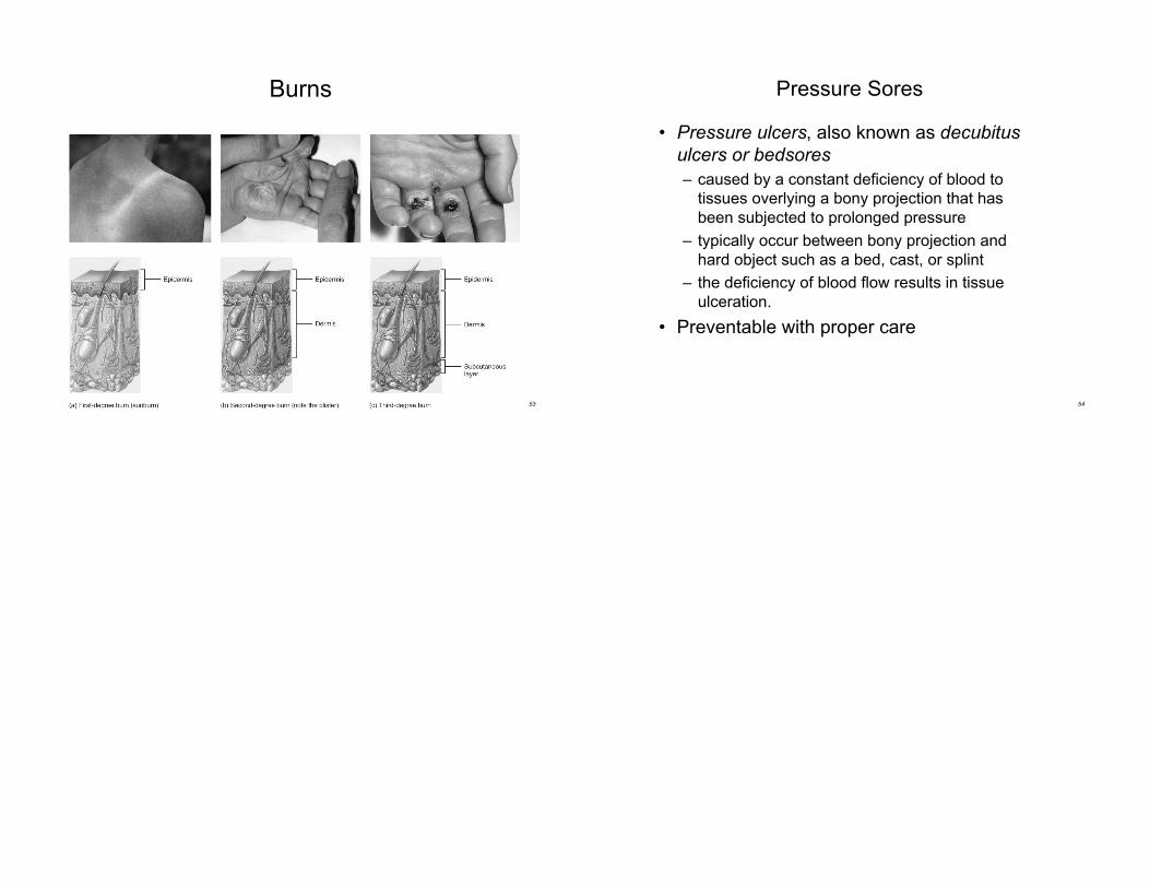
Types of Burns
• First-degree
– only epidermis (sunburn)
• Second-degree
– destroys entire epidermis & part of dermis
– fluid-filled blisters separate epidermis & dermis
– accessory structures are not damaged
– heals without grafting in 3 to 4 weeks & may scar