103
CHAPTER 6 EYE AND EAR ASSESSMENT AND PROCEDURES

CHAPTER 6CHAPTER 6EYE AND EAR ASSESSMENT
AND PROCEDURES

Elsevier items and derived items © 2008 by Saunders, an imprint of Elsevier Inc. 2
PRETESTPRETEST
True or False1. The outer layer of the eye is composed of white
connective tissue known as the sclera.
2. A person who is farsighted has a condition known as myopia.
3. An optometrist can perform eye surgery.
4. The Snellen eye test is conducted at a distance of 20 feet.
5. The eustachian tube connects the nasopharynx to the inner ear.
Elsevier items and derived items © 2008 by Saunders, an imprint of Elsevier Inc. 3
PRETEST, CONT.PRETEST, CONT.
True or False6. An eye instillation may be performed to treat an
eye infection.
7. The function of cerumen is to inhibit the growth of pathogens.
8. The most specific type of hearing test is the tuning fork test.
9. Serous otitis media can result in a conductive hearing loss.
10. An ear instillation may be performed to treat an ear infection.
Elsevier items and derived items © 2008 by Saunders, an imprint of Elsevier Inc. 4
Content OutlineContent Outline
1. MA is responsible for performing a variety of eye and ear assessments and procedures
2. Visual acuity test: screening test to detect deficiencies in vision
3. Hearing test: use of tuning fork or audiometera. Audiometer: instrument that emits sound
waves at various frequencies
b. MA should be alert to signs that indicate the patient might be having difficulty hearing
Introduction to Eye and Ear Assessment
Elsevier items and derived items © 2008 by Saunders, an imprint of Elsevier Inc. 5
Introduction to Eye and Ear Assessment, cont.
Introduction to Eye and Ear Assessment, cont.
4. Color vision assessment
a. Requires specially prepared colored plates
b. Color blindness: inability to distinguish certain colors
• Red and green most common
Elsevier items and derived items © 2008 by Saunders, an imprint of Elsevier Inc. 6
Introduction to Eye and Ear Assessment, cont.
Introduction to Eye and Ear Assessment, cont.
5. Eye and ear irrigations and instillations
a. Irrigation: washing a body canal with a flowing solution
b. Instillation: dropping a liquid into a body cavity

Elsevier items and derived items © 2008 by Saunders, an imprint of Elsevier Inc. 7
The EyeThe Eye
1. Eye has three layers:
a. Sclera: outer layer
• Composed of tough, white, fibrous connective tissue
• Front of sclera is modified to form cornea
– Cornea: transparent covering over the colored part of the eye
Structure of the Eye
Elsevier items and derived items © 2008 by Saunders, an imprint of Elsevier Inc. 8
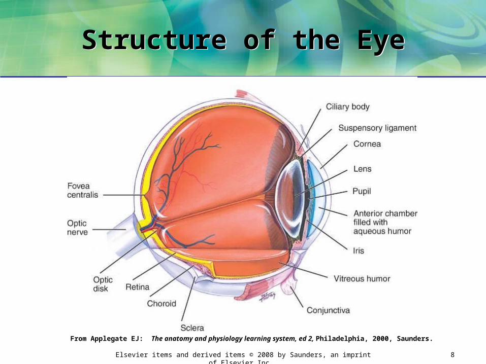
Structure of the EyeStructure of the Eye
From Applegate EJ: The anatomy and physiology learning system, ed 2, Philadelphia, 2000, Saunders.

Elsevier items and derived items © 2008 by Saunders, an imprint of Elsevier Inc. 9
Structure of the Eye, cont. Structure of the Eye, cont.
b. Choroid: middle layer
• Composed of many blood vessels and is highly pigmented
– Blood vessels: nourish the eye
– Pigment: absorbs stray light rays
• Front part of choroid specialized into:
– Ciliary body: muscles that control shape of the lens
– Suspensory ligaments: suspend lens in place
1) Lens: focuses light rays on the retina
Elsevier items and derived items © 2008 by Saunders, an imprint of Elsevier Inc. 10
Structure of the Eye, cont.Structure of the Eye, cont.
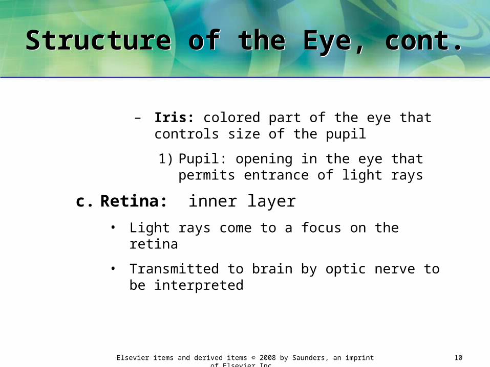
– Iris: colored part of the eye that controls size of the pupil
1) Pupil: opening in the eye that permits entrance of light rays
c. Retina: inner layer
• Light rays come to a focus on the retina
• Transmitted to brain by optic nerve to be interpreted
Elsevier items and derived items © 2008 by Saunders, an imprint of Elsevier Inc. 11
Structure of the Eye, cont. Structure of the Eye, cont.
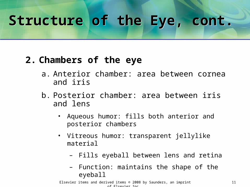
2. Chambers of the eye
a. Anterior chamber: area between cornea and iris
b. Posterior chamber: area between iris and lens
• Aqueous humor: fills both anterior and posterior chambers
• Vitreous humor: transparent jellylike material
– Fills eyeball between lens and retina
– Function: maintains the shape of the eyeball

Elsevier items and derived items © 2008 by Saunders, an imprint of Elsevier Inc. 12
Structure of the Eye, cont. Structure of the Eye, cont.
3. Conjunctiva: membrane that lines eyelids and covers front of the eye, except for the cornea
a. Conjunctiva covering the sclera: transparent
• Allows white sclera to show through
Elsevier items and derived items © 2008 by Saunders, an imprint of Elsevier Inc. 13
Visual AcuityVisual Acuity
1. Visual acuity: acuteness or sharpness of vision
2. Normal visual acuity:
a. Can see clearly
b. Able to distinguish fine details
• Both close up and at a distance

Elsevier items and derived items © 2008 by Saunders, an imprint of Elsevier Inc. 14
Errors of Refraction Errors of Refraction
3. Errors of refraction: most common cause of defects in visual acuity
a. Refraction: bending of the parallel light rays coming into the eye so they can be focused on the retina
Elsevier items and derived items © 2008 by Saunders, an imprint of Elsevier Inc. 15
Errors of Refraction, cont. Errors of Refraction, cont.
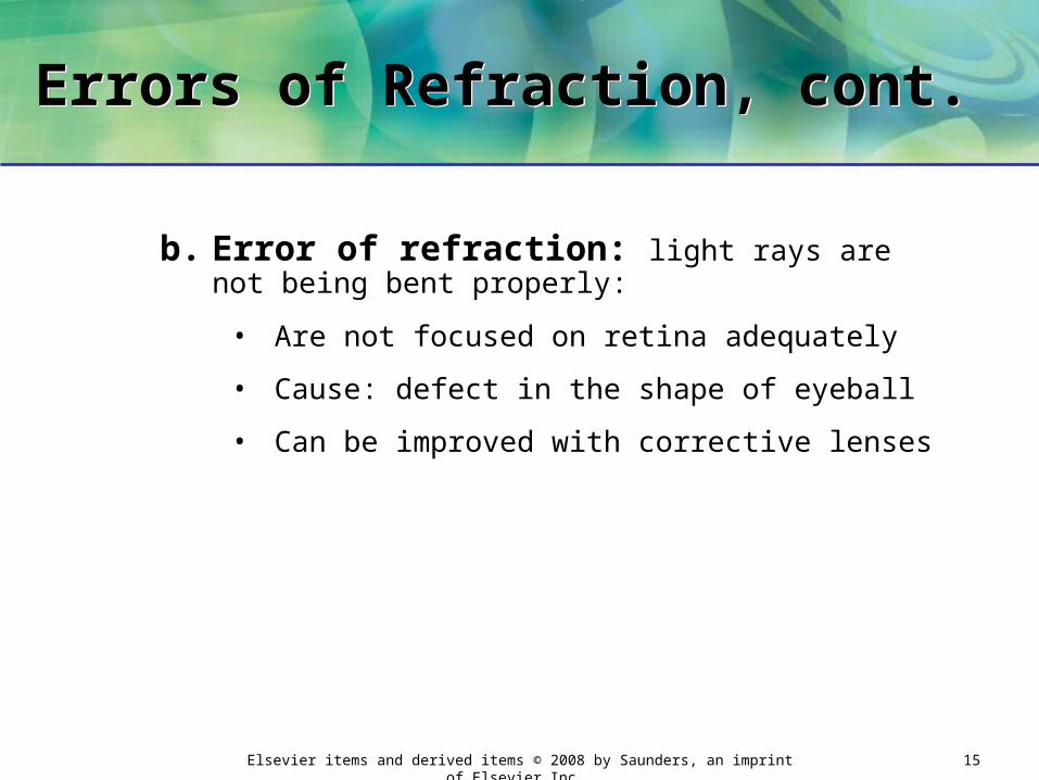
b. Error of refraction: light rays are not being bent properly:
• Are not focused on retina adequately
• Cause: defect in the shape of eyeball
• Can be improved with corrective lenses

Elsevier items and derived items © 2008 by Saunders, an imprint of Elsevier Inc. 16
Myopia Myopia
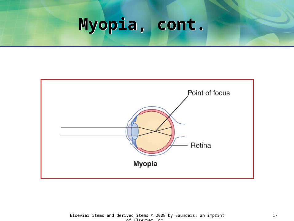
c. Myopia (nearsighted): eyeball too long from front to back
• Causes light rays to focus in front of retina
– Difficulty seeing objects at a distance
– May squint and have HA from eye strain
– Corrective lenses (eyeglasses, contact lenses) or laser surgery: can correct condition
1) Cause light rays to focus on retina
Elsevier items and derived items © 2008 by Saunders, an imprint of Elsevier Inc. 17
Myopia, cont. Myopia, cont.

Elsevier items and derived items © 2008 by Saunders, an imprint of Elsevier Inc. 18
Hyperopia Hyperopia
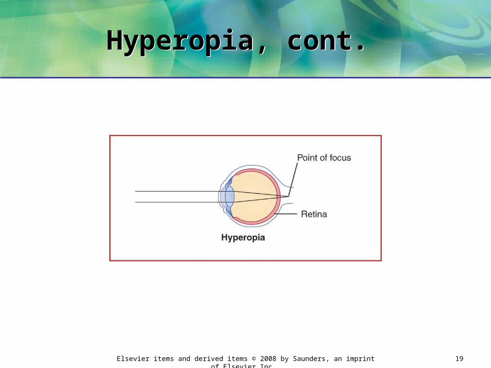
d. Hyperopia (farsighted): eyeball too short from front to back
• Causes light rays to focus behind the retina
– Difficulty viewing objects at a reading or working distance
– May have blurring, headache, and eye strain while performing close-up tasks
– Corrective lenses can correct condition
1) Cause light rays to focus on retina
Elsevier items and derived items © 2008 by Saunders, an imprint of Elsevier Inc. 19
Hyperopia, cont. Hyperopia, cont.

Elsevier items and derived items © 2008 by Saunders, an imprint of Elsevier Inc. 20
Presbyopia Presbyopia
e. Presbyopia: decrease in elasticity of lens
• Usually begins after age 40
• Results in a decreased ability to focus clearly on close objects

Elsevier items and derived items © 2008 by Saunders, an imprint of Elsevier Inc. 21
Eye Specialists Eye Specialists
4. Types of eye specialists
a. Ophthalmologist: medical doctor specializing in diagnosis and treatment of diseases and disorders of the eye
• Prescribes ophthalmic and systemic medications
• Performs eye surgery
Elsevier items and derived items © 2008 by Saunders, an imprint of Elsevier Inc. 22
Eye Specialists, cont. Eye Specialists, cont.
b. Optometrist: licensed primary health care provider who has expertise in measuring visual acuity and prescribing corrective lenses
• Can diagnose and treat of disorders and diseases of the eye
• Prescribes ophthalmic medications
• Not a physician: cannot prescribe systemic medications or perform eye surgery
c. Optician: professional who interprets and fills prescription for eyeglasses and contact lenses

Elsevier items and derived items © 2008 by Saunders, an imprint of Elsevier Inc. 23
Assessment of Distance Visual Acuity (DVA)
Assessment of Distance Visual Acuity (DVA)
1. Used to diagnose myopia
a. Along with other tests
2. Snellen eye chart: most often used

Elsevier items and derived items © 2008 by Saunders, an imprint of Elsevier Inc. 24
Assessment of Distance Visual Acuity (DVA), cont.
Assessment of Distance Visual Acuity (DVA), cont.
3. Types of charts
a. Letters in decreasing sizes
• Used for school-aged children and adults

Elsevier items and derived items © 2008 by Saunders, an imprint of Elsevier Inc. 25
Assessment of Distance Visual Acuity (DVA), cont.
Assessment of Distance Visual Acuity (DVA), cont.
b. Capital letter E in decreasing sizes (arranged in different directions)
• Used for preschool children, non-English-speaking people, nonreaders
c. Pictures of familiar objects
• Used for preschoolers
• Less accurate because some children are unable to identify objects

Elsevier items and derived items © 2008 by Saunders, an imprint of Elsevier Inc. 26
Snellen Big E ChartSnellen Big E Chart
Elsevier items and derived items © 2008 by Saunders, an imprint of Elsevier Inc. 27
Conducting a Snellen TestConducting a Snellen Test
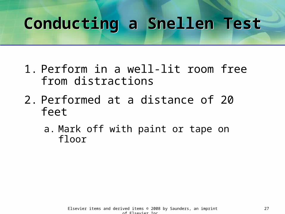
1. Perform in a well-lit room free from distractions
2. Performed at a distance of 20 feet
a. Mark off with paint or tape on floor

Elsevier items and derived items © 2008 by Saunders, an imprint of Elsevier Inc. 28
Conducting a Snellen Test, cont.
Conducting a Snellen Test, cont.
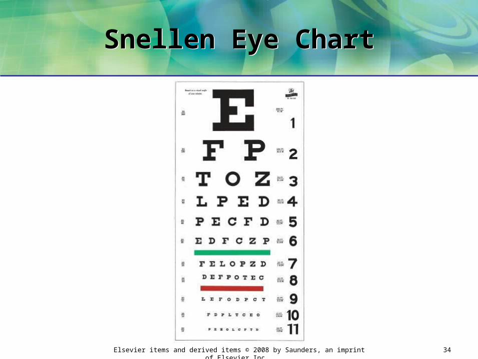
3. Two numbers next to each row of letters
a. Number above line: distance at which test is conducted (20 feet)
b. Number below line: distance from which a person with normal visual acuity can read the row of letters
4. Normal DVA: 20/20
a. Person can read what supposed to read at 20 feet
Elsevier items and derived items © 2008 by Saunders, an imprint of Elsevier Inc. 29
Conducting a Snellen Test, cont.
Conducting a Snellen Test, cont.
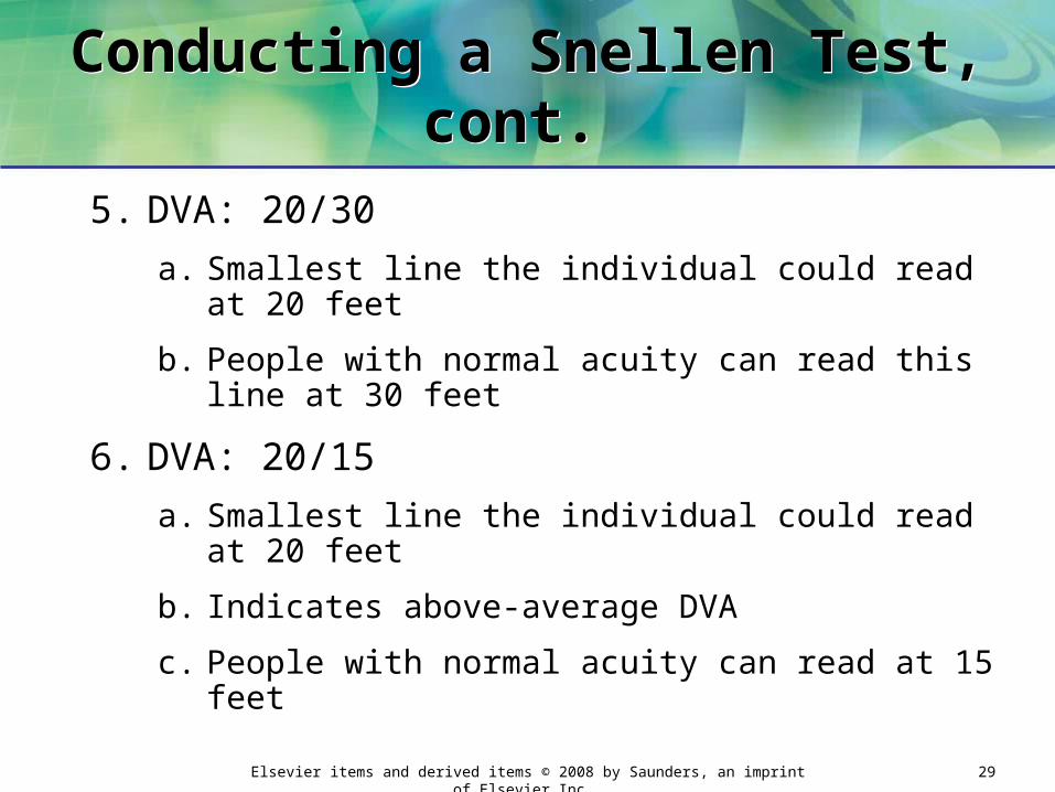
5. DVA: 20/30
a. Smallest line the individual could read at 20 feet
b. People with normal acuity can read this line at 30 feet
6. DVA: 20/15
a. Smallest line the individual could read at 20 feet
b. Indicates above-average DVA
c. People with normal acuity can read at 15 feet

Elsevier items and derived items © 2008 by Saunders, an imprint of Elsevier Inc. 30
Conducting a Snellen Test, cont.
Conducting a Snellen Test, cont.
7. Acuity of each eye measured separately
a. Traditionally beginning with right eye
8. If patient wears eyeglasses or contact lenses (except reading glasses)
a. Keep them on during test
b. Record in chart that corrective lenses were worn
c. Also record if corrective lenses were not being worn

Elsevier items and derived items © 2008 by Saunders, an imprint of Elsevier Inc. 31
Snellen Eye Test Snellen Eye Test
Elsevier items and derived items © 2008 by Saunders, an imprint of Elsevier Inc. 32
Conducting a Snellen Test, cont.
Conducting a Snellen Test, cont.
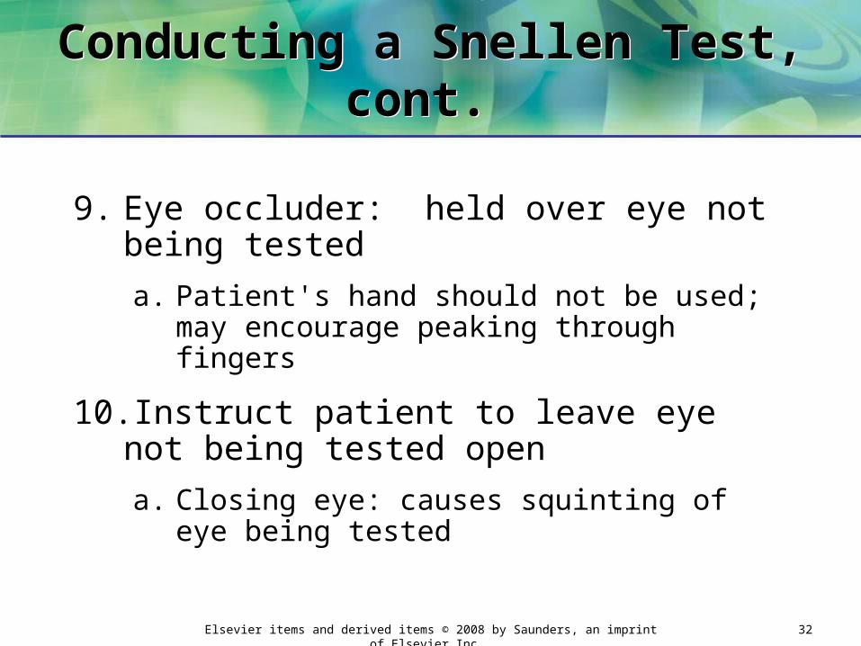
9. Eye occluder: held over eye not being tested
a. Patient's hand should not be used; may encourage peaking through fingers
10.Instruct patient to leave eye not being tested open
a. Closing eye: causes squinting of eye being tested

Elsevier items and derived items © 2008 by Saunders, an imprint of Elsevier Inc. 33
Conducting a Snellen Test, cont.
Conducting a Snellen Test, cont.
Elsevier items and derived items © 2008 by Saunders, an imprint of Elsevier Inc. 34
Snellen Eye ChartSnellen Eye Chart

Elsevier items and derived items © 2008 by Saunders, an imprint of Elsevier Inc. 35
Assessing Distance Visual Acuity in Preschoolers
Assessing Distance Visual Acuity in Preschoolers
1. Snellen Big E chart used
2. Completely explain procedure to child
a. Tell child you will be playing a pointing game
b. Do not force child: results will be inaccurate
Elsevier items and derived items © 2008 by Saunders, an imprint of Elsevier Inc. 36
Assessing Distance Visual Acuity in Preschoolers, cont.
Assessing Distance Visual Acuity in Preschoolers, cont.
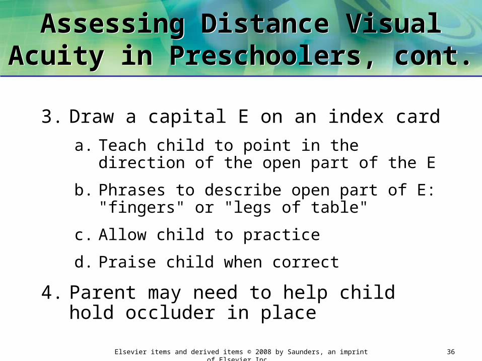
3. Draw a capital E on an index card
a. Teach child to point in the direction of the open part of the E
b. Phrases to describe open part of E: "fingers" or "legs of table"
c. Allow child to practice
d. Praise child when correct
4. Parent may need to help child hold occluder in place

Elsevier items and derived items © 2008 by Saunders, an imprint of Elsevier Inc. 37
Snellen Big E ChartSnellen Big E Chart
Elsevier items and derived items © 2008 by Saunders, an imprint of Elsevier Inc. 38
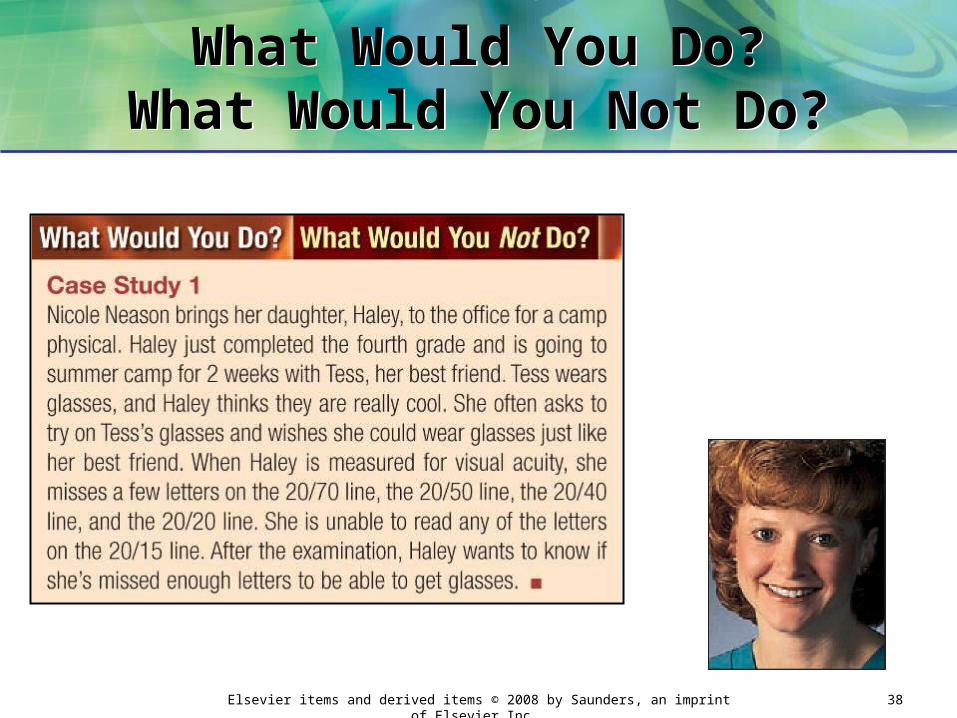
What Would You Do?What Would You Not Do?
What Would You Do?What Would You Not Do?

Elsevier items and derived items © 2008 by Saunders, an imprint of Elsevier Inc. 39
What Would You Do?What Would You Not Do?
What Would You Do?What Would You Not Do?

Elsevier items and derived items © 2008 by Saunders, an imprint of Elsevier Inc. 40
Assessment of Near Visual Acuity (NVA)
Assessment of Near Visual Acuity (NVA)
1. Assesses patient's ability to read objects close up
2. Used to detect hyperopia and presbyopia
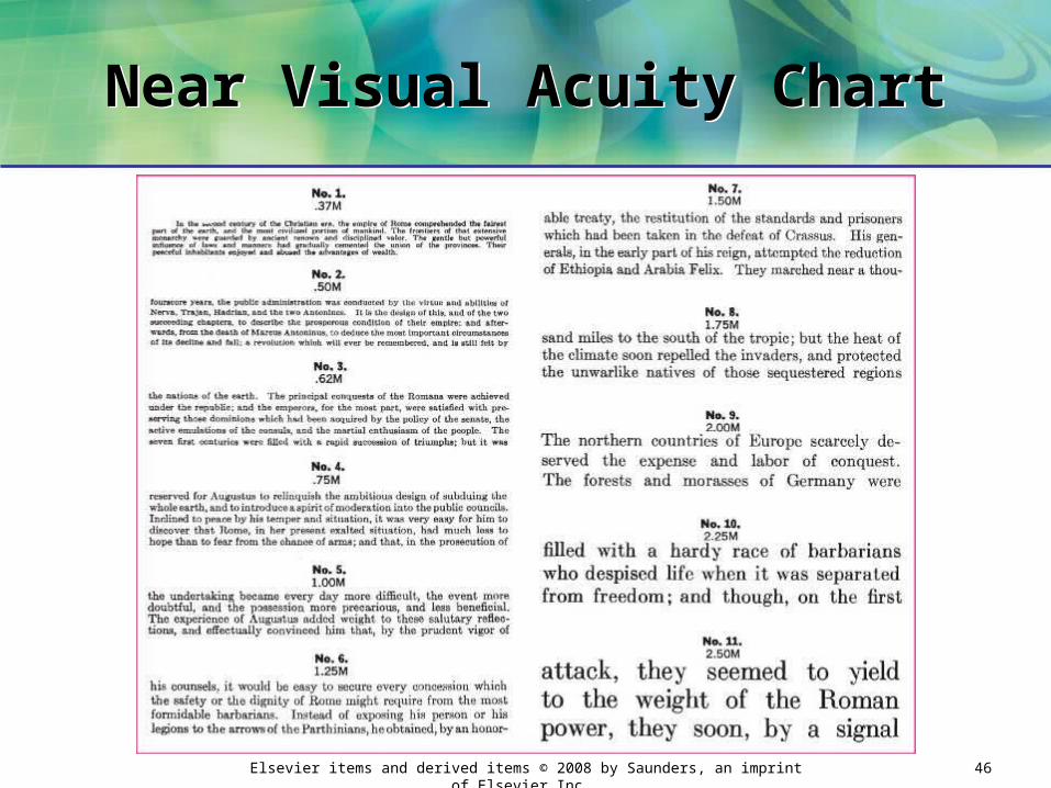
3. NVA card: contains different sizes of type:
a. Ranging from size of newspaper headline down to very small print

Elsevier items and derived items © 2008 by Saunders, an imprint of Elsevier Inc. 41
Assessment of Near Visual Acuity (NVA)
Assessment of Near Visual Acuity (NVA)
4. Available in variety of forms:
a. Printed paragraphs
b. Printed words
c. Pictures

Elsevier items and derived items © 2008 by Saunders, an imprint of Elsevier Inc. 42
Assessment of Near Visual Acuity (NVA), cont.
Assessment of Near Visual Acuity (NVA), cont.
5. To perform NVA testing:
a. Perform test in well-lit room free of distractions
b. Patient holds card at a distance of 14 to 16 inches
c. Reading glasses should be worn (if patient uses them)
d. Each eye tested separately
Elsevier items and derived items © 2008 by Saunders, an imprint of Elsevier Inc. 43
Assessment of Near Visual Acuity (NVA), cont.
Assessment of Near Visual Acuity (NVA), cont.
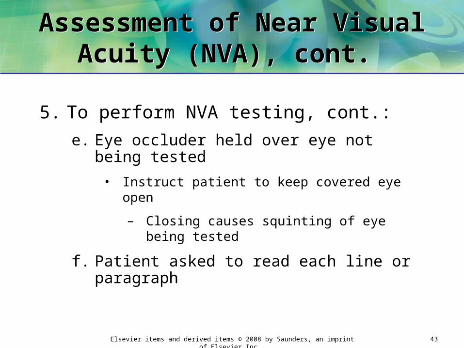
5. To perform NVA testing, cont.:
e. Eye occluder held over eye not being tested
• Instruct patient to keep covered eye open
– Closing causes squinting of eye being tested
f. Patient asked to read each line or paragraph

Elsevier items and derived items © 2008 by Saunders, an imprint of Elsevier Inc. 44
Assessment of Near Visual Acuity (NVA), cont.
Assessment of Near Visual Acuity (NVA), cont.
g. Observe patient for unusual symptoms
• Squinting, tilting of head, watering of eyes
– Indicates patient having difficult reading card
h. Patient continues until reaching smallest line that can be read

Elsevier items and derived items © 2008 by Saunders, an imprint of Elsevier Inc. 45
Assessment of Near Visual Acuity (NVA), cont.
Assessment of Near Visual Acuity (NVA), cont.
i. Record results as smallest type patient could read with each eye
• Recording based on type of test card used
j. Also record:
• Date and time
• If corrective lenses worn
• Unusual symptoms exhibited by patient
Elsevier items and derived items © 2008 by Saunders, an imprint of Elsevier Inc. 46
Near Visual Acuity ChartNear Visual Acuity Chart
Elsevier items and derived items © 2008 by Saunders, an imprint of Elsevier Inc. 47
Assessment of Color VisionAssessment of Color Vision
1. Classification of defects in color vision:
a. Congenital defect: most common
• Inherited (present at birth)
• Most often affects males
b. Acquired defect: acquired after birth
• Eye injury
• Disease
• Certain drugs
Elsevier items and derived items © 2008 by Saunders, an imprint of Elsevier Inc. 48
Assessment of Color Vision, cont.
Assessment of Color Vision, cont.
2. Color vision tests detect congenital color vision defects
a. Often performed in medical office
3. Basic color vision screening test:
a. Ask patient to identify red and green lines on Snellen chart

Elsevier items and derived items © 2008 by Saunders, an imprint of Elsevier Inc. 49
Ishihara TestIshihara Test
1. Detects:
a. Total congenital color blindness
b. Red-green color blindness
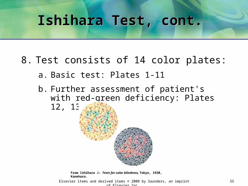
2. Series of plates: colored dots forming a numeral against a background of dots of contrasting colors
3. Patients with normal color vision: read appropriate numeral

Elsevier items and derived items © 2008 by Saunders, an imprint of Elsevier Inc. 50
Ishihara Color PlatesIshihara Color Plates
From Ishihara J: Tests for color blindness, Tokyo, 1920, Kanehara.

Elsevier items and derived items © 2008 by Saunders, an imprint of Elsevier Inc. 51
Ishihara Test, cont. Ishihara Test, cont.
4. Patients with defects read dots as:
a. Not forming a number at all
b. Forming a different number
Elsevier items and derived items © 2008 by Saunders, an imprint of Elsevier Inc. 52
Ishihara Test, cont. Ishihara Test, cont.
5. First plate can be read correctly by all patients
a. Used to explain test procedure to patient
6. Plates with winding colored lines:
a. For patients unable to identify numbers by name
b. Patient asked to trace line formed by the dots
c. Used for preschoolers and non-English-speaking persons

Elsevier items and derived items © 2008 by Saunders, an imprint of Elsevier Inc. 53
Ishihara Test, cont. Ishihara Test, cont.
7. Conduct test in quiet room
a. Illuminated by natural daylight
• Bright sunlight can change shades of color on plates
– Can cause inaccurate test results
b. Can use electric light
• Should adjust to resemble natural daylight

Elsevier items and derived items © 2008 by Saunders, an imprint of Elsevier Inc. 54
Ishihara Test, cont. Ishihara Test, cont.
Elsevier items and derived items © 2008 by Saunders, an imprint of Elsevier Inc. 55
Ishihara Test, cont. Ishihara Test, cont.
8. Test consists of 14 color plates:
a. Basic test: Plates 1-11
b. Further assessment of patient's with red-green deficiency: Plates 12, 13, and 14
From Ishihara J: Tests for color blindness, Tokyo, 1920, Kanehara.

Elsevier items and derived items © 2008 by Saunders, an imprint of Elsevier Inc. 56
Ishihara Test, cont. Ishihara Test, cont.
9. Interpretation of results
a. Normal color vision: 10 or more plates read correctly
b. Color vision deficiency: 7 or fewer plates read correctly
10.Defect in color vision: patient referred to ophthalmologist or optometrist
a. For additional assessment
• Use of more precise color vision tests
Elsevier items and derived items © 2008 by Saunders, an imprint of Elsevier Inc. 57
Eye IrrigationEye Irrigation
1. Washing the eye with a flowing solution
2. Purpose
a. Cleanse the eye by washing away:
• Foreign particles
• Ocular discharges
• Harmful chemicals
b. Relieve inflammation though application of heat
c. Apply an antiseptic solution

Elsevier items and derived items © 2008 by Saunders, an imprint of Elsevier Inc. 58
Eye Irrigation, cont.Eye Irrigation, cont.

Elsevier items and derived items © 2008 by Saunders, an imprint of Elsevier Inc. 59
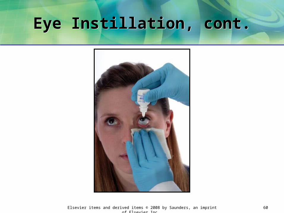
Eye InstillationEye Instillation
1. Dropping of a liquid into lower conjunctival sac
2. Purpose
a. Treat eye infections (with medications)
b. Soothe an irritated eye
c. Dilate the pupil
d. Anesthetize during eye examination or treatment
Elsevier items and derived items © 2008 by Saunders, an imprint of Elsevier Inc. 60
Eye Instillation, cont.Eye Instillation, cont.

Elsevier items and derived items © 2008 by Saunders, an imprint of Elsevier Inc. 61
Eye Instillation, cont. Eye Instillation, cont.
2. Medications instilled in eye may come in the form of:
a. Liquid (ophthalmic drops)
• Usually dispensed in a flexible bottle with an attached dropper
b. Ointment
• Dispensed in a small metal tube with tip for applying medication

Elsevier items and derived items © 2008 by Saunders, an imprint of Elsevier Inc. 62
What Would You Do?What Would You Not Do?
What Would You Do?What Would You Not Do?

Elsevier items and derived items © 2008 by Saunders, an imprint of Elsevier Inc. 63
What Would You Do?What Would You Not Do?
What Would You Do?What Would You Not Do?
Elsevier items and derived items © 2008 by Saunders, an imprint of Elsevier Inc. 64
The EarThe Ear
1. Functions in hearing and maintaining equilibrium
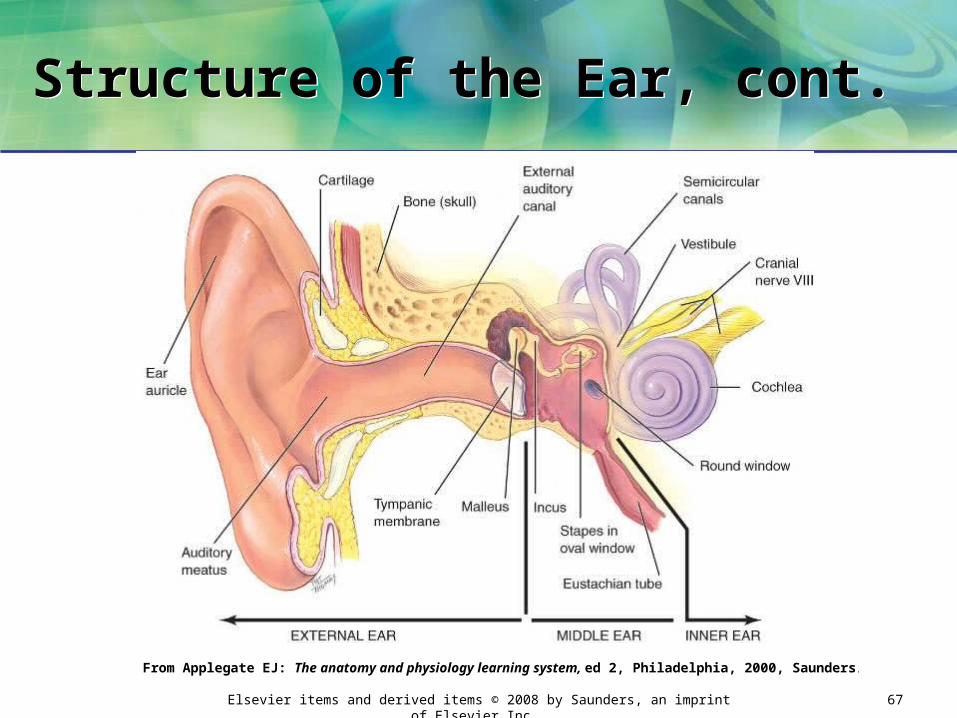
2. Consists of three divisions:
a. External ear
• Auricle (pinna): flap of cartilage covered with skin that
– Projects from side of head
– Receives and collects sound waves and directs them toward the external auditory canal
Structure of the Ear
Elsevier items and derived items © 2008 by Saunders, an imprint of Elsevier Inc. 65
Structure of the Ear, cont. Structure of the Ear, cont.
• External auditory canal: extends from auricle to tympanic membrane
– Also known as external ear canal
– External auditory meatus: opening of canal
– 1 inch long in adult
– Lined with skin that contains nerve endings, fine hairs, glands
1) Glands secrete cerumen: lubricates and protects ear canal

Elsevier items and derived items © 2008 by Saunders, an imprint of Elsevier Inc. 66
Structure of the Ear, cont. Structure of the Ear, cont.
– Canal has an S-shaped curve as it leads inward
1) Canal must be straightened during:
• Tympanic membrane: located at end of the canal
– Pearly gray semitransparent membrane
– Receives sound waves
a) Otoscopic examination
b) Ear instillation
c) Ear irrigation
d) Aural temperature measurement
Elsevier items and derived items © 2008 by Saunders, an imprint of Elsevier Inc. 67
Structure of the Ear, cont. Structure of the Ear, cont.
From Applegate EJ: The anatomy and physiology learning system, ed 2, Philadelphia, 2000, Saunders.
Elsevier items and derived items © 2008 by Saunders, an imprint of Elsevier Inc. 68
Structure of the Ear, cont. Structure of the Ear, cont.
b. Middle ear: air-filled cavity
• Contains three small bones (ossicles)
– Malleus
– Incus
– Stapes
• Eustachian tube: connects middle ear to the nasopharynx
– Stabilizes air pressure between the external atmosphere and the middle ear
Elsevier items and derived items © 2008 by Saunders, an imprint of Elsevier Inc. 69
Structure of the Ear, cont. Structure of the Ear, cont.
c. Inner ear
• Cochlea: essential organ of hearing
• Semicircular canals: maintain equilibrium
Elsevier items and derived items © 2008 by Saunders, an imprint of Elsevier Inc. 70
Assessment of Hearing AcuityAssessment of Hearing Acuity
1. Part of complete PE
2. Person can have hearing loss and not be aware of it
a. Early detection and treatment: may prevent permanent hearing loss
Elsevier items and derived items © 2008 by Saunders, an imprint of Elsevier Inc. 71
Assessment of Hearing AcuityAssessment of Hearing Acuity
3. Person with normal hearing: can hear frequencies of normal speech
a. Ranges from 300 to 4000 Hz (hertz or cycles per second)
4. Patients who exhibit hearing loss:
a. Referred to otolaryngologist or audiologist
Elsevier items and derived items © 2008 by Saunders, an imprint of Elsevier Inc. 72
Conductive Hearing LossConductive Hearing Loss
1. Conductive hearing loss: results when there is a physical interference with normal conduction of sound waves through external and middle ear
a. Most common type of hearing loss
b. Amount of sound reaching inner ear is less than normal
Elsevier items and derived items © 2008 by Saunders, an imprint of Elsevier Inc. 73
Conductive Hearing Loss, cont. Conductive Hearing Loss, cont.
c. Conductive loss in external ear
• Caused by an obstruction of external ear canal:
– Impacted cerumen
– External otitis (swimmer's ear)
– Foreign bodies
– Benign growths (polyps)

Elsevier items and derived items © 2008 by Saunders, an imprint of Elsevier Inc. 74
Conductive Hearing Loss, cont. Conductive Hearing Loss, cont.
d. Conductive loss in middle ear
• Caused by an obstruction in middle ear:
– Serous otitis media (fluid in middle ear)
– Acute otitis media (infection in middle ear)
– Perforated tympanic membrane
– Otosclerosis
Elsevier items and derived items © 2008 by Saunders, an imprint of Elsevier Inc. 75
Conductive Hearing Loss, cont.
Conductive Hearing Loss, cont.
e. Cause of conductive loss: often detected by examining the ear canal with otoscope
• Hearing frequently restored by:
– Removing obstruction (e.g., cerumen)
– Treating the disorder (e.g., serous otitis media)
Elsevier items and derived items © 2008 by Saunders, an imprint of Elsevier Inc. 76
Sensorineural Hearing LossSensorineural Hearing Loss
2. Sensorineural hearing loss: results from damage to inner ear or auditory nerve
a. Sound is conducted normally through outer and middle ear structures
b. Problem with perception of sound waves: results in a hearing deficit

Elsevier items and derived items © 2008 by Saunders, an imprint of Elsevier Inc. 77
Sensorineural Hearing Loss, cont.
Sensorineural Hearing Loss, cont.
• Causes
– Hereditary factors
– Intense noise exposure over period of time
– Tumors
– Changes from normal aging process
– Ototoxicity caused by certain medications
– Infectious diseases (measles, mumps, meningitis)
3. Mixed hearing loss: combination of both conductive and sensorineural loss
Elsevier items and derived items © 2008 by Saunders, an imprint of Elsevier Inc. 78
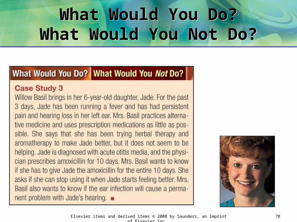
What Would You Do?What Would You Not Do?
What Would You Do?What Would You Not Do?

Elsevier items and derived items © 2008 by Saunders, an imprint of Elsevier Inc. 79
What Would You Do?What Would You Not Do?
What Would You Do?What Would You Not Do?
Elsevier items and derived items © 2008 by Saunders, an imprint of Elsevier Inc. 80
Hearing Acuity TestsHearing Acuity Tests
1. Include:
a. Simple gross screening test
b. Qualitative tests: tuning fork
c. Highly sensitive tests: audiometry
Elsevier items and derived items © 2008 by Saunders, an imprint of Elsevier Inc. 81
Hearing Acuity Tests, cont. Hearing Acuity Tests, cont.
2. Important to test only one ear at a time
a. Hearing deficit may exist only in one ear
3. Ear not being tested: blocked by an earplug or masked
a. Masking: presentation of sound to ear not being tested
• So patient's response is based only on hearing in ear being tested

Elsevier items and derived items © 2008 by Saunders, an imprint of Elsevier Inc. 82
Gross Screening Test Gross Screening Test
4. Gross Screening Test
a. Used to identify a very large hearing impairment
b. Whisper test: patient asked to repeat simple word or series of numbers
• Whispered from a distance of 1 to 2 feet

Elsevier items and derived items © 2008 by Saunders, an imprint of Elsevier Inc. 83
Tuning Fork Tests Tuning Fork Tests
5. Tuning Fork Tests
a. Provide a general assessment of hearing acuity
b. Use of tuning fork with frequency of 512 or 1024 Hz
• These frequencies fall within range of normal speech
Elsevier items and derived items © 2008 by Saunders, an imprint of Elsevier Inc. 84
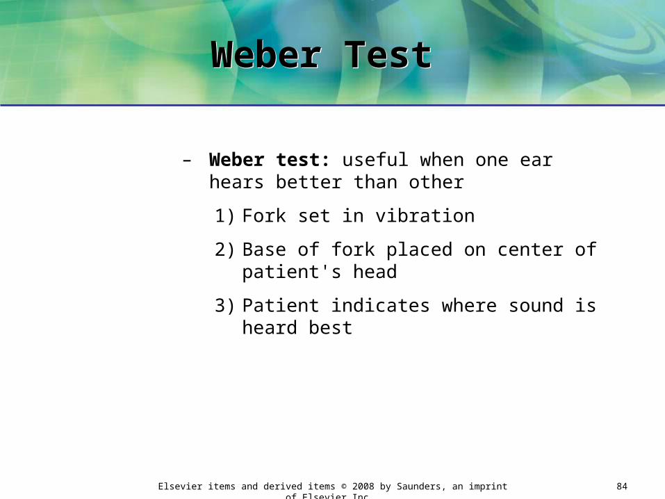
Weber Test Weber Test
– Weber test: useful when one ear hears better than other
1) Fork set in vibration
2) Base of fork placed on center of patient's head
3) Patient indicates where sound is heard best

Elsevier items and derived items © 2008 by Saunders, an imprint of Elsevier Inc. 85
Weber Test, cont. Weber Test, cont.

Elsevier items and derived items © 2008 by Saunders, an imprint of Elsevier Inc. 86
Weber Test, cont. Weber Test, cont.
1) Results of Weber test:
a) Normal hearing: patient hears sounds equally in both ears
b) Conductive hearing loss: patient hears sound better in problem ear
c) Sensorineural hearing loss: patient does not hear the sound as well in problem ear
Elsevier items and derived items © 2008 by Saunders, an imprint of Elsevier Inc. 87
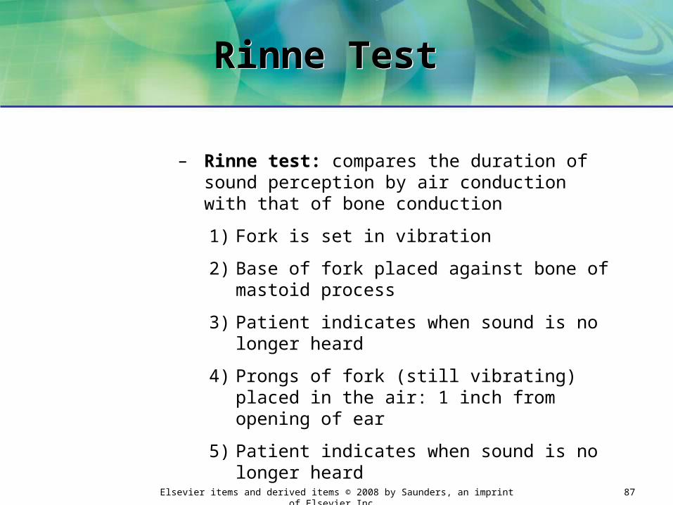
Rinne Test Rinne Test
– Rinne test: compares the duration of sound perception by air conduction with that of bone conduction
1) Fork is set in vibration
2) Base of fork placed against bone of mastoid process
3) Patient indicates when sound is no longer heard
4) Prongs of fork (still vibrating) placed in the air: 1 inch from opening of ear
5) Patient indicates when sound is no longer heard
Elsevier items and derived items © 2008 by Saunders, an imprint of Elsevier Inc. 88
Rinne Test, cont. Rinne Test, cont.
Elsevier items and derived items © 2008 by Saunders, an imprint of Elsevier Inc. 89
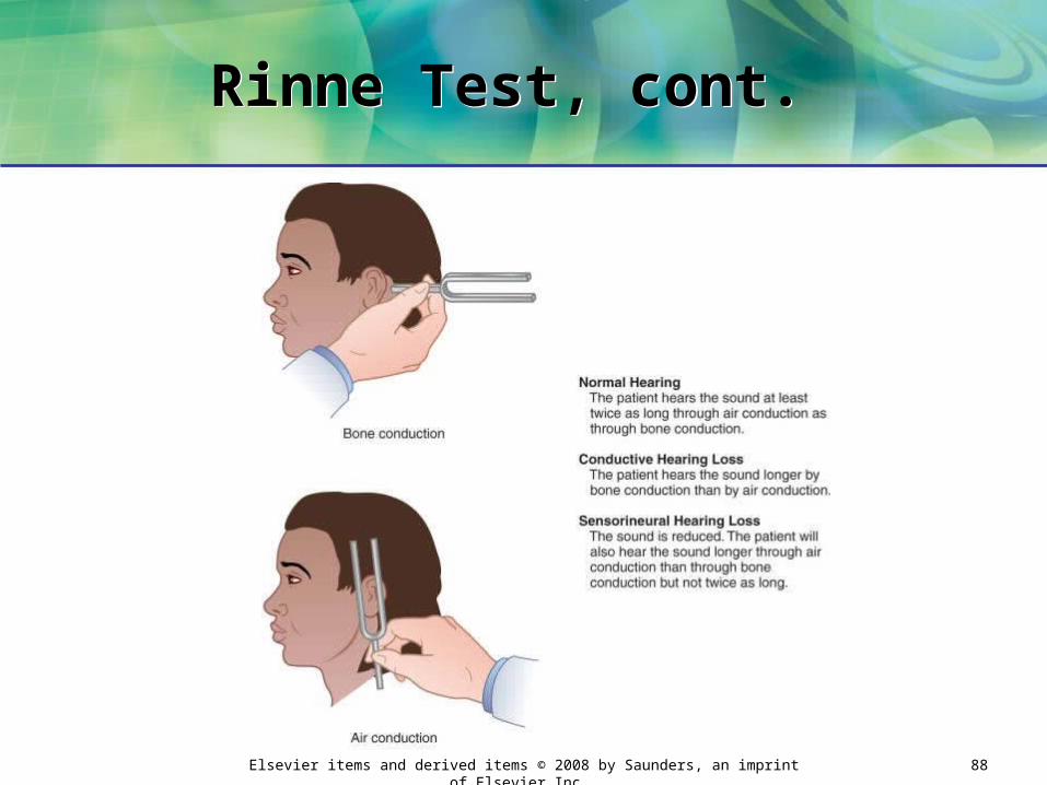
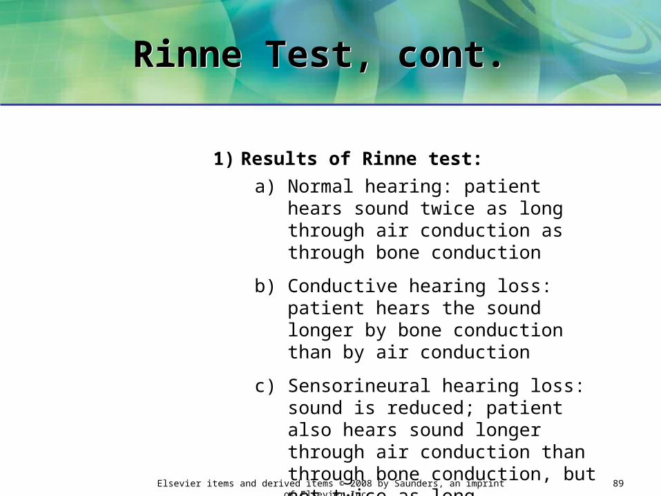
Rinne Test, cont. Rinne Test, cont.
1) Results of Rinne test:
a) Normal hearing: patient hears sound twice as long through air conduction as through bone conduction
b) Conductive hearing loss: patient hears the sound longer by bone conduction than by air conduction
c) Sensorineural hearing loss: sound is reduced; patient also hears sound longer through air conduction than through bone conduction, but not twice as long

Elsevier items and derived items © 2008 by Saunders, an imprint of Elsevier Inc. 90
Audiometry Audiometry
6. Audiometry: measurement of hearing acuity using an audiometer
a. Audiometer: an instrument that quantitatively measures the various frequencies of sound waves
b. Provides information on:
• How extensive hearing loss is
• Which frequencies are involved

Elsevier items and derived items © 2008 by Saunders, an imprint of Elsevier Inc. 91
Audiometer Audiometer
Courtesy GSI [Grayson-Stadler], Milford, NH.

Elsevier items and derived items © 2008 by Saunders, an imprint of Elsevier Inc. 92
Audiometry, cont. Audiometry, cont.
c. To perform test:
• Conduct test in quiet room
• Headphones placed snugly over ears
• Each ear assessed separately
• Audiometer delivers a single frequency at a time
– Starts with low frequencies (250 to 500 Hz) and goes to high frequencies (6000 to 8000 Hz)
• Patient signals when sound is heard
• Results plotted on a graph (audiogram)
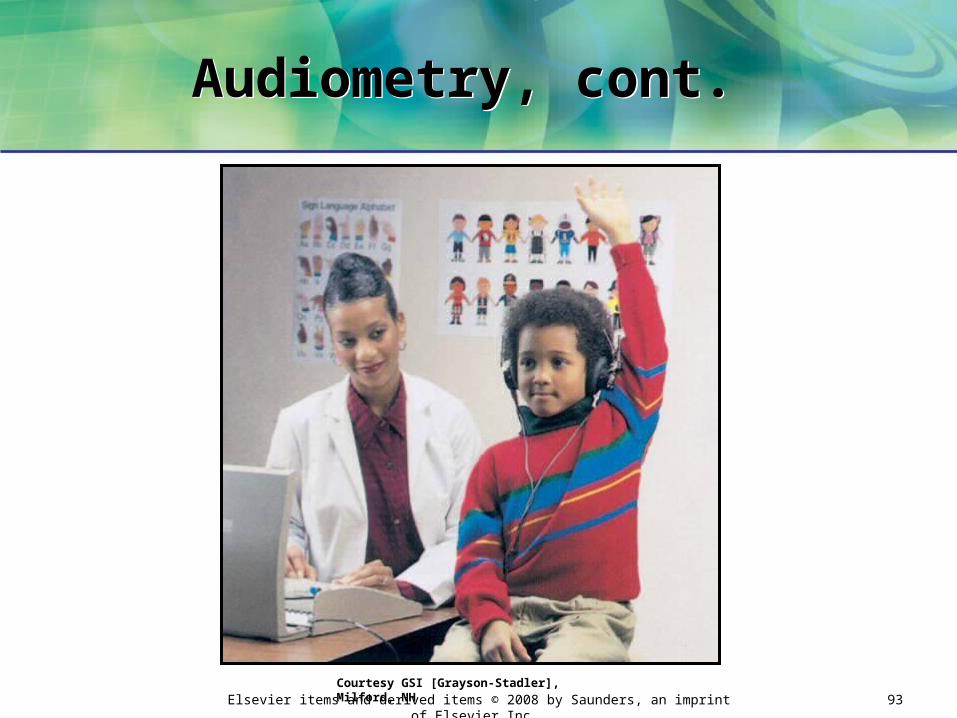
Elsevier items and derived items © 2008 by Saunders, an imprint of Elsevier Inc. 93
Audiometry, cont. Audiometry, cont.
Courtesy GSI [Grayson-Stadler], Milford, NH

Elsevier items and derived items © 2008 by Saunders, an imprint of Elsevier Inc. 94
TympanometryTympanometry
7. Tympanometry: helps determine cause of hearing loss
a. Not a hearing test
b. Tympanometer: earpiece attached to an electronic device
Courtesy GSI [Grayson-Stadler], Milford, NH
Elsevier items and derived items © 2008 by Saunders, an imprint of Elsevier Inc. 95
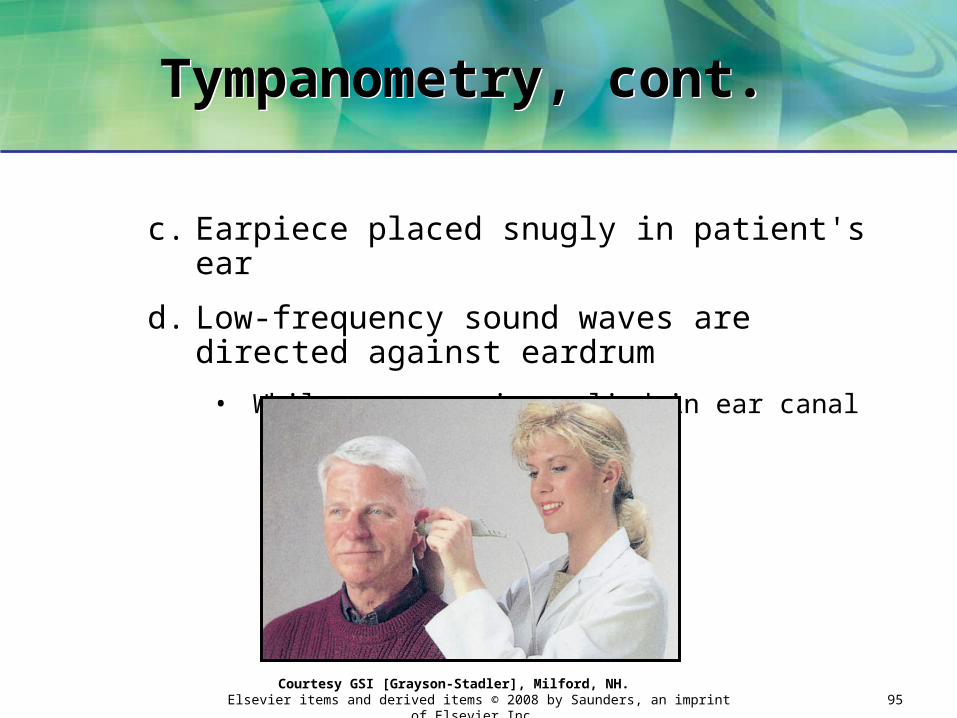
Tympanometry, cont. Tympanometry, cont.
c. Earpiece placed snugly in patient's ear
d. Low-frequency sound waves are directed against eardrum
• While pressure is applied in ear canal
Courtesy GSI [Grayson-Stadler], Milford, NH.

Elsevier items and derived items © 2008 by Saunders, an imprint of Elsevier Inc. 96
Tympanometry, cont. Tympanometry, cont.
e. Normal ear: eardrum exhibits mobility in response to pressure
f. Fluid present in middle ear: eardrum will not move (remains stiff)
• Used to diagnose serous otitis media
– Common cause of temporary hearing loss in children
g. Results printed on a graphic readout: tympanogram

Elsevier items and derived items © 2008 by Saunders, an imprint of Elsevier Inc. 97
Ear IrrigationEar Irrigation
1. Washing of the external auditory canal with a flowing solution
Elsevier items and derived items © 2008 by Saunders, an imprint of Elsevier Inc. 98
Ear Irrigation, cont. Ear Irrigation, cont.
2. Performed to:
a. Cleanse external ear canal to remove:
• Cerumen
• Discharge
• Foreign body
b. Relieve inflammation by applying antiseptic solution
c. Apply heat to ear

Elsevier items and derived items © 2008 by Saunders, an imprint of Elsevier Inc. 99
Ear Irrigation, cont. Ear Irrigation, cont.
3. Impacted cerumen must be softened before removal:
• By instilling warm mineral oil or hydrogen peroxide (10 to 15 minutes)
4. Do not perform irrigation if tympanic membrane is perforated
• Could result in severe irritation or infection of middle ear

Elsevier items and derived items © 2008 by Saunders, an imprint of Elsevier Inc. 100
Ear InstillationEar Instillation
1. Dropping of a liquid into the external auditory canal
a. Usually dispensed in a flexible plastic
container with an attached dropper
2. Performed to:
a. Soften impacted cerumen
b. Combat infection with antibiotic eardrops
c. Relieve pain

Elsevier items and derived items © 2008 by Saunders, an imprint of Elsevier Inc. 101
Ear Instillation, cont.Ear Instillation, cont.

Elsevier items and derived items © 2008 by Saunders, an imprint of Elsevier Inc. 102
POSTTESTPOSTTEST
True or False1. The function of the lens is to permit the entrance
of light rays into the eye.
2. Visual acuity refers to sharpness of vision.
3. Presbyopia is a decrease in the elasticity of the lens due to the aging process.
4. An optician fills prescriptions for eyeglasses.
5. The Snellen Big E chart is used with school-aged children.

Elsevier items and derived items © 2008 by Saunders, an imprint of Elsevier Inc. 103
POSTTEST, CONT.POSTTEST, CONT.
True or False6. The most common color vision defects are
congenital in nature.
7. The cochlea functions in maintaining equilibrium.
8. The range of frequencies for normal speech is 300 to 4000 Hz.
9. Intense noise can result in a sensorineural hearing loss.
10. Tympanometry is used to diagnose patients with auditory nerve damage.


